Injuries to the lower extremity II Aree Tanavalee MD Associate Professor Department of Orthopaedics Faculty of Medicine Chulalongkorn University
|
|
|
- Augustine Hopkins
- 6 years ago
- Views:
Transcription

1 Injuries to the lower extremity II Aree Tanavalee MD Associate Professor Department of Orthopaedics Faculty of Medicine Chulalongkorn University

2 Topics Fracture of the shaft of the femur Fractures around the knee Knee dislocation and fracture dislocation Fractures of tibia and fibular Fractures around the ankle Fracture and fracture dislocation of the foot


3 Common symptoms and signs of fractures Pain Deformity Shortening Swelling Ecchymosed Loss of function Open injury Gross finding of fractures Injury to lower extremity

4 Radiographic evaluation for fractures At least, 2 different planes of Fx site Includes joint above and below Some types of Fx, special views Sometimes, 2 different times Sometimes, calls second opinion


5 Complications of fractures General Delayed union Nonunion Malunion Shortening Infection Severe Neurovascular injuries Compartment syndrome Fat embolism Adult respiratory distress syndrome (ARDS)


6 Fat embolism Common in Fx of long bone and pelvis Multiple Fxs >> single Fx Respiratory insufficiency Usually manifests within 48 hr Clinical Fever Tachepnea Tachycardia Alters consciousness Treatment Respiratory support Early Fx stabilization

7 Compartment syndrome Impaired circulation and function of tissues within a closed space Most common: closed Fx in Leg, forearm Irreversible damage to muscles if > 6 hr Irreversible damage to nerves if > 12 hr Clinical Pain out of proportion, pain with passive ROM Discolation Paresthesia Paralysis Pulseless


8 Compartment syndrome Risks injuries Fx tibia Knee dislocation Crush injury: leg, foot Prolong Fx with vascular compromised Treatment Fasciotomy

9 Type of fractures Closed No wound connects to Fx site Open Grade 1 Simple fracture Wound less than 1 cm Grade 2 Moderate-severe fracture Wound bet 2-10 cm Grade 3 Severe fracture Wound > 10 cm Loss of skin coverage Vascular compromised


10 Fracture of the shaft of the femur General The strongest and longest bone Canal widening: proximal and distal Gluteal and psoas muscles Proximal third: flex, external rotate, abduct Adductors Varus deformity Gastrocnemius muscles Supracondylar: distal part: posterior angulation


11 Fracture of the shaft of the femur Mechanisms of injury General Major trauma High-energy injury Pathological Lesser degree of trauma Often: metaphysis-shaft junction Common mechanisms of Fx Bending load >> transverse Fx
 Transverse Oblique (angle> 30 deg to transverse line) Spiral Segmental")
12 Fracture of the shaft of the femur Type of fractures (according to geometry of Fx line) Transverse Oblique (angle> 30 deg to transverse line) Spiral Segmental Comminution

13 Fracture of the shaft of the femur Symptoms and signs Common S&S of fracture Radiographic evaluation Standard: AP, lateral, CXR Including: hip and knee joint Findings: fracture

14 Fracture of the shaft of the femur Initial evaluation and management Live support: as major traumatized patient Assessment of associated orthopaedic injuries Pelvic fracture Hip fracture Ligamentous injury: around the knee Neurovascular injury Immobilization
15 Fracture of the shaft of the femur Complications Commom Fx complications Fat embolism ARDS (multiple Fx)
16 Fracture of the shaft of the femur Treatment Nonoperative Traction Cast brace

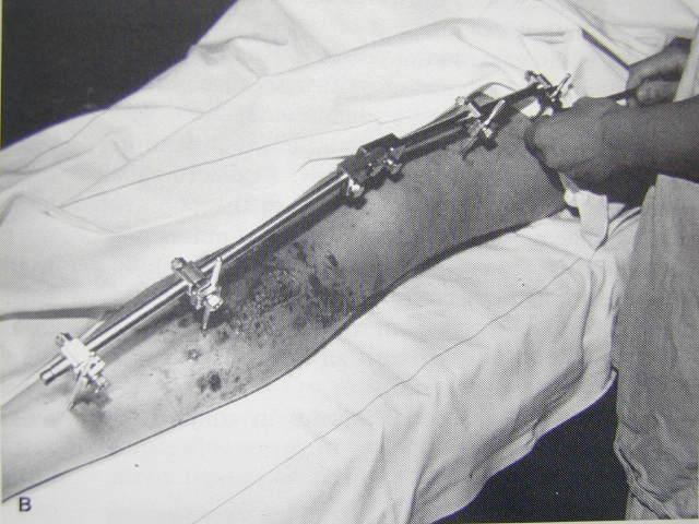
17 Fracture of the shaft of the femur Treatment Operative Plating Intramedullary nailing External fixator

18 Fracture of the shaft of the femur Traction Commonly used in the past Now, indicated in Fracture in children Temporary purpose Surgery is limited Type Skeleton Proximal tibia Distal femur Skin
19 Fracture of the shaft of the femur Traction Disadvantages Limited rotational control of fracture Limb-length discrepancy Loss of range of motion (ROM) Prolonged hospitalization Pin tract infection

20 Fracture of the shaft of the femur Cast brace Provides external support effect Permits progressive weight bearing Prerequisites Good reduction Traction until pain swelling have subsided Indicated in Distal third fracture

21 Fracture of the shaft of the femur Cast brace Disadvantages Limb-length discrepancy Varus angulation Limb-length discrepancy Limited area of usage


22 Fracture of the shaft of the femur Operative treatment Immediate fracture stability More anatomical reduction Early ambulation and ROM Less hospitalization
23 Fracture of the shaft of the femur Plating No need for special instruments Favorable results Ipsilateral neck-shaft fracture (same side) Disadvantages Extensive tissue exposure Higher complications than nailing


24 Fracture of the shaft of the femur Intramedullary nailing Treatment of choice Load-sharing implant Predictable shaft alignment Early recovery Type Open nailing Closed nailing

25 Fracture of the shaft of the femur Intramedullary nailing Closed nailing Interlocking nailing Less invasive Minimal surgical surgical scar Rapid fracture healing

26 Fracture of the shaft of the femur External fixator Open fracture: need wound care Severe comminution Marked contaminated wound Temporary device


27 Fracture of the shaft of the femur Disadvantages of operative treatment Infection Nonunion Delayed union Loss of fixation Others surgical complications


28 Fracture of the shaft of the femur Postoperative rehabilitation Early mobilization Muscle activity: following stability Progressive weight bearing: stable Delayed weight bearing: less stability, proximal or distal fracture

29 Fractures around the knee Fracture of the distal femur Fractures of the tibial plateau Fractures of the tibial spine and intercondylar eminence Fracture of the patella


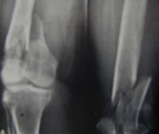
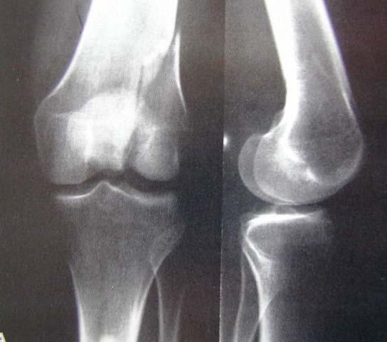
30 Fracture of the distal femur General Supracondylar area 5 cm above the flare of metaphysis Intercondylar area Require careful neurovascular assessment Distal part: posterior angulation Joint Tibiofemoral joint (TF) Patellofemoral joint (PF)

31 Fracture of the distal femur Mechanisms of injuries High energy trauma in young patients Low energy trauma in the elderly Symptoms and signs Pain around the knee Swelling around the knee Tenderness over the fracture site Radiographic evaluation Usually standard x-ray views

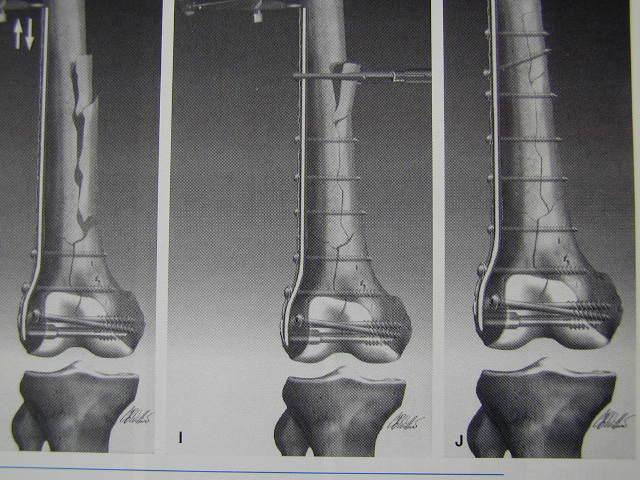
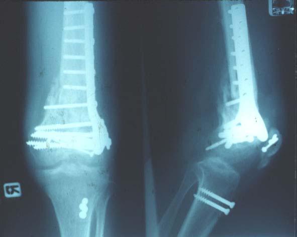
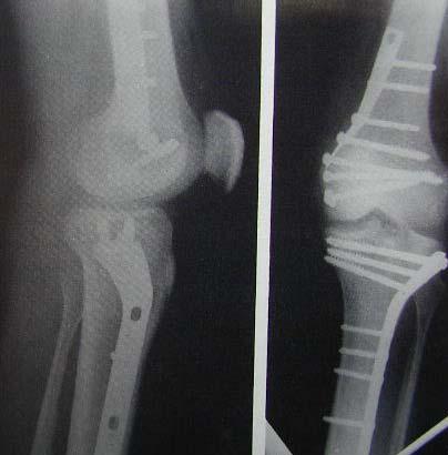
32 Fracture of the distal femur Treatment Goal: restore joint surface and alignment Nonoperative Nondisplaced Traction 4-6 wk Cast brace with NWB and early ROM Operative Significant displaced Implant Condylar blade plate Condylar sliding nail-plate Intramedullary nail
33


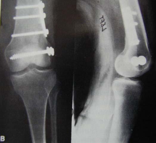
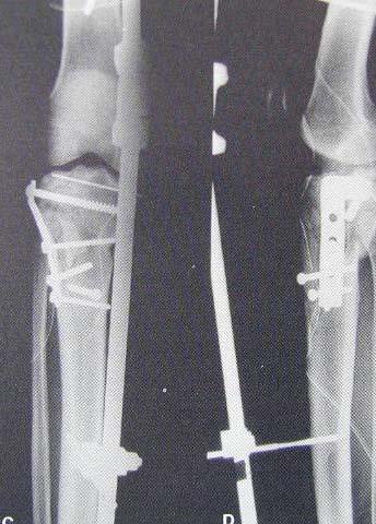
34 Fractures of the tibial plateau General Concomitant ligament injuries Depression and displacement Mechanisms Varus or valgus force with axial loading Symptoms and signs Pain around the knee Swelling around the knee Tenderness over the fracture site Valgus or varus deformity
35 Fractures of the tibial plateau Nonoperative Long leg cast (LLC) Brace with NWB and early ROM Traction Operative Screw or pin Plate and screws Rehabilitation Non weight bearing 6-8 weeks Partial weight bearing until 12 weeks Full weight bearing after 12 weeks
36

37 Fractures of the tibial plateau Complications Common Fx complications Peroneal nerve injury Popliteal artery injury Compartment syndrome Associate injuries Meniscal injury 15% Cruciate ligament and collateral ligament injuries 22%

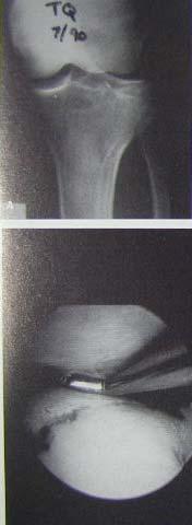
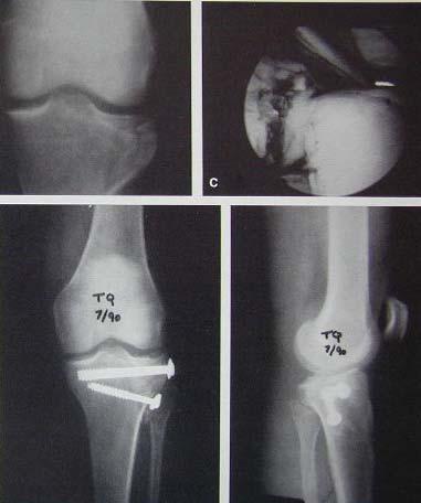
38 Fractures of the tibial spine and intercondylar eminence Mechanism of injury: tibial spine Knee twisting Mechanism of injury: intercondylar eminence Hyperflexion Hyperextension Valgus-varus force


39 Fractures of the tibial spine and intercondylar eminence Symptoms and signs Pain and swelling of the knee A block to full extension Treatment Nonoperative Most fractures Long leg cast in full extension 4-6 wk Operative Arthrotomy and screw fixation Arthroscopy and screw fixation Complication Fragment becomes a loose body

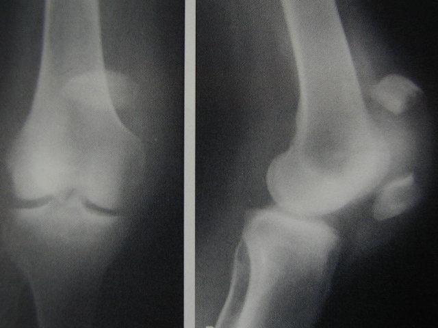
40 Fracture of the patella General The largest sesamoid bone Accessory ossification center Superolateral corner Named bipatite patetta Increases the extensor mechanism Protect the femoral condyles Forces across PF joint Daily activity: >3 times body weight Stair climbing and deep squatting: >7 times body weight

41 Fracture of the patella Mechanisms of injury Direct injuries: direct force Pattern:Comminuted Usually minimal displacement Indirect injuries: muscle forces Pattern:Transverse Combined injuries Pattern:Comminuted with displaced Symptoms and signs Pain and tenderness at the anterior of the knee Skin contusion: direct injuries Ability to extend the knee Depends on the continuity of the extensor mechanism
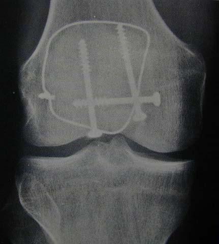
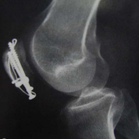
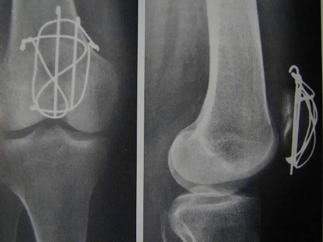
42 Fracture of the patella Treatment Nonoperative Within 2-3 mm displacement or stepping Cylinder cast for 4-6 wk Operative More than 2-3 mm displacement or stepping ORIF Circlage wiring Tension band wiring Patellectomy

43
44

45 Fracture of the patella Complications Common Fx complications Results after treatment Late OA change of the PF joint Painful retained hardware

46 Knee dislocation and fracture dislocation Knee dislocation Patella dislocation Fracture dislocation Injury to lower extremity

47 Knee dislocation Description Position of the distal relates to the proximal Type of dislocation Anterior Posterior Medial Lateral Rotatory


48 Knee dislocation Anterior Hyperextension injury Injury to PCL, ACL and popliteal vessels Common Posterior Common Medial Varus injury Injury to lateral structures


49 Knee dislocation Lateral Valgus injury Injury to medial structures, cruciate ligaments Rotatory Posterolateral Often associate with peroneal nerve injury
50 Knee dislocation Symptoms and signs Gross distortion of the knee Instability after reduction May have neurological deficit (35% of cases) Most common: common peroneal nerve May have vascular compromise Most common: popliteal artery
51 Knee dislocation Treatment Principle Operative better than conservative Emergency vascular assessment Torn; vascular repair or graft Repairs ligament if possible Nerve assessment Torn: repair
52 Knee dislocation Postoperative care Establishes ROM as early as possible At 4-6 wk: begins muscle strengthening Prognosis Depends on the extent of neurovascular injury
53 Patellar dislocation General Common in female Symptoms and signs Pain Distorted knee anatomy Limited ROM in flexed position Treatment Closed reduction (CR) Cylinder cast 4-6 wk Vastus medialis strengthening
54 Fracture dislocation around the knee General Combination of fractures and dislocation Symptoms and signs Injury to lower extremity The same as major fracture around the knee Distorted knee anatomy Treatment immediate CR and immobilization Definite fracture treatment
55 Fractures of tibia and fibular General Anterior compartment Ankle and foot dorsiflexion Deep peroneal N Lateral compartment Foot plantaflexion and eversion Sup peroneal N Superficial posterior compartment Foot plantaflexion Deep posterior compartment Foot plantaflexion and inversion Posterior tibial N Posterior tibial vessels


56 Fractures of the tibia Mechanism of injury of tibial shaft Fx Direct Indirect Penetrating Symptoms and signs of tibial shaft Fx Usually obvious deformed leg Pain Swelling Associated neurovascular injuries


57 Fractures of the tibia Treatment of tibial shaft Fx Nonoperative Criteria Angulation < 10 deg Rotation < 10 deg Shortening <1 cm Apposition 50% Closed reduction Long leg cast At 4 wk: progressive WB At12 wk: full WB


58 Fractures of the tibia Treatment of tibial shaft Fx Operative Indications Failed nonoperative Multiple Fx Associated vascular problems Type External fixator Plate and screws Intramedullary nail


59 Fractures of the tibia Complications Common Fx complications Vascular injury Compartment syndrome Nerve injury
60 Fractures of the fibular General Most are associated with fx of the tibia Isolate fx results from direct injury Local signs and symptoms Difficulty in walking Treatment Mild: elastic bandage support Mod to sev: SLC or brace for pain relief Ambulation Progressive weight bearing as possible Remove cast within 6 wk

61 Fractures around the ankle Fracture of the tibial plafond Ankle fractures Ankle dislocation and fracture dislocation


62 Fracture of the tibial plafond Definition Fx of the distal tibia extending into the ankle May be called pilon fracture Treatment Nonoperative No signification articular displacement Closed reduction and LLC Non weight bearing for 6 wk Progressive weight bearing until wk Operative Signification articular displacement Plate and screws Distraction ring


63 Ankle fractures General Deltoid ligament Between med malleolus and talus Posterior tibial lip Posterior part of tibia The third malleolus Distal tibiofibular syndesmosis Maintains ankle stability Symptoms and signs Pain Swelling Deformity Difficult or unable weight bearing


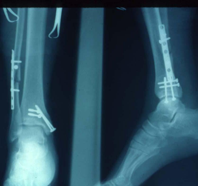
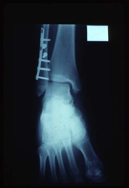
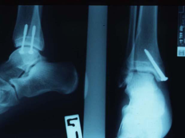
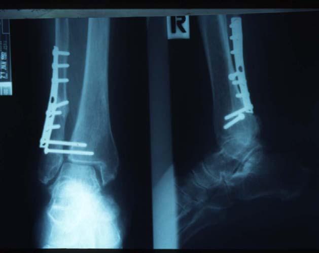
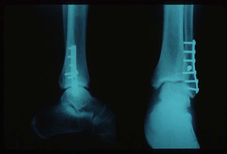
64 Ankle fractures investigation X-ray standard AP and lateral X-ray mortise view Treatment Nonoperative Minimal displaced CR and short leg cast 6-8 wk Operative Displaced Internal fixation Plating Screws Tension band wiring
65


66 Ankle dislocation and fracture dislocation General Injury to lower extremity Combination of fractures around the ankle and dislocation Associated fractures Malleoli Talus Distal tibia Require neurovascular assessment
67 Ankle dislocation and fracture dislocation Symptoms and signs Common symptoms and signs of fracture Distorted ankle anatomy May associated neurovascular deficits Treatment immediate CR and immobilization Definite fracture treatment Injury to lower extremity
68 Fractures of the foot and fracture dislocations Stress and neuropathic fractures Fracture of the talus Dislocation around the talus Fracture of the calcaneus Fractures of the tarsals and joint injuries Fractures of the metatarsals and phalanges


69 Stress fractures Excessive, repetitive stress applied to bone Most common: 2 nd metatarsal, calcaneus Mild to moderate pain and/or swelling Diagnosis X-ray: from 2 wk Bone scan: from 2 days Treatment Nonoperative: SLC 4-6 wk

70 Neuropathic fractures Charcot joint Associated with DM, peripheral nerve diseases Initiating event is fracture around the joint Rapid joint destruction without pain Usually,patients present late Treatment Nonoperative: SLC with non weight bearing until heal


71 Fracture of the talus Neck, head, body and process Fracture of the talar neck Hyperdorsiflexion injury May associated dislocation Subtalar joint Ankle joint Talonavicular joint Treatment Nonoperative Operative
 Skin necrosis Posttraumatic")
72 Frcacture of the talus Treatment Nonoerative No displacement, no dislocation Short leg cast (SLC) 8-12 wk Displaced and/or dislocation CR and SLC 8-12 wk Operative Screws or K-wires fixation and cast Complications Common Fx complications Avascular necrosis (AVN) Skin necrosis Posttraumatic OA
73 Dislocation around the talus Subtalar dislocation Inversion & eversion injuries to the foot Common S&S of dislocation Compromised neurovascular function CR if failed open reduction SLC 4 wk Talar dislocation Most are open injuries Reduction with soft tissue management SLC (may be with pins) 6 wk Results in AVN

 25-40 deg Calcaneal axial")
74 Fracture of the calcaneus General The most common tarsal bone Fx Thin cortex with cancellous bone inside Support the body weight Mechanisms of injury Most are falling from heights Associated spinal injury Radiographic findings Standard AP, lateral Bohler s angle (tuber angle) deg Calcaneal axial view

 Plate")
75 Fracture of the calcaneus Treatment Nonoperative Non or minimal displaced SLC 6-8 wk Operative Percutaneous pin and SLC Open reduction and internal fixation (ORIF) Plate Screws Staples


76 Fractures of the tarsals and joint injuries General Midfoot 3 cuneiforms Navicular Accessory navicular Cuboid Midtarsal joint (Chopart s joint) Talonavicular Calcaneocuboid Tarsometatarsal joint (Linfranc s joint)


77 Fractures of the tarsals and joint injuries Diagnosis Usually overlooked Less appreciated Treatment Tarsal bones Usually nonoperative SLC 4-6 wk Midtarsal joint CR and/or pin and SLC Arthrodesis in late case Tarsometarsal joint Requires adequate Rx CR and pinning ORIF

78 Fractures of the metatarsals and phalanges General Usually nonoperative treatment SLC 4-6 wk Some conditions Open Fx with problem of skin etc. Posterior splint Operative treatment Soft tissue management Displaced Fx Longitudinal K-wire Small screws


79 Fractures of the metatarsals and phalanges Common Fx Ballet dancer Fx Spiral Fx at distal of 5 th metatarsal Treatment: SLC 6 wk Jones Fx (Dancer Fx) Fx base of 5 th metatarsal Treatment: SLC 6 wk Phalanges Fx Posterior splint 4 wk
Surgery-Ortho. Fractures of the tibia and fibula. Management. Treatment of low energy fractures. Fifth stage. Lec-6 د.
 Fifth stage Lec-6 د. مثنى Surgery-Ortho 28/4/2016 Indirect force: (low energy) Fractures of the tibia and fibula Twisting: spiral fractures of both bones Angulatory: oblique fractures with butterfly segment.
Surgical Care at the District Hospital. EMERGENCY & ESSENTIAL SURGICAL CARE
 Surgical Care at the District Hospital 1 18 Orthopedic Trauma Key Points 2 18.1 Upper Extremity Injuries Clavicle Fractures Diagnose fractures from the history and by physical examination Treat with a
Peggers Super Summaries: Foot Injuries
 Lisfranc Injury ANATOMY Roman arch with recessed 2 nd MT base AP medial side of intermediate cuneiform to 2 nd MT base Oblique medial side of lateral cuneiform with 3 rd MT base and 4 th with medial boarder
Hip joint Type: Articulating bones:
 Ana (242 ) Hip joint Type: Synovial, ball & socket Articulating bones: Formed between head of femur and lunate surface of acetabulum of hip bone. Capsule: it is a strong fibrous sleeve connecting the articulating
Foot Injuries. Dr R B Kalia
 Foot Injuries Dr R B Kalia Overview Dramatic impact on the overall health, activity, and emotional status More attention and aggressive management Difficult appendage to study and diagnose. Aim- a stable
Recognizing common injuries to the lower extremity
 Recognizing common injuries to the lower extremity Bones Femur Patella Tibia Tibial Tuberosity Medial Malleolus Fibula Lateral Malleolus Bones Tarsals Talus Calcaneus Metatarsals Phalanges Joints - Knee
7/23/2018 DESCRIBING THE FRACTURE. Pattern Open vs closed Location BASIC PRINCIPLES OF FRACTURE MANAGEMENT. Anjan R. Shah MD July 21, 2018.
 BASIC PRINCIPLES OF FRACTURE MANAGEMENT Anjan R. Shah MD July 21, 2018 DESCRIBING THE FRACTURE Pattern Open vs closed Location POLL OPEN HOW WOULD YOU DESCRIBE THIS FRACTURE PATTERN? 1 Spiral 2 Transverse
The Dance Hall by Vincent van Gogh,1888
 The Dance Hall by Vincent van Gogh,1888 Articulations of the pelvic girdle Lumbosacral joints, sacroiliac joints & pubic symphysis The remaining joints of the lower limb Hip joint Knee joint Tibiofibular
Case. 5 year old with 2 weeks leg pain and now refusing to walk + Fevers, lower leg swelling, warmth Denies and history of trauma or wounds
 Case 5 year old with 2 weeks leg pain and now refusing to walk + Fevers, lower leg swelling, warmth Denies and history of trauma or wounds Exam I: Swelling over entire tibia extending to foot P: Tenderness
X-Ray Rounds: (Plain) Radiographic Evaluation of the Ankle.
 Radiographic Evaluation of the Ankle.") X-Ray Rounds: (Plain) Radiographic Evaluation of the Ankle www.fisiokinesiterapia.biz Anatomy Complex hinge joint Articulations among: Fibula Tibia Talus Tibial plafond Distal tibial articular surface
X-Ray Rounds: (Plain) Radiographic Evaluation of the Ankle www.fisiokinesiterapia.biz Anatomy Complex hinge joint Articulations among: Fibula Tibia Talus Tibial plafond Distal tibial articular surface
Vasu Pai FRACS, MCh, MS, Nat Board Ortho Surgeon Gisborne
 Vasu Pai FRACS, MCh, MS, Nat Board Ortho Surgeon Gisborne FRACTURE MANAGEMENT I Simple closed fracture : Complete or Incomplete Stable or unstable II Open fracture III Multiple fracture IV Polytrauma Fractures
Talus Fractures: When and Why on Screws and Plates
 Talus Fractures: When and Why on Screws and Plates Frank A. Liporace, MD Associate Professor Director of Orthopaedic Research New York University / Hospital for Joint Diseases, NY, NY Director Orthopaedic
Tibial Shaft Fractures
 Tibial Shaft Fractures Mr Krishna Vemulapalli Consultant Orthopaedics Surgeon Queens & King George Hospitals Queens Hospital 14/03/2018 Google Maps Map data 2018 Google 10 km Orthopaedics Department Covers
Fractures and dislocations around elbow in adult
 Lec: 3 Fractures and dislocations around elbow in adult These include fractures of distal humerus, fracture of the capitulum, fracture of the radial head, fracture of the olecranon & dislocation of the
radiologymasterclass.co.uk
 http://radiologymasterclass.co.uk Hip X-ray anatomy - Normal AP (anterior-posterior) Shenton's line is formed by the medial edge of the femoral neck and the inferior edge of the superior pubic ramus Loss
Femoral Shaft Fracture
 Femoral Shaft Fracture The femoral shaft is well padded with muscles(an advantage in protecting the bone from all but the most powerful forces)but the disadvantage is that fractures are often severely
Lower Extremity Dislocations: Management and Triage on the Field
 Lower Extremity Dislocations: Management and Triage on the Field Scott J Tarantino, MD Towson Orthopaedic Associates, Towson, MD None Disclsures Purpose To provide you with knowledge which may guide you
MIDFOOT INJURIES-ARE WE UNDERTREATING IT? Mr Rajiv Limaye Mr Prasad Karpe University Hospital of North Tees 3 rd Foot and Ankle Symposium
 MIDFOOT INJURIES-ARE WE UNDERTREATING IT? Mr Rajiv Limaye Mr Prasad Karpe University Hospital of North Tees 3 rd Foot and Ankle Symposium Introduction Increasing sports injuries RTA and traumatic injuries
General Concepts. Growth Around the Knee. Topics. Evaluation
 General Concepts Knee Injuries in Skeletally Immature Athletes Zachary Stinson, M.D. Increased rate and ability of healing Higher strength of ligaments compared to growth plates Continued growth Children
The Lower Limb II. Anatomy RHS 241 Lecture 3 Dr. Einas Al-Eisa
 The Lower Limb II Anatomy RHS 241 Lecture 3 Dr. Einas Al-Eisa Tibia The larger & medial bone of the leg Functions: Attachment of muscles Transfer of weight from femur to skeleton of the foot Articulations
Fractures of Lower extremity
 www.fisiokinesiterapia.biz Fractures of Lower extremity FRACTURES OF FEMORAL NECK Anatomy Neck-Shaft Angle: In the anteroposterior roentgenogram, it is the angle between the long axis of the femoral neck
Knee Dislocation: Spectrum of Injury, Evolution of Treatment & Modern Outcomes
 Knee Dislocation: Spectrum of Injury, Evolution of Treatment & Modern Outcomes William M Weiss, MD MSc FRCSC Orthopedic Surgery & Rehabilitation Sports Medicine, Arthroscopy & Extremity Reconstruction
Introduction. The primary function of the ankle and foot is to absorb shock and impart thrust to the body during walking.
 The ankle 1 Introduction The primary function of the ankle and foot is to absorb shock and impart thrust to the body during walking. OSTEOLOGRY The term ankle refers primarily to the talocrural joint,
.org. Tibia (Shinbone) Shaft Fractures. Anatomy. Types of Tibial Shaft Fractures
 Shaft Fractures. Anatomy. Types of Tibial Shaft Fractures") Tibia (Shinbone) Shaft Fractures Page ( 1 ) The tibia, or shinbone, is the most common fractured long bone in your body. The long bones include the femur, humerus, tibia, and fibula. A tibial shaft fracture
Tibia (Shinbone) Shaft Fractures Page ( 1 ) The tibia, or shinbone, is the most common fractured long bone in your body. The long bones include the femur, humerus, tibia, and fibula. A tibial shaft fracture
Injuries of the Foot and Ankle. Introduction. Introduction 10/2/2009. Bryan Lapinski, MD
 Injuries of the Foot and Ankle Bryan Lapinski, MD Introduction The average person takes 1 million steps per year Approximately 30 bones in the foot and ankle are subjected to forces of 3 7 times body weight
OPERATIVE TREATMENT OF THE INTERCONDYLAR FRACTURE OF THE FEMUR
 OPERATIVE TREATMENT OF THE INTERCONDYLAR FRACTURE OF THE FEMUR S G Chee, K S Lam, B K Tay, N Balachandran SYNOPSIS Operative treatment of 28 intercondylar fractures of femur were done from 98 to 985. The
CASE ONE CASE ONE. RADIAL HEAD FRACTURE Mason Classification. RADIAL HEAD FRACTURE Mechanism of Injury. RADIAL HEAD FRACTURE Imaging
 CASE ONE An eighteen year old female falls during a basketball game, striking her elbow on the court. She presents to your office that day with a painful, swollen elbow that she is unable to flex or extend
The Knee. Prof. Oluwadiya Kehinde
 The Knee Prof. Oluwadiya Kehinde www.oluwadiya.sitesled.com The Knee: Introduction 3 bones: femur, tibia and patella 2 separate joints: tibiofemoral and patellofemoral. Function: i. Primarily a hinge joint,
Foot and Ankle Systems Coding Reference Guide
 Foot and Ankle Systems Coding Reference Guide Physician Arthrodesis 27870 Arthrodesis, ankle, open 27871 Arthrodesis, tibiofibular joint, proximal or distal 28705 Arthrodesis; pantalar 28715 Arthrodesis;
CHAPTER 8: THE BIOMECHANICS OF THE HUMAN LOWER EXTREMITY
 CHAPTER 8: THE BIOMECHANICS OF THE HUMAN LOWER EXTREMITY _ 1. The hip joint is the articulation between the and the. A. femur, acetabulum B. femur, spine C. femur, tibia _ 2. Which of the following is
Osteosynthesis involving a joint Thomas P Rüedi
 Osteosynthesis involving a joint Thomas P Rüedi How to use this handout? The left column contains the information given during the lecture. The column at the right gives you space to make personal notes.
Clarification of Terms
 Clarification of Terms The plantar aspect of the foot refers to the role or its bottom The dorsal aspect refers to the top or its superior portion The ankle and foot perform three main functions: 1. shock
FIBULAR & SYNDESMOSIS MALUNIONS
 FIBULAR & SYNDESMOSIS MALUNIONS MICHAEL P. CLARE, MD FLORIDA ORTHOPAEDIC INSTITUTE TAMPA, FL USA MORTISE INHERENTLY UNSTABLE LATERAL MALLEOLUS ACTS AS BUTTRESS / POST RESIST LATERAL TRANSLATION OF TALUS
Radiographic Evaluation of Calcaneal Fractures. Kali Luker, PGY-1
 Radiographic Evaluation of Calcaneal Fractures Kali Luker, PGY-1 Anatomy Extraarticular Fractures Involve body, anterior process or tuberosity Treated with immobilization and NWB x 6 wks UNLESS Displaced
Copyright 2012 by The McGraw-Hill Companies, Inc. All rights reserved. McGraw-Hill/Irwin
 CHAPTER 8: THE LOWER EXTREMITY: KNEE, ANKLE, AND FOOT KINESIOLOGY Scientific Basis of Human Motion, 12 th edition Hamilton, Weimar & Luttgens Presentation Created by TK Koesterer, Ph.D., ATC Humboldt State
Copyright 2004, Yoshiyuki Shiratori. All right reserved.
 Ankle and Leg Evaluation 1. History Chief Complaint: A. What happened? B. Is it a sharp or dull pain? C. How long have you had the pain? D. Can you pinpoint the pain? E. Do you have any numbness or tingling?
Pediatric Tibia Fractures Key Points. Christopher Iobst, MD
 Pediatric Tibia Fractures Key Points Christopher Iobst, MD Goals Bone to heal Return to full weight bearing Acceptable alignment rule of 10s 10 degrees of varus 8 degrees of valgus 12 degrees of procurvatum
PRONATION-ABDUCTION FRACTURES
 C H A P T E R 1 2 PRONATION-ABDUCTION FRACTURES George S. Gumann, DPM (The opinions of the author should not be considered as reflecting official policy of the US Army Medical Department.) Pronation-abduction
Basic Care of Common Fractures Utku Kandemir, MD
 Basic Care of Common Fractures Utku Kandemir, MD Assistant Clinical Professor Trauma & Sports Medicine Dept. of Orthopaedic Surgery UCSF / SFGH History Physical Exam Radiology Treatment History Acute trauma
The pilon tibiale fracture
 The pilon tibiale fracture Thomas Beck Spitalzentrum Oberwallis OTC Trauma course september 2017 xxx I have no financial relationships with commercial entities that produce healthcare related products.
Hany El-Rashidy and Anand Vora
 Chapter 194 Lisfranc Injuries Chapter 194 Lisfranc Injuries Hany El-Rashidy and Anand Vora 8 ICD-9 CODE 838.03 Lisfranc (Tarsometatarsal) Fracture-Dislocation Key Concepts The Lisfranc joint represents
Knee Injury Assessment
 Knee Injury Assessment Clinical Anatomy p. 186 Femur Medial condyle Lateral condyle Femoral trochlea Tibia Intercondylar notch Tibial tuberosity Tibial plateau Fibula Fibular head Patella Clinical Anatomy
CURRENT TREATMENT OPTIONS
 CURRENT TREATMENT OPTIONS Fix single column or both: Always fix both. A study by Svend-Hansen corroborated the poor results associated with isolated medial malleolar fixation in bimalleolar ankle fractures.
Paul Alley MD,DPM,MS,FACS,FAAOS,BFD Eby Orthopaedics,Jasper,Indiana
 Paul Alley MD,DPM,MS,FACS,FAAOS,BFD Eby Orthopaedics,Jasper,Indiana Very common Bone=fractures Description (cracked,broke,busted,or smashed) A=anatomic area of bone eg: head,neck,shaft B=bone involved
Therapeutic Foot Care Certificate Program Part I: Online Home Study Program
 Therapeutic Foot Care Certificate Program Part I: Online Home Study Program 1 Anatomy And Terminology Of The Lower Extremity Joan E. Edelstein, MA, PT, FISPO Associate Professor of Clinical Physical Therapy
Balanced Body Movement Principles
 Balanced Body Movement Principles How the Body Works and How to Train it. Module 3: Lower Body Strength and Power Developing Strength, Endurance and Power The lower body is our primary source of strength,
Outline. Ankle/Foot Anatomy Ankle Sprains Ottawa Ankle Rules DDx: The Sprain That Wasn t
 Ankle Injuries Outline Ankle/Foot Anatomy Ankle Sprains Ottawa Ankle Rules DDx: The Sprain That Wasn t Anatomy: Ankle Mortise Bony Anatomy Lateral Ligament Complex Medial Ligament Complex Ankle Sprains
Fractures of the shoulder girdle, elbow and fractures of the humerus. H. Sithebe 2012
 Fractures of the shoulder girdle, elbow and fractures of the humerus H. Sithebe 2012 Fractures of the Clavicle (mid-shaft). Fractures of the clavicle Fractures of the clavicle Treatment- conservative.
Rehabilitation after Total Elbow Arthroplasty
 Rehabilitation after Total Elbow Arthroplasty Total Elbow Atrthroplasty Total elbow arthroplasty (TEA) Replacement of the ulnohumeral articulation with a prosthetic device. Goal of TEA is to provide pain
Multiapical Deformities p. 97 Osteotomy Concepts and Frontal Plane Realignment p. 99 Angulation Correction Axis (ACA) p. 99 Bisector Lines p.
 p. 99 Bisector Lines p.") Normal Lower Limb Alignment and Joint Orientation p. 1 Mechanical and Anatomic Bone Axes p. 1 Joint Center Points p. 5 Joint Orientation Lines p. 5 Ankle p. 5 Knee p. 5 Hip p. 8 Joint Orientation Angles
Normal Lower Limb Alignment and Joint Orientation p. 1 Mechanical and Anatomic Bone Axes p. 1 Joint Center Points p. 5 Joint Orientation Lines p. 5 Ankle p. 5 Knee p. 5 Hip p. 8 Joint Orientation Angles
ACL Athletic Career. ACL Rupture - Warning Features Intensive pain Immediate swelling Locking Feel a Pop Dead leg Cannot continue to play
 FIMS Ambassador Tour to Eastern Europe, 2004 Belgrade, Serbia Montenegro Acute Knee Injuries - Controversies and Challenges Professor KM Chan OBE, JP President of FIMS Belgrade ACL Athletic Career ACL
DEPARTMENT OF TRAUMATOLOGY AND HAND SURGERY INSTITUTE OF MUSCULOSKELETAL SURGERY ANKLE AND FOOT INJURIES
 DEPARTMENT OF TRAUMATOLOGY AND HAND SURGERY INSTITUTE OF MUSCULOSKELETAL SURGERY ANKLE AND FOOT INJURIES Presenter: Dr George Ayerh ENGLISH PROGRAM LECTURES EN_11/A - 2018 TOPICS I. Part: Ankle & Foot
Pediatric Fractures. Objectives. Epiphyseal Complex. Anatomy and Physiology. Ligaments. Bony matrix
 1 Pediatric Fractures Nicholas White, MD Assistant Professor of Pediatrics Eastern Virginia Medical School Attending, Pediatric Emergency Department Children s Hospital of The King s Daughters Objectives
Post test for O&P 2 Hrs CE. The Exam
 Post test for O&P 2 Hrs CE The Exam This examination is taken in "open book" format. That means you are free to answer the questions after research or discussion with your fellow workers. We feel this
5 COMMON INJURIES IN THE FOOT & ANKLE
 5 COMMON INJURIES IN THE FOOT & ANKLE MICHAEL P. CLARE, MD FLORIDA ORTHOPAEDIC INSTITUTE TAMPA, FL USA MECHANISM OF INJURY HOW DID IT HAPPEN? HIGH ENERGY VS LOW ENERGY DIRECTION OF FORCES INVOLVED LIVING
No disclosures relevant to this topic Acknowledgement: some clinical pictures were obtained from the OTA fracture lecture series and AO fracture
 CALCANEUS FRACTURES No disclosures relevant to this topic Acknowledgement: some clinical pictures were obtained from the OTA fracture lecture series and AO fracture lecture series INCIDENCE 2% of all fractures
Computed Tomographic Imaging of Foot and Ankle trauma
 Computed Tomographic Imaging of Foot and Ankle trauma Dr. Tudor H. Hughes M.D., FRCR Department of Radiology University of California School of Medicine San Diego, California CT of Foot and Ankle Trauma
1/19/2018. Winter injuries to the shoulder and elbow. Highgate Private Hospital (Whittington Health NHS Trust)
") Winter injuries to the shoulder and elbow Omar Haddo Consultant Orthopaedic Surgeon, Shoulder, Elbow, Hand & Wrist Specialist MBBS, BmedSci, FRCS(Orth) Highgate Private Hospital (Whittington Health NHS
Winter injuries to the shoulder and elbow Omar Haddo Consultant Orthopaedic Surgeon, Shoulder, Elbow, Hand & Wrist Specialist MBBS, BmedSci, FRCS(Orth) Highgate Private Hospital (Whittington Health NHS
Joints of the Lower Limb II
 Joints of the Lower Limb II Lecture Objectives Describe the components of the knee and ankle joint. List the ligaments associated with these joints and their attachments. List the muscles acting on these
The Leg. Prof. Oluwadiya KS
 The Leg Prof. Oluwadiya KS www.oluwadiya.sitesled.com Compartments of the leg 4 Four Compartments: 1. Anterior compartment Deep fibular nerve Dorsiflexes the foot and toes 2. Lateral Compartment Superficial
PILON FRACTURES Mechanism of injury
 PILON FRACTURES The term pilon is from the French language and refers to a pestle and Plafond, meaning ceiling in French. Ruedi's obtained best results were obtained by open reduction and internal fixation
4/28/2010. Fractures. Normal Bone and Normal Ossification Bone Terms. Epiphysis Epiphyseal Plate (physis) Metaphysis
 Metaphysis") Fractures Normal Bone and Normal Ossification Bone Terms Epiphysis Epiphyseal Plate (physis) Metaphysis Diaphysis 1 Fracture Classifications A. Longitudinal B. Transverse C. Oblique D. Spiral E. Incomplete
Fractures Normal Bone and Normal Ossification Bone Terms Epiphysis Epiphyseal Plate (physis) Metaphysis Diaphysis 1 Fracture Classifications A. Longitudinal B. Transverse C. Oblique D. Spiral E. Incomplete
Figure 7: Bones of the lower limb
 BONES OF THE APPENDICULAR SKELETON The appendicular skeleton is composed of the 126 bones of the appendages and the pectoral and pelvic girdles, which attach the limbs to the axial skeleton. Although the
Bones of Lower Limb. Dr. Heba Kalbouneh Associate Professor of Anatomy and Histology
 Bones of Lower Limb Dr. Heba Kalbouneh Associate Professor of Anatomy and Histology Bones of the lower limb Hip Bone Made up of 3 bones: 1) Ilium (flat), superior in position 2) Ischium (L), postero-inferior
Calcaneal Fractures: Lateral Extensile Incision
 Calcaneal Fractures: Lateral Extensile Incision AS Flemister JR, MD University of Rochester Disclosures I have no financial disclosures 1/27/2016 2 Mechanism Axial Loading Fall From Height MVA BAD SOFT
Financial Disclosure. Medial Collateral Ligament
 Matthew Murray, M.D. UTHSCSA Sports Medicine Financial Disclosure Dr. Matthew Murray has no relevant financial relationships with commercial interests to disclose. Medial Collateral Ligament Most commonly
EVOS MINI with IM Nailing
 Case Series Dr. John A. Scolaro EVOS MINI with IM Nailing A series of studies Introduction Intramedullary nailing has become the standard for many long bone fractures. Fracture reduction prior to nail
Orthopedics in Motion Tristan Hartzell, MD January 27, 2016
 Orthopedics in Motion 2016 Tristan Hartzell, MD January 27, 2016 Humerus fractures Proximal Shaft Distal Objectives 1) Understand the anatomy 2) Epidemiology and mechanisms of injury 3) Types of fractures
Index. B Backslap technique depth assessment, 82, 83 diaphysis distal trocar, 82 83
 Index A Acromial impingement, 75, 76 Aequalis intramedullary locking avascular necrosis, 95 central humeral head, 78, 80 clinical and functional outcomes, 95, 96 design, 77, 79 perioperative complications,
KNEE DISLOCATION. The most common injury will be an anterior dislocation, and this usually results from a hyperextension mechanism.
 KNEE DISLOCATION Introduction Dislocation of the knee is a severe injury associated with major soft tissue injury and a high incidence of damage to the popliteal artery. There is displacement of the tibia
Pelvic Girdle
 ARTICULATIONS OF LOWER EXTREMITY Pages 429-437 Pelvic Girdle formed by connection of the hip bones and the sacrum Sacroiliac Joints compound joints synovial joint - anterior, between the auricular surfaces
BONES JOINTS MUSCLES OF THE LOWER LIMB
 BONES JOINTS MUSCLES OF THE LOWER LIMB LOWER LIMB: BONES LOWER LIMB GLUTEAL REGION consists of 6 major segments: FEMORAL REGION (THIGH) KNEE REGION LEG REGION TALOCRURAL REGION (ANKLE) FOOT REGION LOWER
Anatomage Table Instructors Guide- Lower Limb
 The Lower Limb Anatomage Table Instructors Guide- Lower Limb Table of Contents Lower Limb 1- The Skeletal System...3 1: Hip Bone...3 2: Hip Joint and Femur...4 3: Patella and Knee Joint...7 4: Tibia, Fibula,
5/31/2018. Ipsilateral Femoral Neck And Shaft Fractures. Ipsilateral Neck-Shaft Fractures Introduction. Ipsilateral Neck-Shaft Fractures Introduction
 Ipsilateral Femoral Neck And Shaft Fractures Exchange Nailing For Non- Union Donald Wiss MD Cedars-Sinai Medical Center Los Angeles, California Introduction Uncommon Injury Invariably High Energy Trauma
بسم هللا الرحمن الرحيم
 بسم هللا الرحمن الرحيم Laboratory RHS 221 Manual Muscle Testing Theory 1 hour practical 2 hours Dr. Ali Aldali, MS, PT Department of Physical Therapy King Saud University Talocrural and Subtalar Joint
Mohammad Ayati,M.D Department of Orthopaedics, Yazd University of Medical Science.
 IN THE NAME OF GOD Mohammad Ayati,M.D Department of Orthopaedics, Yazd University of Medical Science. Devastating injury resulting from : high-energy usually from MVC or fall from height commonly a dashboard
Dr Nabil khouri MD. MSc. Ph.D
 Dr Nabil khouri MD. MSc. Ph.D Foot Anatomy The foot consists of 26 bones: 14 phalangeal, 5 metatarsal, and 7 tarsal. Toes are used to balance the body. Metatarsal Bones gives elasticity to the foot in
Leg. Dr. Heba Kalbouneh Associate Professor of Anatomy and Histology
 Leg Dr. Heba Kalbouneh Associate Professor of Anatomy and Histology Skin of the Leg Cutaneous Nerves Medially: The saphenous nerve, a branch of the femoral nerve supplies the skin on the medial surface
Anatomy of Foot and Ankle
 Anatomy of Foot and Ankle Surface anatomy of the ankle & foot Surface anatomy of the ankle & foot Medial orientation point medial malleous sustentaculum tali tuberosity of navicular TA muscle TP muscle
RADIOGRAPHY OF THE ANKLE and LOWER LEG
 RADIOGRAPHY OF THE ANKLE and LOWER LEG Patient Position: ANKLE AP Projection Part Position: True Slight to place foot s long axis Center to Central Ray: to IR Midway Note: Ankle joint is to tips of malleoli
Traumatic Injuries to the Foot and Ankle
 Traumatic Injuries to the Foot and Ankle Dr. Joseph N. Daniel Clinical Associate Professor of Orthopaedic Surgery Foot and Ankle Service, The Rothman Institute Thomas Jefferson University Hospital Philadelphia,
Basic Principles of Fractures & Easily Missed Fractures. Mr Irfan Merchant Trauma & Orthopaedic Registrar Bedford Hospital, East of England
 Basic Principles of Fractures & Easily Missed Fractures Mr Irfan Merchant Trauma & Orthopaedic Registrar Bedford Hospital, East of England Objectives Types Fracture Patterns Fracture Healing Assessing
Fractures of the Hand in Children Which are simple? And Which have pitfalls??
 Fractures of the Hand in Children Which are simple? And Which have pitfalls?? Kaye E Wilkins DVM, MD Professor of Orthopedics and Pediatrics Departments of Orthopedics and Pediatrics University of Texas
HIGH-ENERGY TRAUMA OF THE LOWER EXTREMITY NORDIC FORUM 2016, AARHUS
 HIGH-ENERGY TRAUMA OF THE LOWER EXTREMITY NORDIC FORUM 2016, AARHUS Ken F. Linnau, MD, MS Emergency Radiology Harborview Medical Center University of Washington Seattle, WA ACKNOWLEDGMENTS Nicole Kansier,
Sports Injuries of the Foot and Ankle. Mark McEleney, MD University of Iowa College of Medicine Refresher Course for the Family Physician 4/4/2018
 Sports Injuries of the Foot and Ankle Mark McEleney, MD University of Iowa College of Medicine Refresher Course for the Family Physician 4/4/2018 I. Objectives A. By the end of the lecture attendees will
Arthroscopy Of the Ankle.
 Arthroscopy Of the Ankle www.fisiokinesiterapia.biz Ankle Arthroscopy Anatomy Patient setup Portal placement Procedures Complications Anatomy Portals Anterior Anteromedial Anterolateral Anterocentral Posterior
ROTATIONAL PILON FRACTURES
 CHAPTER 31 ROTATIONAL PILON FRACTURES George S. Gumann, DPM The opinions and commentary of the author should not be construed as refl ecting offi cial U.S. Army Medical Department policy. Pilon injuries
PEM GUIDE CHILDHOOD FRACTURES
 PEM GUIDE CHILDHOOD FRACTURES INTRODUCTION Skeletal injuries account for 10-15% of all injuries in children; 20% of those are fractures, 3 out of 4 fractures affect the physis or growth plate. Always consider
Treatment of malunited fractures of the ankle
 Treatment of malunited fractures of the ankle A LONG-TERM FOLLOW-UP OF RECONSTRUCTIVE SURGERY I. I. Reidsma, P. A. Nolte, R. K. Marti, E. L. F. B. Raaymakers From Academic Medical Center, Amsterdam, Netherlands
2017 Resident Advanced Trauma Techniques Course COMPLICATIONS / CHALLENGES MALUNIONS/DEFORMITY
 2017 Resident Advanced Trauma Techniques Course COMPLICATIONS / CHALLENGES MALUNIONS/DEFORMITY What is a Malunion? Definition: a fracture that has healed in a nonanatomic (i.e. deformed) position Must
AAP Boot Camp KNEE AND ANKLE EXAM
 AAP Boot Camp KNEE AND ANKLE EXAM Disclosures I have no relevant financial relationships with the manufacturers of any commercial products and or providers of commercial services discussed in this CME
Ankle Ligament Injury: Don t Worry- It s Only a Sprain Wes Jackson MD Orthopaedic Foot & Ankle
 Ankle Ligament Injury: Don t Worry- It s Only a Sprain Wes Jackson MD Orthopaedic Foot & Ankle Outline I. Epidemiology II. Classification and Types of Sprains III. Anatomy IV. Clinical Assessment and Imaging
Goals. Initial management skeletal trauma. Physical Exam ABC OF PRIMARY CARE MEDICINE FRACTURE MANAGEMENT 12/4/2010
 ABC OF PRIMARY CARE MEDICINE FRACTURE MANAGEMENT Brian Feeley, MD UCSF Sports Medicine and Shoulder Surgery Goals Discuss common fractures and initial management, treatment guidelines Let your patients
Posterior Tibialis Tendon Dysfunction & Repair
 1 Posterior Tibialis Tendon Dysfunction & Repair Surgical Indications and Considerations Anatomical Considerations: The posterior tibialis muscle arises from the interosseous membrane and the adjacent
The Lower Limb VII: The Ankle & Foot. Anatomy RHS 241 Lecture 7 Dr. Einas Al-Eisa
 The Lower Limb VII: The Ankle & Foot Anatomy RHS 241 Lecture 7 Dr. Einas Al-Eisa Ankle joint Synovial, hinge joint Allow movement of the foot in the sagittal plane only (1 degree of freedom): dorsiflexion:
Fractures of the tibia shaft treated with locked intramedullary nail Retrospective clinical and radiographic assesment
 ARS Medica Tomitana - 2013; 4(75): 197-201 DOI: 10.2478/arsm-2013-0035 Șerban Al., Botnaru V., Turcu R., Obadă B., Anderlik St. Fractures of the tibia shaft treated with locked intramedullary nail Retrospective
Index. orthopedic.theclinics.com. Note: Page numbers of article titles are in boldface type.
 Index Note: Page numbers of article titles are in boldface type. A Acetabular fractures thromboembolic disease after, 341 Achilles tendon rupture ACL. See Anterior cruciate ligament (ACL) Adolescent idiopathic
Contents SECTION 1: GENERAL TRAUMA AND RECONSTRUCTIVE HIP SURGERY
 SECTION 1: GENERAL TRAUMA AND RECONSTRUCTIVE HIP SURGERY 1. Acetabular and Pelvic Fractures...3 2. Acetabular Orientation (Total Hips)...6 3. Acetabular Osteotomy...7 4. Achilles Tendon Ruptures...9 5.
Fracture and Dislocation of Metacarpal Bones, Metacarpophalangeal Joints, Phalanges, and Interphalangeal Joints ( 1-Jan-1985 )
") In: Textbook of Small Animal Orthopaedics, C. D. Newton and D. M. Nunamaker (Eds.) Publisher: International Veterinary Information Service (www.ivis.org), Ithaca, New York, USA. Fracture and Dislocation
In: Textbook of Small Animal Orthopaedics, C. D. Newton and D. M. Nunamaker (Eds.) Publisher: International Veterinary Information Service (www.ivis.org), Ithaca, New York, USA. Fracture and Dislocation
It is formed by fusion of 3 bones: I. Ilium (superior bone). II. Pubis (antero-inferior bone). III. Ischium (postero-inferior bone).
. II. Pubis (antero-inferior bone). III. Ischium (postero-inferior bone).") It is formed by fusion of 3 bones: I. Ilium (superior bone). II. Pubis (antero-inferior bone). III. Ischium (postero-inferior bone). Pubis Acetabulum Ana (242 ) The three constituent of bones of the hip
It is formed by fusion of 3 bones: I. Ilium (superior bone). II. Pubis (antero-inferior bone). III. Ischium (postero-inferior bone). Pubis Acetabulum Ana (242 ) The three constituent of bones of the hip