Alexander A Schult, M.D., FCCP. October 21, 2017 Revised 1/10/18
|
|
|
- Lorraine Carmel Stewart
- 5 years ago
- Views:
Transcription

1 Alexander A Schult, M.D., FCCP October 21, 2017 Revised 1/10/18
2 Identifying normal anatomy Identifying various pathologic states Identifying placement of hardware Identifying limitations of portable CXR
3

4
5
6

7

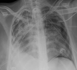
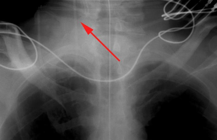
8 In the supine, intubated patient the first sign of barotrauma is often subcutaneous emphysema.
9
10
11
12
13
14
15

16

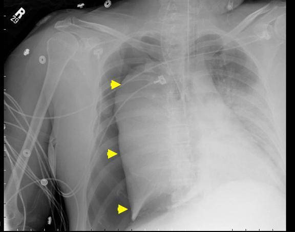
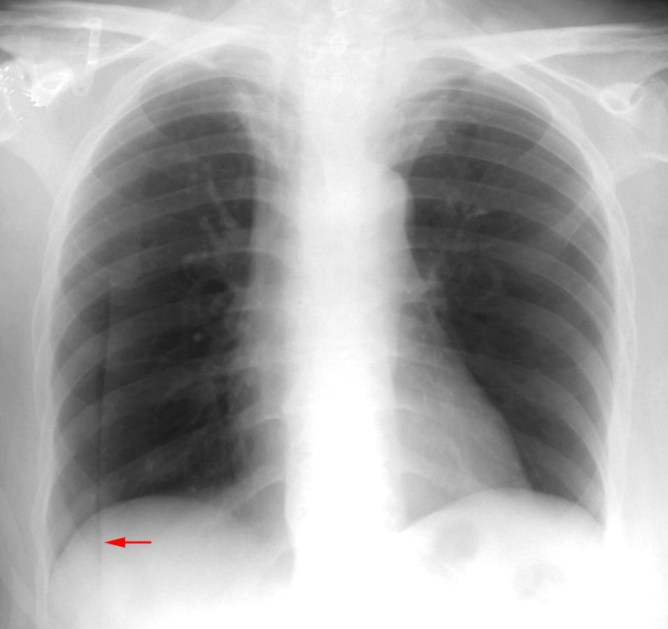
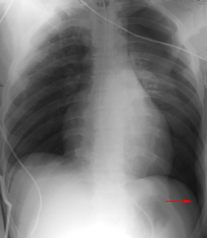
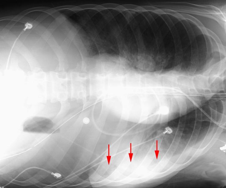
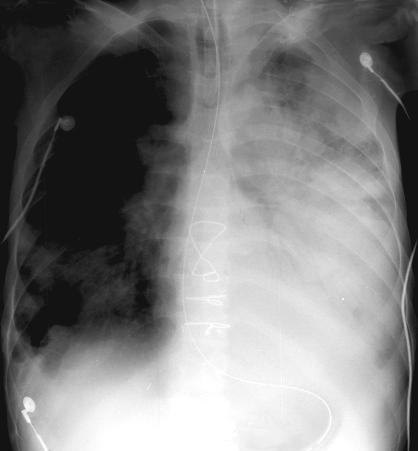
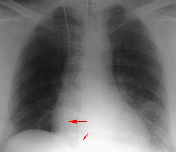
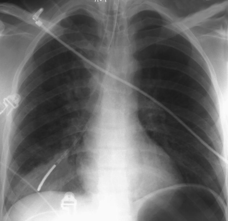
17 Tension Mediastinal shift is usually seen in a tension pneumothorax, but the use of PEEP may prevent this from occurring. The most reliable sign of tension pneumothorax is depression of a hemidiaphragm. Other signs of tension pneumothorax include shifting of the heart border, the superior vena cava, and the inferior vena cava. The shifting of these structures can lead to decreased venous return.
18
19
20 How much fluid must accumulate before you expect to see changes in the supine patient's chest x-ray? 5 ml 50 ml > 500 ml

21 How much fluid must accumulate before you expect to see changes in the supine patient's chest x-ray? 5 ml 50 ml > 500 ml
22
23
24 How much fluid must collect before costophrenic blunting is visible in the erect patient? ml ml ml > 500 ml
25 How much fluid must collect before costophrenic blunting is visible in the erect patient? ml ml ml > 500 ml
26
27
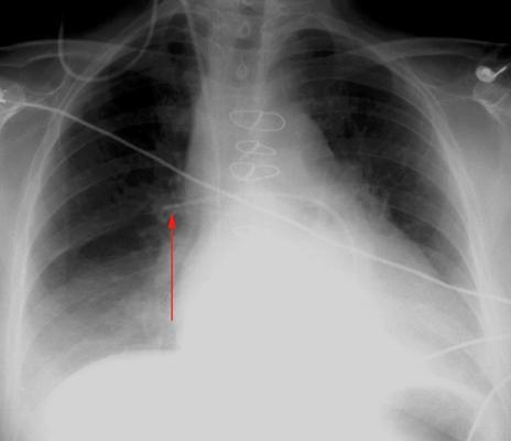
28 The rate of serious malposition of endotracheal tubes has been reported to be between 12-15%. Given that frequency, daily chest x-rays in intubated patients are recommended (maybe). The ideal position for endotracheal tubes is in the midtrachea, 5 cm from the carina, when the head is neither flexed nor extended. This allows for movement of the tip with head movements. The minimal safe distance from the carina is 2 cm.
29

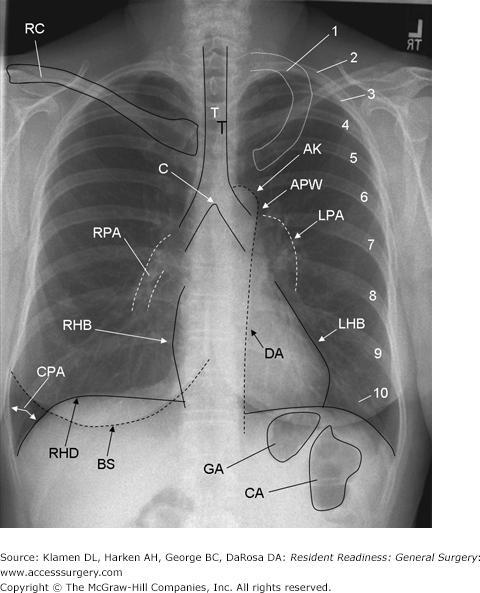
30 95% of patients' carinas project over the T5, T6, or T7 vertebral bodies.
31
32
33
34

35
36
37

38
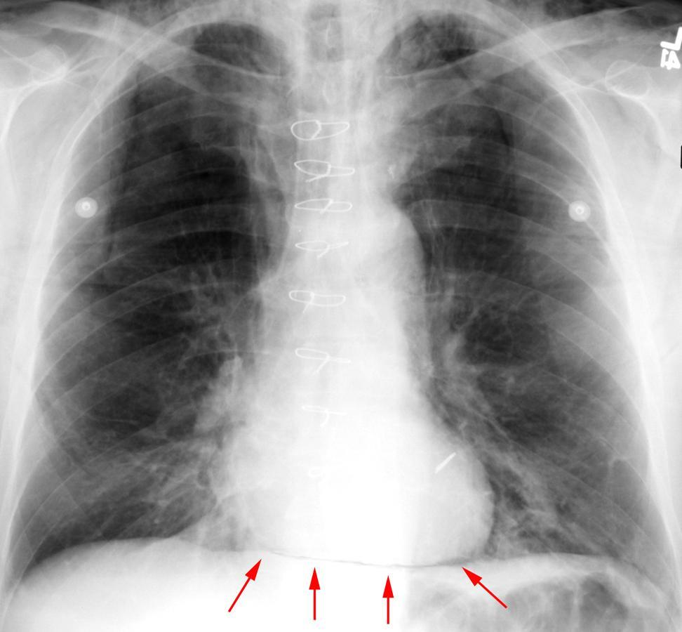
39
40

41

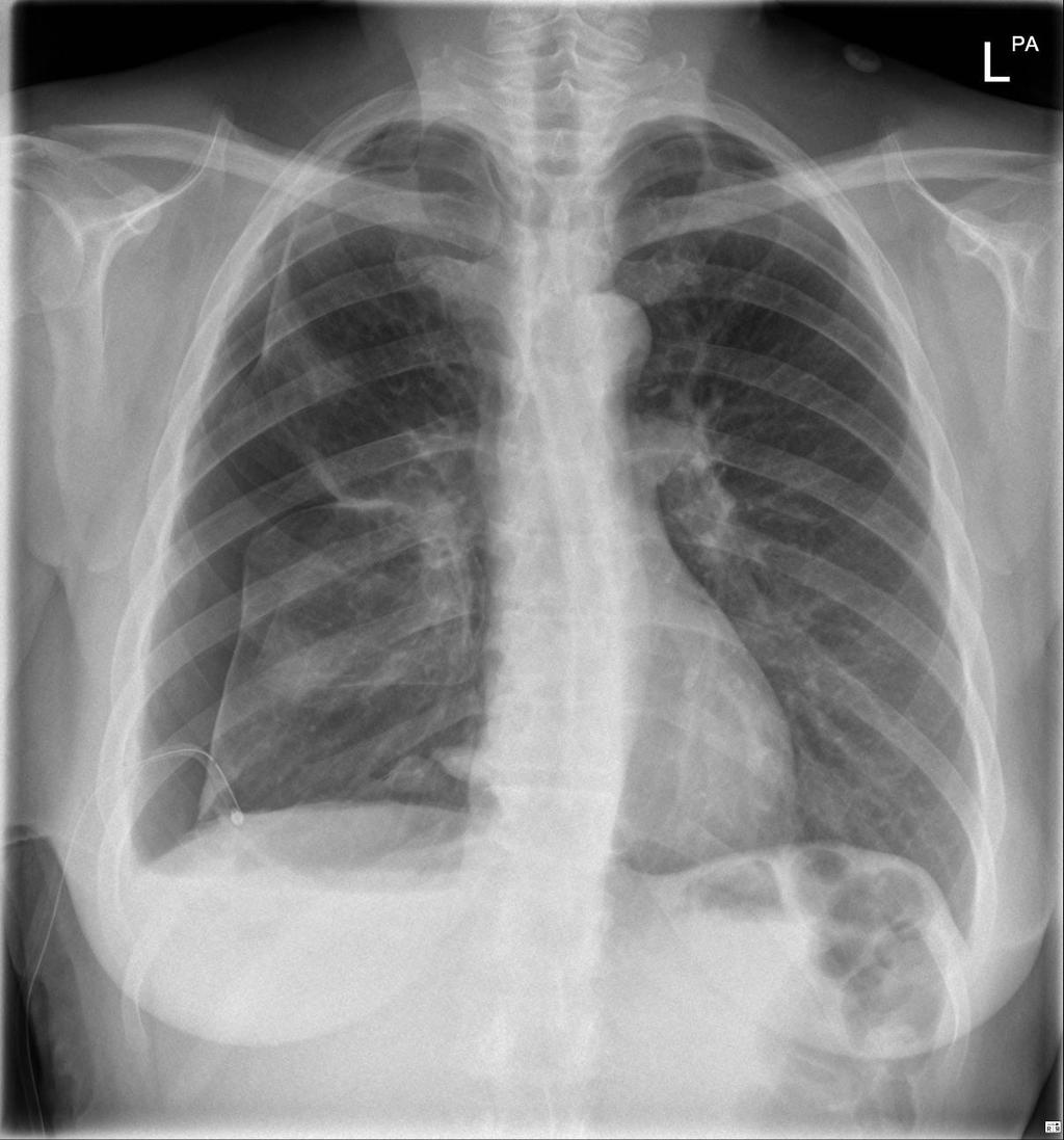
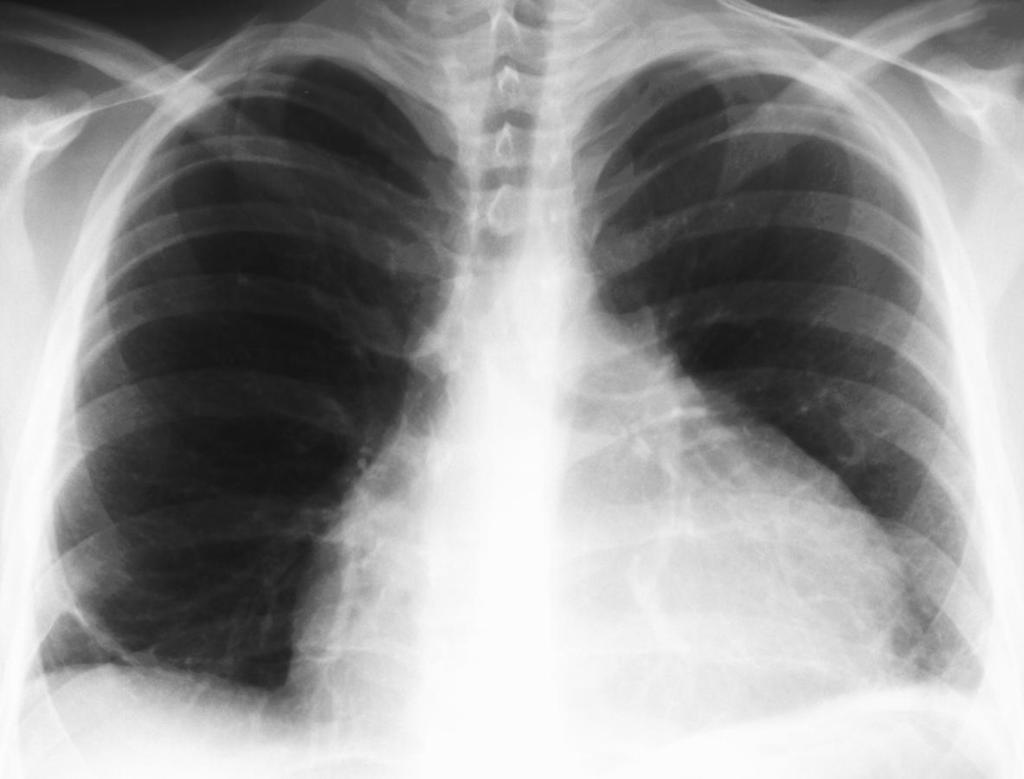
42 No cardiomegaly No pleural effusions No Kerley B-lines
43

44

45 A retrosternal crunch is sometimes auscultated. (Hamman s)

46 Post pericardiocentesis Continuous diaphragm sign
47
48

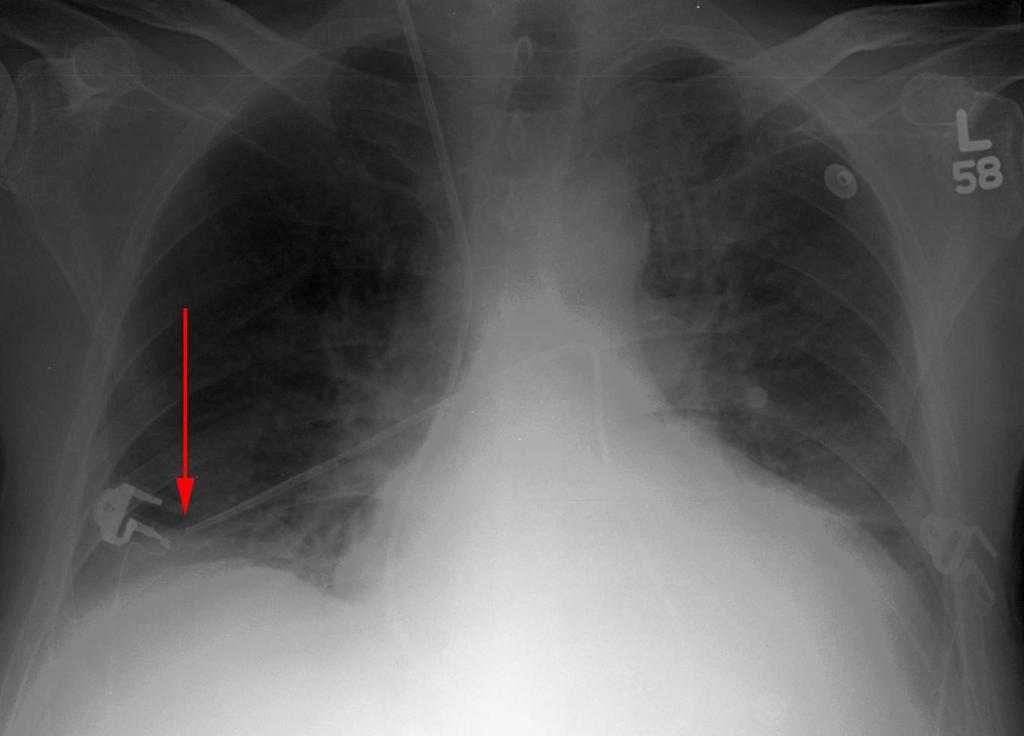
49 The catheter should be in the region of the aortic isthmus or left main bronchus and above the origins of the celiac trunk and superior mesenteric artery
50
51 1. Which of the following statements is true? -Most ICU chest films demonstrate significant findings. -ICU chest films are usually taken in the Posteroanterior (PA) view. -ICU chest films are both sensitive and specific. -The target to film distance is unimportant. 2. On which one or more of the following patients do daily portable x-rays need to be taken? -75 year old woman with a left subclavian central line. -Intubated 51 year old male suffering from COPD. -24 year old man status post appendectomy with two peripheral IV lines. -89 year old woman in the ICU for severe congestive heart failure.
52 1. Which of the following statements is true? -Most ICU chest films demonstrate significant findings. -ICU chest films are usually taken in the Posteroanterior (PA) view. -ICU chest films are both sensitive and specific. -The target to film distance is unimportant. 2. On which one or more of the following patients do daily portable x-rays need to be taken? -75 year old woman with a left subclavian central line. -Intubated 51 year old male suffering from COPD. -24 year old man status post appendectomy with two peripheral IV lines. -89 year old woman in the ICU for severe congestive heart failure.

53 Quiz What are the other major abnormalities? Is the TLC position correct?
54 4. Ambient light can reduce the detectability of lesions on chest film. -True -False 5. Supine positioning of the patient affects which one(s) of the following? -Cephalization of the pulmonary vasculature -The appearance of a pneumothorax -Widens mediastinum -The appearance of pneumonia
55 4. Ambient light can reduce the detectability of lesions on chest film. -True -False 5. Supine positioning of the patient affects which one(s) of the following? -Cephalization of the pulmonary vasculature -The appearance of a pneumothorax -Widens mediastinum -The appearance of pneumonia

56
4/16/2017. Learning Objectives. Interpretation of the Chest Radiograph. Components. Production of the Radiograph. Density & Appearance
 Interpretation of the Arthur Jones, EdD, RRT Learning Objectives Identify technical defects in chest radiographs Identify common radiographic abnormalities This Presentation is Approved for 1 CRCE Credit
Case 1. A 35-year-old male presented with fever, cough, and purulent sputum for one week. This was his CXR (Fig. 1.1). What is the diagnosis?
. What is the diagnosis?") 1 Interpreting Chest X-Rays CASE 1 Fig. 1.1 Case 1. A 35-year-old male presented with fever, cough, and purulent sputum for one week. This was his CXR (Fig. 1.1). What is the diagnosis? CASE 1 Interpreting
1 Interpreting Chest X-Rays CASE 1 Fig. 1.1 Case 1. A 35-year-old male presented with fever, cough, and purulent sputum for one week. This was his CXR (Fig. 1.1). What is the diagnosis? CASE 1 Interpreting
Lines and tubes. 1 Nasogastric tubes Endotracheal tubes Central lines Permanent pacemakers Chest drains...
 Lines and tubes 1 Nasogastric tubes... 15 2 Endotracheal tubes.... 19 3 Central lines... 21 4 Permanent pacemakers.... 25 5 Chest drains... 30 This page intentionally left blank 1 Nasogastric tubes Background
Shedding Light on Neonatal X-rays. Objectives. Indications for X-Rays 5/14/2018
 Shedding Light on Neonatal X-rays Barbara C. Mordue, MSN, NNP-BC Neonatal Nurse Practitioner LLUH Children s Hospital, NICU Objectives Utilize a systematic approach to neonatal x-ray interpretation Identify
Chest X rays and Case Studies. No disclosures. Outline 5/31/2018. Carlo Manalo, M.D. Department of Radiology Loma Linda University Children s Hospital
 Chest X rays and Case Studies Carlo Manalo, M.D. Department of Radiology Loma Linda University Children s Hospital No disclosures. Outline Importance of history Densities delineated on radiography An approach
Interpreting thoracic x-ray of the supine immobile patient: Syllabus
 Interpreting thoracic x-ray of the supine immobile patient: Syllabus Johannes Godt Dep. of Radiology and Nuclear Medicine Oslo University Hospital Ullevål NORDTER 2017, Helsinki Content - Why bedside chest
Chest X-ray Interpretation
 Chest X-ray Interpretation Introduction Routinely obtained Pulmonary specialist consultation Inherent physical exam limitations Chest x-ray limitations Physical exam and chest x-ray provide compliment
10/17/2016. Nuts and Bolts of Thoracic Radiology. Objectives. Techniques
 Nuts and Bolts of Thoracic Radiology October 20, 2016 Carleen Risaliti Objectives Understand the basics of chest radiograph Develop a system for interpreting chest radiographs Correctly identify thoracic
X-Rays. Kunal D Patel Research Fellow IMM
 X-Rays Kunal D Patel Research Fellow IMM The 12-Steps } 1: Name 2: Date 3: Old films 4: What type of view(s) 5: Penetration } Pre-read 6: Inspiration 7: Rotation Quality Control 8: Angulation 9: Soft tissues
Chest X-ray (CXR) Interpretation Brent Burbridge, MD, FRCPC
 Interpretation Brent Burbridge, MD, FRCPC") Chest X-ray (CXR) Interpretation Brent Burbridge, MD, FRCPC An approach to reviewing a chest x-ray will create a foundation that will facilitate the detection of abnormalities. You should create your own
Chest X-ray (CXR) Interpretation Brent Burbridge, MD, FRCPC An approach to reviewing a chest x-ray will create a foundation that will facilitate the detection of abnormalities. You should create your own
PATIENT DATA EVALUATION AND RECOMMENDATION: IMAGING STUDIES
 PATIENT DATA EVALUATION AND RECOMMENDATION: IMAGING STUDIES Robert Harwood, MSA, RRT-NPS Objectives At the end of this presentation the student should be able to: Describe the indications of a chest radiograph.
Radiological Anatomy of Thorax. Dr. Jamila Elmedany & Prof. Saeed Abuel Makarem
 Radiological Anatomy of Thorax Dr. Jamila Elmedany & Prof. Saeed Abuel Makarem Indications for Chest x - A chest x-ray may be used to diagnose and plan treatment for various conditions, including: Diseases/Fractures
B-I-2 CARDIAC AND VASCULAR RADIOLOGY
 (YEARS 1 3) CURRICULUM FOR RADIOLOGY 13 B-I-2 CARDIAC AND VASCULAR RADIOLOGY KNOWLEDGE To describe the normal anatomy of the heart and vessels including the lymphatic system as demonstrated by radiographs,
Undergraduate Teaching
 Prof. James F Meaney Undergraduate Teaching Chest X-Ray Understanding the normal anatomical by reference to cross sectional imaging Radiology? It s FUN! Cryptic puzzle Sudoku (Minecraft?) It s completely
Elderly Man with Dyspnoea
 Asia Pacific Family Medicine, 2004; 3 (1-2): 46-50 RADIOLOGY SERIES Elderly Man with Dyspnoea Wei-Yang LIM Faculty of Medicine National University of Singapore, Singapore Wilfred CG PEH Singapore Health
X-Rays. Prepared by Prof.Dr. Magda Hassab Allah Assist.lecturer Marwa Al Hady
 X-Rays Prepared by Prof.Dr. Magda Hassab Allah Assist.lecturer Marwa Al Hady CHEST X-RAYS Normal Chest X-ray Comments on chest X ray includes examination of 1- Bony cage (ribs,clavicles &vertebral column
PIXHOOK/iSTOCK. 40 l Nursing2014 l January. Copyright 2014 Lippincott Williams & Wilkins. Unauthorized reproduction of this article is prohibited.
 PIXHOOK/iSTOCK 40 l Nursing2014 l January 2.3 ANCC CONTACT HOURS Chest X-ray interpretation NOT JUST BLACK AND WHITE By William Pezzotti, MSN, RN, ACNP-BC, CEN CHEST X-RAYS (CXRs) are one of the oldest
Disclosure. Clinical Chest Radiography Interpretation Part I
 Clinical Chest Radiography Interpretation Part I Anthony M. Angelow, PhD(c), MSN, ACNPC, AGACNP-BC, CEN Associate Lecturer, Fitzgerald Health Education Associates Clinical practice Division of Trauma Surgery
A Curriculum in Cardiothoracic Radiology for Medical Students, with Goals and Objectives 1
 Radiologic Education A Curriculum in Cardiothoracic Radiology for Medical Students, with Goals and Objectives 1 Jannette Collins, MD, MEd, Gautham P. Reddy, MD, Brian F. Mullan, MD, Hrudaya P. Nath, MD,
Introduction to Chest CT Interpretation. Objectives 8/28/2017
 Introduction to Chest CT Interpretation Deborah Stein ACNP BC, CCRN NP Education Specialist Department of Anesthesia and Critical Care Medicine August 28, 2017 Objectives Basic Principles Thoracic Anatomy
Neonatal Chest X-Ray Interpretation
 CHAPTER 7 Neonatal Chest X-Ray Interpretation Prof. Praveen Kumar Neonatal unit, Department of Pediatrics, PGIMER, Chandigarh Learning Objectives At the end of this session, you should be able to: 1. Schematically
Lecture 2: Clinical anatomy of thoracic cage and cavity II
 Lecture 2: Clinical anatomy of thoracic cage and cavity II Dr. Rehan Asad At the end of this session, the student should be able to: Identify and discuss clinical anatomy of mediastinum such as its deflection,
Introduction to Chest Radiography
 Introduction to Chest Radiography RSTH 366: DIAGNOSTIC TECHNIQUES Alan Alipoon BS, RCP, RRT Instructor Department of Cardiopulmonary Sciences 1 Introduction Discovered in 1895 by Wilhelm Roentgen Terminology
X-rays. Dr Will Dooley
 X-rays Dr Will Dooley Plan Chest X-Rays Abdominal X-Rays Exam approach Presentation skills EMQ EMQ- answers Chest X-Ray - Systematic Approach D R Details RIP Image Quality +/- OBVIOUS ABNORMALITY A B C
Lecturer: Ms DS Pillay ROOM 2P24 25 February 2013
 Lecturer: Ms DS Pillay ROOM 2P24 25 February 2013 Thoracic Wall Consists of thoracic cage Muscle Fascia Thoracic Cavity 3 Compartments of the Thorax (Great Vessels) (Heart) Superior thoracic aperture
Imaging of Thoracic Trauma: Tips and Traps. Arun C. Nachiappan, MD Associate Professor of Clinical Radiology University of Pennsylvania
 Imaging of Thoracic Trauma: Tips and Traps Arun C. Nachiappan, MD Associate Professor of Clinical Radiology University of Pennsylvania None Disclosures Objectives Describe blunt and penetrating traumatic
Objectives. What is a Chest X Ray? CXR Workshop. Definition (diagnostic tool/internal PE) Types. Cost
 Types. Cost") Objectives CAPA 2011 Christy Wilson, PA C Georgia Lung Associates Identify the radiographic landmarks on a chest radiograph Recognize identifiers of poor quality on the chest radiograph Outline an approach
Objectives CAPA 2011 Christy Wilson, PA C Georgia Lung Associates Identify the radiographic landmarks on a chest radiograph Recognize identifiers of poor quality on the chest radiograph Outline an approach
Approach to CXR. Terminology. 1.Identification. Greg Blecher SCH Respir Fellow. Correct patient Correct date and time Correct examination
 Approach to CXR Greg Blecher SCH Respir Fellow From Rob Posteraro http://home.earthlink.net/~rhpos/cxr_interpret.txt.html ; http://home.earthlink.net/~rhpos/cxr_main.txt.html) Approach to viewing Chest
UERMMMC Department of Radiology. Basic Chest Radiology
 UERMMMC Department of Radiology Basic Chest Radiology PHYSICS DENSITIES BONE SOFT TISSUES WATER FAT AIR TELEROENTGENOGRAM Criteria for an Ideal Chest Radiograph 1. Upright 2. Posteroanterior View 3. Full
Interactive Lecture. Lecture 7 - Interactive. Radiology of cardiorespiratory disease. Editing File. Done By. Color Coding Important Notes Extra
 Lecture 7 - Interactive 436 Teams Interactive Lecture Radiology of cardiorespiratory disease Done By Team Leaders: Khalid Alshehri Hanin Bashaikh Team Members: Ghaida Alsaeed Maha Alissa Nawwaf AlHarbi
Advances in MDCT of Thoracic Trauma
 Baltic Congress of Radiology, Riga 2010 Advances in MDCT of Thoracic Trauma Robert A. Novelline, MD Professor of Radiology, Harvard Medical School Director of Emergency Radiology, Massachusetts General
Children are not small adults Children are Not Small Adults Anatomic considerations Pliable bony & cartilagenous structures - Significant thoracic inj
 PEDIATRIC CHEST TRAUMA Children are not small adults Role of imaging Spectrum of injury Children are not small adults Children are Not Small Adults Anatomic considerations Pliable bony & cartilagenous
Learning Radiology: Recognizing the Basics. Text with Student Consult Online Access Code
 Learning Radiology: Recognizing the Basics. Text with Student Consult Online Access Code Herring, W ISBN-13: 9780323074445 Table of Contents 1. Recognizing Anything The "colorful" world of radiology A
Caroline Polley, BSN, RN, VA-BC Clinical Specialist BD
 Caroline Polley, BSN, RN, VA-BC Clinical Specialist BD Disclosures The speaker is a employee of BD. (Please consult BD product for any indications, contraindications, hazards, warnings, cautions and instructions
Case N 1. Anterior thoracic paint with increasing dyspnea for few days. No cough. Decrease of cardiac sounds. Courtesy Dr Van den Homberg-Tanzania
 Case N 1 Anterior thoracic paint with increasing dyspnea for few days. No cough. Decrease of cardiac sounds Courtesy Dr Van den Homberg-Tanzania Case N 1 Enlargment of cardiac silhouette. Notice the symetry
Radiological conference. Left upper lobe collapse. Citation Hong Kong Practitioner, 1998, v. 20 n. 9, p
 Title Radiological conference. Left upper lobe collapse Author(s) Wong, LLS; Peh, WCG Citation Hong Kong Practitioner, 1998, v. 20 n. 9, p. 513-517 Issued Date 1998 URL http://hdl.handle.net/10722/44672
Chest Roentgenography for Cardiovascular Evaluation
 34 Chest Roentgenography for Cardiovascular Evaluation ERIC MANHEIMER Definition and Basic Science These topics are presented in Chapter 48, Chest Roentgenography for Pulmonary Evaluation. Technique and
100 Chest X Rays for Study Group. by Dr. Suneet Khurana
 100 Chest X Rays for Study Group by Dr. Suneet Khurana Approach to - Chest X Ray (shadow of the viscera on a photographic plate) Gas appears Black Fat appears Dark Grey Water Appears as Light Grey Bone
Chapter 2 Cardiac Interpretation of Pediatric Chest X-Ray
 Chapter 2 Cardiac Interpretation of Pediatric Chest X-Ray Ra-id Abdulla and Douglas M. Luxenberg Key Facts The cardiac silhouette occupies 50 55% of the chest width on an anterior posterior chest X-ray
Thoracic Trauma The Spectrum
 Thoracic Trauma The Spectrum Joseph Mathew Consultant, s & Emergency dept. 2 Thoracic Trauma Responsible for 20-25% of all deaths attributed to trauma. Contributing cause of death in an additional 25%
Cardiac Radiography. Jared D. Christensen, M.D.
 Cardiac Radiography Jared D. Christensen, M.D. Cardiac radiography Jared D. Christensen, M.D. Overview Basic Concepts Technique Normal anatomy Cases Technique 3 Standard Views Posterior-Anterior (PA) Anterior-Posterior
The External Anatomy of the Lungs. Prof Oluwadiya KS
 The External Anatomy of the Lungs Prof Oluwadiya KS www.oluwadiya.com Introduction The lungs are the vital organs of respiration Their main function is to oxygenate the blood by bringing inspired air into
RADIOLOGY FOR FINALS THE ULTIMATE REVISION GUIDE
 RADIOLOGY FOR FINALS THE ULTIMATE REVISION GUIDE By Lina Fazlanie (4th year medical student) & Ian Bickle (Radiology SpR) This short focused revision guide features 20 of the most popular x-rays that are
A pictorial review of thoracic imaging of intensive care patients
 A pictorial review of thoracic imaging of intensive care patients Poster No.: C-1003 Congress: ECR 2010 Type: Educational Exhibit Topic: Chest Authors: E. Y. P. Lee, H. C. Mathias; Cardiff/UK Keywords:
J Somerville and V Grech. The chest x-ray in congenital heart disease 2. Images Paediatr Cardiol Jan-Mar; 12(1): 1 8.
: 1 8.") IMAGES in PAEDIATRIC CARDIOLOGY Images Paediatr Cardiol. 2010 PMCID: PMC3228330 The chest x-ray in congenital heart disease 2 J Somerville and V Grech Paediatric Department, Mater Dei Hospital, Malta Corresponding
IMAGES in PAEDIATRIC CARDIOLOGY Images Paediatr Cardiol. 2010 PMCID: PMC3228330 The chest x-ray in congenital heart disease 2 J Somerville and V Grech Paediatric Department, Mater Dei Hospital, Malta Corresponding
Dr. Weyrich G07: Superior and Posterior Mediastina. Reading: 1. Gray s Anatomy for Students, chapter 3
 Dr. Weyrich G07: Superior and Posterior Mediastina Reading: 1. Gray s Anatomy for Students, chapter 3 Objectives: 1. Subdivisions of mediastinum 2. Structures in Superior mediastinum 3. Structures in Posterior
Ultrasound in the ICU
 Ultrasound in the ICU Kristine E. W. Breyer, MD Assistant Professor Anesthesia & Critical Care Medicine UCSF DISCLOSURES: NONE Definition The Ultrasound Exam Types & Uses Training Clinical Examples Objectives
Focused Assessment Sonography of Trauma (FAST) Scanning Protocol
 Scanning Protocol") Focused Assessment Sonography of Trauma (FAST) Scanning Protocol Romolo Gaspari CHAPTER 3 GOAL OF THE FAST EXAM Demonstrate free fluid in abdomen, pleural space, or pericardial space. EMERGENCY ULTRASOUND
Focused Assessment Sonography of Trauma (FAST) Scanning Protocol Romolo Gaspari CHAPTER 3 GOAL OF THE FAST EXAM Demonstrate free fluid in abdomen, pleural space, or pericardial space. EMERGENCY ULTRASOUND
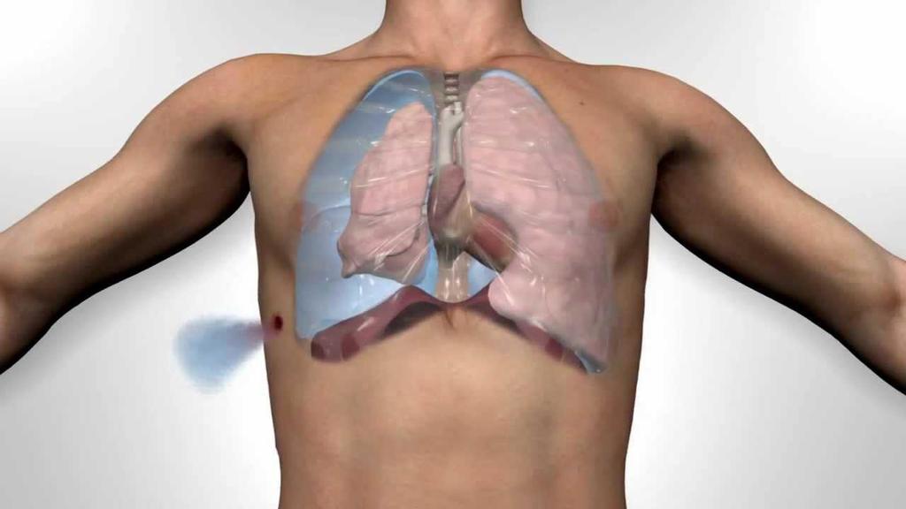
Pneumothorax. Defined as air in the pleural space which can occur through a number of mechanisms
 Pneumothorax Defined as air in the pleural space which can occur through a number of mechanisms Traumatic pneumothorax Penetrating chest trauma Common secondary to bullet or knife penetration Chest tube
TRACE A DROP OF BLOOD FROM RIGHT EAR TO LEFT OCULOMOTOR NERVE
 TRACE A DROP OF BLOOD FROM RIGHT EAR TO LEFT OCULOMOTOR NERVE KEY: TRACE A DROP OF BLOOD FROM RIGHT EAR TO LEFT OCULOMOTOR NERVE RIGHT EAR RIGHT ATRIUM LEFT SUBCLAVIAN ARTERY RIGHT EXTERNAL JUGULAR VEIN
Tubes and lines in neonatal chest radiograph
 Tubes and lines in neonatal chest radiograph Poster No.: C-1008 Congress: ECR 2014 Type: Educational Exhibit Authors: R. TUMMA, N. AHMED, V. Prasad ; Hyderabad/IN, 1 2 1 1 2 HYDERABAD, ANDHRA PRADESH/IN
Do you want to be an excellent Radiologist? - Focus on the thoracic aorta on lateral chest image!!!
 The lateral chest radiograph: Challenging area around the thoracic aorta!!! Do you want to be an excellent Radiologist? - Focus on the thoracic aorta on lateral chest image!!! Dong Yoon Han 1, So Youn
Introduction to Radiology for TB Nurses
 Introduction to Radiology for TB Nurses Juzar Ali, MD; FRCP(C); FCCP May 4, 2018 Essential Skills for the TB Nurse Case Manager Little Rock, AR May 3 4, 2017 Juzar Ali, MD; FRCP(C); FCCP has the following
FUNDAMENTALS OF CXR INTERPRETATION THE BASICS
 FUNDAMENTALS OF CXR INTERPRETATION THE BASICS PART I QUALITY ASSESSMENT 1 PATIENT-DEPENDENT FACTORS 3 REVIEW OF IMPORTANT ANATOMY 7 LUNGS AND PLEURA 11 DIAPHRAGMS 13 BONES AND SOFT TISSUES 14 A BRIEF LOOK
TB Radiology for Nurses Garold O. Minns, MD
 TB Nurse Case Management Salina, Kansas March 31-April 1, 2010 TB Radiology for Nurses Garold O. Minns, MD April 1, 2010 TB Radiology for Nurses Highway Patrol Training Center Salina, KS April 1, 2010
CT of Acute Thoracic Aortic Syndromes Stuart S. Sagel, M.D.
 CT of Acute Thoracic Aortic Syndromes Stuart S. Sagel, M.D. Thoracic Aortic Aneurysms Atherosclerotic Dissection Penetrating ulcer Mycotic Inflammatory (vasculitis) Traumatic Aortic Imaging Options Catheter
H. Mitchell Shulman MDCM FRCPC CSPQ Assistant Professor, Dept. of Surgery, McGill Medical School Attending Physician, Royal Victoria Hospital,
 H. Mitchell Shulman MDCM FRCPC CSPQ Assistant Professor, Dept. of Surgery, McGill Medical School Attending Physician, Royal Victoria Hospital, Montreal General Hospital, McGill University Health Center
pulmonary and critical care pearls
 pulmonary and critical care pearls A 63-Year-Old Woman With Subcutaneous Emphysema Following Endotracheal Intubation* Bassam Hashem, MD; James K. Smith, MD, FCCP; and W. Bruce Davis, MD, FCCP (CHEST 2005;
Shades of Gray Interpretation of Perioperative Imaging
 Stanford Hospital and Clinics DEPARTMENT OF CARDIOTHORACIC SURGERY-THORACIC AORTIC SURGERY UNIT FALK CARDIOVASCULAR RESEARCH CENTER STANFORD, CALIFORNIA 94305-5407 MICHAEL SHEEHAN, MSN, RNFA, NPC TELEPHONE
Radiology of the respiratory disease
 Radiology of the respiratory disease [ Color index: Important Notes Extra ] [ Editing file Feedback Share your notes Shared notes ] Resources: - 435 Slides - 434 Team - 435 Notes Done by: - Mai Alageel
INDEPENDENT LUNG VENTILATION
 INDEPENDENT LUNG VENTILATION Giuseppe A. Marraro, MD Director Anaesthesia and Intensive Care Department Paediatric Intensive Care Unit Fatebenefratelli and Ophthalmiatric Hospital Milan, Italy gmarraro@picu.it
Chest and cardiovascular
 Module 1 Chest and cardiovascular A. Doss and M. J. Bull 1. Regarding the imaging modalities of the chest: High resolution computed tomography (HRCT) uses a slice thickness of 4 6 mm to identify mass lesions
A - Airway. Check patient details - First name, surname, date of birth.
 How to read a chest x-ray a step by step approach SSppeecci ificc Raaddi iool looggi iccaal l Chheecckkl lisst t This article is an attempt to give the reader guidance how to read a chest x-ray. There
CHEST INJURY PULMONARY CONTUSION
 CHEST INJURY PULMONARY CONTUSION Introduction Pulmonary contusion refers to blunt traumatic lung parenchymal injury which results in oedema and haemorrhaging into alveolar spaces. It may also result in
Chest XRay interpretation INTERPRETATIONS Identifications: Name & Date Technical evaluation Basic Interpretations
 Chest XRay interpretation INTERPRETATIONS Identifications: Name & Date Technical evaluation Basic Interpretations TECHNICAL EVALUATION 1. Projection: AP/PA view To differentiate between AP & PA films,
Over the Wire Technique vs. Modified Seldinger Technique in Insertion of PICC
 Over the Wire Technique vs. Modified Seldinger Technique in Insertion of PICC Deniz Kasikci Department of Radiology, Jena University Hospital Friedrich-Schiller-University, Jena, Germany Disclosure Speaker
BELLWORK page 343. Apnea Dyspnea Hypoxia pneumo pulmonary Remember the structures of the respiratory system 1
 BELLWORK page 343 Apnea Dyspnea Hypoxia pneumo pulmonary respiratory system 1 STANDARDS 42) Review case studies that involve persons with respiratory disorders, diseases, or syndromes. Citing information
Tubes, Lines & Drains: Using chest radiography to assess support devices
 Tubes, Lines & Drains: Using chest radiography to assess support devices Ryan Pouliot, HMS Gillian Lieberman, M.D. Core Clerkship in Radiology May 2010 Objectives To understand the importance & develop
Ultrasound. FAST Focused Assessment with Sonography in Trauma
 Ultrasound FAST Focused Assessment with Sonography in Trauma Rohit Patel, MD University of Florida Health Director, Critical Care Ultrasound Surgical ICU Center for Intensive Care Gainesville, Florida
Interpretation of the chest radiograph Elizabeth Puddy MB ChB FCARCSI Catherine Hill MB ChB MRCP FRCR
 Interpretation of the chest radiograph Elizabeth Puddy MB ChB FCARCSI Catherine Hill MB ChB MRCP FRCR The traditional technique used in the acquisition and development of a chest radiograph uses methods
Large veins of the thorax Brachiocephalic veins
 Large veins of the thorax Brachiocephalic veins Right brachiocephalic vein: formed at the root of the neck by the union of the right subclavian & the right internal jugular veins. Left brachiocephalic
Tracheal stenosis in infants and children is typically characterized
 Slide Tracheoplasty for Congenital Tracheal Stenosis Peter B. Manning, MD Tracheal stenosis in infants and children is typically characterized by the presence of complete cartilaginous tracheal rings and
CHEST TRAUMA. Dr Naeem Zia FCPS,FACS,FRCS
 CHEST TRAUMA Dr Naeem Zia FCPS,FACS,FRCS Learning objectives Anatomy of chest wall and thoracic viscera Physiology of respiration and nerve pathways for pain Enumerate different thoracic conditions requiring
Dana Alrafaiah. - Moayyad Al-Shafei. -Mohammad H. Al-Mohtaseb. 1 P a g e
 - 6 - Dana Alrafaiah - Moayyad Al-Shafei -Mohammad H. Al-Mohtaseb 1 P a g e Quick recap: Both lungs have an apex, base, mediastinal and costal surfaces, anterior and posterior borders. The right lung,
Collaborative Stage. Site-Specific Instructions - LUNG
 Slide 1 Collaborative Stage Site-Specific Instructions - LUNG In this presentation, we are going to review the AJCC Cancer Staging criteria for the lung primary site. Slide 2 Reading Assignments As each
PATIENT CHARACTERISTICS AND PREOPERATIVE DATA (ecrf 1).
.") PATIENT CHARACTERISTICS AND PREOPERATIVE DATA (ecrf 1). 1 Inform Consent Date: / / dd / Mmm / yyyy 2 Patient identifier: Please enter the 6 digit Patient identification number from your site patient log
PATIENT CHARACTERISTICS AND PREOPERATIVE DATA (ecrf 1). 1 Inform Consent Date: / / dd / Mmm / yyyy 2 Patient identifier: Please enter the 6 digit Patient identification number from your site patient log
Research Article Can we predict the Position of Central Venous Catheter Tip Following Cannulation of Internal Jugular Vein?
 Cronicon OPEN ACCESS ANAESTHESIA Research Article Can we predict the Position of Central Venous Catheter Tip Following Cannulation of Internal Jugular Vein? Pradeep Marur Venkategowda 1, Surath Manimala
EMERGENCIES IN THE CHEST. Michal Kozub
 EMERGENCIES IN THE CHEST Michal Kozub CHEST IMAGING CHEST IMAGING 1.Consolidation - any pathologic process that fills the alveoli with fluid, pus, blood, cells (including tumor cells) or other substances
Mediastinum It is a thick movable partition between the two pleural sacs & lungs. It contains all the structures which lie
 Dr Jamila EL medany OBJECTIVES At the end of the lecture, students should be able to: Define the Mediastinum. Differentiate between the divisions of the mediastinum. List the boundaries and contents of
Pitfalls in Shortness of Breath
 Pitfalls in Shortness of Breath Stuart Swadron, MD FRCPC FACEP Vice-Chair of Education and Program Director Department of Emergency Medicine Los Angeles County-University of Southern California Medical
SWISS SOCIETY OF NEONATOLOGY. Selective bronchial intubation for unilateral PIE
 SWISS SOCIETY OF NEONATOLOGY Selective bronchial intubation for unilateral PIE October 2000 2 Berger TM, Jöhr M, Neonatal and Pediatric Intensive Care Unit (BTM), Kinderspital Luzern, Department of Anesthesiology
Lung Cancer - Suspected
 Lung Cancer - Suspected Shared Decision Making Lung Cancer: http://www.enhertsccg.nhs.uk/ Patient presents with abnormal CXR Lung cancer - clinical presentation History and Examination Incidental finding
Sectional Anatomy Quiz - III
 Sectional Anatomy - III Rashid Hashmi * Rural Clinical School, University of New South Wales (UNSW), Wagga Wagga, NSW, Australia A R T I C L E I N F O Article type: Article history: Received: 30 Jun 2018
Lung & Pleura. The Topics :
 Lung & Pleura The Topics : The Trachea. The Bronchi. The Brochopulmonary Segments. The Lungs. The Hilum. The Pleura. The Surface Anatomy Of The Lung & Pleura. The Root & Hilum. - first of all, the lung
Top Tips for Pleural Disease in 2012
 Top Tips for Pleural Disease in 2012 The unilateral pleural effusion on the Post Take Ward Round Pleural Effusion on CXR Bedside ultrasound + Pleural aspirate Empyema Nil evidence infection Admit IV antibiotics
Manage TB Dr. A. Chitrakumar Madras Medical College and RGGGH Institute of Thoracic Medicine, Chennai
 Manage TB Dr. A. Chitrakumar Madras Medical College and RGGGH Institute of Thoracic Medicine, Chennai Lecture 16 Radiology in diagnosis of Tuberculosis Session 01 So, welcome to the session Radiology in
Critical Care Monitoring. Indications. Pleural Space. Chest Drainage. Chest Drainage. Potential space. Contains fluid lubricant
 Critical Care Monitoring Indications 1-2- 2 Pleural Space Potential space Contains fluid lubricant Can fill with air, blood, plasma, serum, lymph, pus 3 1 Pleural Space Problems when contain abnormal substances:
Trauma Activation 7/18/17
 Blunt Rupture of the Thoracic Duct after Severe Thoracic Trauma Samuel Brown, MD Trauma Activation 7/18/17 53 year old male, rear end MVC, exited vehicle and was struck by a semi truck. Denies LOC, complaints
Foundation in Critical Care Nursing. Airway / Respiratory / Workbook
 Foundation in Critical Care Nursing Airway / Respiratory / Workbook Airway Anatomy: Please label the following: Tongue Larynx Epiglottis Pharynx Trachea Vertebrae Oesophagus Where is the ET (endotracheal)
BOGOMOLETS NATIONAL MEDICAL UNIVERSITY DEPARTMENT OF HUMAN ANATOMY. Guidelines. Module 2 Topic of the lesson Aorta. Thoracic aorta.
 BOGOMOLETS NATIONAL MEDICAL UNIVERSITY DEPARTMENT OF HUMAN ANATOMY Guidelines Academic discipline HUMAN ANATOMY Module 2 Topic of the lesson Aorta. Thoracic aorta. Course 1 The number of hours 3 1. The
The Chest X-ray for Cardiologists
 Mayo Clinic & British Cardiovascular Society at the Royal College of Physicians, London : 21-23-October 2013 Cases-Controversies-Updates 2013 The Chest X-ray for Cardiologists Michael Rubens Royal Brompton
RS LAB EXAM - LABS : Anatomy Lab : - In the following model, num 6 is pointing at? Answer : middle lobe of right lung
 RS LAB EXAM Exam Details : - Num of qus : 60 qus - Duration : 60 min - LABS : Anatomy Lab : - In the following model, num 6 is pointing at? Answer : middle lobe of right lung - In the following model,
THE VESSELS OF BLOOD CIRCULATION
 THE VESSELS OF BLOOD CIRCULATION scientistcindy.com /the-vessels-of-blood-circulation.html NOTE: You should familiarize yourself with the anatomy of the heart and have a good understanding of the flow
CRITICAL THINKING QUESTIONS AND ANSWERS AND CYCLE 2 LAB EXAM TEMPLATE. There are two main mechanisms that work in conjunction to return the blood
 CRITICAL THINKING QUESTIONS AND ANSWERS AND CYCLE 2 LAB EXAM TEMPLATE There are two main mechanisms that work in conjunction to return the blood THE CARDIAC PUMP 1) The forward pull(vis a fronte) This
3 Circulatory Pathways
 40 Chapter 3 Circulatory Pathways Systemic Arteries -Arteries carry blood away from the heart to the various organs of the body. -The aorta is the longest artery in the body; it branches to give rise to
10/14/2018 Dr. Shatarat
 2018 Objectives To discuss mediastina and its boundaries To discuss and explain the contents of the superior mediastinum To describe the great veins of the superior mediastinum To describe the Arch of
Procedure: Chest Tube Placement (Tube Thoracostomy)
") Procedure: Chest Tube Placement (Tube Thoracostomy) Basic Information: The insertion and placement of a chest tube into the pleural cavity for the purpose of removing air, blood, purulent drainage, or
Procedure: Chest Tube Placement (Tube Thoracostomy) Basic Information: The insertion and placement of a chest tube into the pleural cavity for the purpose of removing air, blood, purulent drainage, or
Double Superior Vena Cava; A Benign Cause of Widened Mediastenum and Implication on Venous Central Access
 ISPUB.COM The Internet Journal of Endovascular Medicine Volume 2 Number 1 Double Superior Vena Cava; A Benign Cause of Widened Mediastenum and Implication on Venous H Enuh, A Patel, A Chaudry, K Diaz,
Anatomy notes-thorax.
 Anatomy notes-thorax. Thorax: the part extending from the root of the neck to the abdomen. Parts of the thorax: - Thoracic cage (bones). - Thoracic wall. - Thoracic cavity. ** The thoracic cavity is covered
disease, bronchopulmonary dysplasia, pulmonary hypoplasia and congenital diaphragmatic hernia.
 Neonatal Chest Imaging - What the Nurse Should Know Expires Monday, April 30, 2018 Nursing Michael J. Diament, M.D. Objectives 1. Describe a good technique for positioning a neonate for the purpose of