Ultrasound. FAST Focused Assessment with Sonography in Trauma
|
|
|
- Doris Baldwin
- 6 years ago
- Views:
Transcription


1 Ultrasound FAST Focused Assessment with Sonography in Trauma Rohit Patel, MD University of Florida Health Director, Critical Care Ultrasound Surgical ICU Center for Intensive Care Gainesville, Florida
2 A few points about didactic lectures Hands on instruction better explained in Bedside Videos Reading material important to cover aspects not discussed in this lecture portion Important to mix hands on Active Learning with the reading/didactic material to best learn ultrasound application
3 Introduction First used in Europe 1970's, ATLS since 1997, later incorporated into surgery and emergency medicine residency curriculums Objective: detect free fluid in pericardium and intraperitoneal in the setting of trauma Alternatives: CT, DPL, OR, Observation; ultrasound has higher specificity for therapeutic laparotomy than DPL Combination algorithmic approach seems best McKenney, Journal of Trauma, 2001
4 FAST and E-FAST FAST exam and pleural fluid assessment Coined at international consensus conference in 1996 to describe an integrated, goal directed, bedside examination to detect fluid FAST detects fluid as low as 100 ml but commonly cited as 250 to 620 ml Sensitivity 79% Specificity 99% Branney, SW. J Trauma Stengel, D. Radiology
5 History of FAST Focused abdominal sonography in trauma Focused assessment with sonography in trauma
6 Focused Questions Is there free fluid in the abdomen? Is there fluid in the pericardium? Extended FAST: Is there fluid in the thorax? Is there a pneumothorax?
7 Anatomy Right paracolic gutter - Morrison's pouch to pelvis Left paracolic gutter - not as deep as right and phrenocolic ligament blocks fluid movement Morrison's pouch (hepatorenal recess) - space between Glisson's capsule on liver and Gerota's fascia of kidney Splenorenal recess - between spleen and Gerota's fascia Rectovesicle pouch - pocket formed by reflection of peritoneum from rectum to bladder; pouch of Douglass in female


8 Probe selection and location Phased array 5 MHz or Abdominal probes (bigger footprint) Noble et al. Textbook. Manual of Emergency and Critical Care Ultrasound.


9 Right upper quadrant Noble et al. Textbook. Manual of Emergency and Critical Care Ultrasound.

10 Right upper quadrant Noble et al. Textbook. Manual of Emergency and Critical Care Ultrasound.
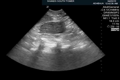
11 Kidney
12 Kidney
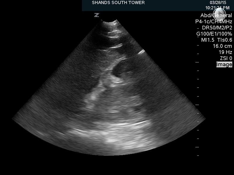
13 Diaphragm--> Kidney

14 Right upper quadrant Kidney diaphragm--> pleural fluid--> <--free fluid

15 Left upper quadrant More Posterior and Superior than RUQ Noble et al. Textbook. Manual of Emergency and Critical Care Ultrasound.
16 Left upper quadrant Noble et al. Textbook. Manual of Emergency and Critical Care Ultrasound.

17 Suprapubic Noble et al. Textbook. Manual of Emergency and Critical Care Ultrasound.
18 Suprapubic Noble et al. Textbook. Manual of Emergency and Critical Care Ultrasound.


19 The heart Noble et al. Textbook. Manual of Emergency and Critical Care Ultrasound.


20 Back to the pump

21 Pericardial fluid > Pleural fluid >
22 Pericardial fluid > Pericardial fluid >
23 Effusion around the pump Tamponade or not? Hallmark: RV free wall inversion, best recognized during diastole Right atrial inversion during systole (more common and early finding) Increased respiratory variation of mitral or aortic inflow velocities (greater than 25%) Dilated inferior vena cava with decreased inspiratory collapse ASE Committee Recomendations. Am Soc Echocardiography. 2005

24 Pericardial fluid >
25 Pericardial fluid > RV < Pericardial fluid >

26 FAST and e-fast If fluid found, move up one or two costal spaces, lung sometimes seen floating in pleural fluid Sensitivity 92% and Specificity 100% in detecting hemothorax in the Emergency Department, can detect 20 ml of pleural fluid VS supine CXR needs 175 ml Head slightly elevated can help accumulate fluid just above diaphragm McEwan K. Emerg Med J Sisley, AC. J Trauma. 1998
")
27 pleural fluid (could be hemothorax in trauma setting) >
28 Ultrasound for pneumothorax First described in a horse in 1986 In a normal lung, the visceral and parietal pleura are closely associated, and ultrasound shows shimmering or sliding at the pleural interface during respiration; absence of this indicates a pneumothorax In trauma, US shown to be more than twice as sensitive for detecting occult pneumothorax with similarly high specificity (98%) Comet tails are ultrasound artifacts that arise when ultrasound encounters a small air fluid interface Kirkpatrick, J Trauma 2004
29 Ultrasound for pneumothorax Chest radiography? US relies on fact that free air is lighter than normal aerated lung tissue, accumulates in nondependent areas of thoracic cavity Multiple studies show ultrasound to be more sensitive than supine chest radiography (CT gold standard) Sensitivity 86 to 100% Specificity 92 to 100% Negative predictive value of 100% (Lichtenstein study) Zhang study: sensitivity 86% vs 27% AND time to obtain study 2.3 minutes vs 19.9 minutes Zhang M. Crit Care Lichtenstein. Chest. 1995
30 Ultrasound for pneumothorax Supine High frequency linear array best Midclavicular line at third through fifth intercostal space to ID pleural line, but should look through several intercostal spaces Lung point: area where pneumothorax interfaces with chest wall Lichtenstein D. Intensive Care Med. 2000


31 Lung Sliding Parietal and visceral pleura can be seen sliding to each other Graphically depicted using M-mode Absence can also be seen in COPD bleb, consolidated pneumonia, atelectasis, main stem intubation

32 Pleural line with sliding >

33 Pleural line without sliding < optimal depth to evaluate is 3-7 cm >
34 Focused Questions Is there free fluid in the abdomen? Is there fluid in the pericardium? Extended FAST: Is there fluid in the thorax? Is there a pneumothorax?
Objectives. The Extended FAST Exam. Focused Assessment e With Sonography In. Trauma (FAST)
") Northern California Emergency Ultrasound Course Objectives The Extended FAST Exam Rimon Bengiamin, MD, RDMS UC SF Discuss the components of the EFAST exam Evaluate the utility of the EFAST Review how to
Northern California Emergency Ultrasound Course Objectives The Extended FAST Exam Rimon Bengiamin, MD, RDMS UC SF Discuss the components of the EFAST exam Evaluate the utility of the EFAST Review how to
Focused Assessment with Sonography in Trauma (FAST) UC Irvine School of Medicine
 UC Irvine School of Medicine") Focused Assessment with Sonography in Trauma (FAST) UC Irvine School of Medicine Purpose of FAST exam Quickly evaluate patient s status in emergency situations Blunt or penetrating trauma Visualize fluid
Focused Assessment with Sonography in Trauma (FAST) UC Irvine School of Medicine Purpose of FAST exam Quickly evaluate patient s status in emergency situations Blunt or penetrating trauma Visualize fluid
Background & Indications
 Teresa S. Wu, MD, FACEP Director, EM Ultrasound Program & Fellowship Co-Director, Simulation Based Training Program & Fellowship Maricopa Medical Center Simulation Curriculum Director Associate Professor,
A Practical Approach to Ultrasound Assessment of Respiratory Distress
 A Practical Approach to Ultrasound Assessment of Respiratory Distress Yanick Beaulieu, MD, FRCPC Director, Bedside Ultrasound Curriculum Division of Cardiology and Critical Care Hôpital du Sacré-Coeur
Lung ultrasound in the critically ill patient Pleural Effusions
 Lung ultrasound in the critically ill patient Pleural Effusions Rohit Patel, MD University of Florida Health Director, Critical Care Ultrasound Surgical ICU Center for Intensive Care Gainesville, Florida
Lung ultrasound in the critically ill patient BASICS
 Lung ultrasound in the critically ill patient BASICS Rohit Patel, MD University of Florida Health Director, Critical Care Ultrasound Surgical ICU Center for Intensive Care Gainesville, Florida Introduction
Extended FAST Exam. Goal of Trauma Care. Golden Hour of Trauma
 Extended FAST Exam Goal of Trauma Care Golden Hour of Trauma Best INITIAL screening modality in trauma efast 2014 LLSA Article (ACEP Policy Statement) Level B Recommendation: In hemodynamically unstable
Focused Assessment Sonography of Trauma (FAST) Scanning Protocol
 Scanning Protocol") Focused Assessment Sonography of Trauma (FAST) Scanning Protocol Romolo Gaspari CHAPTER 3 GOAL OF THE FAST EXAM Demonstrate free fluid in abdomen, pleural space, or pericardial space. EMERGENCY ULTRASOUND
Focused Assessment Sonography of Trauma (FAST) Scanning Protocol Romolo Gaspari CHAPTER 3 GOAL OF THE FAST EXAM Demonstrate free fluid in abdomen, pleural space, or pericardial space. EMERGENCY ULTRASOUND
Chest Ultrasound: Pneumothorax
 WINFOCUS BASIC ECHO (WBE) Chest Ultrasound: Pneumothorax Mark Hamlin, MD, MS Associate Professor of Anesthesiology and Surgery University of Vermont College of Medicine Co-Director of Surgical Critical
EFAST. Extended Focussed Assessment with Sonography for Trauma. Ultrasound Logbook. Name
 EFAST Extended Focussed Assessment with Sonography for Trauma Ultrasound Logbook ame Contents EFAST Accreditation Requirements 25 Abdominal Aorta Report Forms 3 Formative Assessments 1 Summative Assessment
FAST Focused Assessment with Sonography in Trauma
 FAST Focused Assessment with Sonography in Trauma Wilma Rodriguez Mojica,MD,FACR Professor of Radiology UPR School of Medicine Ultrasound Section - Radiological Sciences Department OBJECTIVES Understand
Certificate in Clinician Performed Ultrasound (CCPU) Syllabus. Extended Focussed Abdominal Scan for Trauma (E-FAST)
 Syllabus. Extended Focussed Abdominal Scan for Trauma (E-FAST)") Certificate in Clinician Performed Ultrasound (CCPU) Syllabus Extended Focussed Abdominal Scan for Trauma (E-FAST) Page 1 of 6 01/17 ACN 001 679 161 ABN 64 001 679 Extended Focussed Abdominal Scan for
Certificate in Clinician Performed Ultrasound (CCPU) Syllabus Extended Focussed Abdominal Scan for Trauma (E-FAST) Page 1 of 6 01/17 ACN 001 679 161 ABN 64 001 679 Extended Focussed Abdominal Scan for
Background Focused Assessment with Sonography in Trauma. Johann Baptist Dormagen, MD, PhD
 Focused Assessment with Sonography in Trauma Johann Baptist Dormagen, MD, PhD Unit of Abdominal and Oncologic Radiology Department of Radiology and Nuclear Medicine Oslo University Hospital, Norway 8 th
NON INVASIVE LIFE SAVERS. Ultrasound in PICU
 VOL 1 NO.1 Jan March 2014 54 Table 1. Selected Applications of Point-of-Care Ultrasonography, According to Medical Specialty. Specialty Ultrasound Applications Anesthesia Cardiology Guidance for vascular
Lung sonography in the diagnosis of pneumothorax.
 Lung sonography in the diagnosis of pneumothorax. Poster No.: C-0526 Congress: ECR 2011 Type: Educational Exhibit Authors: K. Stefanidis, K. Vintzilaios, D. D. Cokkinos, E. Antypa, S. Dimopoulos, S. Nanas,
POCUS for the Internist: Lungs & Pericardial Effusions
 POCUS for the Internist: Lungs & Pericardial Effusions Jeremy S. Boyd, MD, FACEP Asst. Professor of Emergency Medicine Vanderbilt University Medical Illustrations courtesy of Robinson Ferre, MD, FACEP
Abdominal Ultrasonography
 Abdominal Ultrasonography David A. Masneri, DO, FACEP, FAAEM Assistant Professor of Emergency Medicine Assistant Director, Emergency Medicine Residency Medical Director, Operational Medicine Division Center
Ultrasound basics Part 1
 Ultrasound basics Part 1 'Ultrasound enhanced critical care medicine' Rohit Patel, MD University of Florida Health Director, Critical Care Ultrasound Surgical ICU Center for Intensive Care Gainesville,
Contents& & & 1.! Ultrasound&basics& 1! 2.! Image&generation& 15!
 A l i n e press é % % % Contents& & & 1. Ultrasound&basics& 1 1.1. What,is,ultrasound?, 1 1.2. Ultrasound,probes,send,and,receive,ultrasound, 3 1.3. How,does,ultrasound,behave,travelling,through,tissue?,
A Frame of Reference for Anatomical Study. Anatomy and Physiology Mr. Knowles Chapter 1 Liberty Senior High School
 A Frame of Reference for Anatomical Study Anatomy and Physiology Mr. Knowles Chapter 1 Liberty Senior High School Anatomical Terms of Direction and Position Created for communicating the direction and
The Role of the FAST exam in the EDRU
 The Role of the FAST exam in the EDRU A. Robb McLean, MD, MHCM Vice Chair of Clinical Operations, Department of Emergency Medicine Joint Trauma Conference June 20, 2017 Disclosures Goals Describe the performance,
The Human Body: An Overview of Anatomy. Anatomy. Physiology. Anatomy - Study of internal and external body structures
 C H A P T E R 1 The Human Body: An Orientation An Overview of Anatomy Anatomy The study of the structure of the human body Physiology The study of body function Anatomy - Study of internal and external
Pediatric Lung Ultrasound (PLUS) In Diagnosis of Community Acquired Pneumonia (CAP)
 In Diagnosis of Community Acquired Pneumonia (CAP)") Pediatric Lung Ultrasound (PLUS) In Diagnosis of Community Acquired Pneumonia (CAP) Dr Neetu Talwar Senior Consultant, Pediatric Pulmonology Fortis Memorial Research Institute, Gurugram Study To compare
Pediatric Lung Ultrasound (PLUS) In Diagnosis of Community Acquired Pneumonia (CAP) Dr Neetu Talwar Senior Consultant, Pediatric Pulmonology Fortis Memorial Research Institute, Gurugram Study To compare
Pericardial Diseases. Smonporn Boonyaratavej, MD. Division of Cardiology, Department of Medicine Chulalongkorn University
 Pericardial Diseases Smonporn Boonyaratavej, MD Division of Cardiology, Department of Medicine Chulalongkorn University Cardiac Center, King Chulalongkorn Memorial Hospital 21 AUGUST 2016 Pericardial
Ultrasound in the ICU
 Ultrasound in the ICU Kristine E. W. Breyer, MD Assistant Professor Anesthesia & Critical Care Medicine UCSF DISCLOSURES: NONE Definition The Ultrasound Exam Types & Uses Training Clinical Examples Objectives
ORIGINAL ARTICLE. Role of Ultrasound in Evaluation of Undifferentiated Shock in ICU Settings
 Journal of The Association of Physicians of India Vol. 66 August 2018 13 Role of Ultrasound in Evaluation of Undifferentiated Shock in ICU Settings Tanvi Vaidya 1*, Pradeep D costa 2, Satish Pande 3 ORIGINAL
Basic of Ultrasound Physics E FAST & Renal Examination. Dr Muhammad Umer Ihsan MBBS,MD, DCH CCPU,DDU1,FACEM
 Basic of Ultrasound Physics E FAST & Renal Examination Dr Muhammad Umer Ihsan MBBS,MD, DCH CCPU,DDU1,FACEM What is Sound? Sound is Mechanical pressure waves What is Ultrasound? Ultrasounds are sound waves
Emergency Ultrasound. Case ECG. Potential diagnoses. An 80-year-old man in shock. ED patient with non traumatic undifferentiated hypotension
 Emergency Ultrasound An 80-year-old man in shock Case 80/M Present with chest pain Found low BP 62/44 at triage Resus. Room Pulse 86 Temp 35.6, SpO2 95% (RA) No history of drug overdose Past health PAF,
The FAST Exam! Dr. David Easton MD FRCPC Critical Care and Emergency Medicine University of Manitoba Canada
 The FAST Exam! Dr. David Easton MD FRCPC Critical Care and Emergency Medicine University of Manitoba Canada Dr. David Easton MD FRCPC Assistant Professor Section of Critical Care and Emergency Medicine
Focused Assessment With Sonography for Trauma Examination Reexamining the Importance of the Left Upper Quadrant View
 ORIGINAL RESEARCH Focused Assessment With Sonography for Trauma Examination Reexamining the Importance of the Left Upper Quadrant View Kathleen M. O Brien, MD, Lori A. Stolz, MD, Richard Amini, MD, Austin
AAENP US WORKSHOP 2/25/17
 Know the components of the Rapid Ultrasound for Shock & Hypotension & Extended Focused Assessment Sonography in Trauma & how they can help quickly determine diagnosis. Be comfortable obtaining and interpreting
Caudal Edge of the Liver in the Right Upper Quadrant (RUQ) View Is the Most Sensitive Area for Free Fluid on the FAST Exam
 View Is the Most Sensitive Area for Free Fluid on the FAST Exam") Original Research Caudal Edge of the Liver in the Right Upper Quadrant (RUQ) View Is the Most Sensitive Area for Free Fluid on the FAST Exam Viveta Lobo, MD* Michelle Hunter-Behrend, MD* Erin Cullnan,
Original Research Caudal Edge of the Liver in the Right Upper Quadrant (RUQ) View Is the Most Sensitive Area for Free Fluid on the FAST Exam Viveta Lobo, MD* Michelle Hunter-Behrend, MD* Erin Cullnan,
This appendix was part of the submitted manuscript and has been peer reviewed. It is posted as supplied by the authors.
 This appendix was part of the submitted manuscript and has been peer reviewed. It is posted as supplied by the authors. - Figure S1: The four quadrant approach lung ultrasound at the bedside. * The anterolateral
Small animal point of care ultrasound techniques
 Small animal point of care ultrasound techniques The role of veterinary point of care ultrasound in determining the presence or absence of specific pathologies is examined by Jantina McMurray DVM; Søren
Pulmonary Ultrasound in Emergency Medicine and Critical Care
 Pulmonary Ultrasound in Emergency Medicine and Critical Care www.rmgultrasound.com Author: Virginia M Stewart, MD RDMS RDCS RDMSK Dr Stewart is a practicing Emergency Physician in Eastern Virginia, USA.
B-I-2 CARDIAC AND VASCULAR RADIOLOGY
 (YEARS 1 3) CURRICULUM FOR RADIOLOGY 13 B-I-2 CARDIAC AND VASCULAR RADIOLOGY KNOWLEDGE To describe the normal anatomy of the heart and vessels including the lymphatic system as demonstrated by radiographs,
Internal Injury Documentation Guidelines
 Internal Injury Documentation Guidelines General Open Wound of Thorax Injury to Heart Identify episode of care Initial Subsequent Sequela Laterality Sequela of injury Place of occurrence of injury Activity
Advanced Imaging Practice CSB068
 Advanced Imaging Practice CSB068 Week 1 Peer Review - Evaluation of work by one or more people of similar competence to the producer - A form of self-regulation about improving quality and upholding standards
ASSESSING THE PLAIN ABDOMINAL RADIOGRAPH M A A M E F O S U A A M P O F O
 ASSESSING THE PLAIN ABDOMINAL RADIOGRAPH M A A M E F O S U A A M P O F O Introduction The abdomen (less formally called the belly, stomach, is that part of the body between the thorax (chest) and pelvis,
Point of Care Ultrasound (PoCUS)
") Point of Care Ultrasound (PoCUS) Competency Assessment Forms AORTA Competency A Focussed Assessment of the Aorta (AAA) Guidance Please follow this guidance as closely as possible to ensure consistency
Point of Care Ultrasound (PoCUS) Competency Assessment Forms AORTA Competency A Focussed Assessment of the Aorta (AAA) Guidance Please follow this guidance as closely as possible to ensure consistency
Point-of-Care Ultrasound Closer look at the Inferior Vena Cavae &
 Point-of-Care Ultrasound Closer look at the Inferior Vena Cavae & Brief Introduction to Gross Systolic Function Omar S. Darwish, MS, DO Certified in Point-of-Care Ultrasound Hospitalist University of California,
Peritoneum: Def. : It is a thin serous membrane that lines the walls of the abdominal and pelvic cavities and clothes the viscera.
 Peritoneum: Def. : It is a thin serous membrane that lines the walls of the abdominal and pelvic cavities and clothes the viscera. Layers of the peritoneum: 1. Outer Layer ( Parietal Peritoneum) : lines
Imaging of Thoracic Trauma: Tips and Traps. Arun C. Nachiappan, MD Associate Professor of Clinical Radiology University of Pennsylvania
 Imaging of Thoracic Trauma: Tips and Traps Arun C. Nachiappan, MD Associate Professor of Clinical Radiology University of Pennsylvania None Disclosures Objectives Describe blunt and penetrating traumatic
Ex. 1 :Language of Anatomy
 Collin College BIOL 2401 : Human Anatomy & Physiology Ex. 1 :Language of Anatomy The Anatomical Position Used as a reference point when referring to specific areas of the human body Body erect Head and
Unusual new signs of pneumothorax at lung ultrasound
 Unusual new signs of pneumothorax at lung ultrasound Volpicelli et al. Volpicelli et al. Critical Ultrasound Journal 2013, 5:10 Volpicelli et al. Critical Ultrasound Journal 2013, 5:10 SHORT COMMUNICATION
Certificate in Clinician Performed Ultrasound (CCPU) Syllabus. Lung
 Syllabus. Lung") Certificate in Clinician Performed Ultrasound (CCPU) Syllabus Lung Page 1 of 8 01/17 Lung Syllabus Purpose: This unit is designed to cover the theoretical and practical curriculum for lung ultrasound in
Certificate in Clinician Performed Ultrasound (CCPU) Syllabus Lung Page 1 of 8 01/17 Lung Syllabus Purpose: This unit is designed to cover the theoretical and practical curriculum for lung ultrasound in
STERNUM. Lies in the midline of the anterior chest wall It is a flat bone Divides into three parts:
 STERNUM Lies in the midline of the anterior chest wall It is a flat bone Divides into three parts: 1-Manubrium sterni 2-Body of the sternum 3- Xiphoid process The body of the sternum articulates above
Case 1. A 35-year-old male presented with fever, cough, and purulent sputum for one week. This was his CXR (Fig. 1.1). What is the diagnosis?
. What is the diagnosis?") 1 Interpreting Chest X-Rays CASE 1 Fig. 1.1 Case 1. A 35-year-old male presented with fever, cough, and purulent sputum for one week. This was his CXR (Fig. 1.1). What is the diagnosis? CASE 1 Interpreting
1 Interpreting Chest X-Rays CASE 1 Fig. 1.1 Case 1. A 35-year-old male presented with fever, cough, and purulent sputum for one week. This was his CXR (Fig. 1.1). What is the diagnosis? CASE 1 Interpreting
Bedside Sonographic Diagnosis of Pneumothorax in Pediatric Patients: A Preliminary Report Chia-Wang Tang 1, Kai-Sheng Hsieh 1 1
 ORIGINAL ARTICLE Bedside Sonographic Diagnosis of in Pediatric Patients: A Preliminary Report Chia-Wang Tang 1, Kai-Sheng Hsieh 1 1 Division of Pediatric Pulmonology, Department of Pediatrics, Kaohsiung
Thoracic Ultrasound: Pictorial review of pneumothorax, the fastest and easiest method to diagnose.
 Thoracic Ultrasound: Pictorial review of pneumothorax, the fastest and easiest method to diagnose. Poster No.: C-1588 Congress: ECR 2014 Type: Educational Exhibit Authors: J. A. Guirola, V. Mayoral Campos,
Certificate in Clinician Performed Ultrasound (CCPU) Syllabus. Lung
 Syllabus. Lung") Certificate in Clinician Performed Ultrasound (CCPU) Syllabus Lung Page 1 of 8 12/15 Lung Syllabus Purpose: This unit is designed to cover the theoretical and practical curriculum for lung ultrasound in
Certificate in Clinician Performed Ultrasound (CCPU) Syllabus Lung Page 1 of 8 12/15 Lung Syllabus Purpose: This unit is designed to cover the theoretical and practical curriculum for lung ultrasound in
Introduction. Chapter 1. Structure and Function. Introduction. Anatomy and Physiology Integrated. Anatomy and Physiology Integrated Anatomy
 Introduction Chapter 1 An Introduction to A&P Study strategies crucial for success Attend all lectures, labs, and study sessions Read your lecture and laboratory assignments before going to class or lab
Intro Case. Outline What We ll Cover. What we won t cover. Cardiac Ultrasound and The RUSH Exam: Bedside Ultrasound in Resuscitation and Shock
 Cardiac Ultrasound and The RUSH Exam: Bedside Ultrasound in Resuscitation and Shock Justin Davis, MD, MPH, RDMS Associate Physician Subchief for Emergency Ultrasound Services Kaiser Oakland Medical Center
Point-of-Care Ultrasound Guide for Landmarks, Recording, and Report Content. TJUH/MHD EM Ultrasound Division 2012
 Point-of-Care Ultrasound Guide for Landmarks, Recording, and Report Content TJUH/MHD EM Ultrasound Division 2012 Table of Contents 1 - Objectives 2 - Procedural 3 - AAA 4 - Abdominal OB 5 - Transvaginal
Ultrasound in critical care
 Stephen Wilson Bsc MBChB MRCP FRCA Andrew Mackay MBChB, FRCA, EDIC, FFICM Matrix Reference 2C01 Key points Focused ultrasound (US) studies can supplement physical examination of critically ill patients.
Chapter 13. Injuries to the Thorax and Abdomen
 Chapter 13 Injuries to the Thorax and Abdomen Anatomy Review Thoracic cage has 12 pairs of ribs. The first 7 pairs connect directly to sternum. Pairs 8 through 10 connect via common costal cartilage. Pairs
Dana Alrafaiah. - Moayyad Al-Shafei. -Mohammad H. Al-Mohtaseb. 1 P a g e
 - 6 - Dana Alrafaiah - Moayyad Al-Shafei -Mohammad H. Al-Mohtaseb 1 P a g e Quick recap: Both lungs have an apex, base, mediastinal and costal surfaces, anterior and posterior borders. The right lung,
Background & Indications Probe Selection
 Teresa S. Wu, MD, FACEP Director, EM Ultrasound Program & Fellowship Co-Director, Simulation Based Training Program & Fellowship Associate Program Director, EM Residency Program Maricopa Medical Center
Bedside ultrasound - Lung ultrasound in the Intensive Care Unit
 Bedside ultrasound - Lung ultrasound in the Intensive Care Unit Kishore K. Pichamuthu, Professor, Department of Critical Care, Christian Medical College, Vellore. Summary In an ICU setting, ultrasonographic
Certificate in Clinician Performed Ultrasound (CCPU) Syllabus. Lung
 Syllabus. Lung") Certificate in Clinician Performed Ultrasound (CCPU) Syllabus Lung ASUM Quality CCPU Syllabi Released: 21 March 2013 Approved by: CEO Lung Purpose: This unit is designed to cover the theoretical and practical
Certificate in Clinician Performed Ultrasound (CCPU) Syllabus Lung ASUM Quality CCPU Syllabi Released: 21 March 2013 Approved by: CEO Lung Purpose: This unit is designed to cover the theoretical and practical
This lab activity is aligned with Visible Body s A&P app. Learn more at visiblebody.com/professors
 1 This lab activity is aligned with Visible Body s A&P app. Learn more at visiblebody.com/professors 2 PRE-LAB EXERCISES: A. Watch the video 29.1 Heart Overview and make the following observations: 1.
Anatomy II ANAT 302. Course Description
 Anatomy II ANAT 302 Course Description This course provides the students with lectures and comprehensive overview of the gross anatomy of the components of the respiratory, cardiovascular, digestive and
Biology 218 Human Anatomy. Adapted from Martini Human Anatomy 7th ed. Chapter 1 Foundations: An Introduction to Anatomy
 Adapted from Martini Human Anatomy 7th ed. Chapter 1 Foundations: An Introduction to Anatomy Introduction Anatomy The study of external structures The study of internal structures The study of the relationship
Date Lab Pd. Lecture Notes (57)
") Name SECTION OBJECTIVES Describe the locations of the major body cavities List the organs located in each major body cavity Name the membranes associated with the thoracic and abdominopelvic cavities Name
Name SECTION OBJECTIVES Describe the locations of the major body cavities List the organs located in each major body cavity Name the membranes associated with the thoracic and abdominopelvic cavities Name
Introduction to The Human Body
 1 Introduction to The Human Body FOCUS: The human organism is often examined at seven structural levels: chemical, organelle, cell, tissue, organ, organ system, and the organism. Anatomy examines the structure
The Human Body: An Orientation
 The Human Body: An Orientation Body standing upright Anatomical Position feet slightly apart palms facing forward thumbs point away from body Directional Terms Superior and inferior toward and away from
ACTIVITY 9: BLOOD AND HEART BLOOD
 ACTIVITY 9: BLOOD AND HEART OBJECTIVES: 1) How to get ready: Read Chapters 21 & 22, McKinley et al., Human Anatomy, 4e. All text references are for this textbook. Read dissection instructions BEFORE YOU
FAST (Focused Assessment With Sonography in Trauma) Accurate for Cardiac and Intraperitoneal Injury in Penetrating Anterior Chest Trauma
 Accurate for Cardiac and Intraperitoneal Injury in Penetrating Anterior Chest Trauma") Article FAST (Focused Assessment With Sonography in Trauma) Accurate for Cardiac and Intraperitoneal Injury in Penetrating Anterior Chest Trauma Vivek S. Tayal, MD, Michael A. Beatty, MD, John A. Marx,
Article FAST (Focused Assessment With Sonography in Trauma) Accurate for Cardiac and Intraperitoneal Injury in Penetrating Anterior Chest Trauma Vivek S. Tayal, MD, Michael A. Beatty, MD, John A. Marx,
Lecturer: Ms DS Pillay ROOM 2P24 25 February 2013
 Lecturer: Ms DS Pillay ROOM 2P24 25 February 2013 Thoracic Wall Consists of thoracic cage Muscle Fascia Thoracic Cavity 3 Compartments of the Thorax (Great Vessels) (Heart) Superior thoracic aperture
Anatomical Terminology
 Anatomical Terminology Dr. A. Ebneshahidi Anatomy Anatomy : is the study of structures or body parts and their relationships to on another. Anatomy : Gross anatomy - macroscopic. Histology - microscopic.
Mousa Salah. Dr. Mohammad Al. Mohtasib. 1 P a g e
 8 Mousa Salah Dr. Mohammad Al. Mohtasib 1 P a g e In the previous lecture we talked about the peritoneum, and we said that the peritonium is a serous sac, and it consists of two layers, visceral and parietal.
Welcome to ANAT 10A! What is Anatomy? Different levels of Anatomy The Language of Anatomy Pearson Education, Inc.
 Welcome to ANAT 10A! What is Anatomy? Different levels of Anatomy The Language of Anatomy Introduction Anatomy means to dissect: (ANAT 10A) The study of internal & external body structures The study of
#1 - Chapter 1 - Anatomy. General Anatomical Terms The Anatomical Position
 #1 - Chapter 1 - Anatomy General Anatomical Terms The Anatomical Position The anatomical position is a stance in which a person stands erect with the feet flat on the floor and close together, arms at
Anatomy of the spleen. Oluwadiya KS
 Anatomy of the spleen Oluwadiya KS www.oluwadiya.com Introduction The spleen is an ovoid, usually purplish, pulpy mass about the size and shape of one's fist. It is the largest lymphoid tissue in the body
This is not a required assignment but it is recommended.
 SU 12 Name: This is not a required assignment but it is recommended. BIO 116 - Anatomy & Physiology II Practice Assignment 2 - The Respiratory and Cardiovascular Systems 1. The exchange of oxygen and carbon
Identification of lung sliding: a basic ultrasound technique with a steep learning curve
 SIGNA VITAE 2013; 8(1): 31-35 ORIGINAL Identification of lung sliding: a basic ultrasound technique with a steep learning curve MATEJ STRNAD SABINA ZADEL ZALIKA KLEMENC-KETIS MATEJ STRNAD ( ) SABINA ZADEL
LESSON 16. By Jeffrey R. Stowell, MD; Ross Kessler, MD; and Scott Lotz, MD. Reviewed by Joshua S. Broder, MD, FACEP
 Shock Waves Ultrasonographic Assessment of Shock LESSON 16 By Jeffrey R. Stowell, MD; Ross Kessler, MD; and Scott Lotz, MD Dr. Stowell is the associate director of the Adult Emergency Ultrasound Program
Archiving in Qpath Defining Adequate
 General Archiving Information for QPath Users As you become familiar with Qpath and how to archive your clips you will want to be sure you are capturing good quality clips for review. The properly captured,
THE DESCENDING THORACIC AORTA
 Intercostal Arteries and Veins Each intercostal space contains a large single posterior intercostal artery and two small anterior intercostal arteries. The anterior intercostal arteries of the lower spaces
Realistic simulation in Point of Care Ultrasound
 Realistic simulation in Point of Care Ultrasound 2 Preparing for PoCUS... BodyWorks Eve is an ultra-realistic female patient simulator designed for interactive and immersive Point of Care Ultrasound (PoCUS)
LAB 1: INTRODUCTION TO ANATOMY AND PHYSIOLOGY
 LAB 1: INTRODUCTION TO ANATOMY AND PHYSIOLOGY ANSWERS TO Pre- Lab Assignments Pre-Lab Activity 1: 1. b 2. a. 3 b. 7 c. 5 d. 6 e. 4 f. 1 g. 8 h. 2 i. 10 j. 9 3. a. frontal b. cervical c. antecubital d.
LIVING ANATOMY: IMPLICATIONS OF RESPIRATION CONVOCATION MARCH 16, 2019 PAMELA L. WILSON, D.O.
 LIVING ANATOMY: IMPLICATIONS OF RESPIRATION CONVOCATION MARCH 16, 2019 PAMELA L. WILSON, D.O. I believe you are taught anatomy in our school more thoroughly than any other school to date, because we want
Dr.Susan Maskel. BIO 105 Western Connecticut State University A&P I BIO 211 Naugatuck Valley Community College A&P I.
 1 Please wait 20 seconds before starting slide show. Mouse click to advance. Arrow keys etc.also work. Dr.Susan Maskel BIO 105 Western Connecticut State University A&P I BIO 211 Naugatuck Valley Community
Initially for cardiac echo Subsequent studies non-cardiac applications
 No disclosures But Heavy accent Initially for cardiac echo Subsequent studies non-cardiac applications 1973: Goldberg et al in JCUS 30 mediastinal masses in pts. age 1-84 yrs. 1977: Kangarloo et al in
Ultrasound-guided Aspiration of the Iatrogenic Pneumothorax Caused by Paravertebral Block
 Case Report Korean J Pain 2012 January; Vol. 25, No. 1: 33-37 pissn 2005-9159 eissn 2093-0569 http://dx.doi.org/10.3344/kjp.2012.25.1.33 Ultrasound-guided Aspiration of the Iatrogenic Pneumothorax Caused
Certificate in Clinician Performed Ultrasound (CCPU) Syllabus. Rapid Cardiac Echo (RCE)
 Syllabus. Rapid Cardiac Echo (RCE)") Certificate in Clinician Performed Ultrasound (CCPU) Syllabus Rapid Cardiac Echo (RCE) Purpose: Rapid Cardiac Echocardiography (RCE) This unit is designed to cover the theoretical and practical curriculum
Certificate in Clinician Performed Ultrasound (CCPU) Syllabus Rapid Cardiac Echo (RCE) Purpose: Rapid Cardiac Echocardiography (RCE) This unit is designed to cover the theoretical and practical curriculum
THE BASICS OF POINT-OF-CARE ULTRASOUND FOR THE GENERAL PEDIATRICIAN
 THE BASICS OF POINT-OF-CARE ULTRASOUND FOR THE GENERAL PEDIATRICIAN Table of Contents Section 1. Introduction and Knobology 2 Section 2. Pulmonary POC Ultrasound 8 Section 3. efast 14 Section 4. Skin/Soft
Guidelines, Policies and Statements D5 Statement on Abdominal Scanning
 Guidelines, Policies and Statements D5 Statement on Abdominal Scanning Disclaimer and Copyright The ASUM Standards of Practice Board have made every effort to ensure that this Guideline/Policy/Statement
Bi100 Chapter 1 Introduction to Human Anatomy and Physiology
 Bi100 Chapter 1 Introduction to Human Anatomy and Physiology Anatomy and Physiology A. Anatomy deals with the structure (morphology) of the body and its parts; in other words, what are things called? B.
2 Blunt Abdominal Trauma
 2 Blunt Abdominal Trauma Ricardo Ferrada, Diego Rivera, and Paula Ferrada Pearls and Pitfalls Patients suffering a high-energy trauma have solid viscera rupture in the abdomen and/or aortic rupture in
Breakout Session: Transesophageal Echocardiography
 Breakout Session: Transesophageal Echocardiography Doris Ockert, MD Andrew Schroeder, MD University of Wisconsin School of Medicine and Public Health Jutta Novalija, MD, PhD Medical College of Wisconsin
The faculty will include physicians with international reputations as outstanding ultrasound educators.
 Ultrasound Courses Course Description Whether you re a beginner or a seasoned sonographer, this year s AAEM pre-conference ultrasound course will be worth your time. We will be offering a half day course
OVERVIEW. Need for USG. Weaning assessment. Mechanics of USG. Pneumonia / VAP. Principles of lung USG. Prone position ventilation assessment
 OVERVIEW Need for USG Mechanics of USG Principles of lung USG BLUE protocol Alveolar syndrome Interstitial syndrome Weaning assessment Pneumonia / VAP Prone position ventilation assessment ETT positioning
(FAST) Peter Logan FRCS(Ed) FFAEM FACEM David Lewis FRCS FFAEM. Focused Assessment with Sonography for Trauma
 Peter Logan FRCS(Ed) FFAEM FACEM David Lewis FRCS FFAEM. Focused Assessment with Sonography for Trauma") Focused Assessment with Sonography for Trauma (FAST) Peter Logan FRCS(Ed) FFAEM FACEM David Lewis FRCS FFAEM 1 (FAST) Introduction Physical examination of the abdomen in blunt trauma is subjective and
Focused Assessment with Sonography for Trauma (FAST) Peter Logan FRCS(Ed) FFAEM FACEM David Lewis FRCS FFAEM 1 (FAST) Introduction Physical examination of the abdomen in blunt trauma is subjective and
ΚΑΡΔΙΟΛΟΓΟΣ EUROPEAN ACCREDITATION IN TRANSTHORACIC AND TRANSESOPHAGEAL ECHOCARDIOGRAPHY
 1 ΚΑΡΔΙΟΛΟΓΟΣ EUROPEAN ACCREDITATION IN TRANSTHORACIC AND TRANSESOPHAGEAL ECHOCARDIOGRAPHY 2 Constrictive pericarditis (CP) is characterized by impaired ventricular filling due to a stiffened or noncompliant
Courtesy of Dr. Susan Maskel Western Connecticut State University
 Courtesy of Dr. Susan Maskel Western Connecticut State University 1 2 SECTION vs. PLANE SECTION = a cut PLANE: when the section is made through the body wall or through an organ, it is made along an imaginary
BELLWORK page 343. Apnea Dyspnea Hypoxia pneumo pulmonary Remember the structures of the respiratory system 1
 BELLWORK page 343 Apnea Dyspnea Hypoxia pneumo pulmonary respiratory system 1 STANDARDS 42) Review case studies that involve persons with respiratory disorders, diseases, or syndromes. Citing information
Alexander A Schult, M.D., FCCP. October 21, 2017 Revised 1/10/18
 Alexander A Schult, M.D., FCCP October 21, 2017 Revised 1/10/18 Identifying normal anatomy Identifying various pathologic states Identifying placement of hardware Identifying limitations of portable CXR
THE THORACIC WALL. Boundaries Posteriorly by the thoracic part of the vertebral column. Anteriorly by the sternum and costal cartilages
 THE THORACIC WALL Boundaries Posteriorly by the thoracic part of the vertebral column Anteriorly by the sternum and costal cartilages Laterally by the ribs and intercostal spaces Superiorly by the suprapleural
Diagnostic Bedside Ultrasound for the Hospitalist
 Diagnostic Bedside Ultrasound for the Hospitalist Trevor Jensen MD MS Assistant Professor, UCSF Nima Afshar MD Associate Professor, UCSF Diagnostic Bedside Ultrasound AKA Point-of-Care Ultrasound (POCUS)