Ultrasound Guided Peripheral Intravenous Access
|
|
|
- Randell Douglas
- 5 years ago
- Views:
Transcription


1 Ultrasound Guided Peripheral Intravenous Access J. Christian Fox, MD, RDMS, FACEP, FAAEM, FAIUM Professor and Interim Chair of Emergency Medicine Director of Instructional Ultrasound University of California, Irvine School of Medicine

2 Ultrasound Guidance eadily visualizes vascular tructures rovides real-time eyes proves accuracy hortens time ecreases complications
3 Ultrasound Guided Procedures Vascular access Bladder aspiration Pericardiocentesis Nerve blocks Paracentesis Foreign bodies Thoracentesis Lumbar Puncture Abscess localization Fracture reduction
4 Ultrasound Guidance Decreases Complications Less arterial punctures Less pneumothoraces Less needle stick attempts Less needle redirects Increased Patient Comfort


5 Ultrasound guidance during central line insertion prevents complications Shojania KG, Duncan BW, McDonald KM, et al. Making health care safer: a critical analysis of patient safety practices. Evid Rep Technol Assess (Summ) 2001;(42):i-x,


6 Do we put in less central lines after implementation of an ultrasound-guided peripheral intravenous access program?
7 Ultrasound Guided Peripheral Lines - Methods Retrospective cohort searching the electronic patient database Total number of patients that had a CVC placed was recorded from 2006 to 2011 The rate of CVC placement was obtained by dividing that number by the total number of patients treated in the ED ED Residents and ED technicians (received 2 hours of training) were placing the peripheral lines Ann Emerg Med. Feb 2013
8 Ultrasound Guided Peripheral Lines - Results Overall rate of CVC decreased by 80%, from 0.81% in 2006 to 0.16% in 2011 Ward: decreased by 4.8% per month (95% CI 4.2% to 5.3%) Telemetry, decreased by 4.4% per month (95% CI 3.6% to 5.1%) Discharged home decreased by 7.6% ( 95% CI 6.2% to 9.1%) By the last year of the study, there were no CVC placed in patients directly discharged home from the ED Ann Emerg Med. Feb 2013
9 Ann Emerg Med. Feb 2013

10 Vessel Selection

11 Vessel Selection Superficial vs. deep veins Avoid using deep veins because they underlie more fascia and can contribute to compartment syndrome if infiltration occurs
12 Vessel Selection Looking with the ultrasound start at the AC and move down the arm, or start at the wrist (thumb side) and move up the arm Go as distal on the arm as possible so the next IV attempt can go above that site Likewise, avoid being below previously used sites


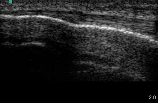
13 Soft and Hard Tissue Epidermis Hypodermis Subcutaneous fascia Muscle Bone


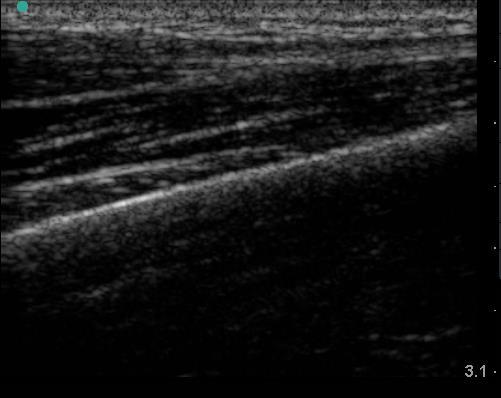
14 Tendons Readily identifiable by ultrasound dense connective tissue is more hyperechoic than muscle Fibrillar pattern Can be evaluated throughout range of motion


15 Muscle Imaging on Ultrasound Muscle appears hypoechoic Hyperechoic linear fibrillar striae seen within muscle Hyperechoic connective tissue surrounds muscle bundles Long-Axis View


16 Muscle Imaging on Ultrasound Muscle appears hypoechoic Hyperechoic punctate striae within muscle Bone appears very dense and echogenic Short-Axis View
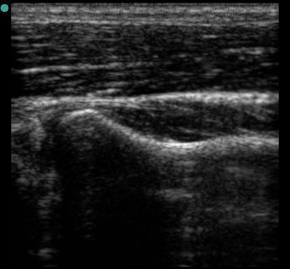
17 Bone

18 What do Nerves look like on Ultrasound? Move probe quickly back and forth along suspected nerve If it disappears after a short distance it's not a nerve If it bifurcates its likely a nerve

19 Discriminating Vascular Structures Color Doppler can easily discriminate vascular structures from other targets Holding the probe still will obviate pulsation in arteries, pressing with the probe will cause patent veins to collapse


20 Beam Angle B-Mode versus Doppler Perpendicular offers the best reflection for B-Mode Doppler B Mode


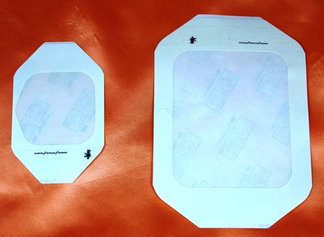
21 Cover the probe with a Tegaderm
 Gives 48+ hours of coverage vs.")
22 Prevent Infection Perform hand hygiene where PPE and always use Chlorhexidine to prep when starting the IV (in the IV start kit) Gives 48+ hours of coverage vs. <4 hours with alcohol alone

23 Proper Transducer Hand Placement Holding the probe correctly allows for better vein visualization Very lightly scan the patient to reduce vessel compression by holding probe with thumb and index finger (C Grip)


24 Apply Tourniquet

25 Angiocath Length Although a catheter is fully advanced, the entire catheter does not reside in the vessel. A portion of the catheter is left in the tissue and is required to reach the vessel. When using ultrasound we encourage the use of a longer IV (1 ¾ ) catheter so that >50% of the catheter is in the vessel post placement, this will help reduce infiltrations.

26 Peripheral Technique Requires use of longer angiocatheter


27 Indicator towards the procedure doer s left Going left to right under the probe will have the same effect on the screen

28 Short Axis Technique Locate vein in short axis Vein is easily compressible

29 Short Axis Technique As long as vessel is CENTERED on screen then it must be underneath CENTER of probe
30 Short Axis Technique As long as vessel is CENTERED on screen then it must be underneath CENTER of probe

31 Short Axis Technique As long as vessel is CENTERED on screen then it must be underneath CENTER of probe

32 Short Axis Technique As long as vessel is CENTERED on screen then it must be underneath CENTER of probe

33
34 Out of Plane Issue
35 In vs Out

36

37 Why Not Start in Long Axis Dr. Fox?
")
38 Long Axis (In Plane) Technique

39 In Plane Success Vein and needle both in-plane with ultrasound beam
40 In Plane Technique with Catheter Advancement Vein and needle both in-plane with ultrasound beam

41
42 Take Home Points Strong evidence in the literature confirms superiority of ultrasound guidance versus landmark approach Use linear high frequency transducer with sterile covering Use longer angiocatheter (1.75 inches) Aim probe indicator to operator s left Center vein under probe and aim needle for the center of the probe Troubleshoot difficult lines using in-plane technique
Bedside Ultrasound for DVT. Linear Probe. Leg Veins
 Bedside Ultrasound for DVT J. Christian Fox, MD, RDMS, FAAEM, FAIUM Director of Emergency Ultrasound Fellowship University of California, Irvine Jchrsitianfox@gmail.com Linear Probe High frequency transducer
Background & Indications Probe Selection
 Teresa S. Wu, MD, FACEP Director, EM Ultrasound Program & Fellowship Co-Director, Simulation Based Training Program & Fellowship Associate Program Director, EM Residency Program Maricopa Medical Center
Ultrasound Guidance Needle Techniques
 Ultrasound Guidance Needle Techniques Dr TANG Ho-ming AED/UCH USG Guidance Needle Techniques Commonly used in EM 1. Vessel cannulation-peripheral & central 2. Foreign body removal 3. Peripheral nerve/plexus
INTRODUCTION. Getting the best scan. Choosing a probe. Choosing the frequency
 Getting the best scan Choosing a probe Select the most appropriate probe for the particular scan required. s vary in their: operating frequency range higher ultrasound frequencies provide better discrimination
Sterile Technique & IJ/Femoral Return Demonstration
 Sterile Technique & IJ/Femoral Return Demonstration Sterile Technique Description: This is a return demonstration checklist used to evaluate participants in the simulated hands on skills portions for certification
Brachial plexus blockade within the interscalene groove involves local anesthetic
 Interscalene Brachial Plexus Block- How I do it. Part 1 of a 2 part discussion on technique. Stuart Grant Professor of Anesthesiology Duke University Medical Center Durham NC Brachial plexus blockade within
Ultrasound Guided Injections
 Ultrasound Guided Injection Technique More accurate injections Better Results! 1 Benefits: Increased Level of Certainty ie : really know how accurate PRP/Prolotherapy Avoid damage to articular cartilage
Background & Indications Probe Selection
 Teresa S. Wu, MD, FACEP Director, EM Ultrasound Program & Fellowship Co-Director, Simulation Based Training Program & Fellowship Associate Program Director, EM Residency Program Maricopa Medical Center
Principles of Ultrasound. Cara C. Prideaux, M.D. University of Utah PM&R Sports Medicine Fellow March 14, 2012
 Principles of Ultrasound Cara C. Prideaux, M.D. University of Utah PM&R Sports Medicine Fellow March 14, 2012 None Disclosures Outline Introduction Benefits and Limitations of US Ultrasound (US) Physics
Index. Note: Page numbers of article titles are in boldface type.
 Note: Page numbers of article titles are in boldface type. A Abscess(es) localization of for incision and drainage ultrasound-guided, 288 290 musculoskeletal sonographic appearance of, 256 periorbital
Abdominal Ultrasound
 Abdominal Ultrasound Imaging Control Buttons Depth The organ imaged should take up 3/4 of the screen Frequency = Penetration Use high frequencies (harmonics) for fluid filled and superficial structures
Central Venous Line Insertion
 Central Venous Line Insertion Understand the indications and risks of CVC insertion Understand and troubleshoot the seldinger technique Understand available sites and select the appropriate site for clinical
Ultrasound Guided Lower Extremity Blocks
 Ultrasound Guided Lower Extremity Blocks CONTENTS: 1. Femoral Nerve Block 2. Popliteal Nerve Block Updated December 2017 1 1. Femoral Nerve Block Indications Surgery involving the knee, anterior thigh,
Certificate in Clinician Performed Ultrasound (CCPU) Syllabus. Vascular Access (venous (peripheral and central) and arterial)
 Syllabus. Vascular Access (venous (peripheral and central) and arterial)") Certificate in Clinician Performed Ultrasound (CCPU) Syllabus Vascular Access (venous (peripheral and central) and arterial) Page 1 of 8 04/16 Vascular Access (venous (peripheral and central) and arterial)
Certificate in Clinician Performed Ultrasound (CCPU) Syllabus Vascular Access (venous (peripheral and central) and arterial) Page 1 of 8 04/16 Vascular Access (venous (peripheral and central) and arterial)
Terminology Tissue Appearance
 By Marc Nielsen, MD Advantages/Disadvantages Generation of Image Ultrasound Machine/Transducer selection Modes of Ultrasound Terminology Tissue Appearance Scanning Technique Real-time Portable No ionizing
Basics of US Regional Anaesthesia. November 2008
 Basics of US Regional Anaesthesia November 2008 Essential Physics HIGH frequency = great resolution but poor penetration LOW frequency = poor resolution but great penetration Potential Advantages of US
Arterial Line Insertion Pre Reading
 PROCEDURE ACCREDITATION THE CANBERRA HOSPITAL EMERGENCY DEPARTMENT Arterial Line Insertion Pre Reading Indications Requirement for continuous blood pressure monitoring (all patients on pressors, inotropes,
Imbibe Bone marrow aspiration needle. Operative technique
 Imbibe Bone marrow aspiration needle Operative technique Imbibe Bone Marrow Aspiration Needle Imbibe Bone marrow aspiration needle Contents 1. Smart design... 3 2. Operative technique... 4 Posterior iliac
ULTRASOUND GUIDED NERVE BLOCKS
 2 ULTRASOUND GUIDED NERVE BLOCKS Elizabeth Kwan, MD UCSF High Risk Emergency Medicine 2014 1 Instructors Kristin Berona Reza Danesh Sally Graglia Daniel Kievlan Starr Knight Allison Mulcahy Carmen Partida
High resolution ultrasound scanner for skin imaging
 High resolution ultrasound scanner for skin imaging Christine Turlat Sales Director Atys medical 17 Parc d Arbora 69510 SOUCIEU EN JARREST Atys company Principle of ultrasound imaging DERMCUP Normal image
3/20/2017. Disclosures. Ultrasound Fundamentals. Ultrasound Fundamentals. Bone Anatomy. Tissue Characteristics
 Disclosures Images of ultrasound equipment in this presentation are not an endorsement Fundamentals of Musculoskeletal Ultrasound Physics and Knobology Shane A. Shapiro, M.D. Assistant Professor Orthopedic
Ultrasound Guided Vascular Access. 7/25/2016
 Ultrasound Guided Vascular Access 7/25/2016 www.ezono.com 1 Objectives Indications for insertion of central and peripheral lines Complications associated with procedures Role of ultrasound in vascular
Successful IV Starts Revised February 2014
 Successful IV Starts Revised February 2014 Why Intravenous Therapy? Used for access to the body s circulation Indications: Administer fluids, blood, medications, and nutrition Obtain laboratory specimens
The Essentials Tissue Characterization and Knobology
 The Essentials Tissue Characterization and Knobology Randy E. Moore, DC, RDMS RMSK No relevant financial relationships Ultrasound The New Standard of Care Musculoskeletal sonography has become the standard
Ultrasonography of the Neck as an Adjunct to FNA. Nicole Massoll M.D.
 Ultrasonography of the Neck as an Adjunct to FNA Nicole Massoll M.D. Basic Features of Head and Neck Ultrasound and Anatomy Nicole Massoll M.D. University of Arkansas for Medical Sciences, Little Rock
WELCOME! Introduction to Bedside Ultrasound
 WELCOME! Introduction to Bedside Ultrasound TEACHERS University of California-Irvine School of Medicine Nathan Molina nathan.d.molina@gmail.com Trevor Plescia taplescia90@gmail.com Jack Silva jpsilva42@gmail.com
The role of ultrasound duplex in endovenous procedures
 The role of ultrasound duplex in endovenous procedures Neophytos A. Zambas MD, PhD Vascular Surgeon Polyclinic Ygia, Limassol, Cyprus ΚΕΑΕΧ ΚΥΠΡΙΑΚΗ ΕΤΑΙΡΕΙΑ ΑΓΓΕΙΑΚΗΣ ΚΑΙ ΕΝΔΑΓΓΕΙΑΚΗΣ ΧΕΙΡΟΥΡΓΙΚΗΣ Pre
USRA OF THE LOWER EXTREMITY
 USRA OF THE LOWER EXTREMITY Christian R. Falyar, CRNA, DNAP Department of Nurse Anesthesia Virginia Commonwealth University Disclosure Statement of Financial Interest I, Christian Falyar, DO NOT have a
Needle tip visualization during ultrasound-guided vascular access: short-axis vs long-axis approach
 American Journal of Emergency Medicine (2010) 28, 343 347 www.elsevier.com/locate/ajem Brief Report Needle tip visualization during ultrasound-guided vascular access: short-axis vs long-axis approach Michael
The Elbow 3/5/2015. The Elbow Scanning Sequence. * Anterior Joint (The anterior Pyramid ) * Lateral Epicondyle * Medial Epicondyle * Posterior Joint
 * Lateral Epicondyle * Medial Epicondyle * Posterior Joint") Scanning Sequence * Anterior Joint (The anterior Pyramid ) * Lateral Epicondyle * Medial Epicondyle * Posterior Joint Anterior Elbow Pyramid Courtesy of Jay Smith, MD. Vice chair PMR Mayo Clinic Rochester,
Scanning Sequence * Anterior Joint (The anterior Pyramid ) * Lateral Epicondyle * Medial Epicondyle * Posterior Joint Anterior Elbow Pyramid Courtesy of Jay Smith, MD. Vice chair PMR Mayo Clinic Rochester,
Ultrasound-guided supraclavicular block
 THE JOURNAL OF NEW YORK SCHOOL J u l y 2009 V o l u m e OF REGIONAL ANESTHESIA 1 3 Ultrasound-guided supraclavicular block Arthur Atchabahian, MD Department of Anesthesiology, St. Vincent Medical Center,
Needle visualization with ZONARE ultrasound systems
 Needle visualization with ZONARE ultrasound systems This material provides a general overview of ultrasound guided needle imaging and techniques and is not intended to replace formal training or education
Bedside Emergency Ultrasound For Deep Venous Thrombosis
 Bedside Emergency Ultrasound For Deep Venous Thrombosis Michael Blaivas, MD, MBA(candidate) FACEP, FAIUM Professor of Medicine University of South Carolina School of Medicine AIUM Third Vice President
CHAPTER 5 Femoral Nerve Block. Arun Nagdev, MD Mike Mallin, MD, RDCS, RDMS
 CHAPTER 5 Femoral Nerve Block Arun Nagdev, MD Mike Mallin, MD, RDCS, RDMS SECTION 1 Introduction An ultrasound-guided femoral nerve block (USFNB) can be a rapid and definitive tool for pain control for
Abdominal Ultrasonography
 Abdominal Ultrasonography David A. Masneri, DO, FACEP, FAAEM Assistant Professor of Emergency Medicine Assistant Director, Emergency Medicine Residency Medical Director, Operational Medicine Division Center
Clinical Protocols of the Anesthesiology Department at the Dartmouth-Hitchcock Medical Center: Techniques for lower extremity nerve blocks.
 Clinical Protocols of the Anesthesiology Department at the Dartmouth-Hitchcock Medical Center: Techniques for lower extremity nerve blocks. Authors from DHMC: Brian D. Sites, MD. Assistant Professor of
KINGSTON GENERAL HOSPITAL NURSING POLICY AND PROCEDURE
 KINGSTON GENERAL HOSPITAL NURSING POLICY AND PROCEDURE SUBJECT Sample (Adult): Advanced Competency (AC) for Nurses (Registered Nurses and Registered Practical Nurses) PAGE 1 of 5 ORIGINAL ISSUE 1985 January
Dr. prakruthi Dept. of anaesthesiology, Rrmch, bangalore
 CENTRAL VENOUS CATHETERIZATION Dr. prakruthi Dept. of anaesthesiology, Rrmch, bangalore OBJECTIVES Introduction Indications and Contraindications Complications Technique Basic principles Specifics by Site
Manual of Emergency and Critical Care Ultrasound
 Manual of Emergency and Critical Care Ultrasound Second Edition Manual of Emergency and Critical Care Ultrasound Second Edition Vicki E. Noble MD, RDMS, FACEP Director, Division of Emergency Ultrasound,
Point-of-Care Ultrasound Guide for Landmarks, Recording, and Report Content. TJUH/MHD EM Ultrasound Division 2012
 Point-of-Care Ultrasound Guide for Landmarks, Recording, and Report Content TJUH/MHD EM Ultrasound Division 2012 Table of Contents 1 - Objectives 2 - Procedural 3 - AAA 4 - Abdominal OB 5 - Transvaginal
We are IntechOpen, the world s leading publisher of Open Access books Built by scientists, for scientists. International authors and editors
 We are IntechOpen, the world s leading publisher of Open Access books Built by scientists, for scientists 4,000 116,000 120M Open access books available International authors and editors Downloads Our
FHS Appendicitis US Protocol
 FHS Appendicitis US Protocol Reviewed By: Shireen Khan, MD; Sarah Farley, MD; Anna Ellermeier, MD Last Reviewed: May 2018 Contact: (866) 761-4200 **NOTE for all examinations: 1. If documenting possible
Basic of Ultrasound Physics E FAST & Renal Examination. Dr Muhammad Umer Ihsan MBBS,MD, DCH CCPU,DDU1,FACEM
 Basic of Ultrasound Physics E FAST & Renal Examination Dr Muhammad Umer Ihsan MBBS,MD, DCH CCPU,DDU1,FACEM What is Sound? Sound is Mechanical pressure waves What is Ultrasound? Ultrasounds are sound waves
Ultrasound Guided Regional Nerve Blocks
 Ultrasound Guided Regional Nerve Blocks In the country of the blind the one eyed man is King -Deciderius Erasmus (1466-1536) Objectives Benefits of Regional Anesthesia Benefits of US guidance Role of ultrasound
For exam: VL DUPLEX EXTREMITY VEINS UNILAT LT
 For exam: VL DUPLEX EXTREMITY VEINS UNILAT LT - 8870390 METHOD/TECHNIQUE: The veins of the left upper extremity were studied at multiple For exam: VL DUPLEX EXTREMITY VEINS UNILAT RT - 8870400 METHOD/TECHNIQUE:
Learning Objectives. Ultrasound for the Primary Care Provider. Portable Ultrasound: Laptops, Tablets, Plug-in Probes, and Pocket devices
 Learning Objectives Ultrasound for the Primary Care Provider Richard Hoppmann, MD, FACP University of South Carolina School of Medicine Assess the main components and functions of a portable ultrasound
Musculoskeletal Ultrasound Fundamentals
 Fundamentals Benjamin D. Levine, M.D. Associate Professor of Radiology Musculoskeletal Imaging Dept. of Radiological Sciences UCLA Health System I. Image Optimization II. Image Interpretation Artifacts
Jefferson Tower Task Trainer List
 Jefferson Tower Task Trainer List Table of Contents Blue Phantom Ultrasound Central Line Training Model 2 Blue Phantom Femoral Vascular Access Training Model 3 Blue Phantom Thoracentesis Ultrasound Training
RadRx Your Prescription for Accurate Coding & Reimbursement Copyright All Rights Reserved.
 Interventional Radiology Coding Case Studies Prepared by Stacie L. Buck, RHIA, CCS-P, RCC, CIRCC, AAPC Fellow President & Senior Consultant Week of October 22, 2018 Paracentesis & Transjugular Liver Biopsy
Musculoskeletal Ultrasound: Basics, Utility, and Clinical Applications
 Musculoskeletal Ultrasound: Basics, Utility, and Clinical Applications Andrew Lavigne, MD, FRCPC Physical Medicine and Rehabilitation CSCN Diplomat (EMG) Dip Sport Medicine Eugene Maida, MD, PGY-4 Resident
MODULE 9 ARTERIAL AND VENOUS CATHETERIZATION. Robert B. McLafferty M.D. Southern Illinois University
 MODULE 9 ARTERIAL AND VENOUS CATHETERIZATION Robert B. McLafferty M.D. Southern Illinois University I. OBJECTIVES By the end of this laboratory session the residents should be able to A. Identify the anatomic
MODULE 2 THE LABORATORY RAT
 University Animal Care Committee LABORATORY ANIMAL BIOMETHODOLOGY WORKSHOP MODULE 2 THE LABORATORY RAT SUBSTANCE ADMINISTRATION AND BLOOD COLLECTION Substance Administration: Subcutaneous injection Intramuscular
L o o k L i s t e n F e e l S c a n. Your Pocus Cards For Your Every Day Scanning.
 L o o k L i s t e n F e e l S c a n Your Pocus Cards For Your Every Day Scanning E-FAST Extended Focused Assessment by Sonography in Trauma Subcostal Heart View Pleural Sliding on M-mode (Sea-shore sign)
ISUOG Basic Training Distinguishing between Normal & Abnormal Appearances of the Long Bones & Extremities
 ISUOG Distinguishing between Normal & Abnormal Appearances of the Long Bones & Extremities Learning objectives At the end of the lecture you will be able to: Describe how to obtain the planes required
Session 2: Ultrasonography for Primary Care Clinicians Learning Objectives
 Session 2: Ultrasonography for Primary Care Clinicians Learning Objectives 1. Assess the main components and functions of a portable ultrasound unit. 2. Identify three clinical applications of portable
Learning Objectives. Frequency: resolution and depth. The Evolution of Ultrasound Technology. Systems are smaller and portable
 9:45 10:45am Ultrasound for the PCP SPEAKER Richard Hoppmann, MD, FACP Presenter Disclosure Information The following relationships exist related to this presentation: Richard Hoppmann, MD, FACP, has no
Urgent Cases and Foreign Bodies
 Urgent Cases and Foreign Bodies Catherine J. Brandon, MD, MS University of Michigan Ann Arbor, MI, USA Introduction: Patients added on to the schedule from the emergency department or as urgent add-on
Probe Selection A high frequency (7-12 MHz) linear array transducer should be used to visualize superficial structures (Image 1).
 linear array transducer should be used to visualize superficial structures (Image 1).") ! Teresa S. Wu, MD, FACEP Director, Emergency Ultrasound Program & Fellowships Co-Director, Women s Imaging Fellowship Maricopa Medical Center Associate Professor, Emergency Medicine Director, Simulation
! Teresa S. Wu, MD, FACEP Director, Emergency Ultrasound Program & Fellowships Co-Director, Women s Imaging Fellowship Maricopa Medical Center Associate Professor, Emergency Medicine Director, Simulation
Focused Musculoskeletal Ultrasound
 Focused Musculoskeletal Ultrasound David Lewis Consultant Emergency Medicine Ipswich (Club Doctor, Ipswich Town FC) Advanced Emergency Ultrasound Objectives! General principles! Musculoskeletal anatomy!
inerve Guide to Nerves 2009
 inerve Guide to Nerves 2009 A guide to self learning and self assessment Context: The following guide is intended to help interpret the sono-anatomy and follow a systematic stepwise approach to the practice
Carotid Doppler: Doppler wave forms obtained from the common, external and internal carotid arteries. As well as the vertebral and subclavian
 Competency Carotid Doppler: Doppler wave forms obtained from the common, external and internal carotid arteries. As well as the vertebral and subclavian arteries. Preferred angle is 60 degrees or less.
Home Health Foundation, Inc. To create more permanent IV access for patients undergoing long term IV therapy.
 PROCEDURE ORIGINAL DATE: 06/99 Revised Date: 09/02 Home Health Foundation, Inc. SUBJECT: PURPOSE: MIDLINE CATHETER INSERTION To create more permanent IV access for patients undergoing long term IV therapy.
BEDSIDE ULTRASOUND BEDSIDE ULTRASOUND. Deep Vein Thrombosis. Probe used
 BEDSIDE ULTRASOUND Part 2 Diagnosis of deep vein thrombosis Kishore Kumar Pichamuthu, Professor, Department of Critical Care, CMC, Vellore Summary: Deep vein thrombosis (DVT) is a problem encountered in
Diagnostic Ultrasound. Sutiporn Khampunnip, M.D.
 Diagnostic Ultrasound Sutiporn Khampunnip, M.D. Definition of Ultrasound Ultrasound is simply sound waves, like audible sound. High-frequency sound and refers to mechanical vibrations above 20 khz. Human
Introduction to Ultrasound Examination of the Hand and upper
 Introduction to Ultrasound Examination of the Hand and upper Emil Dionysian, M.D. Ultrasound of upper ext. Upside Convenient Opens another exam dimension Can be like a stethoscope Helps 3-D D visualization
How Safe Is the Ultrasonographically-Guided Peripheral Internal Jugular Line?
 GENERAL MEDICINE/BEST AVAILABLE EVIDENCE How Safe Is the Ultrasonographically-Guided Peripheral Internal Jugular Line? Michael Gottlieb, MD, RDMS*; Frances M. Russell, MD, RDMS *Corresponding Author. E-mail:
Transfemoral Amputation
 Transfemoral Amputation Pre-Op: 42 year old male who sustained severe injuries in a motorcycle accident. Note: he is a previous renal transplant recipient and is on immunosuppressive treatments. His injuries
LOWER EXTREMITY VENOUS COMPRESSION ULTRASOUND. CPT Stacey Good, DO Emergency Medicine Ultrasound Fellow Madigan Army Medical Center
 LOWER EXTREMITY VENOUS COMPRESSION ULTRASOUND CPT Stacey Good, DO Emergency Medicine Ultrasound Fellow Madigan Army Medical Center Learning Objectives Setup and patient positioning for optimizing success
Central Venous Access Devices. Stephanie Cunningham Amy Waters
 Central Venous Access Devices Stephanie Cunningham Amy Waters 5 Must Know Facts About CVAD s 1) What are CVAD s? 2) What are CVAD s used for? 3) How are these devices put in? 4) What are the complications
Abdominal Ultrasound : Aorta, Kidneys, Bladder
 Abdominal Ultrasound : Aorta, Kidneys, Bladder Nilam J. Soni, MD, MSc Associate Professor of Medicine Divisions of Hospital Medicine and Pulmonary/Critical Care Medicine Department of Medicine University
Disclosure. Pre-Procedural Considerations. Transducer Selection. Sterile Procedure. Sterile Procedure. Ultrasound Guided Foot and Ankle Injections
 Ultrasound Guided Foot and Ankle Injections Disclosure No relevant financial relationships exist Shane A. Shapiro, M.D. Assistant Professor, Orthopedic Surgery Mayo Clinic Florida @ShaneShapiroMD 2012
Sutter Medical Center, Sacramento Department of Emergency Medicine - Delineation of Privileges
 INITIAL: [ ] RENEWED: [ ] ADDITIONAL: [ ] Privileges are granted for Sutter Medical Center, Sacramento and exercise of privileges is based on the type of care, treatment and services provided at each facility.
Shane A. Shapiro, M.D. Assistant Professor, Orthopedic Surgery Mayo Clinic 2012 MFMER slide MFMER slide-3
 Ultrasound Foot and Ankle Pathology Disclosures None relevant Shane A. Shapiro, M.D. Assistant Professor, Orthopedic Surgery Mayo Clinic Florida @ShaneShapiroMD 2012 MFMER slide-2 Foot and Ankle Fundamentals
AIUM Practice Parameter for the Use of Ultrasound to Guide Vascular Access Procedures
 1 2 3 4 5 6 7 8 9 10 11 12 13 14 15 16 17 18 19 20 21 22 23 24 25 26 27 28 29 30 31 32 33 34 35 36 37 38 39 40 AIUM Practice Parameter for the Use of Ultrasound to Guide Vascular Access Procedures Parameter
Applicant s Name First Middle Last
 Delineation of Privileges Internal Medicine Effective from (date) to (date) at (hospital name) Applicant s Name First Middle Last Instructions 1. Review Basic Minimum Requirements to make sure you qualify
Objectives. Hepatobiliary Ultrasound: Anatomy, Technique, Pathology. RUQ: Normal Anatomy. Emergency Ultrasound: Gallbladder Location
 Hepatobiliary Ultrasound: Anatomy, Technique, Pathology Laleh Gharahbaghian, MD FAAEM Associate Director, EM Ultrasound Co-Director, EM Ultrasound Fellowship Stanford University Medical Center Seric Cusick,
FOR IMMEDIATE RELEASE
 FOR IMMEDIATE RELEASE Hitachi to launch ALOKA ARIETTA 850, the flagship model of the ARIETTA series diagnostic of ultrasound platforms This premium model features superior image quality, seamless workflow,
HIGH FREQUENCY ULTRASOUND EVALUATION OF PERIPHERAL NERVES ULTRAEMG
 HIGH FREQUENCY ULTRASOUND EVALUATION OF PERIPHERAL NERVES ULTRAEMG 5-11-17 Jeffrey A. Strakowski, MD Clinical Associate Professor, Dept of PM&R The Ohio State University Associate Director of Medical Education,
Surgery Under Regional Anesthesia
 Surgery Under Regional Anesthesia Jean Daniel Eloy, MD Assistant Professor Residency Program Director Rutgers-New Jersey Medical School Rutgers The State University of New Jersey Peripheral Nerve Block
Arterial Access for Diagnosis and Intervention T-Woei Tan, MD, FACS
 Arterial Access for Diagnosis and Intervention T-Woei Tan, MD, FACS Assistant Professor of Surgery Vascular Endovascular Surgery Louisiana State University Health - Shreveport Disclosures None Objective
EMS Subspecialty Certification Review Course. Conflict of Interest Disclosure. Learning Objectives
 EMS Subspecialty Certification Review Course Cardiovascular 1.4.2.2 Placement of peripheral IV lines 1.4.2.2.1 Access or Placement of Central Venous Lines in the field 1.4.2.2.2 Intraosseous lines 1.4.2.2.3.
Chapter 64 Administration of Injectable Medications
 Chapter 64 Administration of Injectable Medications Injectable Medications Injection or intravenous (IV) infusion More invasive than administration by mouth, rectum, or through the skin Common methods
Job Task Analysis for ARDMS Abdomen Data Collected: June 30, 2011
 Job Task Analysis for ARDMS Abdomen Data Collected: June 30, 2011 Reported: Analysis Summary for: Abdomen Examination Survey Dates 06/13/2011-06/26/2011 Invited Respondents 6,000 Surveys with Demographics
IV therapy. By: Susan Mberenga, RN, MSN. Copyright 2016, 2013, 2010, 2006, 2002 by Saunders, an imprint of Elsevier Inc.
 IV therapy By: Susan Mberenga, RN, MSN 1 IV Therapy Types of solutions Isotonic Hypotonic Hypertonic Caution: Too rapid or excessive infusion of any IV fluid has the potential to cause serious problems
Troubleshooting Technique for Hemodialysis Catheter Insertion
 Troubleshooting Technique for Hemodialysis Catheter Insertion Withoon Ungkitphaiboon Assistant Professor, Department of Surgery, Maha Chakri Sirindhorn Medical Center Srinakharinwirot University Present
Basic Physics of Ultrasound and Knobology
 WELCOME TO UTMB Basic Physics of Ultrasound and Knobology By Daneshvari Solanki, FRCA Laura B. McDaniel Distinguished Professor Anesthesiology and Pain Medicine University of Texas Medical Branch Galveston,
Procedure: Chest Tube Placement (Tube Thoracostomy)
") Procedure: Chest Tube Placement (Tube Thoracostomy) Basic Information: The insertion and placement of a chest tube into the pleural cavity for the purpose of removing air, blood, purulent drainage, or
Procedure: Chest Tube Placement (Tube Thoracostomy) Basic Information: The insertion and placement of a chest tube into the pleural cavity for the purpose of removing air, blood, purulent drainage, or
Do more with ultrasound. Increased Diagnostic Confidence Earlier in the Care Path CASE STUDY KNEE
 Do more with ultrasound. Increased Diagnostic Confidence Earlier in the Care Path CASE STUDY KNEE Better clinical outcomes. Improved efficiencies. Increased patient satisfaction. Increased diagnostic confidence
2013 Coding Changes. Diagnostic Radiology. Nuclear Medicine
 2013 Coding Changes The principal coding changes affecting Radiologists in 2013 occur in the Interventional Radiology Section of the AMA/CPT Manual. As in the past, we continue to see the Relative Update
Bedside Ultrasound for Detection of Deep Vein Thrombosis: the Two-Point Compression Method
 Bedside Ultrasound for Detection of Deep Vein Thrombosis: the Two-Point Compression Method Tom Ashar MD RDMS a, Krishnaraj Jayarama DO, Raymond Yun MD Department of Emergency Medicine, Newark Beth Israel
AACE/ACE Advanced Endocrine Neck Ultrasound Training Course 2016
 AACE/ACE Advanced Endocrine Neck Ultrasound Training Course 2016 This 9mm left inferior nodule should remind us all why we re here! There is no absolute number of images required for documentation
Presentation Menu. Walk-in Slide. Full Presentation. Access. Site. Needle. Flush. Comfort. Monitor. Removing the EZ-IO catheter.
 Presentation Menu Walk-in Slide Full Presentation Access Site Needle Flush Comfort Monitor Removing the EZ-IO catheter Clinical Support Explore. Discover. Examine. Vidacare Workshop Programmes www.vidacare.com
Research Article Comparison of Ultrasound Guided Radial Artery Cannulation with Conventional Palpation Technique
 Cronicon OPEN ACCESS ANAESTHESIA Research Article Comparison of Ultrasound Guided Radial Artery Cannulation with Conventional Palpation Technique Amna 1 *, Saira Mehboob 2, Waqas Alam 3, Amna Gulraze 4
Lateral Elbow Pathology
 Lateral Elbow Pathology Jon A. Jacobson, M.D. Professor of adiology Director, Division of Musculoskeletal adiology University of Michigan Disclosures: Consultant: Bioclinica Advisory Board: GE, Philips
Surface Anatomy and Sonoanatomy for the Occasional Regional Anesthesiologist
 Surface Anatomy and Sonoanatomy for the Occasional Regional Anesthesiologist Edward R. Mariano, M.D., M.A.S. Professor of Anesthesiology, Perioperative & Pain Medicine Stanford University School of Medicine
Introduction to Ultrasound Guided Shoulder Injections. Alison Hall Consultant Sonographer Keele University Cannock Chase Hospital
 Introduction to Ultrasound Guided Shoulder Injections Alison Hall Consultant Sonographer Keele University Cannock Chase Hospital Safe Robust Aim: to provide a service that is Cost effective To enable patients
Ultrasound Guidance for Common Procedures
 18-Levitov_CH18_p230-246.qxd 3/31/10 4:24 PM Page 230 CHAPTER 18 Ultrasound Guidance for Common Procedures Christian H. Butcher, MD, FCCP and Sameh Aziz, MD, FCCP, FACP INTRODUCTION The advent of high-quality,
Neck Ultrasound. Faculty Info: Amy Kule, MD
 Neck Ultrasound Date: Friday, October 19, 2018 Time: 11:00 AM Location: SMALL GROUP LABORATORY SSOM L71 Watch: Ø Neck Ultrasound Scanning Protocol (4:00): https://www.youtube.com/watch?v=zozd2x2ll4q Faculty
1 Fundamentals. Basic Definitions and Physics Principles. Fundamentals
 1 To become versed in the language of ultrasonography, it is necessary to review some of the basic principles of physics. The wave physics principles of ordinary (i.e., audible) sound apply to ultrasound
2013 PHYSICIAN PROCEDURE CODE CHANGES
 2013 PHYSICIAN PROCEDURE CODE CHANGES Page 1 of 7 Effective for dates of service on or after 1/1/2013, refer to the New Codes listed below for billing. The discontinued codes are not valid for billing