Basic Physics of Ultrasound and Knobology
|
|
|
- Gerald Willis
- 6 years ago
- Views:
Transcription


1 WELCOME TO UTMB
2 Basic Physics of Ultrasound and Knobology By Daneshvari Solanki, FRCA Laura B. McDaniel Distinguished Professor Anesthesiology and Pain Medicine University of Texas Medical Branch Galveston, Texas
3 Physics Sound waves with frequencies greater than 20 khz are called ultrasound Ultrasound waves used in medical imaging have frequencies between 2 million to 20 million Hz (2-20MHz) These sound waves are mechanical waves that are created by back and forth displacement of a transducer
4

5 Generation of Ultrasound Voltage is applied to a piezo electric crystal and it expands. When the voltage is removed it contracts. When this is done rapid succession it creates ultrasound This process of transmission and reception occurs 7000 times per second
6 When we use ultraound to locate something the sound waves bounce off the objects. There has to be a way to listen to those sounds Piezo electric crystal that sends out the ultrasound also has the capability of listening to those reflected sounds
7 Physics Ultrasound transducers send out sound waves and then listen for returning echoes Most transducers at this time send out waves only approximately 1% of the time

8 Ultrasound Probe Single piezo electric crystal transmits and receives ultrasounds Ultrasound probe has large number of such piezo electric crystals
9 Transmission of ultrasounds Air is the enemy of ultrasound. It reflects the sound waves away without penetration of the skin so you cannot get an image

10 Transmission of ultrasounds Ultrasound travels well in liquids. So we use thick liquid or jelly between the probe and the skin It keeps air bubbles out and transmits ultrasound through the skin
11 Physics Acoustic impedance is the measure of the opposition that a system presents to an acoustic flow when an acoustic pressure is applied to it. Acoustic impedance is the tendency to resist the passage of ultrasound. Acoustic impedance determines the amount of sound waves transmitted and reflected by the tissues Reflection occurs when the ultrasound beam hits two tissues (areas) having different acoustic impedance Large differences in impedances inhibit useful information
12 Acoustic Impedance Every tissue has a unique property called acoustic impedance It depends on the density of the tissues This then determines the speed of ultrasound in that tissue Acoustic impedance alters the course of the ultrasound waves
13 Acoustic Impedance Material Z (Impedance) Air 400 Fat 1,380,000 Water 1,430,000 Tissue Bone 1,700,00o 7,800,000 Acoustic impedance is the complex representation of acoustic resistance. Acoustic impedance (Z) = density of material (ρ) x speed of sound in the material (v) Z = pv Acoustic impedance determines the amount of sound waves transmitted and reflected by tissues
14 Acoustic Impedance values at the interface Interface Percent Reflection Fat /Muscle 1% Fat / Bone 50% Tissue/Air 100% Reflection occurs when the ultrasound beam hits two tissues (areas) having different acoustic impedances The greater the difference between the acoustic impedances of the two materials at a boundary in the body the greater the amount of reflection
15 What happens to ultrasound waves when they hit the tissues? They are 1. Attenuated 2. Refracted 3. Reflected

16 Attenuation Body absorbs ultrasound energy so some of the waves will not reflect back This is attenuation More the body tissues ultrasound has to cross more it will be attenuated

17 Refraction Ultrasound waves travel to the tissues with different acoustic impedance Some of the waves are REFRACTED (bent) Some of the waves are REFLECTED back to the probe Amount of reflection is difference in acoustic impedance So more the difference in Impedance more is the reflection These reflected sound waves produce the ultrasound image

18 Reflection Ultrasound travels through different tissues and some waves are refracted and others are reflected form each tissue This produces images of different tissues


19 Scattered Reflection Specular Reflection Irregular surfaces like nerves scatter ultrasound in all directions They only reflect a portion of the ultrasound A smooth object like a needle reflects all the ultrasound This is a mirror like reflection
20 Echogenicity It is the ability of the tissues to reflect or transmit ultrasound waves. A visible contrast is seen when the ultrasound travels through tissues with different echogenicity This creates images that are 1. Hyperechoic White image -Reflects most of the waves 2. Hypoechoic Gray image -Allows some reflects others 3. Anechoic Black image -Allows most waves through

21 Hypoech0ic and Hyperechoic images Hypoechoic Gray Hyperechoic Hyperechoic White Hypoechoic

22 Interscalene brachial plexus

23 Ultrasound of the lumbar spine

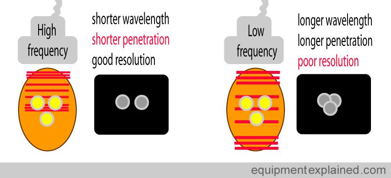
24 Anechoic Images Anechoic Anechoic Black

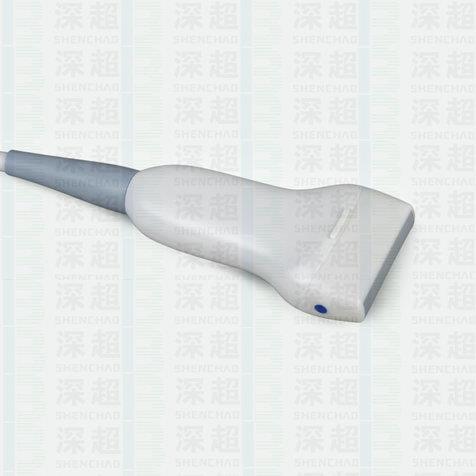
25 Transducers These are ultrasound probes

26 These transducers have different frequencies foot prints Ultrasound probes for regional anesthesia There are two transducers we use for regional anesthesia 1. Linear array 2. Curvilinear array
27 Foot print The footprint refers to the portion of the transducer that comes in contact with the patient This can be linear or curvilinear Curvilinear transducers come with different footprints for use for different purposes
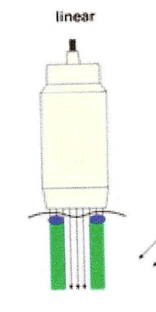
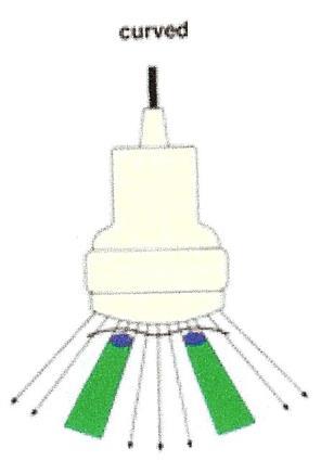
28 Linear transducer gives a square image Linear Curvilinear Curvilinear transducer gives a curved

29 Frequency Ultrasound waves are created by vibration of piezo electric crystals. This creates high pressure and low pressure area that travel forwards Frequency is the number of high pressure and low pressure cycles per second


30 Wavelength Wavelength is the distance between identical points in adjacent cycls of a waveform Wavelength is the distance between identical points in adjacent cycles of a waveform
31 Frequency and Wavelength Wavelength is inversely proportional to frequency Higher frequency has shorter wavelength Lower frequency had longer wavelength This phenomenon affects RESOLUTION
32 Resolution What is resolution? It is the ability to see two things clearly as two things Good resolution allows us to do that Poor resolution makes the picture of the two things blurred and makes it look like one

33 Higher frequency and shorter wavelength produce good resolution Lower frequency and longer wavelength produce poor resolution
34 Depth Ultrasound has to penetrate sufficient depth of the tissues to produce an image Depth of penetration is dependent upon the frequency Higher frequency has shorter depth of penetration Lower frequency has higher depth of penetration
35 Transducers
36 Transducers Linear Transducer High frequency Gives a square image High resolution Good for viewing small areas Good for shallow depth Curvilinear Transducer Low frequency Gives a curved image Lower resolution Good for viewing large area Good for deeper structures
37 Summary
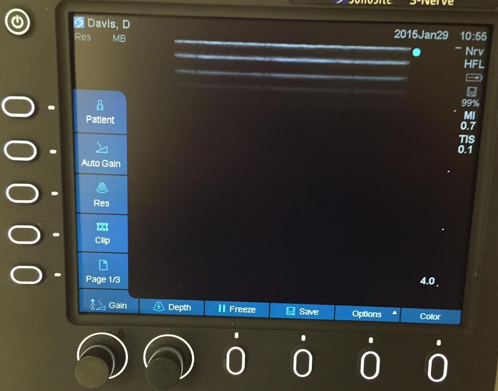
38 Controls you need to know Power button Battery Track ball or mouse pad Zoom Freeze
39 Ultrasound Machines
40 Ultrasound Machines
41 Track ball is like using your mouse on PC. It is used in conjunction with measuring, annotating, and moving Doppler box to the desired location Zoom allows magnification of the ultrasound picture Freeze allows you to freeze the image
42 Terminology you must know Frequency Gain Time gain compensation Depth Focus
43 Gain This function is very similar to a brightness control Echo signal is converted to electronic signal This signal needs to be amplified to produce an image Signal amplification is called gain Gain adjusts acoustic power of the signal
44 Time gain compensation T.G.C. TGC compensates for the effects of attenuation by progressively increasing amplification applied to signals with depth. Buttons for this are sliders and they are usually located on the side Each slider adjusts amplification of 2D mode signal at a specific image depth Sliders for TGC
45 Depth Changing the depth varies the field of view (F.O.V.) Excessively large field of view reduces spatial resolution
46 Focus The point at which the beam is at its narrowest is the focal point or focal zone It is the point of greatest intensity and best lateral resolution.
47 Focus The point at which the beam is at its narrowest It is the point of greatest intensity and best lateral resolution. Focal zone is off Focal zone is right

48 In Plane and Out of Plane Needle Placement

49 Needle visualization Out of plane In Plane
50
51 References 1. asics/ 2. Ultrasound Podcast - Physics Basics YouTube 3.
52 QUESTIONS?
Preamble (disclaimer)
") Preamble (disclaimer) PHYSICS AND PRINCIPLES OF HEAD/NECK ULTRASOUND Joseph C. Sniezek, MD FACS LTC, MC, USA Otolaryngology/H&N Surgery Tripler Army Medical Center 1. I am not a physicist 2. ACS has recommended
Preamble (disclaimer) PHYSICS AND PRINCIPLES OF HEAD/NECK ULTRASOUND Joseph C. Sniezek, MD FACS LTC, MC, USA Otolaryngology/H&N Surgery Tripler Army Medical Center 1. I am not a physicist 2. ACS has recommended
Terminology Tissue Appearance
 By Marc Nielsen, MD Advantages/Disadvantages Generation of Image Ultrasound Machine/Transducer selection Modes of Ultrasound Terminology Tissue Appearance Scanning Technique Real-time Portable No ionizing
Ultrasound Physics and Knobology Alan Macfarlane. Consultant Anaesthetist Glasgow Royal Infirmary
 Ultrasound Physics and Knobology Alan Macfarlane Consultant Anaesthetist Glasgow Royal Infirmary RAPM 2009; 34: 40-46 Ultrasound Proficiency Understanding US image generation and device operation Image
Principles of Ultrasound. Cara C. Prideaux, M.D. University of Utah PM&R Sports Medicine Fellow March 14, 2012
 Principles of Ultrasound Cara C. Prideaux, M.D. University of Utah PM&R Sports Medicine Fellow March 14, 2012 None Disclosures Outline Introduction Benefits and Limitations of US Ultrasound (US) Physics
Diagnostic Ultrasound. Sutiporn Khampunnip, M.D.
 Diagnostic Ultrasound Sutiporn Khampunnip, M.D. Definition of Ultrasound Ultrasound is simply sound waves, like audible sound. High-frequency sound and refers to mechanical vibrations above 20 khz. Human
The Physics of Ultrasound. The Physics of Ultrasound. Claus G. Roehrborn. Professor and Chairman. Ultrasound Physics
 The Physics of Ultrasound Pipe Organ 10-8000 Emission Dog 452-1080 Man 85-1100 Spectrum Bat 10,000-120,000 Porpoise 7000-120,000 Claus G. Roehrborn Professor and Chairman 10 20 Cycles per second Reception
Ultrasound Knobology
 Ultrasound Knobology Raj Dasgupta MD, FACP, FCCP, FASSM Assistant Professor of Clinical Medicine Pulmonary / Critical Care / Sleep Medicine University of Southern California (USC) Objectives Physics of
Ultrasound Principles cycle Frequency Wavelength Period Velocity
 ! Teresa S. Wu, MD, FACEP Director, EM Ultrasound Program & Fellowship Co-Director, Simulation Based Training Program & Fellowship Associate Program Director, EM Residency Program Maricopa Medical Center
1 Fundamentals. Basic Definitions and Physics Principles. Fundamentals
 1 To become versed in the language of ultrasonography, it is necessary to review some of the basic principles of physics. The wave physics principles of ordinary (i.e., audible) sound apply to ultrasound
Introduction to Ultrasound Guided Region Anesthesia
 Introduction to Ultrasound Guided Region Anesthesia Brian D. Sites, MD Dept of Anesthesiology Dartmouth-Hitchcock Medical Center INTRODUCTION Welcome to Introduction to Ultrasound Guided Regional Anesthesia.
Point-of-Care Ultrasound: An Introduction
 Point-of-Care Ultrasound: An Introduction Delegation Teaching Package for Registered Respiratory Therapists and Anesthesia Assistants Developed by: Rob Bryan RRT, AA Edited by: Kelly Hassall RRT, FCSRT,
Ultrasound Physics & Doppler
 Ultrasound Physics & Doppler Endocrine University 2018 Mark Lupo, MD, FACE, ECNU Objectives Review the essential components of ultrasound physics in neck sonography Demonstrate the importance of ultrasound
Ultrasound guidance in regional anesthesia has
 Ultrasound and Regional Anesthesia Artifacts and Pitfall Errors Associated With Ultrasound-Guided Regional Anesthesia. Part I: Understanding the Basic Principles of Ultrasound Physics and Machine Operations
Basic of Ultrasound Physics E FAST & Renal Examination. Dr Muhammad Umer Ihsan MBBS,MD, DCH CCPU,DDU1,FACEM
 Basic of Ultrasound Physics E FAST & Renal Examination Dr Muhammad Umer Ihsan MBBS,MD, DCH CCPU,DDU1,FACEM What is Sound? Sound is Mechanical pressure waves What is Ultrasound? Ultrasounds are sound waves
What is Ultrasound? Resolution Image production Attenuation Imaging modes Ultrasound artifacts... 7
 What is Ultrasound?... 1 Resolution... 3 Image production... 3 Attenuation... 4 Imaging modes... 5 Ultrasound artifacts... 7 0 What is Ultrasound? High frequency sound of frequencies 2-50 MHz is used in
Physical Principles of Ultrasound
 Physical Principles of Ultrasound Grateful appreciation to Richard A. Lopchinsky, MD, FACS and Nancy H. Van Name, RDMS, RTR, and MarleneKattaron, RDMS 2000 UIC All Rights Reserved. Course Objectives Identify
Ultrasound. Principles of Medical Imaging. Contents. Prof. Dr. Philippe Cattin. MIAC, University of Basel. Oct 17th, 2016
 Ultrasound Principles of Medical Imaging Prof. Dr. Philippe Cattin MIAC, University of Basel Contents Abstract 1 Image Generation Echography A-Mode B-Mode M-Mode 2.5D Ultrasound 3D Ultrasound 4D Ultrasound
Sound in medicine. CH.12. Dr.Rajaa أ.م.د. رجاء سهيل جنم جامعة تكريت كلية طب االسنان. General Properties of Sound
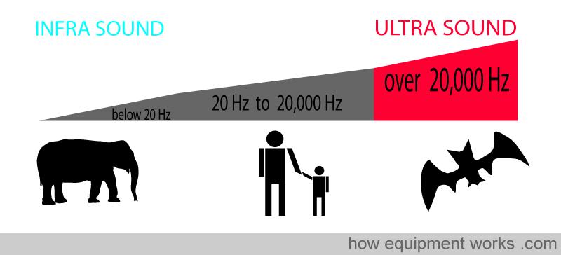
 CH.12. Dr.Rajaa Sound in medicine أ.م.د. رجاء سهيل جنم جامعة تكريت كلية Sound : It is the audible waves of frequency between 20 Hz and 20 khz. Infrasound : refers to the sound of frequency below the normal
3/20/2017. Disclosures. Ultrasound Fundamentals. Ultrasound Fundamentals. Bone Anatomy. Tissue Characteristics
 Disclosures Images of ultrasound equipment in this presentation are not an endorsement Fundamentals of Musculoskeletal Ultrasound Physics and Knobology Shane A. Shapiro, M.D. Assistant Professor Orthopedic
Ultrasound Physics & Terminology
 Ultrasound Physics & Terminology This module includes the following: Basic physics terms Basic principles of ultrasound Ultrasound terminology and terms Common artifacts seen Doppler principles Terms for
Dr Emma Chung. Safety first - Physical principles for excellent imaging
 Safety first - Physical principles for excellent imaging Dr Emma Chung Lecturer in Medical Physics, University of Leicester Clinical Scientist, University Hospitals of Leicester NHS Trust Thanks to Caroline
Introduction & Physics of ED Ultrasound. Objectives. What? - Limited Studies. Who? - ED Docs
 Introduction & Physics of ED Ultrasound Martine Sargent, MD Ultrasound Director, Assistant Professor UCSF Department of Emergency Medicine San Francisco General Hospital & Trauma Center Objectives Who?
ULTRASOUND IMAGING EE 472 F2018. Prof. Yasser Mostafa Kadah
 ULTRASOUND IMAGING EE 472 F2018 Prof. Yasser Mostafa Kadah www.k-space.org Recommended Textbook Diagnostic Ultrasound: Physics and Equipment, 2nd ed., by Peter R. Hoskins (Editor), Kevin Martin (Editor),
What is Ultrasound? What is Ultrasound? B A. Basic Principles of Ultrasound. Basic Principles of Ultrasound. Basic Principles of Ultrasound
 Introduction to Ultrasound Principles Mani Montazemi, RDMS Baylor College of Medicine Division of Maternal-Fetal Medicine Department of Obstetrics and Gynecology Manager, Maternal Fetal Center Imaging
WELCOME! Introduction to Bedside Ultrasound
 WELCOME! Introduction to Bedside Ultrasound TEACHERS University of California-Irvine School of Medicine Nathan Molina nathan.d.molina@gmail.com Trevor Plescia taplescia90@gmail.com Jack Silva jpsilva42@gmail.com
DIGITAL IMAGE PROCESSING IN ULTRASOUND IMAGES
 DIGITAL IMAGE PROCESSING IN ULTRASOUND IMAGES Kamaljeet Kaur Computer Science & Engineering Department Guru Nanak Dev Engg. College, Ludhiana. Punjab-India meetk.89@gmail.com ABSTRACT-- Image processing
ULTRASOUND. OB/Gyn (Core) Ultrasound PIEZOELECTRIC EFFECT. Principles of Ultrasound Physics and Instrumentation. Nathan Pinkney, BS, CDOS
 Ultrasound PIEZOELECTRIC EFFECT. Principles of Ultrasound Physics and Instrumentation. Nathan Pinkney, BS, CDOS") 1 OB/Gyn (Core) Ultrasound Principles of Ultrasound Physics and Instrumentation Nathan Pinkney, BS, CDOS Philadelphia College of Osteopathic Medicine 2016 ULTRASOUND CATEGORIES OF SOUND INFRASOUND = below
1 OB/Gyn (Core) Ultrasound Principles of Ultrasound Physics and Instrumentation Nathan Pinkney, BS, CDOS Philadelphia College of Osteopathic Medicine 2016 ULTRASOUND CATEGORIES OF SOUND INFRASOUND = below
Ultrasound in Anesthesia: Applying Scientific Principles to Clinical Practice
 AANA Journal Course Update for Nurse Anesthetists 3 6 CE Credits* Ultrasound in Anesthesia: Applying Scientific Principles to Clinical Practice Christian R. Falyar, CRNA, DNAP The use of ultrasound as
CONTENTS. Test Number cpd Tanya Reynolds (Nat. Dip. Diag. Rad., B. Tech. Diag. Rad., B. Tech. Ultrasound)
") CONTENTS page 1-15 page 16 BASIC 2-DIMENSIONAL ULTRASOUND PRINCIPLES Multiple Choice Test Test Number cpd 41640 Tanya Reynolds (Nat. Dip. Diag. Rad., B. Tech. Diag. Rad., B. Tech. Ultrasound) Tanya is
CONTENTS page 1-15 page 16 BASIC 2-DIMENSIONAL ULTRASOUND PRINCIPLES Multiple Choice Test Test Number cpd 41640 Tanya Reynolds (Nat. Dip. Diag. Rad., B. Tech. Diag. Rad., B. Tech. Ultrasound) Tanya is
Chapter 14. Imaging Artifacts
 Chapter 14 Image Artifacts The complex physical interactions that occur between an ultrasound beam and human anatomy and the intricate and sophisticated technological components of a sonographic imaging
Development of Ultrasound Based Techniques for Measuring Skeletal Muscle Motion
 Development of Ultrasound Based Techniques for Measuring Skeletal Muscle Motion Jason Silver August 26, 2009 Presentation Outline Introduction Thesis Objectives Mathematical Model and Principles Methods
INTRODUCTION. Getting the best scan. Choosing a probe. Choosing the frequency
 Getting the best scan Choosing a probe Select the most appropriate probe for the particular scan required. s vary in their: operating frequency range higher ultrasound frequencies provide better discrimination
Lesson 03: Sound Wave Propagation and Reflection. This lesson contains 15 slides plus 14 multiple-choice questions.
 Lesson 03: Sound Wave Propagation and Reflection This lesson contains 15 slides plus 14 multiple-choice questions. Accompanying text for the slides in this lesson can be found on pages 8 through 14 in
Introduction to Biomedical Imaging
 Alejandro Frangi, PhD Computational Imaging Lab Department of Information & Communication Technology Pompeu Fabra University www.cilab.upf.edu Basic principles. Comparison to X-rays Ultrasound > 20kHz
Diploma of Medical Ultrasonography (DMU) Physical Principles of Ultrasound and Instrumentation Syllabus
 Physical Principles of Ultrasound and Instrumentation Syllabus") Diploma of Medical Ultrasonography (DMU) Physical Principles of Ultrasound and Instrumentation Syllabus Page 1 of 7 11/18 Candidates are expected to cover all of the content of this syllabus when preparing
Diploma of Medical Ultrasonography (DMU) Physical Principles of Ultrasound and Instrumentation Syllabus Page 1 of 7 11/18 Candidates are expected to cover all of the content of this syllabus when preparing
The Essentials Tissue Characterization and Knobology
 The Essentials Tissue Characterization and Knobology Randy E. Moore, DC, RDMS RMSK No relevant financial relationships Ultrasound The New Standard of Care Musculoskeletal sonography has become the standard
Basic Ultrasound Physics Board Review Questions
 Basic Ultrasound Physics Board Review Questions Sidney K. Edelman, PhD ESP Ultrasound The Woodlands, TX Question 1 What is the wavelength of 2 MHz sound in soft tissue? 1. 1.54 mm 2. 0.75 mm 3. 0.75 cm
Lesson 07: Ultrasound Transducers. This lesson contains 73 slides plus 16 multiple-choice questions.
 Lesson 07: Ultrasound Transducers This lesson contains 73 slides plus 16 multiple-choice questions. This lesson was derived from pages 33 through 42 in the textbook: Ultrasound Transducers Ultrasound Transducers
4.17. RESEARCHING MODELS WITH AN ULTRASONIC ECHOSCOPE
 4.17. RESEARCHING MODELS WITH AN ULTRASONIC ECHOSCOPE Purpose of experiment Determine the main characteristics of ultrasound waves, and the distances and positions of models using an ultrasonic echoscope.
Supplement (videos)
") Supplement (videos) Ruben s tube (sound): http://www.youtube.com/watch?v=gpcquuwqayw Doppler US (diagnostic use): http://www.youtube.com/watch?v=fgxzg-j_hfw http://www.youtube.com/watch?v=upsmenyoju8 High
Supplement (videos) Ruben s tube (sound): http://www.youtube.com/watch?v=gpcquuwqayw Doppler US (diagnostic use): http://www.youtube.com/watch?v=fgxzg-j_hfw http://www.youtube.com/watch?v=upsmenyoju8 High
Image optimization for critical care US
 Image optimization for critical care US 1 Although we assume you are already familiar with focused US in the ED, it might not hurt to revise the basics: Machines & transducers US appearance of normal tissues
Ultrasonography of the Neck as an Adjunct to FNA. Nicole Massoll M.D.
 Ultrasonography of the Neck as an Adjunct to FNA Nicole Massoll M.D. Basic Features of Head and Neck Ultrasound and Anatomy Nicole Massoll M.D. University of Arkansas for Medical Sciences, Little Rock
Application of Phased Array Radar Theory to Ultrasonic Linear Array Medical Imaging System
 Application of Phased Array Radar Theory to Ultrasonic Linear Array Medical Imaging System R. K. Saha, S. Karmakar, S. Saha, M. Roy, S. Sarkar and S.K. Sen Microelectronics Division, Saha Institute of
The 2 nd Cambridge Advanced Emergency Ultrasound Course
 The 2 nd Cambridge Advanced Emergency Ultrasound Course Addenbrooke s Hospital Cambridge Sept 2008 1 2 Faculty! UK! USA! Australia! Toshiba! Emergency Medicine! Radiology 3 Programme! Day 1 Introduction
Basic Physics of Ultrasound in Transesophageal Echocardiography
 SPECIAL ARTICLE IJUTPC Basic Physics of Ultrasound in Transesophageal Echocardiography Basic Physics of Ultrasound in Transesophageal Echocardiography 1 Mary Korula, 2 Ravi Hebballi 1 Senior Consultant,
Routine Quality Assurance Cookbook
 This Cookbook is a companion guide to the AIUM Routine Quality Assurance (QA) for Diagnostic Ultrasound Equipment document, which outlines the basic QA requirements for AIUM-accredited practices. The Guide
Chapter 1. Principles of medical ultrasound. Overview. Background history: first steps to the piezo-electric effect.
 Chapter 1 Principles of medical ultrasound GRAHAM ARTHURS, PATRICK HILL AND TREVOR FRANKEL Overview This chapter provides an introduction to the ultrasound process for trainees in anesthesia and other
4.17. RESEARCHING MODELS WITH AN ULTRASONIC ECHOSCOPE
 4.17. RESEARCHING MODELS WITH AN ULTRASONIC ECHOSCOPE Purpose of experiment Determine the main characteristics of ultrasound waves, and the distances and positions of models using an ultrasonic echoscope.
Concepts of Imaging and Knobology
 Concepts of Imaging and Knobology Pravin Patil, MD FACC FASE Associate Professor of Medicine Director, Cardiovascular Disease Training Program Lewis Katz School of Medicine at Temple University Disclosures
Ultrasound in Peripheral Nerve Interventions
 Ultrasound in Peripheral Nerve Interventions John L. Lin, M.D. Shepherd Center Assistant Clinical Professor Emory University, School of Medicine Outline Ultrasound basics Nerve blocks in physiatric setting
Physics. Norman McDicken Tom Anderson CHAPTER ULTRASOUND. Ultrasound Propagation
 CHPTER 2 Physics Norman McDicken Tom nderson This chapter provides an introduction to the physics of medical ultrasound (US). Several books exist that can be consulted to extend the material presented
Ultrasound Applied Physics
 Ultrasound Applied Physics University of Toronto Department of Medical Imaging Applied Physics Mini-Course #3 2016 Ultrasound Laboratory Manual and Examination Booklet 1/21/2016 Ultrasound Applied Physics
Evaluation of the Quality of Thick Fibre Composites Using Immersion and Air- Coupled Ultrasonic Techniques
 ECNDT 2006 - We.1.6.4 Evaluation of the Quality of Thick Fibre Composites Using Immersion and Air- Coupled Ultrasonic Techniques Kaj K. BORUM, Risø National Laboratory, Materials Research Department, Roskilde,
An Overview of Ultrasound Testing For Lesion Detection in Human Kidney
 Journal of Tomography System & Sensors Application Vol.1, Issue 1, June 2018 An Overview of Ultrasound Testing For Lesion Detection in Human Kidney Aina Fadhilah Abd Rahim 1, Zawin Najah Abd Halim 1, Jaysuman
ULTRASOUND NOMENCLATURE
 Chapter 1: Ultrasound Nomenclature, Image Orientation, and Basic Instrumentation CYNTHIA SIKOWSKI Ultrasound waves are sound waves that have a frequency exceeding 20,000 Hz. When sound waves are transmitted
Ultrasonic Testing Level I:
 Ultrasonic Testing Level I: 1- Sound Wave - Introduction - ASNT Level I - Sound Wave Propagation - Velocity / Frequency / Wave Length - Acoustic Impedance - Energy / Intensity 2- Ultrasound Wave Modes
Breast Imaging Essentials
 Breast Imaging Essentials Module 9 Transcript 2016 ASRT. All rights reserved. Breast Imaging Essentials Module 9 Breast Ultrasound 1. ASRT Animation 2. Welcome Welcome to Module 9 of Breast Imaging Essentials
Flaw Assessment Using Shear wave Phased array Ultrasonic Transducer
 18th World Conference on Nondestructive Testing, 16-20 April 2012, Durban, South Africa Flaw Assessment Using Shear wave Phased array Ultrasonic Transducer Byungsik YOON AUTHOR 1, Hee-Jong LEE CO-AUTHOR
for the Veterinary Technician
 An Overview of Abdominal Ultrasound for the Veterinary Technician Valerie Gates, CVT, VTS (ECC) Learning Objective: The reader should gain a basic understanding of ultrasound, including physics, terminology,
Emergency Medicine Interest Group (EMIG) 2016
 2016") Emergency Medicine Interest Group (EMIG) 2016 Welcome to the flipped classroom (learning objectives summary) for the 2016 Emergency Medicine Interest Group (EMIG) Procedures Workshop. Overview - Tuesday
Emergency Medicine Interest Group (EMIG) 2016 Welcome to the flipped classroom (learning objectives summary) for the 2016 Emergency Medicine Interest Group (EMIG) Procedures Workshop. Overview - Tuesday
Basic Training Programme. 16 Februrary 2018, ROTTERDAM. Pre and Post-Course Test Answers
 Basic Training Programme 16 Februrary 2018, ROTTERDAM Pre and Post-Course Test Answers Your details: Name: Conference registration number/ BT delegate number: Email address: Are you already performing
High resolution ultrasound scanner for skin imaging
 High resolution ultrasound scanner for skin imaging Christine Turlat Sales Director Atys medical 17 Parc d Arbora 69510 SOUCIEU EN JARREST Atys company Principle of ultrasound imaging DERMCUP Normal image
This brief introduction considers the physics needed to get basic ultrasound images from a typical point of care machine.
 AAGBI seminar on Focused echocardiography for anaesthesia Craig Morris Physics This brief introduction considers the physics needed to get basic ultrasound images from a typical point of care machine.
Table of contents. Foreword. Preface. 1 Introduction Historical Perspective 00
 Table of contents Foreword Preface 1 Introduction 00 1.1 Historical Perspective 00 2 Fundamentals of musculoskeletal ultrasound 00 2.1 Frequency and wavelength 00 2.2 Generating ultrasound waves 00 2.3
Diagnostic approach to heart disease
 Diagnostic approach to heart disease Initial work up History Physical exam Chest radiographs ECG Special studies Echocardiography Cardiac catheterization Echocardiography principles Technique of producing
Knobology for Dummies
 Knobology for Dummies Power On/Off Preset button Patient Information Entry Choose preset Transducer probes Connect and disconnect transducer Approach to the patient (machine placement, comfort, draping,
Pulse-Echo Ultrasound Imaging. Resolution in Ultrasound Imaging. Doppler Ultrasound. Resolution vs Penetration. Medical Imaging (EL582/BE620/GA4426)
") Medical Imaging (EL582/BE620/GA4426) Pulse-Echo Ultrasound Imaging Ultrasound Imaging Lecture 2 Daniel (Dan) Turnbull, Ph.D. Skirball Institute and Dept of Radiology NYU School of Medicine (daniel.turnbull@med.nyu.edu)
Medical Imaging (EL582/BE620/GA4426) Pulse-Echo Ultrasound Imaging Ultrasound Imaging Lecture 2 Daniel (Dan) Turnbull, Ph.D. Skirball Institute and Dept of Radiology NYU School of Medicine (daniel.turnbull@med.nyu.edu)
Linear Ultrasonic Wave Propagation in Biological Tissues
 Indian Journal of Biomechanics: Special Issue (NCBM 7-8 March 29) Linear Ultrasonic Wave Propagation in Biological Tissues Narendra D Londhe R. S. Anand 2, 2 Electrical Engineering Department, IIT Roorkee,
Critical Care Ultrasound Study Notes
 Critical Care Ultrasound Study Notes Compiled by David Tripp October 2014 Ultrasound Physics 2 Ultrasound in Tissue 2 Ultrasound Interaction with Tissue 2 Pulsed Ultrasound and Imaging 3 Image Formation
ULTRASONIC ARRAY APPROACH FOR THE EVALUATION OF ELECTROFUSION JOINTS OF POLYETHYLENE GAS PIPING
 ULTRASONIC ARRAY APPROACH FOR THE EVALUATION OF ELECTROFUSION JOINTS OF POLYETHYLENE GAS PIPING H. J. Shin 1, Y. H. Jang 1, J. R. Kwan 2, H. D. Lee 3 1 INDE System Co., Ltd., Suwon, Kyunggi-do, 440-746,
Introduction to Musculoskeletal Ultrasound. Disclosures. Evidence Based Medicine Key References 8/30/2017
 Introduction to Musculoskeletal Ultrasound Johannes Roth MD, PhD, FRCPC, RhMSUS Professor of Pediatrics University of Ottawa Gurjit S Kaeley MBBS, MRCP, RhMSUS Professor of Medicine Division Chief Director
Thyroid Ultrasound Physics and Doppler
 Thyroid Ultrasound Physics and Doppler Advanced AACE-ACE US training course 2017 Dev Abraham MD, MRCP(UK), ECNU, FACE Professor of Medicine, University of Utah No Disclosures Natural Ability to see with
TG-128: Quality Assurance for Prostate Brachytherapy Ultrasound
 TG-128: Quality Assurance for Prostate Brachytherapy Ultrasound STEVEN SUTLIEF DOUG PFEIFFER (HEATHER PIERCE, WENGZHENG FENG, JIM KOFLER) AAPM ANNUAL MEETING 2010 Educational Objectives To describe the
CSB 046 Complementary Imaging Techniques
 CSB 046 Complementary Imaging Techniques - Quizzes are only ultrasound, final includes nuc med and ultrasound Week 1 Intro to Ultrasound Physics - Uses 1 to 20 MHz frequencies, which is way above the sound
Chapter 3. Sonographic Image Interpretation
 Chapter 3 Sonographic Image Interpretation Sonograms are two-dimensional gray-scale images that allow assessment and diagnosis of many anatomic and pathologic changes that can occur in the human body.
The table below shows the density and velocity of waves in two different substances. Density / kg m 3 Velocity / m s 1
 Q1.(a) When ultrasound is incident at an interface between two different media some energy is transmitted and some is reflected. The ratio of the reflected energy intensity I r to the incident energy intensity
Optimising your Doppler settings for an accurate PI. Alison McGuinness Mid Yorks Hospitals
 Optimising your Doppler settings for an accurate PI Alison McGuinness Mid Yorks Hospitals Applications Both maternal uterine and fetal circulations can be studied with doppler sonography Uterine arteries
Basics of US Regional Anaesthesia. November 2008
 Basics of US Regional Anaesthesia November 2008 Essential Physics HIGH frequency = great resolution but poor penetration LOW frequency = poor resolution but great penetration Potential Advantages of US
1. Fig. 1 shows data for the intensity of a parallel beam of X-rays after penetration through varying thicknesses of a material
 1. Fig. 1 shows data for the intensity of a parallel beam of X-rays after penetration through varying thicknesses of a material. intensity / MW m 2 thickness / mm 0.91 0.40 0.69 0.80 0.52 1.20 0.40 1.60
Confident and Conclusive Diagnosis with Flexible Solutions
 Internal Medicine Confident and Conclusive Diagnosis with Flexible Solutions ALPINION MEDICAL SYSTEMS We are Ultrasound Professionals Ultrasound is widely used as the gold-standard for internal medicine
Underwater Acoustic Measurements in Megahertz Frequency Range.
 Underwater Acoustic Measurements in Megahertz Frequency Range. Current State and Prospects of Development in Russia Alexander M. Enyakov,, Many medical applications of underwater acoustic measurements
Endobronchial Ultrasound
 Endobronchial Ultrasound l Armin Ernst Editors Felix J.F. Herth Endobronchial Ultrasound An Atlas and Practical Guide 13 Editors Armin Ernst, MD Chief, Section of Interventional Pulmonology; Director,
Chapter 2 Pitfalls in Musculoskeletal Ultrasound
 Chapter 2 Pitfalls in Musculoskeletal Ultrasound Violeta Maria Vlad MD, PhD Introduction Taking a good ultrasound (US) picture is an art. Interpreting it is a science. This is in fact everything US is
Can you believe that the ultrasonic waves are used for cleaning purpose???
 Introduction (Ultrasonic Cleaning Unit) Can you believe that the ultrasonic waves are used for cleaning purpose??? Learning Objectives On completion of this chapter you will be able to: 1. Describe the
Introduction. Cardiac Imaging Modalities MRI. Overview. MRI (Continued) MRI (Continued) Arnaud Bistoquet 12/19/03
 MRI (Continued) Arnaud Bistoquet 12/19/03") Introduction Cardiac Imaging Modalities Arnaud Bistoquet 12/19/03 Coronary heart disease: the vessels that supply oxygen-carrying blood to the heart, become narrowed and unable to carry a normal amount
Introduction Cardiac Imaging Modalities Arnaud Bistoquet 12/19/03 Coronary heart disease: the vessels that supply oxygen-carrying blood to the heart, become narrowed and unable to carry a normal amount
Improving Methods for Breast Cancer Detection and Diagnosis. The National Cancer Institute (NCI) is funding numerous research projects to improve
 is funding numerous research projects to improve") CANCER FACTS N a t i o n a l C a n c e r I n s t i t u t e N a t i o n a l I n s t i t u t e s o f H e a l t h D e p a r t m e n t o f H e a l t h a n d H u m a n S e r v i c e s Improving Methods for
CANCER FACTS N a t i o n a l C a n c e r I n s t i t u t e N a t i o n a l I n s t i t u t e s o f H e a l t h D e p a r t m e n t o f H e a l t h a n d H u m a n S e r v i c e s Improving Methods for
Ultrasound 10/1/2014. Basic Echocardiography for the Internist. Mechanical (sector) transducer Piezoelectric crystal moved through a sector sweep
 transducer Piezoelectric crystal moved through a sector sweep") Ultrasound Basic Echocardiography for the Internist Carol Gruver, MD, FACC UT Erlanger Cardiology Mechanical wave of compression and rarefaction Requires a medium for transmission Ultrasound frequency
Ultrasound Basic Echocardiography for the Internist Carol Gruver, MD, FACC UT Erlanger Cardiology Mechanical wave of compression and rarefaction Requires a medium for transmission Ultrasound frequency
Ultrasonic Testing of Composite Structures
 I. Introduction Ultrasonic Testing of Composite Structures This section of this work defines ultrasound basic concepts and Ultrasonic Technique. It describes the details of how ultrasonic testing works,
The Evolution and Benefits of Phased Array Technology for the Every Day Inspector
 ECNDT 2006 - Poster 198 The Evolution and Benefits of Phased Array Technology for the Every Day Inspector Dan KASS, Tom NELLIGAN, and Erich HENJES Olympus NDT, Waltham, USA Abstract. Phased arrays were
Ultrasound Evaluation of the Posterior Segment of the Eye A Ready Reckoner
 180 Kerala Journal of Ophthalmology Vol. XX, No. 2 OPHTHALMIC INSTRUMENTATION Ultrasound Evaluation of the Posterior Segment of the Eye A Ready Reckoner Dr. Mahesh G. MS DO DNB FRCSEd., Dr. A. Giridhar
Background & Indications Probe Selection
 Teresa S. Wu, MD, FACEP Director, EM Ultrasound Program & Fellowship Co-Director, Simulation Based Training Program & Fellowship Associate Program Director, EM Residency Program Maricopa Medical Center
Critical Care Ultrasound Seeing is Believing
 Critical Care Ultrasound Seeing is Believing Philip D Lumb, M.B., B.S., MCCM Los Angeles, California USA Objectives Integrate ultrasound into routine critical care practice Introduce HOLA (Holistic Approach)
S1Stephanie J. Doniger
 Section 1 Ultrasound fundamentals Introduction S1Stephanie J. Doniger Pediatric Emergency Medicine is a relatively new field of medicine developed in the 1980s. Since its inception, several advancements
APPLICATION AND DEPLOYMENT OF ADVANCED NDE TECHNIQUES IN HIGH PRESSURE VESSELS
 APPLICATION AND DEPLOYMENT OF ADVANCED NDE TECHNIQUES IN HIGH PRESSURE VESSELS Jeffrey P. Milligan, Daniel T. Peters, Structural Integrity Associates, Inc., USA Many advances in Non-Destructive Examination
Descriptions of NDT Projects Fall 2004 October 31, 2004
 Descriptions of NDT Projects Fall 2004 October 31, 2004 Introduction There are two separate NDT labs in Magister: ULTRA for ultrasound and EDDY for eddy current. Both labs are equipped with mechanical
The Value. Ultrasound System. Professional Concept for. Women s. Health
 The Value Innovative Ultrasound System Professional Concept for Women s Health 9 Beyond Women s Healthcare E-CUBE 9 - Powerful Imaging Performance for All Women s Health Diagnostic Applications Diagnostic
CHARACTERIZATION OF ANNULAR ARRAY TRANSDUCER
 Analele Universităţii de Vest din Timişoara Vol. LV, 2011 Seria Fizică CHARACTERIZATION OF ANNULAR ARRAY TRANSDUCER Luminita Moraru 1, Laura Onose 1, 2, Ana-Maria Chiselev 1 1 Dunărea de Jos University
Regional Anaesthesia
 Regional Anaesthesia Basic Sciences Anaesthetic Course Dr Vassilis ATHANASSOGLOU FRCA Consultant Anaesthetist and Lecturer in Medicine Oxford University Hospitals NHS Trust, UK Geneva, 5th June 2017 Faculty
Physical Principles of Ultrasound
 Physical Principles of Ultrasound Pat F. Fulgham 2 Introduction The use of ultrasound is fundamental to the practice of urology. In order for urologists to best use this technology on behalf of their patients,
Therapeutic Ultrasound
 THERAPEUTIC ULTRASOUND Therapeutic is a form of MECHANICAL energy Mechanical vibration at increasing frequencies is known as sound energy. Below 16Hz, these vibrations are not recognizable as sound The