Acupuncture Treatment in. Spinal Cord Injury. Ben Quadriplegia Debbie Paraplegia
|
|
|
- Allan Gordon
- 5 years ago
- Views:
Transcription
1 Acupuncture Treatment in Spinal Cord Injury Ben Quadriplegia Debbie Paraplegia
2 I. General Consideration 1. spine trauma Spinal cord injury Vertebrae fracture, dislocation or/and compression Quadriplegia, paraplegia Loss of skin sensation or/and muscle movement 2. Case out-line of Ben: lesions to motor central nuclei Case out-line of Debbie: lesions to lower motor neurons Picture: 1 SCI-Cervical vertebrae fracture
3 II. Three Main Effects in this ACU Treatment Approach to SCI 1. The needle is applied as a stimulator: Sensory neurons Neural path reflex mechanism Motor neurons
4 II. Three Main Effects in this ACU Treatment Approach to SCI 2. Activate wound healing potential: Cellular wound healing 1-1 Macrophages 1-2 White blood cells 1-3 Skin regeneration, periosteum/bone stem cells ** Mother and Son law blood nourishes the son and the son moves the blood flow ** Fire burning the mountain Pure reinforcement needle techniques
5 II. Three Main Effects in this ACU Treatment Approach to SCI 3. The needle is used as a scalpel: 1-1 Releases obstruction in neurovascular pathway 1-2 Corrects malformation 1-3 Removal of degenerative proliferation tissues ** Releases the obstructive tissues

6 III. Needle Techniques 1. Skin: Regeneration, receptors - free nerve ending Reticular layer - stretching power

7 Spinal nerves dorsal root ganglia [sensory neurons] Ventral root mixes nerve [motor neurons]
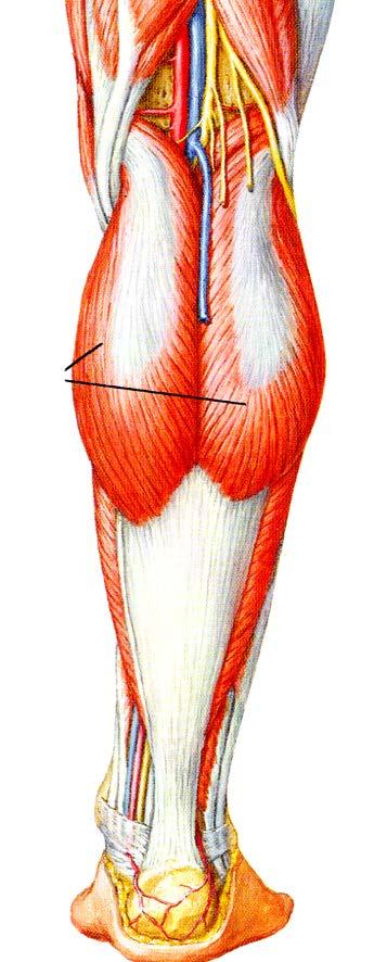
8 III. Needle Techniques 2. Muscles Origin muscles spindles Belly motor end plate [contraction] Insertion to the joint or bone
9 III. Needle Techniques 2. Muscles 1-1. The needle is inserted into spindles where the muscles attach to the joint or bone The needle is inserted into the belly of the muscles The needle is inserted into the insertion of the muscles The needle is inserted obliquely into the fascia tendon and aponeurosis The needle is inserted perpendicularly, into the muscle fibres The needle is inserted between two muscles connective tissues The needle is inserted into the quadriceps of Femoris.

10 III. Needle Techniques 2. Muscles Pure Reinforcement Techniques Fire Burning the Mountain


11 III. Needle Techniques 3. Three Sets of Meridian Points Located on the Vertebral Column: GV, JIAJY and Bladder meridian points GV between two spinous process JIAJI point on the side of spinous process [intervertebral foramen, recurrent meningeal nerve] BL meridian two straight line medial BL11-35, lateral line BL
12 IV. Show video Demonstration Ben and Debbie stand and walk with walking frame
13 Case: Ben Quadriplegia Name: Benjamin Date of birth: 9/2/1987 Male Date of accident: 27/10/2012 Date of starting acupuncture treatment: 12/1/2013
14 Diagnosis: 1-1 Ben was in a severe motor vehicle accident Overstretched flexion and extension of the head and neck caused lesion to the medulla and brainstem neural pathway. 1-2 MRI and CT scan report showed C5-6-7 cervical vertebrae fracture and cord injury 1-3 Clinically Ben was diagnosed C7 (neurological level) ASIA B


15 MRI Fig 1. Ben s X-ray images: just after injury (10/2012) Fig 2. Ben s X-ray images: 4.5 years after injury (16/5/2017)


16
17 10/10/12 23/1/13 30/3/17 L R L R L R Hip flexion Extension Abduction Adduction Opposition Knee flexion Extension Ankle flexion Extension Foot flexion Extension Toe flexion Extension Table 1. Ben s Lower Limb Motor Power L: left, R: right

18 The Lesion Involved the Medulla and Brainstem
19 Clinical Findings The neurological examination indicated the syndrome involved the interruptionof the Red nucleus, Vestibular nucleus, Reticular formation, Medullar oblongata and the neural pathway. The extrapyramidal system is a biological neural network that is part of the motor system causing involuntary movements Extrapyramidal tracts are chiefly found in the recticular formation of the pons and medulla and target neurons in the spinal cord involved in reflexes, locomotion, complex movements, and postural control. The extrapyramidal tracts include parts of the following: Rubospinal tract Pontine reticulospinal tract Medullary reticulospinal tract Lateral vestibulospinal tract Tectospinal tract Those tracts are in turn modulated by various parts of the central nervous system: including Basal ganglia
20 1-1 The Red Nucleus A. The syndrome of lesion to the red nucleus: pectoralis, scapular region, posterior superior serratus, rhomboid, or/and trapezius, arm and hand muscles spasticity. * Pectoralis: Points selection: LU1-2, ST , KI22 KI24, CV15 * Biceps: ACU point LI15, LU1, LU4 PC2, and LU5 PC3. * Posterior Superior Serratus, Rhomboid, Trapezius and Tendons: BL 41 to 47 on the edge of spine of the scapular and points on vertebral column BL11 to BL18.
21 1-2 Reticular Formation A. Neural network spino-reticular tracts connected sensory nerve impulses to cerebellum cerebral cortex and via reticulo-spinal tracts descending to the lower motor neurons in spinal cord B. Vestibulospinal tract assist the reticular formation to maintain muscle tone and balance the body s posture, so when standing and walking, the patient needs to keep the head up and eyes looking forward in a straight position. C. ACU treatment for reticular formation: * Needle insertion stimulates the fingertips, body trunk and extremity points * Moxa stick heats the point for temperature stimulation * Breathing exercises affect the breathing centre and increase reflex activity * Scratching increases joint, muscle and tendon activity * Combining methods may apply
22 1-3 Medulla Oblongata A. Trauma in the anterior median fissure and pyramidal tracts prevents the sensation getting through the olive medulla, then the cortico-spinal tracts cannot send the motor signal to the spinal cord and thus motor movement is impaired. * Point location: GB20 BL11 TH17 TH16 GV15 GV16. * GV JIAJI and BL meridian points targets on trapezius, scalene, rhomboid minor or major and levator-scapula muscles. ACU points GB21 GV14 BL11 BL41TH14 and TH15. * Neck-flexion point selection: LI18, ST5, ST9, ST11, CV22, CV23, TH17

23 Chest and Abdominal Wall Muscle Points A. Pectoralis contraction ACU points: LU1 SP20 PC1 ST16 ST17 KI24 B. Abdominal muscle points: ST , CV , SP12, , GB , Liv14 and KI11,12,13 KI22.
24 Thigh A. Major points are on the lumber-sacral plexuses such as BL22, 23, 25, 26 and BL50,51,52 on sacrum BL27 to BL34, BL53 and, BL54. B. Tendon and muscle linking points such as tensor fascia latae with gluteus maximus fascia and iliotibial tract with vastus lateralis ACU point GB30 GB31 GB32 GB33. C. Muscle attachment surrounding the greater trochanter and hamstring muscles ACU points: BL35, 36, 37, 38, 39, 40. D. Large muscles of the femoris quadriceps and hamstring muscles need to gain equilibrium. Strength especially needs to be gained in the rectus femoris muscles.


25
26 Spinal Nerve, Sympathetic and Parasympathetic Nerve A. Most of SCI patients have lost the function of urination, sexuality and bowel movement. B. The points selection involved Vagus CNX, BL23,24,25,28,32, GV1,2,4, JIAJI on L3 and L5 vertebrae level. C. Abdominal points included CV1,2,3, KI11,12, ST25,28,29.

27

28 6. Five SHU Points 1.1 Points on apex of fingers or toes: 1-2. Jing-well points 1-3. The Web 1-4. The phalangeal joint 1-5. The points on the wrists or ankles 1-6. The muscles below the knee: HE sea points 1.7. The points on palm or planta
29 Thalamus, supplementary cortex area and primary cortex
30 Case: Debbie Paraplegia Name: Debbie Date of birth: 21/07/1962 Female Date of accident: 19/09/2012 Date of starting acupuncture treatment: 12/1/2013 Accident: Coconut tree fell down front of body and crushed her 3:30pm 19/9/2012 in Bali Indonesia

31 1. Diagnosis 1-1. Debbie was diagnosed T11 (neurological level) ASIA A Both of her hips, knees, ankles feet and toes motor power were 0/ The sensation (proprioception) of both her lower limbs were absent. Illustration Spinal cord


32 1. Diagnosis Spine CT on 04/06/13 reviewed: A compression fracture through T12 is noted. The posterio-superior aspect of the vertebral body has retropulsed mildly into the spinal canal. There has been posterior fusion from T10 to L1 with bilateral rods and screws. The T12 vertebral body shows wedged compression. Visualisation of the spinal canal and its contents is obstructed by metallic artefact. The fracture is still relatively acute without obvious healing. The alignment remains, however, satisfactory and there is no spinal canal stenosis. The metallic spinal support was removed on 30/07/2013 at POWH.
33 10/10/12 23/1/13 30/3/17 L R L R L R Hip flexion Extension Abduction Adduction Opposition Knee flexion Extension Ankle flexion Extension Foot flexion Extension Toe flexion Extension Table 2. Debbie s lower limb motor power L-left, R-right

34 2. ACU Treatment Design 1-1 Release post-operative scars 1-2 Treat below spinal cord lesion region lumbar, thighs, legs and feet to toes 1-3 Propose distal part extremities management to avoid degeneration and atrophy 1-4 TCM methods: Point selection are Back-SHU & Front-MU points and foot Five-SHU points. Spina nerves distribution
35 3. Clinical Findings Debbie presented in her wheelchair. Both of her legs had significant muscle atrophy and no resistance in passive movement. The legs were purple in colour with a much lower temperature than other parts of her body. Both of the ankles and feet were swollen. She could not move both legs and had no needle sensation below T11 post-operative scar from T7 to L5. On the front, the abdominal muscles were overstressed and were a mass of contracture. There was no needle sensation below the navel.

36 4. ACU Treatment on the Spine and Abdomen 1-1. BL Meridian, GV and JIAJI Points on the Spine The needle is inserted into the T7 to L5 and down to S4 GV JIAJI. BL meridian BL15 to BL30 and BL43 to BL54 points. The needle is inserted repetitively and gently into the points and retained in the points for 30 minutes.

37 4. ACU Treatment on the Spine and Abdomen 1-2. The Point on the Chest and Abdomen The needle is inserted into the chest and abdominal wall. The point selections are on ST , CV , SP14-16, GB25-27, Liv14, Lu1-2 and KI22. Method: The needle is gently inserted into the point above the peritoneum. The lifting and thrusting needle method is applied, the needle is retained for 30 minutes. After needling; scratching, stretching the chest and abdominal muscles.
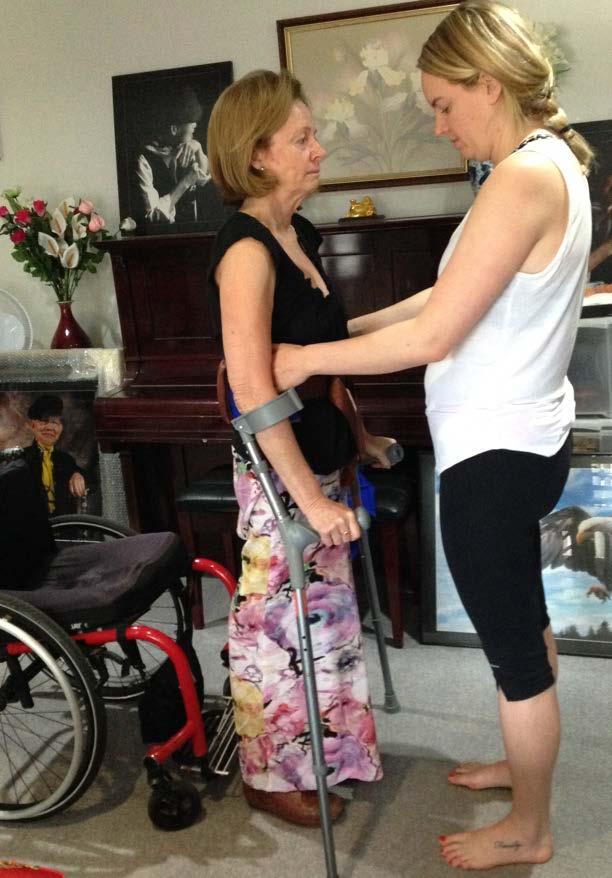
38 5. Standing and Walking Five months after acupuncture treatment, Debbie could stand up with other people s help and could feel the sensation of pain in both of her thighs. Nine months after acupuncture treatment, she could lift both thighs and stand up independently. With another seven months of acupuncture treatment (May 2014), Debbie could walk a few steps with a walking frame.
39 6. Femur Fracture Accident After Debbie was able to walk, she attempted to walk with the walking frame. She tried to move out of her wheelchair quickly without help from others. On 29/9/2014 during the first few steps, Debbie fell (4 months after she could walk with walking frame). This fall completed the bone fracture of her right femur at level 20cm above the knee (Figure 3 A and B arrows indicate the similar point). Debbie did not go to hospital for an operation but instead used acupuncture as the major treatment for the bone fracture.
40 1-1. Fracture Management A. Firstly, using gentle hand traction to let the fractured bone return to the correct postural position, then using an external full leg brace, support and advice, Debbie maintained the leg, knee and ankle in a straight position for the next two months and no exercise was allowed. B. The needles were inserted around the fracture site, including ST and GB34, needling perpendicularly deep into the periosteum. The lifting and thrusting needle method is used and the needles retained in the points for 20 minutes. C. Before needling, moxa stick was used to heat the points. After needling, gentle scratching of the upper and lower lesion section of skin and muscles for increased blood circulation and improvement in wound healing. D. The specialist agreed with the treatment and closely observed the progress.
 and the right thigh regained muscle strength.")
41 6. Femur Fracture Accident Acupuncture treatment was focused only on the right leg bone fracture in the first month after the bone fracture, then needling on both left and right legs followed. With acupuncture treatment, the bone fracture healed (Figure 6 C, D are the X-rays taken about 1.5 years after the fracture indicated by the arrows) and the right thigh regained muscle strength. Slowly, both legs of the patient got stronger and the patient was able to start to stand up and walk again in Feb 2015 (5 months after right leg bone fracture).
42 7. Treatment of the Thigh 1-1. Most of the ACU points on the pelvis are on muscles attached to the bone and joint such as GB27 GB28 origin of the quadriceps and the insertion of the patella ST The thigh muscles are large and strong and are less innervated. Not many ACU points are found there either. The muscles on the hamstring attach to the tuberculosis and sacrum. The BL26 to BL30 and BL36 GB 30 and the Liv points are located along the ischium and pubic area The tensor fascia latae is the strong tendon that supports the hip and the knee. The lesion was located between the ilio-tibialis tendon and vastus lateral muscles, the point is on GB32 GB33.


43
44 8. ACU Treatment Below the Knee When Debbie started ACU treatment, Tai proposed starting treatment on both lower extremities but the treatment achieved no response. Point selection: K1-K2 ST36-GB34 BL60-K3 BL SP6-K8 ST40-41 After needling passive stretching of the foot and leg followed. Moxa stick heats the point and makes a first-degree burn on the ST36 or SP6 and K1 or K2 alteration point on the BL57-59 or BL40. Temporarily after needling no response was shown. Moxa on the skin made it red in colour or blistered (preferably, a hole is gently made in the blister to release the fluid), the skin was palpated and Debbie s leg felt warm.
45 8. ACU Treatment Below the Knee 1-1. Proposal of ACU treatment on 12/05/2016: Only the right leg from the knee down to the foot would be treated. No treatment was provided to the left leg which was to be used as a comparison for the research Both legs are weak, the muscles are in a pre-atrophy condition and there is no muscle resistance on passive lift up and down, no skin sensation or muscle movement. The tone in the muscles of the left leg above the knee and in the thigh is better than in the right.
46 8. ACU Treatment Below the Knee 1-1 In the first three months of intensive ACU treatment of the right leg, the repetitive needling method is used and more needles are retained. Needling mostly focuses on the individual muscles from the origin of the muscles to insertion of the muscles of the tibialis anterior and posterior and the extensor digitorum longus. 1-2 The needle is inserted into the points K6 and BL61, SP6 and BL , which involve the calculus tendon and gastrocnemius soleus muscles. Repetitive needling, scratching and stretching tries to move the tendon and muscles. 1-3 Moxa heats the points and especially provides the first-degree burn on K1 K2 ST36 SP8 BL40 and BL56, which is for prolonged stimulation. 1-4 Gentle scratching and passive movement is provided after the needling and moxa heat.
47 8. ACU Treatment Below the Knee 1-5 After six months of treatment the right leg shows better improvement. The right leg can lift the knee higher than the left and the muscles show tone. The ankle also shows it can move with the leg. The left leg remains the same, it feels cooler. When needling into the points of the right foot during the morning treatment, Debbie reported she could feel tingling in her left foot but not in the right at night. 1-6 Debbie realised there was a big difference in her leg which was getting ACU treatment compared to the one that was not. The gentle ACU treatment on the left leg, did not improve to the same level as the right. Debbie then asked for a strong treatment on the left leg which improved it to the same level as the right. 1-7 On 20/11/2016, when the needle was inserted into plantar points of the right foot, Debbie felt tingling on the left foot and it was hot at night in bed, however, when needling the left foot plantar points K1 and retaining the needle in the point, she felt she wanted to move her right foot but the foot did not move.
48 8. ACU Treatment Below the Knee 1-8 Progress on the legs was slow. It took two years of strong ACU treatment to achieve a weak tone in the leg muscles and another year to regain sensation in the ankle. 1-9 It is only when Debbie was able to mobilise her ankle that she was able to place her body weight on her foot and walk sufficiently. She still needs more practice with her walking and stretching exercises but this will come with time. ** In October 2017, Debbie stands using crutches with the help of her carer and takes a few steps out of her wheelchair.


49


50


51
52
Scapula Spine Lateral edge of clavicle. Medial border Scapula. Medial border of Scapula, between superior angle and root of spine. Scapula.
 Muscle attachments and actions answer sheet Muscle Origins insertions Movements Joints crossed Trapezius Base of skull Spinous process of C7 Thoracic Spine Lateral edge of clavicle Elevation Retraction
Muscles of the Hip 1. Tensor Fasciae Latae O: iliac crest I: lateral femoral condyle Action: abducts the thigh Nerve: gluteal nerve
 Muscles of the Hip 1. Tensor Fasciae Latae O: iliac crest I: lateral femoral condyle Action: abducts the thigh Nerve: gluteal nerve 2. Gluteus Maximus O: ilium I: femur Action: abduct the thigh Nerve:
RN(EC) ENC(C) GNC(C) MN ACNP *** MECHANISM OF INJURY.. MOST IMPORTANT ***
 ENC(C) GNC(C) MN ACNP *** MECHANISM OF INJURY.. MOST IMPORTANT ***") HISTORY *** MECHANISM OF INJURY.. MOST IMPORTANT *** Age of patient - Certain conditions are more prevalent in particular age groups (Hip pain in children may refer to the knee from Legg-Calve-Perthes
HISTORY *** MECHANISM OF INJURY.. MOST IMPORTANT *** Age of patient - Certain conditions are more prevalent in particular age groups (Hip pain in children may refer to the knee from Legg-Calve-Perthes
musculoskeletal system anatomy nerves of the lower limb 2 done by: Dina sawadha & mohammad abukabeer
 musculoskeletal system anatomy nerves of the lower limb 2 done by: Dina sawadha & mohammad abukabeer #Sacral plexus : emerges from the ventral rami of the spinal segments L4 - S4 and provides motor and
Temporalis Elevates & retracts mandible. Masseter Elevates mandible. Sternocleidomastoid Neck flexion. Trapezius Elevates & depresses shoulders
 Anterior Posterior Temporalis Elevates & retracts mandible Masseter Elevates mandible Sternocleidomastoid Neck flexion Trapezius Elevates & depresses shoulders Masseter Elevates mandible Temporalis Elevates
Muscles of the lower extremities. Dr. Nabil khouri MD, MSc, Ph.D
 Muscles of the lower extremities Dr. Nabil khouri MD, MSc, Ph.D Posterior leg Popliteal fossa Boundaries Biceps femoris (superior-lateral) Semitendinosis and semimembranosis (superior-medial) Gastrocnemius
Arterial Blood Supply
 Arterial Blood Supply Brain is supplied by pairs of internal carotid artery and vertebral artery. The four arteries lie within the subarachnoid space Their branches anastomose on the inferior surface of
Prime movers provide the major force for producing a specific movement Antagonists oppose or reverse a particular movement Synergists
 Dr. Gary Mumaugh Prime movers provide the major force for producing a specific movement Antagonists oppose or reverse a particular movement Synergists Add force to a movement Reduce undesirable or unnecessary
The Muscular System. Chapter 10 Part D. PowerPoint Lecture Slides prepared by Karen Dunbar Kareiva Ivy Tech Community College
 Chapter 10 Part D The Muscular System Annie Leibovitz/Contact Press Images PowerPoint Lecture Slides prepared by Karen Dunbar Kareiva Ivy Tech Community College Table 10.14: Muscles Crossing the Hip and
Gross Anatomy of Lower Spinal Cord
 Chapter 13 Spinal Cord, Spinal Nerves and Somatic Reflexes Spinal cord Spinal nerves Somatic reflexes Gross Anatomy of Lower Spinal Cord Meninges of Vertebra & Spinal Cord Spina Bifida Congenital defect
Cadaver Muscular System Practice Practical
 Cadaver Muscular System Practice Practical Station 1 Station 1 1. Specific structure 1. Rectus sheath 2. Red line 2. Linea alba Station 2 Station 2 3. Red muscle 1. Rectus abdominis 4. Red muscle actions
Spinal cord. We have extension of the pia mater below L1-L2 called filum terminale
 Spinal cord Part of the CNS extend from foramen magnum to the level of L1-L2 (it is shorter than the vertebral column) it is covered by spinal meninges. It is cylindrical in shape. It s lower end become
OBJECTIVES. Unit 7:5 PROPERTIES OR CHARACTERISTICS OF MUSCLES. Introduction. 3 Kinds of Muscles. 3 Kinds of Muscles 4/17/2018 MUSCULAR SYSTEM
 OBJECTIVES Unit 7:5 MUSCULAR SYSTEM Compare the three main kinds of muscles by describing the action of each Differentiate between voluntary and involuntary muscles List at least three functions of muscles
Year 2004 Paper one: Questions supplied by Megan
 QUESTION 47 A 58yo man is noted to have a right foot drop three days following a right total hip replacement. On examination there is weakness of right ankle dorsiflexion and toe extension (grade 4/5).
Human Anatomy and Physiology I Laboratory Spinal and Peripheral Nerves and Reflexes
 Human Anatomy and Physiology I Laboratory Spinal and Peripheral Nerves and Reflexes 1 This lab involves the second section of the exercise Spinal Cord, Spinal Nerves, and the Autonomic Nervous System,
The Nervous System: Sensory and Motor Tracts of the Spinal Cord
 15 The Nervous System: Sensory and Motor Tracts of the Spinal Cord PowerPoint Lecture Presentations prepared by Steven Bassett Southeast Community College Lincoln, Nebraska Introduction Millions of sensory
Spinal Cord Organization. January 12, 2011
 Spinal Cord Organization January 12, 2011 Spinal Cord 31 segments terminates at L1-L2 special components - conus medullaris - cauda equina no input from the face Spinal Cord, Roots & Nerves Dorsal root
Lower Limb Nerves. Clinical Anatomy
 Lower Limb Nerves Clinical Anatomy Lumbar Plexus Ventral rami L1 L4 Supplies: Abdominal wall External genitalia Anteromedial thigh Major nerves.. Lumbar Plexus Nerves relation to psoas m. : Obturator n.
I: To describe the pyramidal and extrapyramidal tracts. II: To discuss the functions of the descending tracts.
 Descending Tracts I: To describe the pyramidal and extrapyramidal tracts. II: To discuss the functions of the descending tracts. III: To define the upper and the lower motor neurons. 1. The corticonuclear
Human Anatomy and Physiology I Laboratory
 Human Anatomy and Physiology I Laboratory Gross Anatomy of the Muscular System (Two weeks) 1 This lab involves study of the laboratory exercise Gross Anatomy of the Muscular System. Complete the Review
MUSCLES OF THE LOWER LIMBS
 MUSCLES OF THE LOWER LIMBS Naming, location and general function Dr. Nabil khouri ROLES THAT SHOULD NOT BE FORGOTTEN Most anterior compartment muscles of the hip and thigh Flexor of the femur at the hip
lesser trochanter of femur lesser trochanter of femur iliotibial tract (connective tissue) medial surface of proximal tibia
 medial surface of proximal tibia") LOWER LIMB MUSCLES OF THE APPENDICULAR SKELETON The muscles that act on the lower limb fall into three groups: those that move the thigh, those that move the lower leg, and those that move the ankle, foot,
LOWER LIMB MUSCLES OF THE APPENDICULAR SKELETON The muscles that act on the lower limb fall into three groups: those that move the thigh, those that move the lower leg, and those that move the ankle, foot,
Human Anatomy Lab #7: Muscles of the Cadaver
 Human Anatomy Lab #7: Muscles of the Cadaver Table of Contents: Expected Learning Outcomes.... 1 Introduction...... 1 Identifying Muscles on Yourself.... 2 Muscles of the Anterior Trunk and Arm.. 2 Muscles
Spinal Cord- Medulla Spinalis. Cuneyt Mirzanli Istanbul Gelisim University
 Spinal Cord- Medulla Spinalis Cuneyt Mirzanli Istanbul Gelisim University Spinal Column Supports the skull, pectoral girdle, upper limbs and thoracic cage by way of the pelvic girdle. Transmits body weight
Lumbar Plexus. Ventral rami L1 L4 Supplies: Major nerves.. Abdominal wall External genitalia Anteromedial thigh
 Lower Limb Nerves Lectures Objectives Describe the structure and relationships of the plexuses of the lower limb. Describe the course, relationships and structures supplied for the major nerves of the
Human Anatomy. Spinal Cord and Spinal Nerves
 Human Anatomy Spinal Cord and Spinal Nerves 1 The Spinal Cord Link between the brain and the body. Exhibits some functional independence from the brain. The spinal cord and spinal nerves serve two functions:
Human Anatomy Biology 351
 Human Anatomy Biology 351 Lower Limb Please place your name on the back of the last page of this exam. You must answer all questions on this exam. Because statistics demonstrate that, on average, between
Muscle Energy Technique
 PRACTICE SESSION: Muscle Energy Technique BE AN ARTIST and work out the best way for you to use the Muscle Energy Technique (MET). This technique works best when muscles are shortened. If you try MET on
Role Of The Fitness Professional. Causes of Fitness Related Injuries. The Assessments. Screening & Assessing: A Holistic Approach 2/9/2016
 Screening & Assessing: A Holistic Approach Role Of The Fitness Professional Fitness professionals must assess clientele, but need to understand the difference between medical diagnosis vs fitness limitations.
Muscular System. IB Sports, exercise and health science 1.2
 Muscular System IB Sports, exercise and health science 1.2 Characteristics Common to Contractility-ability to shorten the muscles length Extensibility-ability to lengthen the muscles length Elasticity-muscle
Class Outline: Posterior Anatomy
 Class Outline: Posterior Anatomy 5 minutes Breath of Arrival and Attendance 5 minutes Howdy Partner 35 minutes Posterior Anatomy using Power Point Presentation 5 minutes Overview of skeletal segments 5
Muscles to know. Lab 21. Muscles of the Pelvis and Lower Limbs. Muscles that Position the Lower Limbs. Generally. Muscles that Move the Thigh
 Muscles to know Lab 21 Muscles of the Pelvis, Leg and Foot psoas major iliacus gluteus maximus gluteus medius sartorius quadriceps femoris (4) gracilus adductor longus biceps femoris semitendinosis semimembranosus
Practical 1 Worksheet
 Practical 1 Worksheet ANATOMICAL TERMS 1. Use the word bank to fill in the missing words. reference side stand body arms palms anatomical forward All anatomical terms have a(n) point which is called the
Stretching Exercises for the Lower Body
 Stretching Exercises for the Lower Body Leg Muscles The leg has many muscles that allow us to walk, jump, run, and move. The main muscle groups are: Remember to: Warm-up your muscles first before stretching
In-Depth Foundations: Anatomy Terms to Know
 Be familiar with / able to identify and define all the following parts. The Spine Cranium Vertebrae Cervical, Thoracic, Lumbar Sacrum Coccyx Bones of Upper Body Cranium Mastoid process; Occipital condyle,
The Muscular System PART C. PowerPoint Lecture Slide Presentation by Patty Bostwick-Taylor, Florence-Darlington Technical College
 PowerPoint Lecture Slide Presentation by Patty Bostwick-Taylor, Florence-Darlington Technical College The Muscular System 6 PART C Five Golden Rules of Skeletal Muscle Activity Table 6.2 Muscles and Body
1-Apley scratch test.
 1-Apley scratch test. The patient attempts to touch the opposite scapula to test range of motion of the shoulder. 1-Testing abduction and external rotation( +ve sign touch the opposite scapula, -ve sign
Lower limb summary. Anterior compartment of the thigh. Done By: Laith Qashou. Doctor_2016
 Lower limb summary Done By: Laith Qashou Doctor_2016 Anterior compartment of the thigh Sartorius Anterior superior iliac spine Upper medial surface of shaft of tibia 1. Flexes, abducts, laterally rotates
The Spinal Cord. The Nervous System. The Spinal Cord. The Spinal Cord 1/2/2016. Continuation of CNS inferior to foramen magnum.
 The Nervous System Spinal Cord Continuation of CNS inferior to foramen magnum Simpler than the brain Conducts impulses to and from brain Two way conduction pathway Reflex actions Passes through vertebral
Healing Hands School of Holistic Health. Advanced Circulatory & Sports Massage Class Handouts
 Class Handouts 1 Posterior Trepidations Torso Rock Torso Rock half-step Torso Rock both sides Torso Rock down body Torso Side Stretch Erector Rock Spinal Rock Lumbo Rock Cha Cha Leg Clay Snake Flop Leg
Fig Cervical spinal nerves. Cervical enlargement C7. Dural sheath. Subarachnoid space. Thoracic. Spinal cord Vertebra (cut) spinal nerves
 spinal nerves") Fig. 13.1 C1 Cervical enlargement C7 Cervical spinal nerves Dural sheath Subarachnoid space Thoracic spinal nerves Spinal cord Vertebra (cut) Lumbar enlargement Medullary cone T12 Spinal nerve Spinal nerve
Fig. 13.1 C1 Cervical enlargement C7 Cervical spinal nerves Dural sheath Subarachnoid space Thoracic spinal nerves Spinal cord Vertebra (cut) Lumbar enlargement Medullary cone T12 Spinal nerve Spinal nerve
VCE PHYSICAL EDUCATION WORKBOOK UNIT 1 BODIES IN MOTION NAME:
 VCE PHYSICAL EDUCATION WORKBOOK UNIT 1 BODIES IN MOTION NAME: SKELETAL SYSTEM List the 5 functions of the skeletal system and complete the following table. FUNCTION DESCRIPTION Label the following features
Electrode Placement. Skin Preparation. Frontalis (FRL) (Specific) Temporalis Anterior (TA) (Specific) Sternocleidomastoid (SCM) (Specific)
 (Specific) Temporalis Anterior (TA) (Specific) Sternocleidomastoid (SCM) (Specific)") Electrode Placement Skin Preparation 1) Removing the hair: Shave if necessary 2) Clean the skin: Use a towel or abrasive pad with conductive cleaning paste or alcohol to remove dead skin cells (high impedance)
Electrode Placement Skin Preparation 1) Removing the hair: Shave if necessary 2) Clean the skin: Use a towel or abrasive pad with conductive cleaning paste or alcohol to remove dead skin cells (high impedance)
Role of brainstem in somatomotor (postural) functions
 functions") Role of brainstem in somatomotor (postural) functions (vestibular apparatus) The muscle tone and its regulation VESTIBULAR SYSTEM (Equilibrium) Receptors: Otolith organs Semicircular canals Sensation (information):
Role of brainstem in somatomotor (postural) functions (vestibular apparatus) The muscle tone and its regulation VESTIBULAR SYSTEM (Equilibrium) Receptors: Otolith organs Semicircular canals Sensation (information):
Name this muscle. Name this muscle
 this muscle this muscle Pectoralis Major Pectoralis Minor Serratus anterior Pectoralis minor Serratus anterior this muscle Deltoid: The major abductor of the upper limb this muscle this muscle this muscle
The abdominal muscles:
 Core Body Strength A horse s natural centre of gravity is slightly behind the shoulder and approximately one third of the way down it s body. In order to accommodate the weight of the rider, we need to
Evaluating the Athlete Questionnaire
 Evaluating the Athlete Questionnaire Prior to developing the strength and conditioning training plan the coach should first evaluate factors from the athlete s questionnaire that may impact the strength
Chapter 13: The Spinal Cord and Spinal Nerves
 Chapter 13: The Spinal Cord and Spinal Nerves Spinal Cord Anatomy Protective structures: Vertebral column and the meninges protect the spinal cord and provide physical stability. a. Dura mater, b. Arachnoid,
VCE PHYSICAL EDUCATION WORKBOOK UNIT 1 BODIES IN MOTION NAME:
 VCE PHYSICAL EDUCATION WORKBOOK UNIT 1 BODIES IN MOTION NAME: SKELETAL SYSTEM List the 5 functions of the skeletal system and complete the following table. FUNCTION DESCRIPTION Label the following features
Muscles of the Gluteal Region
 Muscles of the Gluteal Region 1 Some of the most powerful in the body Extend the thigh during forceful extension Stabilize the iliotibial band and thoracolumbar fascia Related to shoulders and arms because
PHYSICAL EDUCATION. 4º E.S.O. 2nd TERM. The skeletal and muscular systems.
 PHYSICAL EDUCATION 4º E.S.O. 2nd TERM. The skeletal and muscular systems. PARTS OF THE BODY Head Torso / Trunk Dorsal: Back Ventral: Thorax y Abdomen Extremities Superior: Arm Forearm Hand Joint: Shoulder
In which arm muscle are intramuscular injections most often given? (not in text)
") AP1 Lab 9 - Muscles of the Arms and Legs Locate the following muscles on the models and on yourself. Recall anatomical position. Directional terms such as anterior, posterior, lateral, etc. all assume
AP1 Lab 9 - Muscles of the Arms and Legs Locate the following muscles on the models and on yourself. Recall anatomical position. Directional terms such as anterior, posterior, lateral, etc. all assume
Anatomy & Physiology. Muscles of the Lower Limbs.
 Anatomy & Physiology Muscles of the Lower Limbs http://www.ishapeup.com/musclecharts.html Muscles of the Lower Limbs Among the strongest muscles in the body. Because pelvic girdle is composed of heavy,
Where should you palpate the pulse of different arteries in the lower limb?
 Where should you palpate the pulse of different arteries in the lower limb? The femoral artery In the femoral triangle, its pulse is easily felt just inferior to the inguinal ligament midway between the
Motor tracts Both pyramidal tracts and extrapyramidal both starts from cortex: Area 4 Area 6 Area 312 Pyramidal: mainly from area 4 Extrapyramidal:
 Motor tracts Both pyramidal tracts and extrapyramidal both starts from cortex: Area 4 Area 6 Area 312 Pyramidal: mainly from area 4 Extrapyramidal: mainly from area 6 area 6 Premotorarea: uses external
ANATYOMY OF The thigh
 ANATYOMY OF The thigh 1- Lateral cutaneous nerve of the thigh Ι) Skin of the thigh Anterior view 2- Femoral branch of the genitofemoral nerve 5- Intermediate cutaneous nerve of the thigh 1, 2 and 3 are
The Lower Limb VI: The Leg. Anatomy RHS 241 Lecture 6 Dr. Einas Al-Eisa
 The Lower Limb VI: The Leg Anatomy RHS 241 Lecture 6 Dr. Einas Al-Eisa Muscles of the leg Posterior compartment (superficial & deep): primary plantar flexors of the foot flexors of the toes Anterior compartment:
The Musculoskeletal system
 Level 3 BTEC Applied Science Summer Homework The Musculoskeletal system Student name:.. Tutor name: 1 Student Instructions This workbook incorporates elements of Unit 8 Learning Aim A: Understand the impact
Gluteal region DR. GITANJALI KHORWAL
 Gluteal region DR. GITANJALI KHORWAL Gluteal region The transitional area between the trunk and the lower extremity. The gluteal region includes the rounded, posterior buttocks and the laterally placed
Chapter 12b. Overview
 Chapter 12b Spinal Cord Overview Spinal cord gross anatomy Spinal meninges Sectional anatomy Sensory pathways Motor pathways Spinal cord pathologies 1 The Adult Spinal Cord About 18 inches (45 cm) long
Human Anatomy Biology 351
 Human Anatomy Biology 351 Lower Limb Please place your name on the back of the last page of this exam. You must answer all questions on this exam. Because statistics demonstrate that, on average, between
River North Pain Management Consultants, S.C., Axel Vargas, M.D., Regional Anesthesiology and Interventional Pain Management.
 River North Pain Management Consultants, S.C., Axel Vargas, M.D., Regional Anesthesiology and Interventional Pain Management. Chicago, Illinois, 60611 Phone: (888) 951-6471 Fax: (888) 961-6471 Clinical
Muscles of Lesson Five. Muscular Nomenclature and Kinesiology - Two. Muscles of Lesson Five, cont. Chapter 16
 Chapter 16 Muscular Nomenclature and Kinesiology - Two Lessons 5-6 Muscles of Lesson Five Iliopsoas (psoas major, iliacus) Hip outward rotators (piriformis, gemellus superior, gemellus inferior, obturator
Lecturer. Prof. Dr. Ali K. Al-Shalchy MBChB/ FIBMS/ MRCS/ FRCS 2014
 Lecturer Prof. Dr. Ali K. Al-Shalchy MBChB/ FIBMS/ MRCS/ FRCS 2014 Dorsal root: The dorsal root carries both myelinated and unmyelinated afferent fibers to the spinal cord. Posterior gray column: Long
Human Anatomy Biology 255
 Human Anatomy Biology 255 Exam #4 Please place your name and I.D. number on the back of the last page of this exam. You must answer all questions on this exam. Because statistics demonstrate that, on average,
Note: Please refer to handout Spinal Plexuses and Representative Spinal Nerves for
 Chapter 13 Outline Note: Please refer to handout Spinal Plexuses and Representative Spinal Nerves for what you need to know from Exhibits 13.1 13.4 I. INTRODUCTION A. The spinal cord and spinal nerves
Certified Personal Trainer Re-Certification Manual
 Certified Personal Trainer Re-Certification Manual Section II 1 Anatomy & Physiology Terms Anatomy and physiology are closely related fields of study: anatomy is the study of form, and physiology is the
Spinal injury. Structure of the spine
 Spinal injury Structure of the spine Some understanding of the structure of the spine (spinal column) and the spinal cord is important as it helps your Neurosurgeon explain about the part of the spine
2/4/2018. Identify the two reasons why muscle cells may go through muscle fatigue. Ch.7 Review. Sternocleidomastoid.
 Ch.7 Review Identify the two reasons why muscle cells may go through muscle fatigue Temporalis Depressor anguli oris Sternocleidomastoid Tibialis anterior 1 Gluteus medius Deltoid Adducts & rotates scapula
Due in Lab weeks because of Thanksgiving Prelab #10. Homework #8. Both sides! Both sides!
 Lab 8 MUSCLES Due in Lab 10 2 weeks because of Thanksgiving Prelab #10 Both sides! Homework #8 Both sides! Refer to Muscles 22-23 Naming of muscles Origin Site of muscle attachment that doesn t move during
EXERCISE PHOTOS, TIPS AND INSTRUCTIONS
 Page 1 of 21 EXERCISE PHOTOS, TIPS AND INSTRUCTIONS Page 2. Squat Page 12. Crab Walks Page 3. Single Leg Squat Page 13. Bench Press Page 4. Split Squat Page 14. Bench Pull Page 5. Deadlift Page 15. Shoulder
S.A.F.E. Elements of Technique. S.A.F.E. is an acronym for strength, alignment, flexibility, and STRENGTH ALIGNMENT FLEXIBILITY ENDURANCE
 Elements of Technique S.A.F.E. S.A.F.E. is an acronym for strength, alignment, flexibility, and endurance. These are all elements that are innate in the study of dance. STRENGTH The amount of control and
Chapter 10: Muscular System: Gross Anatomy
 Chapter 10: Muscular System: Gross Anatomy I. General Principles A. General Terminology 1. Tendons attach 2. What is an aponeurosis? 3. The points of muscle attachment are called & 4. How is the "origin"
The Massage Routine. Start with your client lying face down - Prone Position. Clean YOUR HANDS and CLIENTS FEET using antibacterial wipes
 The Massage Routine Start with your client lying face down - Prone Position Clean YOUR HANDS and CLIENTS FEET using antibacterial wipes!!!! GROUNDING FOR 3 BREATHS TUNE YOUR BREATHING WITH THE CLIENTS!!!
Active-Assisted Stretches
 1 Active-Assisted Stretches Adequate flexibility is fundamental to a functional musculoskeletal system which represents the foundation of movement efficiency. Therefore a commitment toward appropriate
Year 2 MBChB Clinical Skills Session Examination of the Motor System
 Year 2 MBChB Clinical Skills Session Examination of the Motor System Reviewed & ratified by: o o o o Dr D Smith Consultant Neurologist Dr R Davies Consultant Neurologist Dr B Michael Neurology Clinical
Nervous System: Spinal Cord and Spinal Nerves (Chapter 13)
") Nervous System: Spinal Cord and Spinal Nerves (Chapter 13) Lecture Materials for Amy Warenda Czura, Ph.D. Suffolk County Community College Eastern Campus Primary Sources for figures and content: Marieb,
Nervous System: Spinal Cord and Spinal Nerves (Chapter 13) Lecture Materials for Amy Warenda Czura, Ph.D. Suffolk County Community College Eastern Campus Primary Sources for figures and content: Marieb,
Muscle fiber (cell) Blood vessel. Perimysium. Epimysium. Fascicle (wrapped by perimysium) Endomysium (between fibers) Tendon. Bone
 Blood vessel. Perimysium. Epimysium. Fascicle (wrapped by perimysium) Endomysium (between fibers) Tendon. Bone") Figure 6.1 Connective tissue wrappings of skeletal muscle. Blood vessel Muscle fiber (cell) Perimysium Epimysium Fascicle (wrapped by perimysium) Tendon Endomysium (between fibers) Bone Figure 6.15 Superficial
Figure 6.1 Connective tissue wrappings of skeletal muscle. Blood vessel Muscle fiber (cell) Perimysium Epimysium Fascicle (wrapped by perimysium) Tendon Endomysium (between fibers) Bone Figure 6.15 Superficial
Stretching. Back (Latissimus dorsi) "Chicken Wings" Chest (Pec. major + Ant. deltoid) "Superman" Method: Method: 1) Stand tall and maintain proper
 Chicken Wings Chest (Pec. major + Ant. deltoid) Superman Method: Method: 1) Stand tall and maintain proper") Chest (Pec. major + Ant. deltoid) "Chicken Wings" Back (Latissimus dorsi) "Superman" 1) Stand tall and maintain proper 1) Reach hands overhead and lumbar curve. grasp one wrist. 2) Place palms on lower
Chest (Pec. major + Ant. deltoid) "Chicken Wings" Back (Latissimus dorsi) "Superman" 1) Stand tall and maintain proper 1) Reach hands overhead and lumbar curve. grasp one wrist. 2) Place palms on lower
11/15/2018. Temporalis Elevates & retracts mandible. Masseter = Prime mover of jaw closure. Levator scapulae Supraspinatus Clavicle.
 Due in Lab 10 Lab 8 MUSCLES 2 weeks because of Thanksgiving Prelab #10 Both sides! Homework #8 Both sides! Refer to Muscles 22-23 Examples of Origin & Insertion Naming of muscles Origin Site of muscle
Nervous System. Student Learning Objectives:
 Nervous System Student Learning Objectives: Identify the primary parts of the neuron Identify the major structures of the central nervous system Identify the major structures of the peripheral nervous
Chapter 3: Applied Kinesiology. ACE Personal Trainer Manual Third Edition
 Chapter 3: Applied Kinesiology ACE Personal Trainer Manual Third Edition Introduction Kinesiology is the study of the body s infinite number of movements, positions, and postures and is grounded in the
NERVOUS SYSTEM. Academic Resource Center. Forskellen mellem oscillator og krystal
 NERVOUS SYSTEM Academic Resource Center Forskellen mellem oscillator og krystal Overview of the Nervous System Peripheral nervous system-pns cranial nerves spinal nerves ganglia peripheral nerves enteric
lower limb Anterior Compartment: lecture 3 The deep fascia ( fascia lata) divides the thigh into 3 compartments:
 divides the thigh into 3 compartments:") lower limb lecture 3 The deep fascia ( fascia lata) divides the thigh into 3 compartments: 1. Anterior Extensor compartment 2. Medial Adductor compartment 3. Posterior Flexor compartment Anterior Compartment:
lower limb lecture 3 The deep fascia ( fascia lata) divides the thigh into 3 compartments: 1. Anterior Extensor compartment 2. Medial Adductor compartment 3. Posterior Flexor compartment Anterior Compartment:
32b Passive Stretches: Guided Full Body
 32b Passive Stretches: Guided Full Body 32b Passive Stretches: Guided Full Body! Class Outline" 5 minutes" "Attendance, Breath of Arrival, and Reminders " 10 minutes "Lecture:" 25 minutes "Lecture:" 15
HEAD AND NECK PART 2
 HEAD AND NECK PART 2 INTEGRATED CURRICULUM = Integrate Basic Science and Clinical Training 1- ENT PATIENT EXAM IN ICS COURSE - Today and next week - Review/Preview Anatomy underlying ENT exam 2- NEUROANATOMY/NEUROLOGY
The Lower Limb. Anatomy RHS 241 Lecture 2 Dr. Einas Al-Eisa
 The Lower Limb Anatomy RHS 241 Lecture 2 Dr. Einas Al-Eisa The bony pelvis Protective osseofibrous ring for the pelvic viscera Transfer of forces to: acetabulum & head of femur (when standing) ischial
Part 1: Communication between CNS & PNS
 Ch. 6: Peripheral Nervous System Objectives: 1. Communication between CNS & PNS: afferent (sensory) pathway versus efferent (motor) pathway of information. 2. Regulation of somatic (voluntary) motor system
Exercises to restore range of movement: Rotation
 Exercises to restore range of movement: Rotation Start position: Sitting upright with your back supported in a chair. Position your head so it is evenly balanced, looking forward. Avoid allowing your head
Epicranius (frontal belly) Zygomaticus minor. Zygomaticus major Buccinator
 Zygomaticus minor. Zygomaticus major Buccinator") Epicranius (frontal belly) Zygomaticus minor Zygomaticus major Buccinator Masseter Digastric (posterior belly) Stylohyoid Sternocleidomastoid Trapezius Scalenus Omohyoid (inferior belly) Orbicularis oris
Epicranius (frontal belly) Zygomaticus minor Zygomaticus major Buccinator Masseter Digastric (posterior belly) Stylohyoid Sternocleidomastoid Trapezius Scalenus Omohyoid (inferior belly) Orbicularis oris
THE BACK. Dr. Ali Mohsin. Spinal Cord
 Spinal Cord THE BACK Dr. Ali Mohsin The spinal cord is the elongated caudal part of the CNS. It starts as the inferior continuation of the medulla oblongata at the level of foramen magnum, & ends as an
Brain Stem. Nervous System (Part A-3) Module 8 -Chapter 14
 Module 8 -Chapter 14") Nervous System (Part A-3) Module 8 -Chapter 14 Overview Susie Turner, M.D. 1/9/13 Cellular structure of the nervous system Neurons Neuroglia Nervous System Divisions Central nervous system Peripheral nervous
Nervous System (Part A-3) Module 8 -Chapter 14 Overview Susie Turner, M.D. 1/9/13 Cellular structure of the nervous system Neurons Neuroglia Nervous System Divisions Central nervous system Peripheral nervous
Chapter 13. The Nature of Muscle Spindles, Somatic Reflexes, and Posture
 Chapter 13 The Nature of Muscle Spindles, Somatic Reflexes, and Posture Nature of Reflexes A reflex is an involuntary responses initiated by a sensory input resulting in a change in the effecter tissue
Bell Work. How does the muscular system relate to the following organ systems, Respiratory Circulatory Digestive
 Muscular System Bell Work How does the muscular system relate to the following organ systems, Respiratory Circulatory Digestive Exercise Science Standards 8) Review the gross and cellular anatomy and physiology
Lecture VIII. The Spinal Cord, Reflexes and Brain Pathways!
 Reflexes and Brain Bio 3411! Monday!! 1! Readings! NEUROSCIENCE 5 th ed: Review Chapter 1 pp. 11-21;!!Read Chapter 9 pp. 189-194, 198! THE BRAIN ATLAS 3 rd ed:! Read pp. 4-17 on class web site! Look at
STRETCHING. Benefits of stretching
 STRETCHING Benefits of stretching Most individuals and athletes, never take stretching seriously. They have what we refer to as a weight lifting mentality. The misconception is that if you do not feel
The psoas minor is medial to the psoas major. The iliacus is a fan-shaped muscle that when contracted helps bring the swinging leg forward in walking
 1 p.177 2 3 The psoas minor is medial to the psoas major. The iliacus is a fan-shaped muscle that when contracted helps bring the swinging leg forward in walking and running. The iliopsoas and adductor
Human Anatomy, First Edition McKinley & O'Loughlin
 Human Anatomy, First Edition McKinley & O'Loughlin Chapter 8 : Appendicular Skeleton 8-1 Appendicular Skeleton Includes the bones of the upper and lower limbs. The girdles of bones that attach the upper
By Dr. Saeed Vohra & Dr. Sanaa Alshaarawy
 By Dr. Saeed Vohra & Dr. Sanaa Alshaarawy 1 By the end of the lecture, students will be able to : Distinguish the internal structure of the components of the brain stem in different levels and the specific
Muscle Testing of Knee Extensors. Yasser Moh. Aneis, PhD, MSc., PT. Lecturer of Physical Therapy Basic Sciences Department
 Muscle Testing of Knee Extensors Yasser Moh. Aneis, PhD, MSc., PT. Lecturer of Physical Therapy Basic Sciences Department Muscle Testing of Knee Extensors othe Primary muscle Quadriceps Femoris -Rectus