Vitreomacular interface disorders. Ghanbari MD 1393:10:25
|
|
|
- Lucas Booth
- 5 years ago
- Views:
Transcription

1
2 Vitreomacular interface disorders Ghanbari MD 1393:10:25

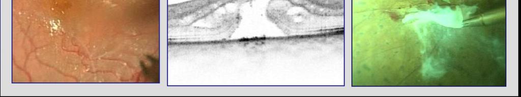
3 Human vitreous after dissection of the sclera, choroid, and retina.

4 Lamellar structure of the posterior vitreous cortex (PVC) in the monkey. V = Vitreous; R = Retina; ILL = Internal Limiting Lamina.

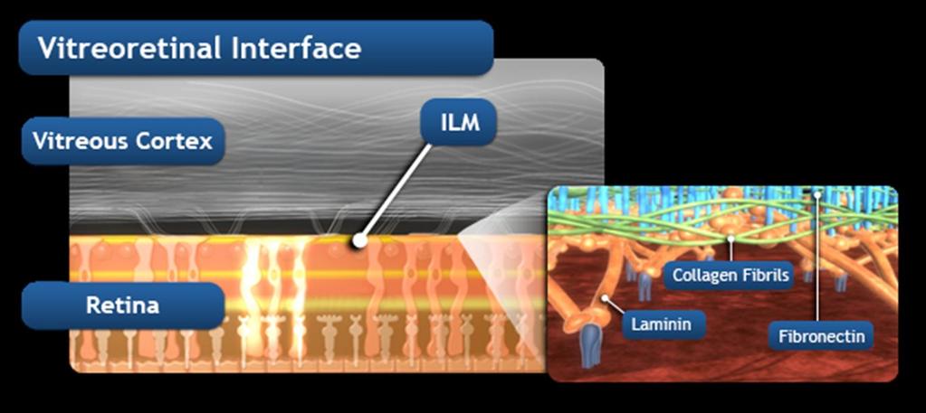
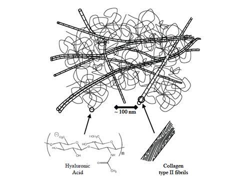
5 Yellow lines represent vitreoretinal interface bonds constituted by laminin, fibronectin, and collagen types VI, VII, XVIII.

6
7
8
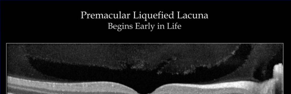
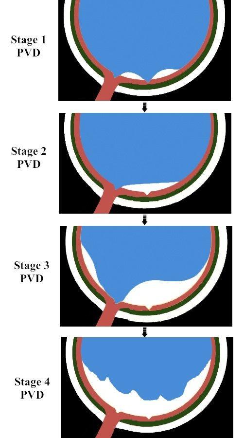
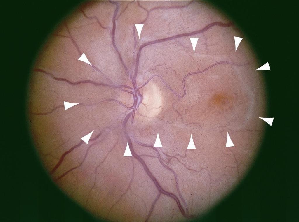
9 Normal aging is accompanied by a number of physiological changes in the vitreous gel. After the age of 40, it undergoes progressive liquefaction (synchisis), with fluid escaping through defects in the posterior vitreous cortex, eg, those at the optic disc or prepapillary hole.
10
11
12

13

14
15
16 Stage 0: No evidence of PVD Stage 1: Perifoveal vitreous detachment with vitreofoveal adhesion. Stage 2: Perifoveal vitreous detachment with no vitreofoveal adhesion. Stage 3: Complet PVD except for vitreopapillary adhesion Stage 4: Complete PVD
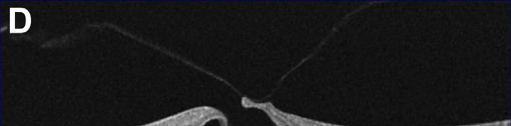
17
18
19
20
21


22 Posterior vitreous detachment (PVD) is the result of a complex and inevitable set of events that occurs as the eye ages. It manifests as gel liquefaction and weakening of vitreoretinal adhesion. Imaging of the VMI with OCT reveals that PVD usually begins in the perifoveal macula.1,2,13
23
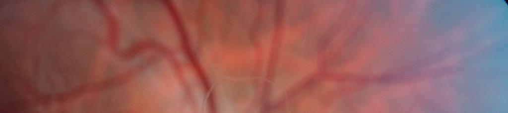
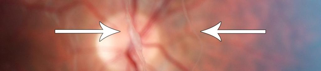
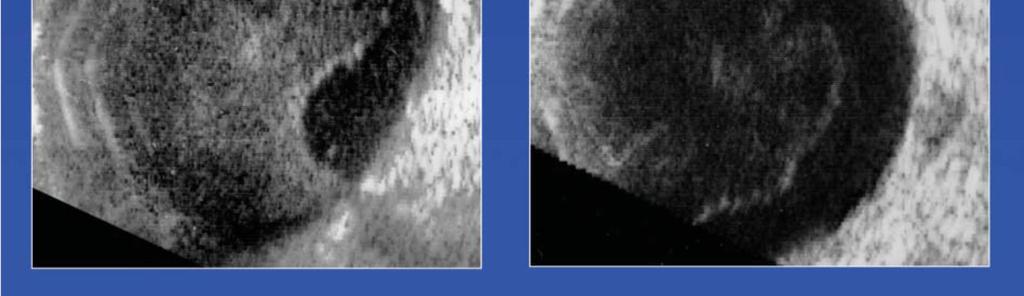
24 The vitreous cortex temporally has a lamellar structure with two layers (arrows). The inner layer is detached from the outer lamellae. In this subject, the vitreous pocket is undetectable in both eyes.
25 Symptoms of PVDs Flashing lights Floaters Acute PVD have 15% retinal tear. PVD with vit. Hemorrhage 50%-70% have retinal tears.
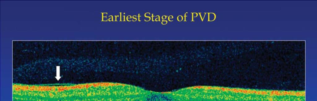
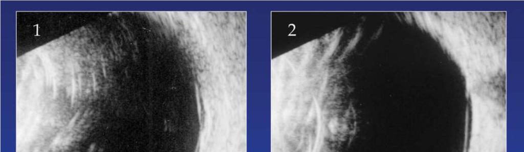
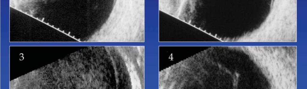
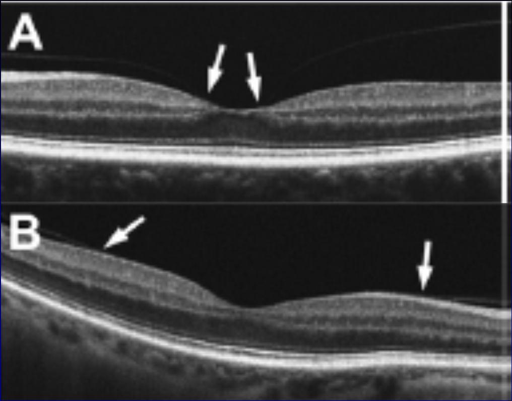
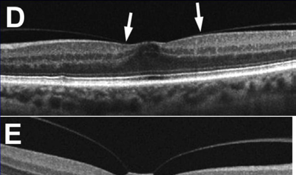
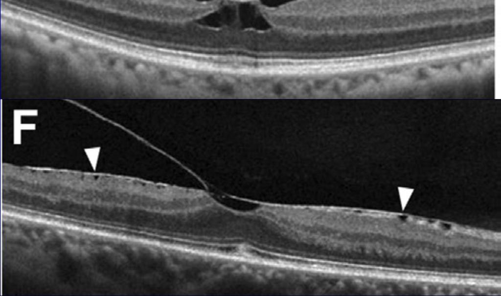
 Earliest detectable evidence for PVD (arrow) in a 64-year-old woman.")
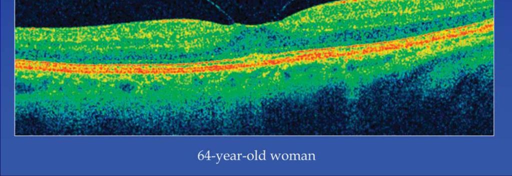
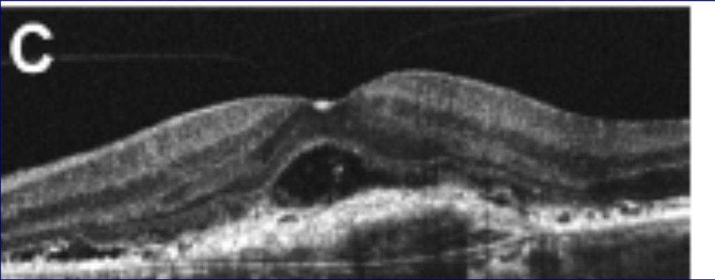
 Perifoveal PVD with focal foveolar vitreous attachment in a 64-yearold woman (note premacular liquefied pocket).")
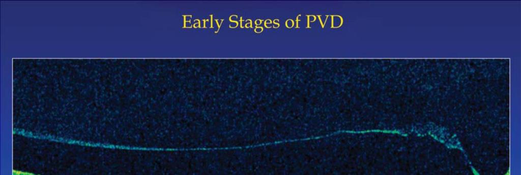
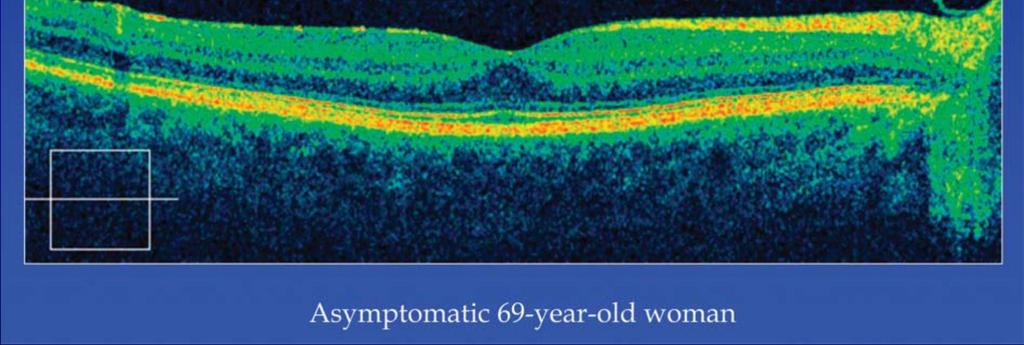
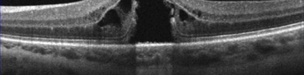
26 Spectral-domain optical coherence tomography images of early stages of posterior vitreous detachment (PVD) in asymptomatic individuals. (Top) Earliest detectable evidence for PVD (arrow) in a 64-year-old woman. (Second row) Evolving perifoveal PVD with broad residual macular adhesion in a 54-year-old man. (Third row) Perifoveal PVD with focal foveolar vitreous attachment in a 64-yearold woman (note premacular liquefied pocket). (Bottom) PVD extending through the entire macular region in a 69-year-old woman.
27
28
29
30 VITREOMACULAR ADHESIONS

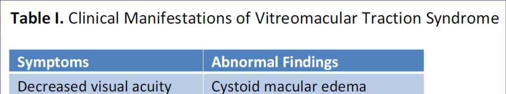
31 VITREOMACULAR TRACTION
32
33 OCT Based Definition and Classification of Vitreomacular Adhesion VMA is the equivalent of a stage 1 PVD Most eyes have complete vitreoretinal adhesion at birth, so the concept of vitreoretinal adhesion and VMA is a normal state.

 Broad")
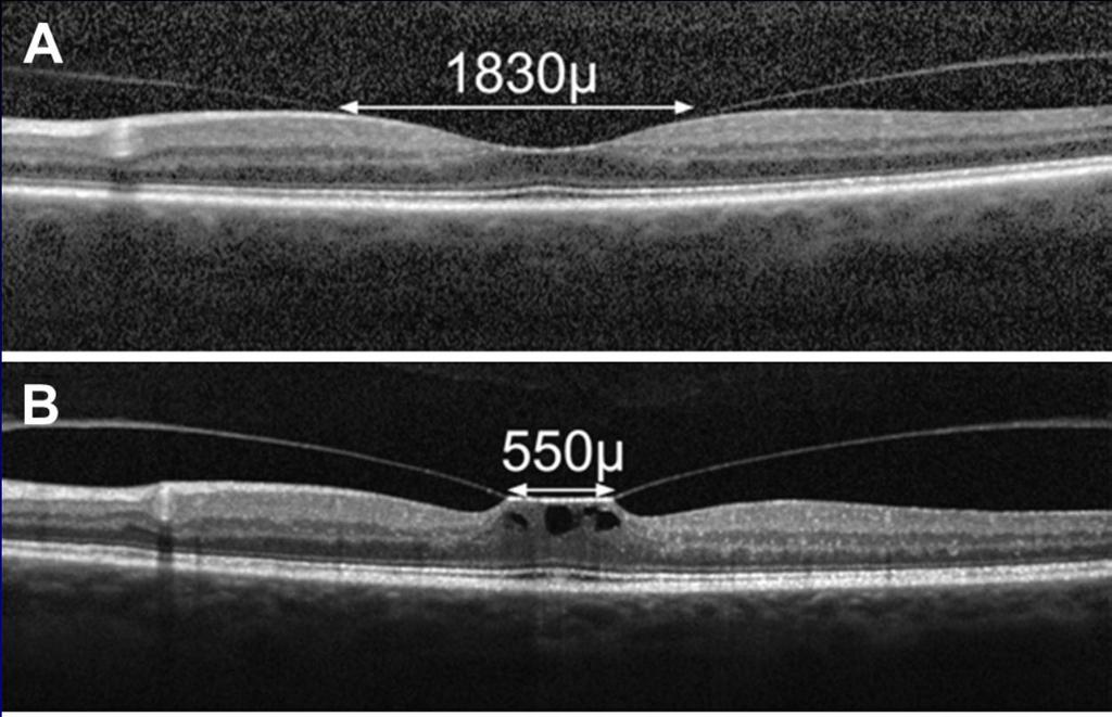
34 VMA (1) Focal ( 1500 µm) (2) Broad (>1500 µm;
35 If detectable retinal anatomic changes occur on OCT, with concurrent vitreous status showing perifoveolar PVD, the eye is characterized as having VMT.
36


37
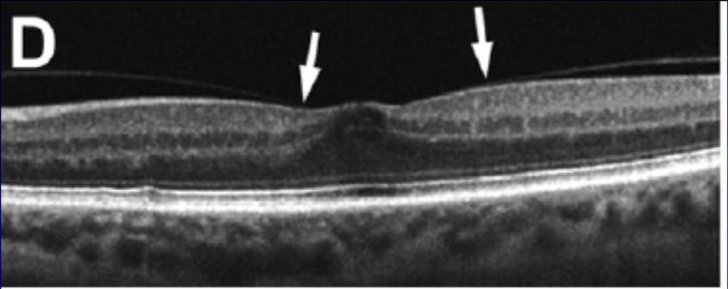
38 OCT Based Definition and Classification of Vitreomacular Traction (1) Evidence of perifoveal vitreous cortex detachment from the retinal surface; (2) macular attachment of the vitreous cortex within a 3-mm radius of the fovea; and (3) Association of attachment with distortion of the foveal surface, intraretinal structural changes, elevation of the fovea above the RPE, or a combination there of, but no fullthickness interruption of all retinal layers.
39 VMT Size: Focal ( 1500 mm) or Broad (>1500 mm) Isolated or concurrent
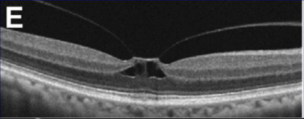
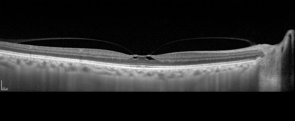
40 Broad areas of attachment with traction can cause: generalized thickening of the macula vascular leakage on FA macular schisis, cystoid macular edema.
41 Focal areas of vitreous attachment Foveal surface distortion. Elevate the foveal floor, pseudocysts within the central macula Combination
42
43 The presence of pseudocysts usually is associated with diminished visual acuity and visual distortion. After release of traction, pseudocysts generally resolve over time with little remaining visual deficit.


44 Spectral-domain optical coherence tomography scan from an eye with neovascular age-related macular degeneration showing associated stage 1 posterior vitreous detachment with broad vitreomacular adhesion zone.
45 Relationship to Epiretinal Membrane Formation. Autopsy studies reveal that residual vitreous remains on the surface of the retina in nearly half of all eyes with PVD. This condition is called vitreoschisis
46 The vitreous cortex temporally has a lamellar structure with two layers (arrows). The inner layer is detached from the outer lamellae. In this subject, the vitreous pocket is undetectable in both eyes.
47 Epiretinal membrane This residual vitreous may proliferate to form an epiretinal membrane (ERM) at any stage of vitreous separation. Clinical studies uniformly show a high incidence of apparent PVD in eyes with macular pucker.
48 ERM composed of : 1. Glial cells 2. laminocytes (histiocytes).
49 CLASSIFICATION OF ERM Grade 0: Cellophane Maculopathy A transparent membrane without distortion of the underlying retina is observed.
50 Grade 1: Crinkled Cellophane Maculopathy As the disease progresses, shrinkage or contraction of the ierm results in irregular wrinkling of the inner layers of the retina.

51
52 Grade 2: Preretinal Macular Fibrosis The ierm is characterized by a thicker and more opaque membrane obscuring the underlying retinal vasculature and a marked full-thickness retinal distortion.
53
54 Retinal edema, Small retinal hemorrhages, Cotton-wool spots, Exudates Opaque membranes.
55 Approximately 80% of patients with Grade 2 ierm will have symptoms of blurred vision and/or metamorphopsia.
56 In general, ierm is a chronic disease, and its onset and progression are usually slow. According to the BlueMountain Eye study, the 5-year cumulative progression rate from Grade 0 to Grade 2 ierm was reported as 9.3%, and the overall progression, regression, and stable rates were 28.6, 25.7, and 38.8%, respectively.
57 INDICATIONS OF TREATMENT Decreased vision Visual symptoms
58 Management Surgical Management of Idiopathic Epiretinal Membrane Trans pars plana vitrectomy and epiretinal membrane peeling have been used in patients with symptomatic visual disturbances as a standard procedure. However, in 10% to 21% of cases, the ERMs recurred, and 3% of recurrent cases required a second surgical intervention.
59 Therefore, additional ILM peeling is advised to achieve the complete removal of epi-ilm and sub-ilm proliferation, thus eliminating the scaffold for further proliferation. Several clinical series reported that ILM peeling seems to give better results than non-ilm peeling.
60 In both groups, equivalent efficacy and safety profiles in terms of final visual outcomes were found, whereas the ERM recurrence rate was lower in the ILM peeling than in the non-ilm peeling group.
61 Pharmaceutical Management of ierm and Associated Ocular Disorders Recent clinical data showed that preoperative cystoid macular edema is correlated to the presence of postoperative, persistent ntraretinal cysts, whereas an increased, preoperative central retinal thickness is correlated to an increased, postoperative central retinal thickness.
62 Topical antiinflammatory agents, including nonsteroidal antiinflammatory drugs, and dorzolamide might possibly be beneficial in the resolution of macular edema after vitrectomy for ierm. Schoenberger et al90 reported that the administration of topical nonsteroidal antiinflammatory drugs as compared with a placebo, resulted in more rapid reduction in macular volume.
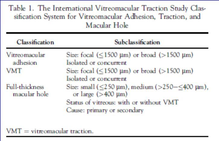
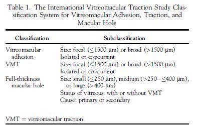
63 Full-Thickness Macular Hole Introduction and Definition: Full-thickness macular hole is an anatomic defect in the fovea featuring interruption of all neural retinal layers from the ILM to the RPE.
64 Optical Coherence TomographyeBased Full-Thickness Macular Hole Classification System Size of Hole, Presence or Absence of Vitreomacular Traction, Cause.
 predict anatomic treatment with both medications and")
65 The OCT-based measurements of minimum hole width (aperture size) predict anatomic treatment with both medications and surgery


66 A small FTMH features an aperture size of less than 250 µm
67 The cutoff for small FTMHs at 250 µm is derived from studies showing that these holes are associated with a small rate of spontaneous closure, have a very high closure rate with vitrectomy.


68 A medium FTMH is defined by aperture size from 250 to 400 µm
69 Studies exploring postsurgical FTMH closure rates by aperture size consistently show a very high anatomic closure rate (>90% in all recent series) with complete removal of residual hyaloid, with or without ILM peeling.
70
71 Nearly half of FTMHs are large (diameter >400 mm) at the time of diagnosis. Vitrectomy with ILM peel is associated with high closure rates (90%e95%), even for these large holes. Without an ILM peel, the vitrectomy success rate is closer to 75%. In the few eyes with large FTMH that have undergone pharmacologic vitreolysis, no anatomic success has been recorded.
72 Status of the Vitreous: Presence or Absence of Vitreomacular Traction. In the OCT-based anatomic system, FTMHs are categorized secondarily according to absence or presence of vitreous attachment Only macular holes with concurrent VMT should be considered for pharmacologic vitreolysis.
73 Primary Versus Secondary Full- Thickness Macular Hole. Full-thickness macular hole can be subdivided into primary and secondary forms. Primary FTMH (VMT)idiopathic) results anomalous PVD (VMT). A secondary FTMH is caused directly by other pathology.
74 secondary FTMH (1) blunt trauma, (2) lightning strike, (3) high myopia (4) macular schisis, (5) macular telangiectasia type 2, (6) wet macular degeneration, (7) macroaneurysm, (8) surgical trauma. (9) diabetic macular edema, (10) retinal vascular occlusions (11)uveitis.
75 It recently was shown that fovea destabilization resulting from tractioninduced damage to the inner fovea, occurring before or coincident with spontaneous vitreofoveal separation, may predispose some eyes to macular hole formation.
76
77 Impending Macular Hole A special circumstance exists when an individual develops FTMH in one eye and OCT reveals VMA or VMT in the fellow eye. Studies show that these fellow eyes are at increased risk for development of FTMH.
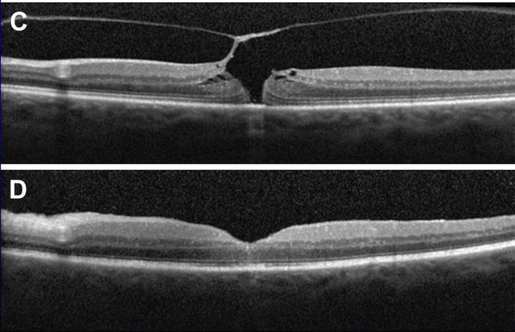
78 LMH Definition: Irregular foveal contour Defect in the inner fovea (may not have actual loss of tissue) Intraretinal splitting (schisis), typically between the outer plexiform and outer nuclear layers Maintenance of an intact photoreceptor layer

79
80 Lamellar macular hole usually progresses slowly and is thought to arise from incomplete FTMH formation, centripetal traction from ERM, or both, although current understanding of LMH evolution and surgical indications remains incomplete. Forces acting on the central macula include an ERM in some patients and varying anteroposterior and tangential forces from the vitreous in others.
81 Lamellar macular hole Most patients with LMH have mild metamorphopsia, limited central vision loss, and stable visual acuity.
82 Surgery for LMH remains controversial, and patient selection is crucial. Results of surgical interventions for LMH are variable, with anywhere between 25% and 75% of patients achieving improved visual outcomes, typically because of peeling of the associated ERM. 57.
83 Pseudomacular hole
84 OCT confirms the diagnosis on the basis of the following 4 characteristics (1) invaginated or heaped foveal edges, (2) concomitant ERM with central opening, (3) steep macular contour to the central fovea with near-normal central foveal thickness, (4) no loss of retinal tissue.
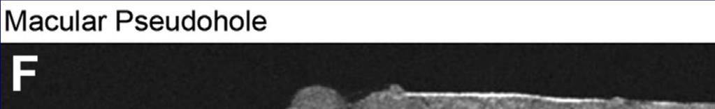

85
86 Perhaps the most characteristic feature of macular pseudohole is the presence of a concomitant ERM on the surface of the macula that distorts the foveal contour into a shape with a steep slope; altered light reflex also is observed commonly
87 Management of macular pseudohole typically is conservative. If the ERM is associated with a significant decline in vision, pars plana vitrectomy with membrane peeling can be performed.
88 Dr Kaiser presented six-month follow-up data on Ocriplasmin for the Treatment of Macular Hole: Phase III Results. The main points included: Approximately 40.6% of patients with full thickness macular hole (FTMH) achieved closure compared with 17% on placebo (p=0.004) Approximately 58% of patients with FTMHs smaller than 250 μm saw closure with ocriplasmin compared with just 20% in placebo patients 27% of patients gained 3 lines in visual acuity after six months of treatment with ocriplasmin, compared with 13% on placebo
89


90

91
VMA at the macula resulting in VMT
 Ocriplasmina for pharmacologic treatment in VMT Teresio Avitabile 1 Introduction PVD is a normal, physiologic process that occurs with aging; however, in some cases, PVD is incomplete Incomplete PVD localized
Vitreomacular Traction: Management
 Miscellaneous Refractive Surgery Vitreomacular Traction: Management Raji K. MS, DNB Raji K. MS, DNB, A.K. Upadhyay MS, S. Waikar MS, DNB, P. Tiwari MBBS Department of Ophthalmology, Command Hospital (WC)
EPIRETINAL MEMBRANE & VITREOMACULAR TRACTION
 EPIRETINAL MEMBRANE & VITREOMACULAR TRACTION Management of ERM and VMT K.V.Chalam,MD,PhD,MBA,FACS Professor and Director of Retina Loma Linda Eye Institute Los Angeles, USA REVIEW ANATOMY The vitreous
Managing the Vitreomacular Interface
 Managing the Vitreomacular Interface A Guide to VMA, VMT, Holes and ERM Anna K. Bedwell, OD, FAAO Indiana University School of Optometry Please silence all mobile devices and remove items from chairs so
Yasser R. Serag, MD Tamer Wasfi, MD El- Saied El-Dessoukey, MD Magdi S. Moussa, MD Anselm Kampik, MD
 Microperimetric Evaluation of Brilliant Blue G- assisted Internal Limiting Membrane Peeling By Yasser R. Serag, MD Tamer Wasfi, MD El- Saied El-Dessoukey, MD Magdi S. Moussa, MD Anselm Kampik, MD The internal
The Foundation WHAT IS THE RETINA? continued next page. RETINA HEALTH SERIES Facts from the ASRS
 The Foundation American Society of Retina Specialists Committed to improving the quality of life of all people with retinal disease. Vitreomacular Traction Syndrome The vitreous humor is a transparent,
evaluation of vitreoretinal adhesions in exudative AMD using optical coherence tomography
 evaluation of vitreoretinal adhesions in exudative AMD using optical coherence tomography Dr. Mahmoud Alaa Abouhusssein, FRCO Lecturer of ophthalmology, Alexandria university Dr. Amir Ramadan Gomaa, MD
An A to Z guide on Epiretinal Membranes (ERMs) Paris Tranos PhD,ICO,FRCS OPHTHALMICA Vitreoretinal & Uveitis Department
 Paris Tranos PhD,ICO,FRCS OPHTHALMICA Vitreoretinal & Uveitis Department") An A to Z guide on Epiretinal Membranes (ERMs) Paris Tranos PhD,ICO,FRCS OPHTHALMICA Vitreoretinal & Uveitis Department Types of ERM Natural history OCT prognostic factors ERM with co-existing pathology
An A to Z guide on Epiretinal Membranes (ERMs) Paris Tranos PhD,ICO,FRCS OPHTHALMICA Vitreoretinal & Uveitis Department Types of ERM Natural history OCT prognostic factors ERM with co-existing pathology
Early diagnosis and treatment of VMT with single Intravitreal Injection of Pharmacologic Vitreolysis. Stratos Gotzaridis MD Athens
 Early diagnosis and treatment of VMT with single Intravitreal Injection of Pharmacologic Vitreolysis Stratos Gotzaridis MD Athens The Vitreous Body Gel composed of 98-99% water 1% macromolecules Glycoproteins
OPTIC DISC PIT Pathogenesis and Management OPTIC DISC PIT
 OPTIC DISC PIT Pathogenesis and Management Abdel-Latif Siam Ain Shams University Cairo Egypt OPTIC DISC PIT Congenital pit is an atypical coloboma usually located on the temporal edge of the disc, associated
OCT Assessment of the Vitreoretinal Relationship in CSME
 December 2007 Sonia Rani John et al. - IFIS 375 ORIGINAL ARTICLE OCT Assessment of the Vitreoretinal Relationship in CSME Dr. Manoj S. DNB FRCS, Dr. Unnikrishnan Nair MS DO FRCS, Dr. Gargi Sathish MS Introduction
Often asymptomatic but can cause a reduction in BCVA and distortion of vision.
 Christopher Wolfe, OD, FAAO, Dipl. ABO Epiretinal Membrane (ERM) and Vitreomacular Traction (VMT) Epiretinal membrane (macular pucker, cellophane maculopathy, premacular fibrosis) consists of a layer of
Vitreo-retinal interface pathologies and fibrinolytic treatment approaches
 Vitreo-retinal interface pathologies and fibrinolytic treatment approaches Constantin J. Pournaras Memorial A. de Rothschild Clinical Research Group La Colline Ophthalmology Center Vitreoretinal Interface
Optical Coherence Tomography in Diabetic Retinopathy. Mrs Samantha Mann Consultant Ophthalmologist Clinical Lead of SEL-DESP
 Optical Coherence Tomography in Diabetic Retinopathy Mrs Samantha Mann Consultant Ophthalmologist Clinical Lead of SEL-DESP Content OCT imaging Retinal layers OCT features in Diabetes Some NON DR features
PREDICTIVE FACTORS OF VISUAL OUTCOME FOR VITREOMACULAR TRACTION SYNDROME AFTER VITRECTOMY
 PREDICTIVE FACTORS OF VISUAL OUTCOME FOR VITREOMACULAR TRACTION SYNDROME AFTER VITRECTOMY Downloaded from https://journals.lww.com/retinajournal by mv7bzw+nz2blpko//cqyhwu2mokppdiwuep6ir1molueskh0dp9rbmb7dum5a2/cp6zifirtq3zbawzt+95f/m61fycawpqbpe8y2wuyzwnns2gw3+gmrxei6x11wu+s
Audit of Macular Hole Surgery, Visual Outcome Prediction on OCT Appearance of Macular Hole
 International Journal of Ophthalmology & Visual Science 2017; 2(4): 93-97 http://www.sciencepublishinggroup.com/j/ijovs doi: 10.11648/j.ijovs.20170204.13 Audit of Macular Hole Surgery, Visual Outcome Prediction
Idiopathic vitreomacular traction and macular hole: a comprehensive review of pathophysiology, diagnosis, and treatment
 OPEN (2013) 27, S1 S21 & 2013 Macmillan Publishers Limited All rights reserved 0950-222X/13 www.nature.com/eye Idiopathic vitreomacular traction and macular hole: a comprehensive review of pathophysiology,
Optical coherence tomography of the vitreoretinal interface in macular hole formation
 1092 St Thomas s Hospital, London V Tanner D S Chauhan T L Jackson T H Williamson Correspondence to: Mr V Tanner, Royal Berkshire Hospital, London Road, Reading RG1 5AN, UK tannerone@aol.com Accepted for
Financial Disclosures
 Financial Disclosures Consultant Genentech, Regeneron, Allergan, Thrombogenics, Optos, and ArcticDx Grant Support Regeneron, Allergan Mathew W. MacCumber, MD, PhD Professor & Assoc. Chair for Research
History/principles of the OCT What does the normal retinal OCT look like Vitreal disorders Retinal/RPE disorders Choroidal disorders
 Nathan Lighthizer, O.D., F.A.A.O. Assistant Professor Assistant Dean for Clinical Care Director of Continuing Education Chief of Specialty Care Clinics Chief of Electrodiagnostics Clinic Oklahoma College
Ocriplasmin for the Treatment of Symptomatic Vitreomacular Adhesion/Traction. Baruch D Kuppermann, MD, PhD
 Ocriplasmin for the Treatment of Symptomatic Vitreomacular Adhesion/Traction Baruch D Kuppermann, MD, PhD Professor of Ophthalmology and Biomedical Engineering; Chief, Service; Vice-Chair, Clinical Research,
Case report 12/10/2014. Delphine Lam ; Dr Mayer Srour Service d ophtalmologie Professeur E.Souied Université Paris Est
 Case report 12/10/2014 Delphine Lam ; Dr Mayer Srour Service d ophtalmologie Professeur E.Souied Medical history Man, 75 years old Complaint: Vision loss in left eye in June 2014 Past ophthalmologic history:
Re)nal and OCT Grand Rounds. What's new in OCT? Principles of AngioVue OCTA. Vascular Imaging No Referral Needed 3/9/18. Spectral Domain: Many Op3ons
nal and OCT Grand Rounds. What's new in OCT? Principles of AngioVue OCTA. Vascular Imaging No Referral Needed 3/9/18. Spectral Domain: Many Op3ons") Spectral Domain: Many Op3ons Re)nal and OCT Grand Rounds Steven Ferrucci, OD, FAAO Chief, Optometry Sepulveda VA Professor, SCCO/MBKU Ease of use Customer support Integra)on of other technology FAF Color
Spectral Domain: Many Op3ons Re)nal and OCT Grand Rounds Steven Ferrucci, OD, FAAO Chief, Optometry Sepulveda VA Professor, SCCO/MBKU Ease of use Customer support Integra)on of other technology FAF Color
The Quick Guide to OCT Mastery 50 Real Cases with Expert Analysis
 OPTICAL COHERENCE TOMOGRAPHY The Quick Guide to OCT Mastery 50 Real Cases with Expert Analysis VOL 1 Sanjay Sharma, MD, FRCS, MSc (Epid), MBA Ophthalmologist, Epidemiologist Queen s University, Canada
Foveal Red Spot, Macular Microhole and Foveal Photoreceptor Defect in the Era of High-Resolution Optical Coherence Tomography
 1:15 PM Foveal Red Spot, Macular Microhole and Foveal Photoreceptor Defect in the Era of High-Resolution Optical Coherence Tomography Edward F. Hall, MD Steven J. Rose, MD Brian P. Connolly, MD Ernest
Treatment Options for VMT and Macular Holes Observation, Surgery, and Pharmacotherapy
 Treatment Options for VMT and Macular Holes Observation, Surgery, and Pharmacotherapy Andrew Moshfeghi, MD, MBA Bascom Palmer Eye Institute Palm Beach Gardens, FL Financial Disclosures Salary/Honoraria:
The Foundation WHAT IS THE RETINA? continued next page. RETINA HEALTH SERIES Facts from the ASRS
 The Foundation American Society of Retina Specialists Committed to improving the quality of life of all people with retinal disease. Epiretinal Membranes (ERMs), also commonly known as cellophane maculopathy
Dehiscence of detached internal limiting membrane in eyes with myopic traction maculopathy with spontaneous resolution
 Hirota et al. BMC Ophthalmology 2014, 14:39 RESEARCH ARTICLE Open Access Dehiscence of detached internal limiting membrane in eyes with myopic traction maculopathy with spontaneous resolution Kazunari
OCT in Diabetic Macular Edema and its Correlation with Flourescein Angiography
 Uvea OCT in Diabetic Macular Edema and its Correlation with Flourescein Angiography Kirti Jaisingh MS Kirti Jaisingh MS, Yashpal Goel* MS, Kshitij Aditya** DO * Guru Nanak Eye Centre, New Delhi ** Baba
8/6/17. Disclosures Aerie Pharmaceuticals Alcon BioTissue Diopsys Optovue Shire
 Nathan Lighthizer, O.D., F.A.A.O. Associate Professor Assistant Dean for Clinical Care Director of Continuing Education Chief of Specialty Care Clinics Oklahoma College of Optometry Tahlequah, OK lighthiz@nsuok.edu
Visual and Anatomical Outcomes of Vitreous Surgery for Large Macular Holes
 March 2009 Raju K.V. et al. - Closed Globe Injuries 31 ORIGINAL ARTICLE Visual and Anatomical Outcomes of Vitreous Surgery for Large Macular Holes Dr. Mahesh G. MS DO DNB FRCSEd, Dr. A. Giridhar MS, Dr.
Χειρουργική Ωχράσ Κηλίδασ. Γ. Γ. Παππάς, Βεληδέιεηο ΓΝ
 Χειρουργική Ωχράσ Κηλίδασ Γ. Γ. Παππάς, Βεληδέιεηο ΓΝ FREQUENCY OF MACULAR SURGERY Incidence= 8 in 100000 (McCannel 2009) Prevalence = between 0.2 (Mitchell 2007) and 3.3 (Baltimore Eye Survey 1996) 2nd
Vitreomacular Traction
 Supplement to March 2014 Rethink Vitreomacular Traction With articles by Pravin U. Dugel, MD Anselm Kampik, MD J. Sebag, MD, FACS, FRCOphth, FARVO Ramin Tadayoni, MD, PhD Sponsored by Alcon The articles
Macular Hole Associated with Vogt-Koyanagi-Harada Disease at the Acute Uveitic Stage
 Published online: September 15, 2015 2015 The Author(s) Published by S. Karger AG, Basel 1663 2699/15/0063 0328$39.50/0 This article is licensed under the Creative Commons Attribution-NonCommercial 4.0
Re(nal and OCT Grand Rounds
 Op#cal Coherence Tomography Op(cal: Light- based Re(nal and OCT Grand Rounds Steven Ferrucci, OD, FAAO Chief, Optometry Sepulveda VA Professor, SCCO/MBKU Coherence: property of light waves in which the
Clinical spectrum of lamellar macular defects including pseudoholes and pseudocysts defined by optical coherence tomography
 Clinical spectrum of lamellar macular defects including pseudoholes and pseudocysts defined by optical coherence tomography J C Chen, 1,2 L R Lee 1,3 1 City Eye Centre, Brisbane, Australia; 2 Institute
CLINICAL COURSE OF VITREOMACULAR ADHESION MANAGED BY INITIAL OBSERVATION
 CLINICAL COURSE OF VITREOMACULAR ADHESION MANAGED BY INITIAL OBSERVATION VISHAK J. JOHN, MD,* HARRY W. FLYNN, JR., MD,* WILLIAM E. SMIDDY, MD,* ADAM CARVER, MD, ROBERT LEONARD, MD, HOMAYOUN TABANDEH, MD,
THE NATURAL HISTORY OF TRACTIONAL CYSTOID MACULAR EDEMA
 THE NATURAL HISTORY OF TRACTIONAL CYSTOID MACULAR EDEMA SOFIA CHARALAMPIDOU, MRCOPHTH,* JOHN NOLAN, PHD, STEPHEN BEATTY, FRCOPHTH* Background: To describe clinical outcomes in a series of patients with
 This article was originally published in the Encyclopedia of the Eye, published by Elsevier, and the attached copy is provided by Elsevier for the author's benefit and for the benefit of the author's institution,
Observation of Posterior Precortical Vitreous Pocket Using Swept-Source Optical Coherence Tomography
 Anatomy and Pathology Observation of Posterior Precortical Vitreous Pocket Using Swept-Source Optical Coherence Tomography Hirotaka Itakura, Shoji Kishi, Danjie Li, and Hideo Akiyama Department of Ophthalmology,
Early surgery preserves more vision for patients with Epiretinal Membranes
 Early surgery preserves more vision for patients with Epiretinal Membranes Rahman R 1, Stephenson J 2 KEYWORDS: Epiretinal membrane, Combined phakovitrectomy, OCT. Addresses: 1 Ms Rubina Rahman*, CalderdaleRoyalHospital,
Ocriplasmin for Treatment of Vitreomacular Traction: An Update
 Ophthalmol Ther (2016) 5:147 159 DOI 10.1007/s40123-016-0062-6 REVIEW Ocriplasmin for Treatment of Vitreomacular Traction: An Update Mohammed Ali Khan. Julia A. Haller Received: July 22, 2016 / Published
Optical Coherence Tomograpic Features in Idiopathic Retinitis, Vasculitis, Aneurysms and Neuroretinitis (IRVAN)
") Columbia International Publishing Journal of Ophthalmic Research (2014) Research Article Optical Coherence Tomograpic Features in Idiopathic Retinitis, Vasculitis, Aneurysms and Neuroretinitis (IRVAN)
Columbia International Publishing Journal of Ophthalmic Research (2014) Research Article Optical Coherence Tomograpic Features in Idiopathic Retinitis, Vasculitis, Aneurysms and Neuroretinitis (IRVAN)
A retrospective nonrandomized study was conducted at 3
 Department of Ophthalmology, Kangbuk Samsung Hospital, Sungkyunkwan University College of Medicine 1, Seoul, Korea Hangil Eye Hospital 2, Incheon, Korea Seoul National University Bundang Hospital 3, Seongnam,
OCT Interpretation. Financial Disclosure. Jay M. Haynie, OD, FAAO. OCT Image Layers 7/21/2014
 OCT Interpretation Jay M. Haynie, OD, FAAO Financial Disclosure I have received honoraria or am on the advisory board for the following companies: Olympia Tacoma Renton Kennewick - Washington Carl Zeiss
Course # Getting to Know Your OCT
 Course # 140 Getting to Know Your OCT Course Title: Lecturer: Getting to Know Your OCT Brad Sutton, OD, FAAO IU School of Optometry Financial Disclosures No financial disclosures Optical Coherence Tomography-OCT
CLINICAL SCIENCES. Optical Coherence Tomography Findings in Myopic Traction Maculopathy
 Optical Coherence Tomography Findings in Myopic Traction Maculopathy Giacomo Panozzo, MD; Andrea Mercanti, MD CLINICAL SCIENCES Objective: To describe the features and incidence of epiretinal traction
ATLAS OF OCT. Retinal Anatomy in Health & Pathology by Neal A. Adams, MD. Provided to you by:
 ATLAS OF OCT Retinal Anatomy in Health & Pathology by Neal A. Adams, MD Provided to you by: Atlas of OCT The OCT Atlas is written by Neal A. Adams, MD, and produced by Heidelberg Engineering, Inc. to help
We are IntechOpen, the world s leading publisher of Open Access books Built by scientists, for scientists. International authors and editors
 We are IntechOpen, the world s leading publisher of Open Access books Built by scientists, for scientists 3,500 108,000 1.7 M Open access books available International authors and editors Downloads Our
Optical coherence tomography-guided classification of epiretinal membranes
 DOI 10.1007/s10792-014-9975-z ORIGINAL PAPER Optical coherence tomography-guided classification of epiretinal membranes Vasileios Konidaris Sofia Androudi Alexandros Alexandridis Anna Dastiridou Periklis
PART 1: GENERAL RETINAL ANATOMY
 PART 1: GENERAL RETINAL ANATOMY General Anatomy At Ora Serrata At Optic Nerve Head Fundoscopic View Of Normal Retina What Is So Special About Diabetic Retinopathy? The WHO definition of blindness is
Review Article Spectral Domain Optical Coherence Tomography in the Diagnosis and Management of Vitreoretinal Interface Pathologies
 Hindawi Publishing Corporation Volume 2012, Article ID 876472, 7 pages doi:10.1155/2012/876472 Review Article Spectral Domain Optical Coherence Tomography in the Diagnosis and Management of Vitreoretinal
When optical coherence tomography (OCT)
") Macular Imaging: SD-OCT in nterior Segment Surgical Practice Many pathologic processes of the macula can be visualized or quantified only with this modality. y Steven G. Safran, MD When optical coherence
Macular Imaging: SD-OCT in nterior Segment Surgical Practice Many pathologic processes of the macula can be visualized or quantified only with this modality. y Steven G. Safran, MD When optical coherence
Macular Hole. Helpline
 Macular Hole The retina is a light-sensitive layer of tissue lining the back of the eye. The macula is a small area at the centre of the retina responsible for all of our central vision, most of our colour
Optical Coherence Tomography s Contribution to the Diagnosis of the Pathologies of the Vitreoretinal Interface in Lomé
 Open Journal of Ophthalmology, 2018, 8, 180-189 http://www.scirp.org/journal/ojoph ISSN Online: 2165-7416 ISSN Print: 2165-7408 Optical Coherence Tomography s Contribution to the Diagnosis of the Pathologies
R&M Solutions
 Mohamed Hosny El-Bradey, MD., Assistant Professor of Ophthalmology, Tanta University. Wael El Haig, MD., Professor of Ophthalmology. Zagazeeg University. 1 Myopic CNV is considered the most common vision
The College of Optometrists - Learning outcomes for the Professional Certificate in Medical Retina
 Learning outcomes for the Professional Certificate in Medical Retina, incorporating diabetic retinopathy screening and age related macular degeneration The professional certificate is a prerequisite to
PROSPECTIVE THREE-DIMENSIONAL ANALYSIS OF STRUCTURE AND FUNCTION IN VITREOMACULAR ADHESION CURED BY PHARMACOLOGIC VITREOLYSIS
 PROSPECTIVE THREE-DIMENSIONAL ANALYSIS OF STRUCTURE AND FUNCTION IN VITREOMACULAR ADHESION CURED BY PHARMACOLOGIC VITREOLYSIS Kevin R. Tozer, BS,* Wolfgang Fink, PhD, ** Alfredo A. Sadun, MD, PhD, FARVO,
Progressive Symptomatic Retinal Detachment Complicating Retinoschisis. Initial Reporting Questionnaire
 Progressive Symptomatic Retinal Detachment Complicating Retinoschisis In association with the British Ophthalmological Surveillance Unit Ethics ref: 13/NW/0037 Initial Reporting Questionnaire Case Definition:
DIABETIC VITRECTOMY INDICATIONS AND TECHNIQUES. steve charles
 DIABETIC VITRECTOMY INDICATIONS AND TECHNIQUES steve charles Traction Retinal Detachment Macula involved TRD TRD with rhegmatogenous component even if extra-macular TTRD Extra-Macular TRD should be observed
Eccentric Macular Hole after Pars Plana Vitrectomy for Epiretinal Membrane Without Internal Limiting Membrane Peeling: A Case Report
 Ophthalmol Ther (2017) 6:391 395 DOI 10.1007/s40123-017-0113-7 CASE REPORT Eccentric Macular Hole after Pars Plana Vitrectomy for Epiretinal Membrane Without Internal Limiting Membrane Peeling: A Case
Optical Coherence Tomography: Pearls for the Anterior Segment Surgeon Basic Science Michael Stewart, M.D.
 Optical Coherence Tomography: Pearls for the Anterior Segment Surgeon Basic Science Michael Stewart, M.D. Disclosure OCT Optical Coherence Tomography No relevant financial relationships I will refer to
VITREOMACULAR UPDATE FOR THE PRIMARY CARE OD
 VITREOMACULAR UPDATE FOR THE PRIMARY CARE OD VITREOMACULAR UPDATE FOR THE PRIMARY CARE OD 1 2 DISCLOSURE STATEMENT I have received lecture honoraria from TearScience. I have no direct financial or proprietary
VITREOPAPILLARY ADHESION IN MACULAR DISEASES ABSTRACT INTRODUCTION METHODS AND MATERIALS
 VITREOPAPILLARY ADHESION IN MACULAR DISEASES BY J. Sebag MD FRCOphth,* Michelle Y. Wang MD, Dieuthu Nguyen MD, AND Alfredo A. Sadun MD PhD ABSTRACT Purpose: The effect of vitreopapillary adhesion (VPA)
Cirrus TM HD-OCT. Details defi ne your decisions
 Cirrus TM HD-OCT Details defi ne your decisions 2 With high-defi nition OCT Carl Zeiss Meditec takes you beyond standard spectral domain Built on 10 years experience at the vanguard of innovation, Carl
Diagnosis and treatment of diabetic retinopathy. Blake Cooper MD Ophthalmologist Vitreoretinal Surgeon Retina Associates Kansas City
 Diagnosis and treatment of diabetic retinopathy Blake Cooper MD Ophthalmologist Vitreoretinal Surgeon Retina Associates Kansas City Disclosures Consulted for Novo Nordisk 2017,2018. Will be discussing
ZEISS AngioPlex OCT Angiography. Clinical Case Reports
 Clinical Case Reports Proliferative Diabetic Retinopathy (PDR) Case Report 969 PROLIFERATIVE DIABETIC RETINOPATHY 1 1-year-old diabetic female presents for follow-up of proliferative diabetic retinopathy
CORRELATION BETWEEN CENTRAL FOVEAL THICKNESS AND VISUAL ACUITY IN PATIENTS WITH IDIOPATHIC VITREOMACULAR TRACTION
 CORRELATION BETWEEN CENTRAL FOVEAL THICKNESS AND VISUAL ACUITY IN PATIENTS WITH IDIOPATHIC VITREOMACULAR TRACTION MEHMET M. UZEL, MD, MEHMET CITIRIK, MD, CAGRI ILHAN, MD, KEMAL TEKIN, MD Purpose: To evaluate
Spontaneous Large Serous Retinal Pigment Epithelial Tear
 This is an Open Access article licensed under the terms of the Creative Commons Attribution-NonCommercial-NoDerivs 3.0 License (www.karger.com/oa-license), applicable to the online version of the article
11/29/2016 MACULAR MALADIES: TYPICAL & ATYPICAL CASES
 MACULAR MALADIES: TYPICAL & ATYPICAL CASES Dawn Pewitt, OD, FAAO Triad Eye Institute, Grove, OK Dpewitt@triadeye.com Disclosure Statement: No financial disclosures COPE 51218-PS Please silence all mobile
THESES ON INCIDENCE, CORRELATION AND NATURAL HISTORY OF EPIRETINAL MEMBRANES SURROUNDING IDIOPATHIC MACULAR HOLES
 THESES ON INCIDENCE, CORRELATION AND NATURAL HISTORY OF EPIRETINAL MEMBRANES SURROUNDING IDIOPATHIC MACULAR HOLES Duration 1YEAR AIM to evaluate the prevalence, correlation and natural history of EPIRETINAL
case profile: Macular Holes
 Early Detection Saves Sight 2012 ISSUE 2 newsletter for optometrists I m pleased to let you know about a new series of resources for optometrists that is being produced by the team at CFEH. The Clinical
The vitreoretinal interface and its role in the pathogenesis of vitreomaculopathies
 English version of "Die vitreoretinale Grenzfläche und ihre Rolle in der Pathogenese vitreomakulärer Erkrankungen" DOI 10.1007/s00347-014-3048-6 Springer-Verlag Berlin Heidelberg 2015 J. Sebag VMR Institute
Outline. Outline. Vitreous Development & Anatomy OPT - 243
 2010 OPT - 243 Vitreous Disorders & Vitreoretinal Disorders of the Posterior Pole I Leo Semes, OD, FAAO 100% 0% 0% 0% 0% Which of these gives the best resolution for studying vitreoretinal disorders of
We are IntechOpen, the world s leading publisher of Open Access books Built by scientists, for scientists. International authors and editors
 We are IntechOpen, the world s leading publisher of Open Access books Built by scientists, for scientists 3,900 116,000 120M Open access books available International authors and editors Downloads Our
IN NICU OCT UTILIZES A CONCEPT KNOWN AS INTERFEROMETRY APPLICATIONS FOR OCT THE PRIMARY USE IN THE EYE - RETINA
 2016 25 YEARS OF OPTICAL COHERENCE TOMOGRAPHY OPTICAL COHERENCE TOMOGRAPHY IN NICU Marcin Stopa, MD, PhD, FEBO Department of Ophthalmology, Chair of Ophthalmology and Optometry. Poznan University of Medical
Moncef Khairallah, MD
 Moncef Khairallah, MD Department of Ophthalmology, Fattouma Bourguiba University Hospital Faculty of Medicine, University of Monastir Monastir, Tunisia INTRODUCTION IU: anatomic form of uveitis involving
Posterior Segment Update
 Posterior Segment Update Featured Speaker: Dr. Kyle Cheatham, FAAO, DIP ABO DISCLOSURE STATEMENT We have no direct financial or proprietary interest in any companies, products or services mentioned in
Vitrectomy for Diabetic Retinopathy The current role of pars plana vitrectomy for diabetic macular edema and proliferative diabetic retinopathy.
 Vitrectomy for Diabetic Retinopathy The current role of pars plana vitrectomy for diabetic macular edema and proliferative diabetic retinopathy. Y R.V. PUL CHN, MD; ND DONLD J. D MICO, MD The incidence
! Reichert, Alcon, Allergan, CZ & Zeavision. Uchino, E. et al. Arch Ophthalmol ! Vitreoschisis (split within the vitreous)! ERM!
! ERM!") Financial disclosure From Print to Practice: PVD a common process with potential for ocular morbidity! I have received lecture honoraria or serve on the advisory boards or speaker s bureaus of:! Reichert,
Financial disclosure From Print to Practice: PVD a common process with potential for ocular morbidity! I have received lecture honoraria or serve on the advisory boards or speaker s bureaus of:! Reichert,
Related Policies None
 Medical Policy BCBSA Ref. Policy: 9.03.30 Last Review: 03/29/2018 Effective Date: 03/29/2018 Section: Other Related Policies None DISCLAIMER Our medical policies are designed for informational purposes
Retrospective study on outcome of macular hole surgery
 Original article Singh S, Byanju R, Pradhan S, Lamichhane G. Bharatpur Eye Hospital,Bharatpur Abstract Introduction: Macular hole is a common and treatable cause of central visual loss. Classic macular
Moving forward with a different perspective
 Moving forward with a different perspective The Leader In Vision Diagnostics Offers A New Perspective Marco has served the eyecare community by offering exceptional lane products and automated high tech
Diabetic Retinopathy. Barry Emara MD FRCS(C) Giovanni Caboto Club October 3, 2012
 Giovanni Caboto Club October 3, 2012") Diabetic Retinopathy Barry Emara MD FRCS(C) Giovanni Caboto Club October 3, 2012 Outline Statistics Anatomy Categories Assessment Management Risk factors What do you need to do? Objectives Summarize the
Diabetic Retinopathy Barry Emara MD FRCS(C) Giovanni Caboto Club October 3, 2012 Outline Statistics Anatomy Categories Assessment Management Risk factors What do you need to do? Objectives Summarize the
What Is O.C.T. and Why Should I Give A Rip? OCT & Me How Optical Coherence Tomography Changed the Life of a Small Town Optometrist 5/19/2014
 OCT & Me How Optical Coherence Tomography Changed the Life of a Small Town Optometrist Email: myoder@wcoil.com Mark A. Yoder, O.D. 107 N. Main Street PO Box 123 Bluffton, OH 45817 @yoderod 115.02 Histoplasma
Optical Coherence Tomography (OCT) in Uveitis Piergiorgio Neri, BMedSc, MD, PhD Head Ocular Immunology Unit
 in Uveitis Piergiorgio Neri, BMedSc, MD, PhD Head Ocular Immunology Unit") The Eye Clinic Polytechnic University of Marche Head: Prof Alfonso Giovannini November, 1991 Optical Coherence Tomography (OCT) in Uveitis Piergiorgio Neri, BMedSc, MD, PhD Head Ocular Immunology Unit
The Eye Clinic Polytechnic University of Marche Head: Prof Alfonso Giovannini November, 1991 Optical Coherence Tomography (OCT) in Uveitis Piergiorgio Neri, BMedSc, MD, PhD Head Ocular Immunology Unit
Consulting Fee: Alcon Laboratories
 Consulting Fee: Alcon Laboratories Pre-Op EMM Post PPV, Forceps EMM & ILM Peeling 25/27 Gauge, Trans-Conjunctival, Sutureless PPV Inside-Out, End-Grasping Forceps Peeling w/ Alcon 25G End-Grasping DSP
Is OCT-A Needed As An Investigative Tool During The Management Of Diabetic Macular Edema
 Is OCT-A Needed As An Investigative Tool During The Management Of Diabetic Macular Edema Ayman M Khattab MD, FRCS Professor of Ophthalmology Cairo University Diabetic Macular Edema (DME) Diabetic macular
Collagens and retinal Müller cells in healthy and diseased vitreoretinal interface Bu, Shao
 University of Groningen Collagens and retinal Müller cells in healthy and diseased vitreoretinal interface Bu, Shao IMPORTANT NOTE: You are advised to consult the publisher's version (publisher's PDF)
Steven Ferrucci, OD. FAAO; Jeffry Gerson, OD, FAAO; Robert Prouty, OD, FAAO; Leo semes OD, FAAO
 PARDON THE OBJECTION: RETINA Steven Ferrucci, OD. FAAO; Jeffry Gerson, OD, FAAO; Robert Prouty, OD, FAAO; Leo semes OD, FAAO 1. Introductions/Disclosures (Ferrucci) 2. The genetics of AMD (Gerson) a. Background
Practical Care of the Cataract Patient with Retinal Disease
 Practical Care of the Cataract Patient with Retinal Disease Brooks R. Alldredge, OD, FAAO Kelly L. Cyr, OD, FAAO The Retina Center Eye Associates of New Mexico 4411 The 25 Way NE, Suite 325 Albuquerque,
Gas for Vitreomacular Traction RCT (Protocol AG) Gas for Macular Hole Single-Arm Study (Protocol AH)
 Gas for Macular Hole Single-Arm Study (Protocol AH)") Gas for Vitreomacular Traction RCT (Protocol AG) Gas for Macular Hole Single-Arm Study (Protocol AH) Protocol AG Chair: Clement Chan, MD Protocol AH Chair: Calvin Mein, MD DRCR.net Protocol AG Randomized
Gas for Vitreomacular Traction RCT (Protocol AG) Gas for Macular Hole Single-Arm Study (Protocol AH) Protocol AG Chair: Clement Chan, MD Protocol AH Chair: Calvin Mein, MD DRCR.net Protocol AG Randomized
OCT Interpretation in Retinal Disease
 OCT Interpretation in Retinal Disease Jay M. Haynie, OD, FAAO Financial Disclosure I have received honoraria or am on the advisory board for the following companies: Carl Zeiss Meditec Advanced Ocular
 measure of your overall performance. An isolated glucose test is helpful to let you know what your sugar level is at one moment, but it doesn t tell you whether or not your diabetes is under adequate control
Intravitreal triamcinolone staining observation of residual undetached cortical vitreous after posterior vitreous detachment
 (2006) 20, 423 427 & 2006 Nature Publishing Group All rights reserved 0950-222X/06 $30.00 www.nature.com/eye Intravitreal triamcinolone staining observation of residual undetached cortical vitreous after
Diabetic Retinopathy
 Diabetic Retinopathy Diabetes can be classified into type 1 diabetes mellitus and type 2 diabetes mellitus, formerly known as insulin-dependent diabetes mellitus, and non-insulin diabetes mellitus, respectively.
3/6/2014. Hoda MH Mostafa MD Associate Professor of Ophthalmology Cairo University. The author has no proprietary interest. Today s Objectives
 Hoda MH Mostafa MD Associate Professor of Ophthalmology Cairo University The author has no proprietary interest Today s Objectives Identify the CLINICAL SCENARIOS IN MACULAR EDEMA where OCT plays a MAJOR
Dermatologic and Ophthalmic Drugs Advisory Committee Meeting Briefing Package. for. Ocriplasmin Intravitreal Injection, 2.5 mg/ml.
 Food and Drug Administration Center for Drug Evaluation and Research Division of Transplant and Ophthalmology Products Dermatologic and Ophthalmic Drugs Advisory Committee Meeting Briefing Package for
OCT Angiography in Primary Eye Care
 OCT Angiography in Primary Eye Care An Image Interpretation Primer Julie Rodman, OD, MS, FAAO and Nadia Waheed, MD, MPH Table of Contents Diabetic Retinopathy 3-6 Choroidal Neovascularization 7-9 Central
Optical Coherence Tomography Findings in Highly Myopic Eyes following Cataract Surgery
 Optical Coherence Tomography Findings in Highly Myopic Eyes following Cataract Surgery Fedra Hajizadeh, MD 1 Mohammad Riazi Esfahani, MD 1,2 Hooshang Faghihi, MD 3 Mehdi Khanlari, MD 4 Abstract Purpose: