OCT Angiography. Financial Disclosures: Pre-Test: Which one is Correct?
|
|
|
- Edith Alexander
- 5 years ago
- Views:
Transcription


1 OCT Angiography Brandon Lujan, MD Medical Director, Casey Reading Center Assistant Professor of Ophthalmology Financial Disclosures: Genentech (Consultant, Grant support, Educational training) UC Berkeley (Patent) No financial relationships with any OCT companies 2016 UCSF Ophthalmology Update Pre-Test: Which one is Correct? 1. OCTA will soon replace Fluorescein Angiography 2. OCTA frequently provides findings key to clinical decision-making David Huang, MD, PhD Yali Jia, PhD Yan Li, PhD Gangjun Liu, Ou Tan, PhD Maolong Tang, Xinbo Zhang, PhD PhD PhD 3. OCTA is a mature technology 4. OCTA is an abbreviation for Optical Coherence Tomography Angiography Acner Camino Benech, PhD Simon S. Gao, PhD Ahmed Hagag, MS Kelsey Hoffman, COA Liang Liu, MD David Poole, COT Denny Romfh, OD Omkar Thaware, OD Kathleen S. Torok, MA Jie Wang, MS Jianlong Yang, PhD Pengxiao Zang, MS 1


2 Superficial Retinal Capillary Plexuses Courtesy of Dave Wilson, MD Casey Eye Institute Nerve Fiber Layer Ganglion Cell Layer Radial NFL GCL INL Retinal Capillary Plexuses Intermediate Inner Nuclear Layer Deep Ern Zi Tan et al,
3 3


4 Optical Coherence Tomography Angiography Decorrelation Signals Simple Standard Deviation Map 4


5 FDA-Approved Commercial OCTA Systems Zeiss Angioplex Several more on the way... Optovue Avanti Zeiss Optovue Angioplex Avanti 45yo CRVO 20/25 5


6 20/25 Baseline Inject 20/20 +6W Observe 20/25 +12W Inject 20/25 +18W Inject +24W 20/16* Observe +24W: Temporal ischemia with new scotoma Inner retinal atrophy Inner and middle retinal ischemia / atrophy +18W +24W 6


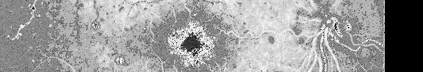
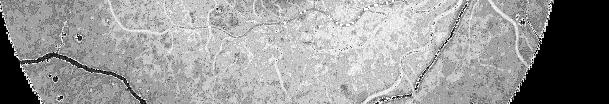
7 OCTA OCTA Angiography Fluorescein Angiography +18W +24W Many capillaries better visualized on OCTA...but not with slow flow! Depth Encoding MacTel Type 2 Phase-Variance OCT Angiography Fluorescein Angiogram 00:37 Courtesy of Philip Rosenfeld, MD, PhD Zeiss AngioPlex Schwartz DM et al,


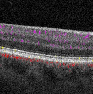
8 Artifacts in OCTA B-scans Consequence of decorrelation "shadows" Artifacts in OCTA Slabs Consequence of decorrelation "shadows" Superficial plexus Intermediate plexus Decorrelation: Real Graph-based segmentation Real Vessels Decorrelation: Artifact Vitreous-ILM Above IPL-INL Below IPL-INL Below sopl RPE-BM Structural OCT Inner retinal flow Outer retinal flow Choroidal flow Zhang M, et al Deep plexus Outer retina Normal Retinal Anatomy suppression by simple Slab Subtraction Graph-based segmentation Superficial plexus Real Vessels Intermediate plexus Fragment suppression by amplitude filtering: -Resolved OCTA Graph-based segmentation Superficial plexus Real Vessels Intermediate plexus Vitreous-ILM Above IPL-INL Below IPL-INL Below sopl RPE-BM Structural OCT Inner retinal flow Outer retinal flow Choroidal flow Zhang M, et al Fragmented Deep plexus OK Outer retina Normal Retinal Anatomy Vitreous-ILM Above IPL-INL Below IPL-INL Below sopl RPE-BM Structural OCT Inner retinal flow Outer retinal flow Choroidal flow Zhang M, et al Deep plexus Outer retina Normal Retinal Anatomy 8


9 PR-OCTA Details Retinal Vascular Anatomy Macular Ischemia in Non-Proliferative DR Superficial Intermediate Deep Inner Case 1 A B C D Case 2 A B C D 3 retinal plexuses show different patterns of vascular pathologies Peripapillary Parafoveal Peripheral Zhang M et al, 2016 Rapid Dynamic CNV Response to Anti-VEGF Inner retinal flow Outer retinal flow (CNV) Anti-VEGF Baseline 2 days 1 week 4 weeks Post-Test: Which one is Correct? 1. OCTA complements Fluorescein Angiography 1 st Rx 2. OCTA provides insights into disease pathogenesis 3. OCTA is a rapidly developing technology 2 nd Rx 1 day 2 weeks 6 weeks 4. OCTA is an abbreviation for Optical Coherence Tomography Angiography 5. All of the Above Huang D et al, 2015 Max response 9

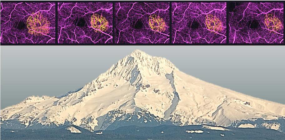


10 Save the date! OCT ANGIOGRAPHY SUMMIT July 21-22, 2017 Portland, Oregon, USA 10
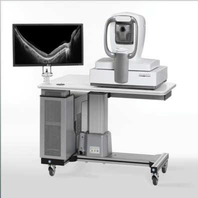
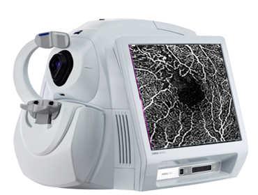
Swept-Source OCT Angiography: SS OCT Angio TM
 Swept-Source OCT Angiography: SS OCT Angio TM Not available in all countries, please check with your distributor. 2015.09 Swept-Source OCT Angiography: SS OCT Angio TM Introduction Optical coherence tomography
Visualize. Analyze. Personalize. OCT + OCTA
 Visualize. Analyze. Personalize. OCT + OCTA A New Approach to Protecting Vision AngioVue OCT Angiography brings valuable new information to clinical practice. Non-invasive visualization of retinal vasculature.
OCT angiography of ONH blood flow in glaucoma
 Hawaiian Eye Meeting 19-25 January 2013 OCT angiography of ONH blood flow in glaucoma David Huang, MD, Weeks Professor of Ophthalmic Research Professor of Ophthalmology & Biomedical Engineering Casey Eye
OCT Angiography. SriniVas Sadda, MD
 OCT Angiography SriniVas Sadda, MD Professor of Ophthalmology Director, Medical Retina Unit Ophthalmic Imaging Unit University of Southern California Los Angeles, California, USA Disclosure Consulting
ZEISS AngioPlex OCT Angiography Overview ZEISS OCT Angiography
 ZEISS AngioPlex OCT Angiography Overview ZEISS OCT Angiography California, ZEISS AngioPlex Ultra-clear visualization of microvascular blood flow using non-invasive OCT angiography 2 AngioPlex OCT Angiography
OCT Angiography: The Next Step in Retinal Imaging Jonathan Zelenak D.O.
 OCT Angiography: The Next Step in Retinal Imaging Jonathan Zelenak D.O. Hillsdale Hospital Michigan State University Overview Evolution of OCT How does OCT angiography work? Clinical examples Potential
Incorporating OCT Angiography Into Patient Care
 Incorporating OCT Angiography Into Patient Care Beth A. Steele, OD, FAAO OCT A: Introduction Isolates microvascular circulation from OCT image data Axial resolution = 5 microns (i.e. fine capillaries visible)
Functional OCT for Glaucoma Evaluation
 Hawaiian Eye Meeting 18-24 January 2014 Functional OCT for Glaucoma Evaluation David Huang, MD, PhD Weeks Professor of Ophthalmic Research Professor of Ophthalmology & Biomedical Engineering Casey Eye
OCT Angiography in Primary Eye Care
 OCT Angiography in Primary Eye Care An Image Interpretation Primer Julie Rodman, OD, MS, FAAO and Nadia Waheed, MD, MPH Table of Contents Diabetic Retinopathy 3-6 Choroidal Neovascularization 7-9 Central
Advances in OCT Murray Fingeret, OD
 Disclosures Advances in OCT Murray Fingeret, OD Consultant Alcon, Allergan, Bausch & Lomb, Carl Zeiss Meditec, Diopsys, Heidelberg Engineering, Reichert, Topcon Currently Approved OCT Devices OCT Devices
10/17/2017. FDA Approved. Zeiss AngioPlex TM Optovue AngioVue TM
 Images retinal microvasculature without dye injection Displays structure and function from a single imaging system Standard of Care-2011 DFE, Fundus Photos, VF 10-2, SD-OCT, FAF, or mferg 2016-AAO Baseline
The leading causes of blindness. ocular circulation: Macular Degeneration
 Taiwan Academy of Ophthalmology Taipei, March 31, 2012 Measurement of Blood Flow in the Retina and Optic Disc with OCT David Huang, MD, Weeks Professor of Ophthalmic Research Professor of Ophthalmology
Is OCT-A Needed As An Investigative Tool During The Management Of Diabetic Macular Edema
 Is OCT-A Needed As An Investigative Tool During The Management Of Diabetic Macular Edema Ayman M Khattab MD, FRCS Professor of Ophthalmology Cairo University Diabetic Macular Edema (DME) Diabetic macular
Visualize. Analyze. Personalize. OCT + OCTA. with
 Visualize. Analyze. Personalize. OCT + OCTA with Avanti Widefield OCT with AngioVue OCTA Imaging Comprehensive Structural and Functional Imaging in a Single Imaging Platform Comprehensive OCT Imaging The
FA vs. OCTA? The status of OCTA, today. Fukuoka, JSOS 2016 Gerd Klose. Korobelnik J Fr Ophthalmol (2015)
") FA vs. OCTA? The status of OCTA, today Korobelnik J Fr Ophthalmol (2015) Fukuoka, JSOS 2016 Gerd Klose 1 2 FA / ICGA a well-founded Gold standard! Benefits Useful for many pathologies High contrast, detailed
FA vs. OCTA? The status of OCTA, today Korobelnik J Fr Ophthalmol (2015) Fukuoka, JSOS 2016 Gerd Klose 1 2 FA / ICGA a well-founded Gold standard! Benefits Useful for many pathologies High contrast, detailed
OPTICAL COHERENCE TOMOGRAPHY ANGIOGRAPHY OF THE RETINA AND OPTIC NERVE. Lindsay B. Howse, OD
 OPTICAL COHERENCE TOMOGRAPHY ANGIOGRAPHY OF THE RETINA AND OPTIC NERVE Lindsay B. Howse, OD drlindsayhowse@gmail.com None. FINANCIAL DISCLOSURES OUTLINE Introduction/How OCTA works OCTA Analysis Advantages
What Is New in Optical Coherence Tomography. Ultrahigh-speed OCT makes 3D better
 OOAA 66 th Meeting & CEI 20 th Birthday Casey Eye Institute, Portland, OR, 17 June 2011 What Is New in Optical Coherence Tomography David Huang, MD, PhD Weeks Professor of Ophthalmic Research Prof. of
Optical Coherence Tomography: Pearls for the Anterior Segment Surgeon Basic Science Michael Stewart, M.D.
 Optical Coherence Tomography: Pearls for the Anterior Segment Surgeon Basic Science Michael Stewart, M.D. Disclosure OCT Optical Coherence Tomography No relevant financial relationships I will refer to
ZEISS AngioPlex OCT Angiography Making the revolutionary, routine.
 ZEISS AngioPlex OCT Angiography Making the revolutionary, routine. The moment that revolutionary insight becomes routine. // OCT ANGIOGRAPHY MADE BY ZEISS CIRRUS with AngioPlex creates a new era in both
Introducing ANGIOVUE ESSENTIAL. Built on the Avanti Widefield OCT Platform. OCT Angiography for Primary Eye Care
 Introducing ANGIOVUE ESSENTIAL Built on the Avanti Widefield OCT Platform OCT Angiography for Primary Eye Care Transform Your View of the Retina OCT Angiography (OCTA) is a quick non-invasive test that
SOUTH-EAST EUROPEAN JOURNAL of OPHTHALMOLOGY 2015; 1 (1) 34 40
 34 40") Review article SOUTH-EAST EUROPEAN JOURNAL of OPHTHALMOLOGY 2015; 1 (1) 34 40 Retinal nerve fiber layer versus peripapillary capillary density assessment A powerful tool for detecting optic nerve head
Review article SOUTH-EAST EUROPEAN JOURNAL of OPHTHALMOLOGY 2015; 1 (1) 34 40 Retinal nerve fiber layer versus peripapillary capillary density assessment A powerful tool for detecting optic nerve head
OCT Angiography The Next Frontier
 Choroid Retina avascular 5/13/2017 OCT Angiography The Next Frontier Pierce Kenworthy OD, FAAO June 9, 2017 OCT Angiography (OCTA) 2016 Non-invasive, motion contrast imaging Represents erythrocyte movement
OCT Interpretation in Retinal Disease
 OCT Interpretation in Retinal Disease Jay M. Haynie, OD, FAAO Financial Disclosure I have received honoraria or am on the advisory board for the following companies: Carl Zeiss Meditec Advanced Ocular
Projection-resolved optical coherence tomographic angiography
 Projection-resolved optical coherence tomographic angiography Miao Zhang, Thomas S. Hwang, J. Peter Campbell, Steven T. Bailey, David J. Wilson, David Huang and Yali Jia * Casey Eye Institute, Oregon Health
What You Should Know About Acute Macular Neuroretinopathy
 What You Should Know About Acute Macular Neuroretinopathy David J. Browning MD, PhD Chong Lee BS Acute macular neuroretinopathy is a condition characterized by the sudden, painless onset of paracentral
Mark Dunbar: Disclosure
 Important Things to Understand About OCT Mark T. Dunbar, O.D., F.A.A.O. Bascom Palmer Eye Institute University of Miami, School of Medicine Mark Dunbar: Disclosure Optometry Advisory Board for: Allergan
Will OCT-Angiography replace FA?
 ASL Roma A PRESIDIO TERRITORIALE NUOVO REGINA MARGHERITA AMBULATORIO PATOLOGIE RETINICHE Resp. Dott.ssa SUSANNA CATALANO CENTRO ITALIANO MACULA Will OCT-Angiography replace FA? Marco Rispoli, Luca di Antonio,
Deeper visualizations for intervening with confidence.
 CIRRUS OCT with AngioPlex from ZEISS Making the revolutionary routine New vascular quantification Deeper visualizations for intervening with confidence. CIRRUS OCT with AngioPlex from ZEISS can be a much
Quantitative OCT Angiography Evaluation of Peripapillary Retinal Circulation after Plaque Brachytherapy
 Quantitative OCT Angiography Evaluation of Peripapillary Retinal Circulation after Plaque Brachytherapy Alison H. Skalet, MD, PhD, 1,2, * Liang Liu, MD, 1, * Christina Binder, MD, PhD, 2 Audra K. Miller,
Optical Coherence Tomography in Diabetic Retinopathy. Mrs Samantha Mann Consultant Ophthalmologist Clinical Lead of SEL-DESP
 Optical Coherence Tomography in Diabetic Retinopathy Mrs Samantha Mann Consultant Ophthalmologist Clinical Lead of SEL-DESP Content OCT imaging Retinal layers OCT features in Diabetes Some NON DR features
Ganglion cell analysis by optical coherence tomography (OCT) Jonathan A. Micieli, MD Valérie Biousse, MD
 Jonathan A. Micieli, MD Valérie Biousse, MD") Ganglion cell analysis by optical coherence tomography (OCT) Jonathan A. Micieli, MD Valérie Biousse, MD Figure 1. Normal OCT of the macula (cross section through the line indicated on the fundus photo)
Ganglion cell analysis by optical coherence tomography (OCT) Jonathan A. Micieli, MD Valérie Biousse, MD Figure 1. Normal OCT of the macula (cross section through the line indicated on the fundus photo)
Angio-OCT. Degenerazione Maculare Legata all Eta. Giuseppe Querques
 Angio-OCT Degenerazione Maculare Legata all Eta Giuseppe Querques Department of Ophthalmology, IRCCS Ospedale San Raffaele, University Vita Salute San Raffaele, Milan, Italy Financial Disclosure ADVISORY
Why Is Imaging Critical in My Uveitis Practice?
 Why Is Imaging Critical in My Uveitis Practice? Dilraj S. Grewal, MD Developed in collaboration Imaging Is the Backbone of Uveitis Workup and Monitoring Treatment Response FP FAF B- scan Multimodal Imaging
Diabetic retinopathy (DR), characterized by capillary nonperfusion,
, characterized by capillary nonperfusion,") Retina Automated Quantification of Nonperfusion in Three Retinal Plexuses Using Projection-Resolved Optical Coherence Tomography Angiography in Diabetic Retinopathy Miao Zhang, 1 Thomas S. Hwang, 1 Changlei
Retina Automated Quantification of Nonperfusion in Three Retinal Plexuses Using Projection-Resolved Optical Coherence Tomography Angiography in Diabetic Retinopathy Miao Zhang, 1 Thomas S. Hwang, 1 Changlei
Disclosures. Definitions. Goals. Imaging and glaucoma 3/22/2016
 Pinakin Davey OD, PhD, FAAO Professor and Director of Research Disclosures Principal investigator for ivue OCT trial Principal investigator Topcon FDA trials for Maestro and OCT 2000 Consultant for Topcon
Non-arteritic anterior ischemic optic neuropathy (NAION) with segmental optic disc edema. Jonathan A. Micieli, MD Valérie Biousse, MD
 with segmental optic disc edema. Jonathan A. Micieli, MD Valérie Biousse, MD") Non-arteritic anterior ischemic optic neuropathy (NAION) with segmental optic disc edema Jonathan A. Micieli, MD Valérie Biousse, MD A 75 year old white woman lost vision in the inferior part of her visual
Non-arteritic anterior ischemic optic neuropathy (NAION) with segmental optic disc edema Jonathan A. Micieli, MD Valérie Biousse, MD A 75 year old white woman lost vision in the inferior part of her visual
OCT Image Analysis System for Grading and Diagnosis of Retinal Diseases and its Integration in i-hospital
 Progress Report for1 st Quarter, May-July 2017 OCT Image Analysis System for Grading and Diagnosis of Retinal Diseases and its Integration in i-hospital Milestone 1: Designing Annotation tool extraction
Il contributo dell'angio-oct: valutazione integrata della componente nervosa e vascolare della malattia glaucomatosa
 SIMPOSIO G.O.A.L. - LE NUOVE FRONTIERE DIAGNOSTICHE E LE LINEE DI INDIRIZZO AMBULATORIALI DEL GLAUCOMA Coordinatore e moderatore: D. Mazzacane Presidente: L. Rossetti Il contributo dell'angio-oct: valutazione
Retinal Capillary Network and Foveal Avascular Zone in Eyes with Vein Occlusion and Fellow Eyes Analyzed With Optical Coherence Tomography Angiography
 Retinal Capillary Network and Foveal Avascular Zone in Eyes with Vein Occlusion and Fellow Eyes Analyzed With Optical Coherence Tomography Angiography The MIT Faculty has made this article openly available.
Case Report Optical Coherence Tomography Angiography of Macular Telangiectasia Type 2 with Associated Subretinal Neovascular Membrane
 Hindawi Case Reports in Ophthalmological Medicine Volume 2017, Article ID 8186134, 4 pages https://doi.org/10.1155/2017/8186134 Case Report Optical Coherence Tomography Angiography of Macular Telangiectasia
Principle of OCT. Reading Between the Lines: OCT Interpretation. Initial Concept. Advantage: High Resolution Cross Section Images
 Principle of OCT Reading Between the Lines: OCT Interpretation Mohammad Rafieetary, OD, FAAO mrafieetary@charlesretina.com Introduction Optical Biopsy Morphologic Evaluation of Live Tissue Measurements
ANSWERING THE WHY? Clinicians discuss the latest imaging technologies for retina practice BY PETER K. KAISER, MD
 Insert to March 2018 Sponsored by MULTI-MODALITY IMAGING: LATEST EVOLUTIONS IN OCTA AND UWF As the array of safe and efficacious medical and surgical options for retinal diseases expands, so does the need
ZEISS AngioPlex OCT Angiography. Clinical Case Reports
 Clinical Case Reports Proliferative Diabetic Retinopathy (PDR) Case Report 969 PROLIFERATIVE DIABETIC RETINOPATHY 1 1-year-old diabetic female presents for follow-up of proliferative diabetic retinopathy
8/6/17. Disclosures Aerie Pharmaceuticals Alcon BioTissue Diopsys Optovue Shire
 Nathan Lighthizer, O.D., F.A.A.O. Associate Professor Assistant Dean for Clinical Care Director of Continuing Education Chief of Specialty Care Clinics Oklahoma College of Optometry Tahlequah, OK lighthiz@nsuok.edu
The diagnostic value of optical coherence tomography angiography in diabetic retinopathy: a systematic review
 https://doi.org/10.1007/s10792-018-1034-8 (0456789().,-volV) (0456789().,-volV) REVIEW The diagnostic value of optical coherence tomography angiography in diabetic retinopathy: a systematic review David
Ganglion cell complex scan in the early prediction of glaucoma
 Original article in the early prediction of glaucoma Ganekal S Nayana Super Specialty Eye Hospital and Research Center, Davangere, Karnataka, India Abstract Objective: To compare the macular ganglion cell
Go With the Flow: An OCT Angiography Primer Lorne Yudcovitch, OD, MS, FAAO
 Go With the Flow: An OCT Angiography Primer Lorne Yudcovitch, OD, MS, FAAO yudcovil@pacificu.edu OCT Angiography (OCTA) History 2000 - First Doppler flowimetry OCT on human retina 2005 Speckle analysis
Dr/ Marwa Abdellah EOS /16/2018. Dr/ Marwa Abdellah EOS When do you ask Fluorescein angiography for optic disc diseases???
 When do you ask Fluorescein angiography for optic disc diseases??? 1 NORMAL OPTIC DISC The normal optic disc on fluorescein angiography is fluorescent due to filling of vessels arising from the posterior
Title: OCT Analysis Workshop: Interpretation of OCT printouts
 Title: OCT Analysis Workshop: Interpretation of OCT printouts Authors: David Yang, OD, FAAO Staff Optometrist, VA Palo Alto Health Care System Associate Clinical Professor, UC Berkeley School of Optometry
Retinal Complications of Obstructive Sleep Apnea A Growing Concern!
 Retinal Complications of Obstructive Sleep Apnea A Growing Concern! Jay M. Haynie, OD, FAAO Financial Disclosure I have received honoraria or am on the advisory board for the following companies: Carl
THE OPHTHALMOLOGIST S NEEDS FOR THE ANALYSIS OF THE RETINA
 biophotonics end-users needs THE OPHTHALMOLOGIST S NEEDS FOR THE ANALYSIS OF THE RETINA Dr Matonti Frédéric CHU Nord / INT AMU Marseille ANATOMY OF THE RETINA ANATOMY OF THE RETINA ANATOMY OF THE RETINA
Pearls, Pitfalls and Advances in Neuro-Ophthalmology
 Pearls, Pitfalls and Advances in Neuro-Ophthalmology Nancy J. Newman, MD Emory University Atlanta, GA Consultant for Gensight Biologics, Santhera Data Safety Monitoring Board for Quark AION Study Medical-legal
OPTIC DISC PIT Pathogenesis and Management OPTIC DISC PIT
 OPTIC DISC PIT Pathogenesis and Management Abdel-Latif Siam Ain Shams University Cairo Egypt OPTIC DISC PIT Congenital pit is an atypical coloboma usually located on the temporal edge of the disc, associated
Diagnosis in AMD. Managing your AMD Patients
 Managing your AMD Patients Robert W. Dunphy, O.D., F.A.A.O. Diagnosis in AMD Have suspicion Identify relative risk Conduct surveillance Biometry Utilize technology to facilitate detection of change / stability
History/principles of the OCT What does the normal retinal OCT look like Vitreal disorders Retinal/RPE disorders Choroidal disorders
 Nathan Lighthizer, O.D., F.A.A.O. Assistant Professor Assistant Dean for Clinical Care Director of Continuing Education Chief of Specialty Care Clinics Chief of Electrodiagnostics Clinic Oklahoma College
SOCT Copernicus REVO. * - Currently import and overlay are avaibale in manual mode only
 SOCT Copernicus REVO Easy Operation (Full auto & Auto mode) Auto alignment (Z-position, C-gate, Focus, Tomogram) Voice guide (support patient through examination) Powerful analysis tools Enhanced tomograms
Glaucoma Diagnosis & Tracking with Optical Coherence Tomography
 Glaucoma Diagnosis & Tracking with Optical Coherence Tomography David Huang, MD, PhD Charles C. Manger III, MD Chair of Corneal Laser Surgery Assoc. Prof. of Ophthalmology & Biomedical Engineering Doheny
The Quick Guide to OCT Mastery 50 Real Cases with Expert Analysis
 OPTICAL COHERENCE TOMOGRAPHY The Quick Guide to OCT Mastery 50 Real Cases with Expert Analysis VOL 1 Sanjay Sharma, MD, FRCS, MSc (Epid), MBA Ophthalmologist, Epidemiologist Queen s University, Canada
OCT Interpretation. Financial Disclosure. Jay M. Haynie, OD, FAAO. OCT Image Layers 7/21/2014
 OCT Interpretation Jay M. Haynie, OD, FAAO Financial Disclosure I have received honoraria or am on the advisory board for the following companies: Olympia Tacoma Renton Kennewick - Washington Carl Zeiss
IN NICU OCT UTILIZES A CONCEPT KNOWN AS INTERFEROMETRY APPLICATIONS FOR OCT THE PRIMARY USE IN THE EYE - RETINA
 2016 25 YEARS OF OPTICAL COHERENCE TOMOGRAPHY OPTICAL COHERENCE TOMOGRAPHY IN NICU Marcin Stopa, MD, PhD, FEBO Department of Ophthalmology, Chair of Ophthalmology and Optometry. Poznan University of Medical
Research Article Diabetic Macular Ischemia Diagnosis: Comparison between Optical Coherence Tomography Angiography and Fluorescein Angiography
 Ophthalmology Volume 2016, Article ID 3989310, 6 pages http://dx.doi.org/10.1155/2016/3989310 Research Article Diabetic Macular Ischemia Diagnosis: Comparison between Optical Coherence Tomography Angiography
Clinical Case Presentation. Branch Retinal Vein Occlusion. Sarita M. Registered Nurse Whangarei Base Hospital
 Clinical Case Presentation on Branch Retinal Vein Occlusion Sarita M. Registered Nurse Whangarei Base Hospital Introduction Case Study Pathogenesis Clinical Features Investigations Treatment Follow-up
Simply the best OCT & OCTA image quality.
 Avanti Widefield OCT with AngioVue OCT Angiography Simply the best OCT & OCTA image quality. Dear Friends of Optovue, Since introducing Spectral Domain OCT to the ophthalmology market in 2006, Optovue
PLEX Elite 9000 from ZEISS Swept-Source OCT
 PLEX Elite 9000 from ZEISS Swept-Source OCT Uncovering the undiscovered. ZEISS PLEX Elite 9000 // INNOVATION MADE BY ZEISS 2 Ultra-wide angiography En face montage Image courtesy of Prof. G. Querques,
Glaucoma Past, Present and Future
 Glaucoma Past, Present and Future Paul A. Weber, M.D. Professor Emeritus Department of Ophthalmology and Visual Science- Havener Eye Institute The Ohio State University Before we talk about glaucoma, I
Longitudinal Changes of Retinal Thicknesses in Branch Retinal Artery Occlusion: Spectral-Domain Optical Coherence Tomography Study METHODS.
 Retina Longitudinal Changes of Retinal Thicknesses in Branch Retinal Artery Occlusion: Spectral-Domain Optical Coherence Tomography Study Min-Su Kim, 1 Kyeung-Min Kim, 1 Hyung-Bin Lim, 1,2 Young-Joon Jo,
November Volume 35 - Issue 11
 November 2015 - Volume 35 - Issue 11 pp: 2161-2431,e67-e72 Editorial Optical Coherence Tomography Angiography Spaide, Richard F.; Fujimoto, James G.; Waheed, Nadia K. Original Study IMAGE ARTIFACTS IN
11/29/2016 MACULAR MALADIES: TYPICAL & ATYPICAL CASES
 MACULAR MALADIES: TYPICAL & ATYPICAL CASES Dawn Pewitt, OD, FAAO Triad Eye Institute, Grove, OK Dpewitt@triadeye.com Disclosure Statement: No financial disclosures COPE 51218-PS Please silence all mobile
Clinical Study Optical Coherence Tomography Angiography in Retinal Vascular Diseases and Choroidal Neovascularization
 Hindawi Publishing Corporation Journal of Ophthalmology Volume 2015, Article ID 343515, 8 pages http://dx.doi.org/10.1155/2015/343515 Clinical Study Optical Coherence Tomography Angiography in Retinal
Measuring of the fovea and foveola using line scans and 3D Macular scans obtained with spectral domain optical coherent tomography.
 Measuring of the fovea and foveola using line scans and 3D Macular scans obtained with spectral domain optical coherent tomography. Vakhrameeva O.A. 1, Moiseenko G.A. 1, Maltsev D.S. 2, Sukhinin M.V. 2,
Chun-Hsiu Liu, Ling-Yuh Kao, Ming-Hui Sun, Wei-Chi Wu, and Henry Shen-Lih Chen
 Hindawi Ophthalmology Volume 2017, Article ID 9632647, 7 pages https://doi.org/10.1155/2017/9632647 Research Article Retinal Vessel Density in Optical Coherence Tomography Angiography in Optic Atrophy
Optical coherence tomography angiography of foveal hypoplasia
 Optical coherence tomography angiography of foveal hypoplasia Kaivon Pakzad-Vaezi, MD 1, Pearse Keane, MD 1,2, João Nobre Cardoso, MD 1, Catherine Egan, MD 1, Adnan Tufail, MD 1. Moorfields Eye Hospital
PART 1: GENERAL RETINAL ANATOMY
 PART 1: GENERAL RETINAL ANATOMY General Anatomy At Ora Serrata At Optic Nerve Head Fundoscopic View Of Normal Retina What Is So Special About Diabetic Retinopathy? The WHO definition of blindness is
Supplementary information Novel VCP modulators mi2gate major pathologies of rd10, a mouse model of re2ni2s pigmentosa
 Supplementary information Novel VCP modulators mi2gate major pathologies of rd1, a mouse model of re2ni2s pigmentosa Hanako Ohashi Ikeda, Norio Sasaoka, Masaaki Koike, Noriko Nakano, Yuki Muraoka, Yoshinobu
OCT in the Diagnosis and Follow-up of Glaucoma
 OCT in the Diagnosis and Follow-up of Glaucoma Karim A Raafat MD. Professor Of Ophthalmology Cairo University Hmmmm! Do I have Glaucoma or not?! 1 Visual Function 100% - N Gl Structure : - 5000 axon /
Optical Coherence Tomograpic Features in Idiopathic Retinitis, Vasculitis, Aneurysms and Neuroretinitis (IRVAN)
") Columbia International Publishing Journal of Ophthalmic Research (2014) Research Article Optical Coherence Tomograpic Features in Idiopathic Retinitis, Vasculitis, Aneurysms and Neuroretinitis (IRVAN)
Columbia International Publishing Journal of Ophthalmic Research (2014) Research Article Optical Coherence Tomograpic Features in Idiopathic Retinitis, Vasculitis, Aneurysms and Neuroretinitis (IRVAN)
High Resolution Imaging in Patients with Retinal Dystrophies
 High Resolution Imaging in Patients with Retinal Dystrophies Ophthalmic Photographers Society Annual Midyear Meeting April 2, 213 Jacque Duncan, M.D. UCSF Department of Ophthalmology How can retinal imaging
Fundus Autofluorescence. Jonathan A. Micieli, MD Valérie Biousse, MD
 Fundus Autofluorescence Jonathan A. Micieli, MD Valérie Biousse, MD The retinal pigment epithelium (RPE) has many important functions including phagocytosis of the photoreceptor outer segments Cone Rod
New Concepts in Glaucoma Ben Gaddie, OD Moderator Murray Fingeret, OD Louis Pasquale, MD
 New Concepts in Glaucoma Ben Gaddie, OD Moderator Murray Fingeret, OD Louis Pasquale, MD New Concepts in Glaucoma Optical Coherence Tomography: Is it necessary and needed to diagnose and monitor glaucoma?
Case Report Peripapillary Intrachoroidal Cavitation in Myopia Evaluated with Multimodal Imaging Comprising (En-Face) Technique
 Technique") Case Reports in Ophthalmological Medicine Volume 2015, Article ID 890876, 5 pages http://dx.doi.org/10.1155/2015/890876 Case Report Peripapillary Intrachoroidal Cavitation in Myopia Evaluated with Multimodal
Case Reports in Ophthalmological Medicine Volume 2015, Article ID 890876, 5 pages http://dx.doi.org/10.1155/2015/890876 Case Report Peripapillary Intrachoroidal Cavitation in Myopia Evaluated with Multimodal
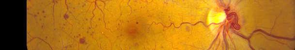
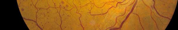
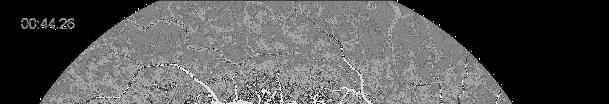
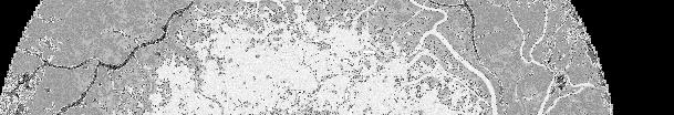
Radial Peripapillary Capillary Network Visualized Using Wide-Field Montage Optical Coherence Tomography Angiography
 Special Issue Radial Peripapillary Capillary Network Visualized Using Wide-Field Montage Optical Coherence Tomography Angiography Tomoko Mase, Akihiro Ishibazawa, Taiji Nagaoka, Harumasa Yokota, and Akitoshi
Comparison of Neovascular Lesion Area Measurements From Different Swept-Source OCT Angiographic Scan Patterns in Age-Related Macular Degeneration
 Retina Comparison of Neovascular Lesion Area Measurements From Different Swept-Source OCT Angiographic Scan Patterns in Age-Related Macular Degeneration Fang Zheng, 1,2 Qinqin Zhang, 3 Elie H. Motulsky,
OCT Angiography: An Upcoming Tool for Diagnosis and Treatment of Retinal Vascular Diseases
 E-ISSN 2454-2784 Recent Advances OCT Angiography: An Upcoming Tool for Diagnosis and Treatment of Retinal Vascular Diseases Purnima Sood 1, Nalini Saxena 2, Dinesh Talwar 3 1 Vitreo-Retina Consultant,
Simposio GOAL. Coordinatore e moderatore: D. Mazzacane Presidente: L. Rossetti
 www.amedeolucente.it Simposio GOAL Coordinatore e moderatore: D. Mazzacane Presidente: L. Rossetti L avanzamento della diagnosi strutturale nel glaucoma: ruolo dell angio-oct Disclosure Consulting Free
Incidental branch retinal artery occlusion on optical coherence tomography angiography presenting as segmental optic atrophy in a child: a case report
 DOI 10.1186/s12886-017-0653-6 CASE REPORT Incidental branch retinal artery occlusion on optical coherence tomography angiography presenting as segmental optic atrophy in a child: a case report Ji Hyung
NIH Public Access Author Manuscript Retin Cases Brief Rep. Author manuscript; available in PMC 2012 January 1.
 NIH Public Access Author Manuscript Published in final edited form as: Retin Cases Brief Rep. 2011 ; 5(1): 46 48. doi:10.1097/icb.0b013e3181cafc49. Intact Retinal Tissue and Retinal Pigment Epithelium
How to Be Efficient and Effective. Disclosure. Topics CASE CM. Case JF 2007 OHTN / POAG? How to Be Efficient and Effective with. with New Technology
 How to Be Efficient and Effective with Disclosure COPE Course ID: 40750 GL Michael Chaglasian has the following disclosures: 1. Advisory Board: Allergan, Inc., Alcon Labs, B+L Carl Zeiss Meditec 2. Research:
Technicians & Nurses Program
 ASCRS ASOA Symposium & Congress Technicians & Nurses Program April 17-21, 2015 San Diego, California Optical Coherence Tomography: Essentials in Anterior and Posterior Segment Imaging Michael Stewart,
International Journal of Health Sciences and Research ISSN:
 International Journal of Health Sciences and Research www.ijhsr.org ISSN: 2249-9571 Original Research Article A Multivariate Analysis of Intravitreal Injection of Anti-VEGF Bevacizumab in the Treatment
Convergence in. Introduction. Case Report: Dr. Piyali SenM.B.B.S, Dr. Abhipsha Saha M.B.B.S, Dr. Anuradha Chandra M.S,FAICO
 Convergence in Dr. Piyali SenM.B.B.S, Dr. Abhipsha Saha M.B.B.S, Dr. Anuradha Chandra M.S,FAICO Introduction non-progressive ophthalmoplegia with or without ptosis affecting part or all of the occulomotor
Method for comparing visual field defects to local RNFL and RGC damage seen on frequency domain OCT in patients with glaucoma.
 Method for comparing visual field defects to local RNFL and RGC damage seen on frequency domain OCT in patients with glaucoma. Donald C. Hood 1,2,* and Ali S. Raza 1 1 Department of Psychology, Columbia
Structural & Functional Optical Coherence Tomography for Glaucoma Diagnosis
 Congreso Argentino de Oftalmologia, May 2009, Buenos Aires Structural & Functional Optical Coherence Tomography for Glaucoma Diagnosis David Huang, MD, PhD Assoc. Prof. of Ophthalmology & Biomedical Engineering
IMAGE ARTIFACTS IN OPTICAL COHERENCE TOMOGRAPHY ANGIOGRAPHY
 IMAGE ARTIFACTS IN OPTICAL COHERENCE TOMOGRAPHY ANGIOGRAPHY The MIT Faculty has made this article openly available. Please share how this access benefits you. Your story matters. Citation Spaide, Richard
Overview. Macular OCT Artifact Study
 Imaging Artifacts Sarah Moyer, CRA, OCT-C Director, Ophthalmic Imaging Kittner Eye Center University of North Carolina Chapel Hill, NC Disclose financial interest now Overview Sarah s Thoughts on Artifacts
The Evaluation of Diabetic Macular Ischemia Using Optical Coherence Tomography Angiography
 Retina The Evaluation of Diabetic Macular Ischemia Using Optical Coherence Tomography Angiography Patrick D. Bradley, 1 Dawn A. Sim, 1 Pearse A. Keane, 1 João Cardoso, 1,2 Rupesh Agrawal, 1 Adnan Tufail,
ARVO 2016 Annual Meeting Abstracts
 146 OCT Angiography 1 Sunday, May 01, 2016 3:15 PM 5:00 PM 6B Paper Session Program #/Board # Range: 947 953 Organizing Section: Retina Program Number: 947 Presentation Time: 3:15 PM 3:30 PM Comparisons
CAPILLARY NETWORK ANOMALIES IN BRANCH RETINAL VEIN OCCLUSION ON OPTICAL COHERENCE TOMOGRAPHY ANGIOGRAPHY
 CAPILLARY NETWORK ANOMALIES IN BRANCH RETINAL VEIN OCCLUSION ON OPTICAL COHERENCE TOMOGRAPHY ANGIOGRAPHY MARCO RISPOLI, MD, MARIA CRISTINA SAVASTANO, MD, PHD, BRUNO LUMBROSO, MD Purpose: To analyze the
The Blood-Retinal Barrier in the Management of Retinal Disease: EURETINA Award Lecture
 Ophthalmologica Euretina Lecture Received: December 29, 2016 Accepted: January 2, 2017 Published online: February 3, 2017 The Blood-Retinal Barrier in the Management of Retinal Disease: EURETINA Award
 Title Optical Coherence Tomography Angiog Retinal Blood Flow in Eyes with Ret Sugahara, Masako; Miyata, Manabu; I Norimoto; Morooka, Satoshi; Ogino, Author(s) Hirashima, Takako; Yoshikawa, Munem Muraoka,
Cirrus TM HD-OCT. Details defi ne your decisions
 Cirrus TM HD-OCT Details defi ne your decisions 2 With high-defi nition OCT Carl Zeiss Meditec takes you beyond standard spectral domain Built on 10 years experience at the vanguard of innovation, Carl