THE RESPIRATORY SYSTEM
|
|
|
- Tabitha Stone
- 6 years ago
- Views:
Transcription
1 THE RESPIRATORY SYSTEM 1. Identify and give functions for each of the following: -larynx -bronchioles -alveoli -trachea -epiglottis -diaphragm and ribs -bronchi -pleural membranes -thoracic cavity 2. Explain the roles of cilia and mucus in the respiratory tract. 3. Explain the relationship between the structure and function of alveoli. 4. Compare and contrast the mechanics of the processes of inhalation and exhalation. 5. Describe the interaction of the lungs, pleural membranes, ribs, and the diaphragm in the breathing process. 6. Explain the roles of carbon dioxide and hydrogen ions in stimulating the breathing/respiratory center in the medulla oblongata. 7. Describe the exchange of carbon dioxide and oxygen during internal and external respiration. 8. List the conditions that cause the equations to proceed in one way at the lungs and the opposite way at the tissues. 9. Distinguish between the transport of CO2 and O2 in the blood by explaining the roles of oxyhemoglobin, carbaminohemoglobin, reduced hemoglobin, and bicarbonate ions. 10. Describe the process of acclimatization. 11. Describe the role of the nervous system in regulating the breathing rate. 1
2 The Respiratory System 2
3 The Respiratory System (Reference notes) Pages Respiration includes all of the following: a) Breathing the inhalation and exhalation of air into and out of the lungs. b) External Respiration exchange of gases between the alveolus and the blood. c) Internal Respiration exchange of gases between tissues and blood. d) Cellular Respiration production of ATP using oxygen in the mitochondrion. 2. Passage of Air As air moves along passages, it is warmed by blood vessels, filtered by nose hairs and cilia, and moistened by the wet surfaces of the passages. Air is 99.5% saturated by the time it reaches the end of the trachea. As air moves out, it cools and loses its moisture depositing it on the lining of the passage. This causes the dripping nose on cold days and the cloud of water droplets when you breathe out. 3. Parts of the Respiratory Tract a) Nose has two nasal cavities containing specialized ciliated cells that act as odor receptors. Nerves lead from these to the brain where impulses are perceived as smell. Mucus traps dirt. Tears produced by the tear glands drain into the nose, which causes the nose to run when crying. b) Pharynx here the food and air cross, with the trachea lying in front of the esophagus. c) Larynx known as the voice box, a triangle-shaped structure whose apex (Adam s apple) is at the front of the neck. The glottis is the opening at the top of the trachea covered by the epiglottis (a flap of tissue). The vocal cords are elastic ligaments that lie at the top of the glottis. Vibration of these causes sound. The length of the vocal cords influences the pitch of the sound. d) Trachea has C shaped cartilaginous rings to keep it open. Lined with cilia to sweep debris up and out. e) Bronchi trachea divides into two bronchi, which divide into smaller branches called bronchioles. Each bronchiole terminates in a multitude of air sacs called alveoli. f) Lungs alveolar sacs are surrounded by blood capillaries to allow for gas exchange. Alveoli contain about 100x the surface area compared to skin. There are two pleural membranes, one covering the lungs and the other lining the pleural cavity. The space between these membranes is the intrapleural space. The pressure in this space is less than one atmosphere an important factor in breathing. If a puncture occurs allowing air into this space, the lung(s) will collapse. Thus it is often said that we breathe by negative pressure. 3

4 Functions of the Pleural Membranes a) seals the pleural cavity. b) maintains negative pressure around the lungs preventing them from collapsing. c) reduces friction. Negative Pressure Breathing Mechanics of Breathing Pages Impulses from the breathing center cause the diaphragm to contract and move down. Intercostal muscles lift the rib cage up and out. 2. The outer pleural membrane moves out with the rib cage and because there is less than one atmosphere of pressure in the intrapleural space, the inner membranes move out as well. (in other words, we breath by negative pressure.) 3. The lungs expand decreasing the pressure in them and air rushes in. This is called inspiration or inhalation. 4. Stretch receptors in the alveoli wall send an inhibitory impulse to the respiratory center in the medulla. Without further stimulation, the intercostal muscles relax and the rib cage moved down and in. The diaphragm also relaxes and moves up. 5. With the increase in pressure in the lungs, air is pushed out. This is called expiration or exhalation. 4
5 Respiratory Lab Page 286 You are going to measure your lung capacity known as the vital capacity. We will compare your results to others in the classroom. Observe the other types of lung capacity/volumes measurable during respiration. Procedure: Each person will have two attempts at measuring their vital capacity. 1. Set the pointer to 0 and place a new mouthpiece on the tube. 2. Inhale normally and then place your mouth over the tube and exhale. Have your partner record the value of the pointer in ml. This is your tidal volume. 3. Reset to 0, take as deep a breath as possible and exhale as much as possible. Record your results in ml. Reset and repeat. This is your vital capacity. 4. Record the number of breaths you take in one minute. Calculation: Your vital capacity can change from day-to-day and especially during different activities throughout the day. Vital Capacity Mass(kg) Most athletes have a value greater than 80 ml/kg. The average person is about ml/kg. Your teacher is probably ml/kg. 5
 4.")
6 Questions: 1. Of what value are the first cries after birth? What information does this tell the doctor?(2) 2. What is the purpose of the residual volume in your lungs? (1) 3. A snorkel has a short tube causing you to swim very close to the surface. Why can they not make snorkels that are 5 metres long? (1) 4. Name three factors that tend to reduce lung efficiency. (3) 6
7 Thinking about the Mechanics of Breathing 1. Is air entering or leaving the lungs between Y and Z? 2. Which is higher in the blood at Z, HbO2 or HbCO2? 3. Would the intercostals begin contracting or relaxing at time X? 4. Do the lungs expand before or after time X? 5. Where is the PCO2 the greatest, at time X, Y, or Z in the lung? 6. What causes (part of the body) the pressure change from Y to Z? 7. Where are the alveolar stretch receptors the most active? 7
8 Pressure in The Lungs A Time Use the graph above to answer the following questions. a) Describe two differences in this person s blood that caused the change at A. b) Give the feedback loop that caused this change. c) List three differences between blood in the pulmonary artery and the carotid artery? 8
9 Respiration The lungs continue to open on inhalation until in the alveolar walls send an impulse to the respiratory center in the inhibiting its action. Then the relaxes and moves up as the intercostal muscles to relax dropping the which the volume of the chest cavity, the pressure there. The lung volume then, which the pressure there and air rushes out. In external respiration, diffuses from the into the and is carried to the body cells. In these reactions, ions in the plasma diffuse into RBC s joining with to produce. Then the enzyme causes it to dissociate into. and have the greatest effect on your breathing rate. When oxygen levels are low in the blood, in the aorta and carotid arteries detect and tell the to turn up the nervous system, which the breathing rate. If the oxygen level remains low for long periods of time, the detects it and releases which tells the bone marrow to increase RBC production. The equations proceed in one direction at the lungs and the opposite at the tissue cells because: 9
10 10

11 THE EXCRETORY SYSTEM 1. Identify and give functions for each of the following: -kidney -ureter -urethra -urinary bladder -renal cortex -renal medulla -renal pelvis 2. List the organs involved with excretion. 3. List the functions of the kidney. 4. Identify and give functions for each of the following: -nephron -Bowman s capsule -afferent/efferent arterioles -proximal convoluted tubule -glomerulus -collecting duct -loop of Henle -distal convoluted tubule 5. Contrast blood in the renal artery and the renal vein with respect to urea and glucose content. 6. Identify the source glands for ADH and Aldosterone and explain how these hormones are regulated in a feedback loop. 7. Relate ADH, Aldosterone, and the nephron to the regulation of water and sodium levels in the blood. 8. Explain the process of CAPD and outline its shortcomings as a replacement for the kidney. 9. Explain how the kidney regulates blood ph. 11
12 Excretion Chapter 16 page 302 ridding the body of the end products of metabolism Excretory Substance: 1. Ammonia arises from deamination (the removal of the amine group from amino acids), it is very toxic and is converted to urea by the liver 2. Uric acid comes from the breakdown of nucleotides, is an insoluble solid, causes gout if it builds up in the joints 3. Creatinine from muscle metabolism 4. Bile pigments from the hemo portion of hemoglobin, forms bile, if it gets into the blood it causes a yellowing of the skin = jaundice 5. Carbon dioxide from Kreb s cycle, most excreted by the lungs by kidney excretes bicarbonate ion 6. Ions these give blood its proper ph, osmotic balance, and electrolyte balance 7. Water is an end product of metabolism (end of ETC, from dehydration synthesis) determines the OP of the blood, too much can cause high blood pressure (hypertension) Organs of Excretion: 12
 Under normal conditions approximately 1600 L of blood flows through a")
13 Below is a simple diagram of the excretion anatomy parts in a human. Study the diagram below and read chapter 16 on Excretion. Fill in the following functions: Urethra Bladder Ureter Structure Function Kidney (made up of 1 million microscopic nephrons) Under normal conditions approximately 1600 L of blood flows through a pair of kidneys each day. Processing this enormous amount of blood yields about 180 L of initial filtrate. Of this, about 99% of the water and nearly all of the sugars, amino acids, vitamins, and other organic molecules are reabsorbed leaving about 1.5 L of urine to be transported daily to the bladder. 13

14 Each kidney has an outer renal cortex and an inner renal medulla. Both regions are supplied with blood by the renal artery and drained by a renal vein. Within the cortex and medulla lie tightly packed excretory tubules and associated blood vessels. The inner renal pelvis collects urine from the excretory tubules and passes it to the urinary bladder Weaving back and forth across the renal cortex and medulla are the nephrons, the functional units of the kidney. Most nephrons are found in the cortical regions but in drier climates it is the juxtamedullary nephrons that work the most. What would be a reason for this? Each nephron consists of a single long tubule and a ball of capillaries called the glomerulus. The blind end of the tubule forms a cup-shaped swelling, called Bowman s capsule, which surrounds the glomerulus. Fluid leaving the blood and entering the tubule network is called filtrate. As the filtrate passes through several regions of the nephron it s concentration changes and it eventually becomes urine. 14

15 Nephron Anatomy Study the diagram of the anatomy of the nephron below: Three major regions of the nephron include the proximal tubule, the loop of Henle (descending/ascending) and the distal tubule. A collecting duct receives urine from many nephrons and transports it to the renal pelvis. Each nephron is supplied with blood by an afferent arteriole, an offshoot of the renal artery that branches and forms the capillaries of the glomerulus. The caps converge as they leave the glomerulus to form the efferent arteriole. Branches of this vessel form the peritubular capillaries, which surround the proximal and distal tubules. Other branches extend downward and form the vasa recta surrounding the loop of Henle. The Steps in Urine Formation 1. Pressure Filtration Blood enters the glomerulus under high pressure. The efferent arteriole can constrict causing the blood pressure in the glomerulus to increase even higher. About 80% of all the small molecules from the blood are filtered out of the blood to enter the first part of the nephron called Bowman s capsule. The fluid is called filtrate. The filtrate contains water and small solutes, such as salts, sugars, amino acids, and nitrogenous wastes (urea). It will eventually be called urine once the concentration of substances change along the nephron. 15


16 2. Selective Reabsorption The filtrate now enters the proximal convoluted tubule (PCT). This is the site of selective reabsorption. The cells that line the PCT are cuboidal epithelial cells. The shape of these cells is conducive to forming a tube and they are lined by microvilli and many mitochondria. Selective reabsorption recovers useful molecules and water from the filtrate and returns them to the body fluids and eventually the blood. Valuable solutes including glucose, certain salts, vitamins, hormones, and amino acids are reabsorbed by active and passive transport. Non-essential solutes and waste are left in the filtrate. 3. Tubular Secretion The blood has another chance to pump wastes into the filtrate at the distal convoluted tubule (DCT). The filtrate is isotonic to blood plasma as it approaches the DCT. Critical wastes in the blood such as creatinine, uric acid, penicillin, and H + ions are pumped into the nephron. From Blood Filtrate to Urine: A Closer Look 16
17 1. Proximal Tubule Reabsorption in the proximal tubule is critical for the recapture of ions, water, and valuable nutrients from the huge volume of initial filtrate. Na + in the filtrate diffuses into the cuboidal epithelial cells lining the tube and is actively pumped into the blood. Cl - passively follow. The resulting NaCl draws water from the filtrate by osmosis and returns it to the blood. Glucose, amino acids, potassium ions, and other essential substances are also actively or passively transported from the filtrate to the blood. 2. Descending limb of the Loop of Henle Reabsorption of water continues as the filtrate moves into the descending limb of the loop of Henle. Here numerous water channels (aquaporins) make the limb permeable to water as it leaves by osmosis and gets picked up by the blood. There are almost no other channels for salt or other small solutes so they stay inside the filtrate in the descending loop. For water to continue to move out of limb by osmosis, the interstitial fluid of the kidney s tissues surrounding the limb increases in solute concentration as water moves further down the descending limb. 3. Ascending limb of the Loop of Henle The filtrate returns to the cortex within the ascending limb. Unlike the descending limb, the ascending limb lacks water channels causing any water in the tube to remain inside. The ascending limb has two specialized regions: a thin segment followed by a thick segment near the distal tubule. As filtrate ascends in the thin segment, NaCl, which becomes concentrated in the descending limb, diffuses out of the permeable tubule into the kidney tissues. This movement helps make the tissues hypertonic the deeper into the medulla the loop goes. In the thick segment of the ascending limb, the movement of NaCl continues into the kidney tissues but active transport pumps NaCl out. As a result of losing salt and but not water, the filtrate becomes progressively more dilute as it moves up to the cortex near the distal tubule. 4. Distal Tubule The distal tubule plays a key role in regulating the acid content of the blood. If the blood is too acidic, H + are pumped out of the blood and HCO 3 - are reabsorbed to make the blood less acidic. If the blood is too alkaline, then H + remain in the blood. Salt and potassium ions are adjusted to their final concentrations in the blood and filtrate. Creatinine, uric acid, and penicillin are pumped into the filtrate. 5. Collecting Duct When the filtrate enters the collecting duct, it is now called urine. The collecting duct is impermeable to water. The walls of the tube are very thick. Water stays inside the tubes unless certain hormones adjust the permeability of the collecting duct. When the kidneys are conserving water, the hypertonic tissues of the kidney pull water out of the nephron and back into the peritubular capillaries. Near the inner medulla, the collecting duct has a high concentration of urea. Up to 44% of all urea can be reabsorbed back into the blood. This really assists the kidney to pull water back into the blood by vastly increasing the solute content of the surrounding tissues. The collecting duct using hormones acts to maintain homeostasis for osmolarity, blood pressure, and blood volume. 17

18 Label the Nephron structures and functions 18
19 Work of the Kidney Water, nitrogenous wastes, nutrients and ions (salts) all leave the by the process of and enter the first part of the tubule called. These molecules then flow into the next part of the tubule called the. From there most of the good materials undergo the process of And re-enter the blood capillaries. In fact, % of and amino acids are taken out of the filtrate by the processes of. Large plasma proteins and formed elements leave the glomerulus. The above two parts of the nephron are found in the of the kidney. The remaining materials then flow down the. As water and dissolved materials flow up the, it remains impermeable to keeping it inside the tube as is actively pumped out. This builds up a concentration of in the renal medulla to a level 4X the concentration found anywhere else in the body. As a result, an filtrate to blood plasma enter the next part called the. Here, another process called occurs to allow the blood to get rid of unwanted H+ as well as penicillin, uric acid and creatinine. Two possibilities: You have no water to drink: The distal convoluted tubule and the are normally impermeable to. However, if the in the hypothalamus measures in the OP of the blood, the hormone is released from the which makes the walls of the distal convoluted tubule and the collecting duct permeable to water causing the water to be and a urine will result. You do not need water: the distal tubule and collecting duct remain to water since no hormone is present and a urine results. After exercising, the renal vein has a(n) ph level compared to the renal artery. 19
 Blood is too alkaline: Fewer H + are excreted and less HCO3 - is reabsorbed. This creates an alkaline urine. When kidney failure occurs in an individual, dialysis may be needed to keep them alive.")
20 ph Control of Blood by the DCT of the Nephron Two possible scenarios: CAPD a) Blood is too acidic: H + and NH3 + are excreted and HCO3 - are reabsorbed. This creates an acidic urine. b) Blood is too alkaline: Fewer H + are excreted and less HCO3 - is reabsorbed. This creates an alkaline urine. When kidney failure occurs in an individual, dialysis may be needed to keep them alive. CAPD stands for continuous ambulatory peritoneal dialysis. Dialysate fluid is dumped into the peritoneal cavity and left for several hours. Wastes diffuse through the walls of the mesenteric and peritoneal capillaries into this fluid. The waste filled fluid is now drained into a bag and replaced with new fluid. The dialysate is isotonic to blood plasma, except for the waste products. Most patients are connected permanently to a surgically implanted catheter. A patient can usually dialyze while sleeping making every-day life more manageable. 20
21 Make a feedback loop below for renin-angiotensin-aldosterone to solve the problem of low BP. Show your teacher when done. Make a feedback loop below for ADH to solve the problem of dehydration. Show your teacher when done. Alcohol blocks the release of ADH = more urine production. Diabetes Insipidus The body does not make enough ADH and the collecting duct remains relatively impermeable to water (water stays inside tubes) producing a hypotonic (watery) urine. 21
22 Blood, Filtrate, and Urine Should the components of blood listed vertically, be associated with the structures, processes, or substances listed horizontally? Place a in the box opposite the component of blood if it should be associated with the structure, process, or substance. Include the % value for anything reabsorbed. Component of blood Afferent arteriole Filtrate Efferent arteriole Reabsorb % Tubular excretion Urine Absent in urine Venous blood Plasma proteins Erythrocytes Leucocytes Glucose Amino Acids Sodium chloride Water Urea Uric acid Penicillin The three basic processes involved in the overall function of excretion are: How does the nephron solve the problem of reabsorbing H 2O by osmosis as less and less H 2O is found in the loop of Henle? Name two locations of active transport pumping out of the tubule network. 22
23 Excretion The three basic processes involved with the production of urine by the nephron are:. The order of the blood vessels around the nephron are. About % of the small molecules, such as the first part of the nephron, called the leave the blood capillary and enter. This filtrate is to the blood. As the filtrate passes through the next part, the, % of the good molecules are reabsorbed back into the blood. A cell in this part would look like: (draw a picture) As the filtrate moves down the, leaves due to the higher salt concentration outside of it. The filtrate is now to the blood. As the filtrate flows up the, is actively transported out. Since this part is to water, the filtrate entering the next part, the, is now to the blood. Due to the active transport of, the At the of the kidney becomes 4X salt concentration., H+ ions, bicarbonate ions, etc. can be dumped during tubular secretion. The last part, the Is normally relatively to water. If the blood s OP, receptors in the detect and cause the release of the hormone 23
24 from the gland. This hormone acts on the and makes it more permeable to water. Water leaves and enters the leaving a urine behind. Name the parts of the nephron where active transport is carried out: Name the parts of the nephron where osmosis occurs: Name the parts of the nephron found within the renal medulla: Explain how the kidney acts to solve the problem of the blood being too acid. List the four functions of the kidney. State the function of any hormones, involved. List six differences between blood in the renal artery and renal vein. Name two substances that are not filtered by the nephron. 24
25 What is a diuretic and why are diuretic drugs prescribed? Give two reasons why the OP increases as you do deep into the renal medulla. What is ANH and state its function? 25
26 Excretory System Matching Try this exercise with your book closed after reading Ch. 16. Bowman s capsule aldosterone ADH afferent arteriole proximal tubule urea osmoreceptors angiotensin medulla glomerulus CAPD ureter distal tubule urethra NaHCO 3 ascending loop of Henle 1. Blood vessels entering glomerulus 2. Nitrogenous waste material 3. Stimulates release of aldosterone 4. Always impermeable to water 5. Loop mostly found in this area 6. Takes urine to bladder 7. Reabsorbed when blood ph is 6 or less 8. Allows for pressure filtration 9. In hypothalamus 10. Site of active transport of glucose 11. Site of tubular excretion 12. Carries urine from the bladder 13. Causes reabsorption of sodium 14. A type of dialysis 15. Released when you are thirsty 16. Permeable to most molecules 26
 prostate gland seminal vesicle 2.")
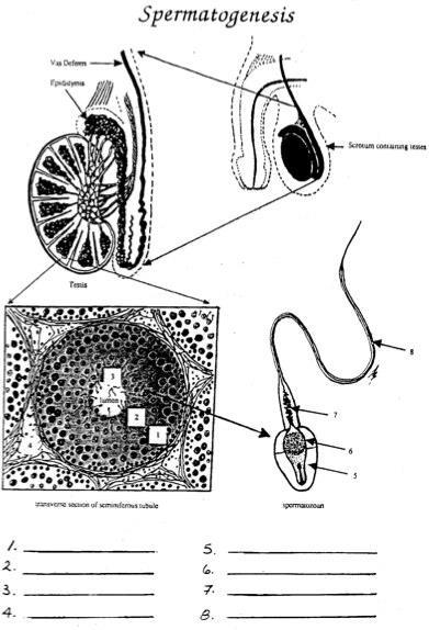
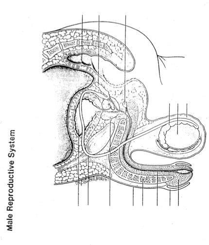
27 THE REPRODUCTIVE SYSTEM 1. Identify and give functions for the following: seminiferous tubules scrotum penis interstitial cells of testis vas deferens epididymis Cowper s gland (bulbourethral) prostate gland seminal vesicle 2. Describe the path of sperm from the seminiferous tubules to the urethral opening. 3. List and state the functions of the components of seminal fluid. 4. Identify the tail, midpiece, head and acrosome of a mature sperm and state their function. 5. Describe the secondary sex characteristics in males. Explain how secretion of testosterone is controlled. 6. Diagram negative feedback loops involving testosterone, inhibin, GnRH, FSH and LH. 7. Identify and give functions for each of the following. urethra oviduct (fallopian tube) ovary follicle corpus luteum clitoris uterus cervix vagina endometrium 8. Describe the functions of estrogen and progesterone. 9. Describe the sequence of events in the ovarian and uterine cycles. 10. Describe the control of these cycles by the following hormones: GnRH, FSH, LH, estrogen and progesterone. 11. Describe the hormonal changes that occur as a result of implantation. (HCG) 12. Discuss various methods of birth control in both males and females. 13. Describe positive feedback loops for oxytocin and estrogen. 27
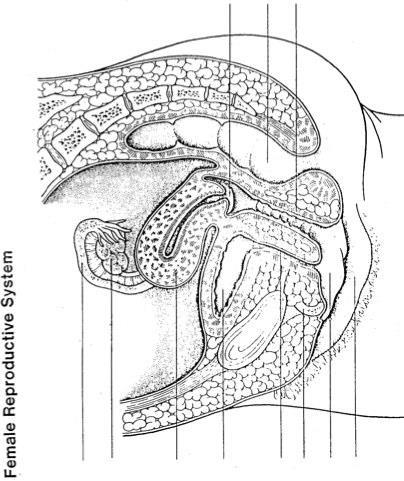
28 The Male Reproductive System The testis lie outside the body cavity since sperm cannot live at 37 o C. The interstitial cells which lie between the seminiferous tubules (which make sperm) make androgens, of which testosterone is one. Once the sperm are made they are stored in the epididymis until they mature. Upon ejaculation, the seminal vesicles, prostate gland and Cowper s gland secrete seminal fluid. Sperm + seminal fluid = semen. The purpose of the alkaline solution is to counteract the acidity of the vagina and male urethra. Sugar is present to provide nutrients for the sperm. Prostaglandins are also present. The hypothalamus causes the anterior pituitary to release FSH and LH, two gonadotropic hormones. Copy the diagram from page 417 (10e) of your text. FSH stimulates the production of sperm while LH promotes the production of testosterone, which causes the development of secondary sex characteristics, such as enlarged vocal cords, a beard, more muscle mass, baldness, sex drive, etc. The Female Reproductive System Females are borne with over 400,000 follicles, each containing an egg. Since eggs age as a woman ages, older women (over 40) have children with more genetic defects than do younger women. Once a month, one follicle matures and releases an egg (if two are released, fraternal twins can result). Look at table on page 420 (10e). Estrogen and progesterone produced by the ovaries are the female sex hormones. They foster the development of the reproductive organs, maintain the uterine cycle, and bring about the secondary sex characteristics in females. Estrogen is largely responsible for the secondary sex characteristics in females, including body hair, wider pelvic girdle and fat distribution. Both progesterone and estrogen are required for breast development. Progesterone is called the pregnancy hormone. Menopause, the period in a woman s life during which the ovarian and uterine cycles cease because the ovaries no longer respond to the gonadotropic hormones. 28
29 29
30 30
31 Female Reproductive Anatomy The female reproductive system is specialized to produce a haploid (1N) gamete, the egg, approximately once a month. It also prepares for he possible fertilization of that egg, by producing a lining in which the egg can implant. This also occurs approximately once a month. The Ovaries and Oviducts The ovaries are the organs, which produce the female gametes (eggs). They are held in the abdomen by ovarian and suspensory ligaments. There are two ovaries, one on the left of the abdomen and one on the right. The outer layer of the ovary, the cortex, contains thousands of follicles. Each follicle consists of a group of cells surrounding an undeveloped egg or oocyte. When a female is born, she has a complete set of undeveloped eggs. No new eggs will be produced during her lifetime. Contrast this to the male, who produces millions of new gametes (sperm) every day! There are about two million of these undeveloped female eggs at birth; however, over the course of a female s reproductive lifespan, only about four hundred will ever mature into eggs. The cells of the follicle provide nourishment for the developing egg. They are also a source of estrogen, a female hormone. A single follicle and egg usually matures about once every twentyeight days. The mature follicle is known as a graffian follicle. When it is mature, the follicle moves to the surface of the ovary, where it blisters out and ruptures, releasing the egg. Release of the egg from the ovary is known as ovulation. The follicle does not degenerate immediately after ovulation. Instead, the follicle cells begin to grow and form the corpus luteum, which secretes estrogen, and another hormone, progesterone. If pregnancy does not occur, the corpus luteum is reabsorbed and ceases hormone production, after about ten days. In other words, females produce a new gland once every month. If fertilization and pregnancy occurs, the corpus luteum persists as a source of hormones for up to six months. After ovulation, the newly released egg passes into the oviduct, or fallopian tube. This structure is basically a thin tube, which leads to the uterus. It is not directly joined to the ovary, and therefore, the egg must be picked up by fingerlike projections found in the opening of the oviduct called fimbria. The fimbria sweep across the surface of the ovary. The beating of the cilia, which line the inside of the oviduct also help to draw in the egg. They also help the egg move along the oviduct towards the uterus. Peristaltic contractions of the smooth muscle walls of the oviduct also help the egg move along. It takes about three days for the egg to travel from the oviduct to uterus. However, the lifespan of the egg is only about six to twenty-four hours. Fertilization, therefore, must occur while the egg is still in the oviduct. 31
32 The Uterus The oviducts enter into the uterus, one from each ovary. You can see from your female reproductive diagrams that the uterus is a pear-shaped organ. The lining of the uterus, the endometrium, consists of two layers. Every month the inner layer grows thicker, and if pregnancy does not occur, this layer is sloughed off during menstruation. The monthly building-up and subsequent sloughing-off of the inner layer of the endometrium constitutes the menstrual cycle. If pregnancy does occur, the fertilized egg (now called a zygote) will implant itself in the endometrium. The fetus therefore, grows and develops from this fertilized egg, in the uterus. The uterus is both strong and elastic. Its walls contain many layers of smooth muscle. Powerful contractions of this muscle tissue will help expel the baby during childbirth. Its elastic properties are needed to accommodate the fetus as it grows in size. The uterus leads into the vagina via an opening in a circular muscle known as the cervix. The cervix I the strongest sphincter muscle in the body. You may recall that a sphincter is a muscle whose contractions close off some opening or tube (such as the pyloric or cardiac sphincter). Why do you think the cervix would need to be so strong? During birth, the cervix dilates to allow the baby to pass into the vagina. By looking at the female anatomy diagram again, you can see that the uterus leads into the vagina at almost a right angle. It is believed that this almost horizontal positioning of the uterus helps a woman carry a baby in the uterus while maintaining a typical upright human posture. The drawback is that childbirth is more difficult in humans than in animals who walk on all fours. The Vagina The vagina is an elastic, muscular tube leading from the bottom of the uterus to the exterior. It is the pathway for a baby to the outside world during childbirth. It also functions as the receptive organ for the penis during sexual intercourse. It connects the external genitalia with the internal features of the female reproductive system. 32
33 Confirm Your Understanding 1. What is the name of the ligaments, which hold the ovaries in place? 2. What is the principal function of the ovaries? 3. Define follicle. 4. What is an oocyte? 5. What is a Graafian follicle? 6. What is ovulation? 7. After ovulation, what happens to the follicle? 8. What is the source of progesterone? 9. Describe what happens to the corpus luteum: a) if the egg is fertilized b) if the egg is not fertilized 10. What is the purpose of an oviduct? 11. Name two ways in which passage of the egg along the oviduct is aided. 33
34 12. True or False. If false, state why. a) The fetus grows and develops in the uterus. b) The endometrium is part of the ovary. c) The cervix is a circular muscle. d) During menstruation, the uterus is shed. 13. Why does fertilization usually occur in the oviduct? 34
35 35
36 36
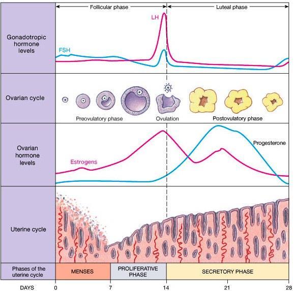
37 HORMONAL CONTROL OF FEMALE REPRODUCTION Hormonal regulation of reproduction in females is more complex than that in males. It involves a cyclic change in the amount of the hormones involved, as well as a cyclic change in the structures involved. Let s first briefly look at the four hormones: 1. Luteinizing Hormone (LH) and Follicle Stimulating Hormone (FSH) You will recall that LH and FSH are two of the trophic hormones released by the anterior pituitary. You may also recall that these two hormones function in the hormonal control of male reproduction. As in the male, their release is stimulated by gonadotropin-releasing hormones from the hypothalamus. 2. Estrogen Estrogen is an important female hormone. Estrogen is secreted by the follicle as it matures in the ovary, and later, when the follicle develops into the corpus luteum, it is secreted by the corpus luteum. So essentially, it is secreted by cells of follicular origin, whether they are a part of the follicle or the corpus luteum. Estrogen is the main hormone that promotes development of female secondary sex characteristics, such as accumulation of body fat under the skin, and the distribution of hair under the arms, on the legs, and in the pubic region. At puberty, secretion of estrogen stimulates growth of the uterus and vagina, and onset of the menstrual cycle. It also causes widening of the pelvic girdle. 3. Progesterone Progesterone is another important female sex hormone. It is produced only in the corpus luteum. It is necessary, along with estrogen, for the development of the breasts. TO SUMMARIZE: LH: FSH: Estrogen: Progesterone: from the anterior pituitary from the anterior pituitary from follicle and corpus luteum (in the ovary) from corpus luteum (in the ovary) For all of the cycles discussed we can assume that we are looking at the cycles of a model female, that is, one whose cycles last exactly 28 days. In reality, there is great variation among cycle times, even from the same woman from month to month. 37
38 The Ovarian Cycle This cycle is concerned with the physical changes that occur in an ovary over 28 days: During days 1 to 13, the follicle is maturing in the ovary. On the 14 th day, ovulation occurs. Day 1 to 14 are known as the follicular phase. Days 15 to 28 are the luteal phase. It is during this time that the corpus luteum develops from the follicle, secretes hormones, and then degenerates. The Menstrual Cycle This cycle is concerned with the physical changes that occur in the endometrium (lining of the uterus) over 28 days. During days 1 to 5, the endometrium is sloughed off, resulting in the flow of menstrual fluid (mense) associated with menstruation. Rebuilding and thickening of the endometrium occurs during days 6 to 13. Remember that ovulation occurs on day 14. Days 14 to 28 see a much greater thickening of the endometrium, accompanied by the development of mucus-secreting glands in the uterus. These two cycles occur at the same time. For example, during days 1 to 5, when the endometrium is degenerating, a new follicle is already starting to mature in the ovary (days 1-5 in the ovarian cycle). It is the ovarian cycle, with its accompanying hormonal changes, which controls the menstrual cycle. Therefore, in the next section, where we consider the cyclic hormonal changes that occur over the 28 days, we shall follow the phases of the ovarian cycle. Hormonal Regulation The female reproductive cycle is governed by cyclic changes in the amounts of the four hormones we looked at above. The level of any one hormone is determined by the relative levels of one or more of the other hormones. Look at the chart provided. A. Follicular Phase (Days 1 to 14) 1. FSH and LH, secreted by the anterior pituitary, stimulate growth and development of a follicle in the ovary. Endometrium is sloughing during the first five days (menstruation). 2. The follicle produces estrogen, which stimulates the start of endometrial rebuilding. 3. Around days 8 to 10, there is a surge of estrogen secretion from the follicle. This causes a corresponding surge in LH and FSH production by the anterior pituitary (positive feedback). LH and FSH peak on day The peak of the LH level causes ovulation on day
39 B. Luteal Phase (Days 15 to 28) 5. The corpus luteum begins to develop form the emptied follicle, under the influence of the still high (but decreasing) LH level. 6. Cells of the corpus luteum secrete estrogen, with the addition of progesterone. Estrogen plus progesterone greatly increase thickness and development of the endometrium. Mucus secreting glands develop. 7. Progesterone plus lower levels of estrogen inhibit LH and FSH secretion by the anterior pituitary (negative feedback compare with step 3). 8. The corpus luteum begins to degenerate, probably as a result of reduced LH and FSH levels. The corpus luteum only degenerates if pregnancy does not occur. 9. Since the corpus luteum is the source of estrogen and progesterone, its degeneration results in a drop-off of the level of these two hormones. C. Follicular Phase (Days 1 to 14) Pregnancy 10. Reduced levels of estrogen and progesterone result in the sloughing off of the endometrium. We are back again at menstruation of days 1 to Because the source of inhibition of LH and FSH production has decreased (ie. Estrogen and progesterone levels dropped-off), their levels start to increase once more. This causes a follicle to begin to develop once more. If fertilization were to occur, the zygote (fertilized egg) would travel down the oviduct into the uterus, where it would embed itself in the endometrium. This process is called implantation. Cells of the zygote then secrete a hormone known as Human Chorionic Gonadotropin, or HCG. HCG causes the corpus luteum to be maintained, and to continue producing estrogen and progesterone. This is important, because then the endometrium is prevented form sloughing off, and in fact is developed even further under stimulation from estrogen and progesterone. Thus, menstruation does not occur in a pregnant woman. HCG is very similar in structure to the hormone LH. For this reason it can inhibit LH and FSH production by the anterior pituitary, through negative feedback. This ensures that no more follicles mature while the woman is pregnant (recall from step 1 that LH and FSH stimulate follicle production). Once the placenta develops, it takes over production of both HCG and progesterone and estrogen. The placenta is the structure across which exchange of nutrients and wastes between the blood of the mother and the fetus takes place. It is formed from both fetal and maternal tissue. 39
40 40
41 Confirm Your Understanding 1. Name the four major hormones of the female reproductive system and the source of each of these hormones. 2. Which hormone is the most important for promoting the development of the secondary sex characteristics? Name three characteristics that estrogen produces. 3. What are the two phases of the ovarian cycle and the days they occur on? 4. What occurs between the two phases and what triggers it? 5. When menstruation occurs during days 1-5 what is happening in the ovarian cycle? 6. What effect does estrogen have on the endometrium during the follicular phase? 7. During the luteal phase, why does the corpus luteum usually begin to degenerate? 8. When the corpus luteum degenerates, which two hormone levels decrease? 41
42 9. What does HCG mean and where does it come from? 10. What effect does HCG have on female reproduction? 11. How does the placenta develop and what does it do? 42
43 Human Reproduction When there is a low level of and hormones, the lining of the endometrium is lost. The first day of bleeding is day (28, 1, 5 pick a number) of the next cycle. In response to the low level of these hormones, the releases the hormone which starts the follicle developing. As the follicle and the contained in it mature the hormone is released which starts the developing a rich blood supply. When estrogen levels are high, and are released. is responsible for ovulation as well as changing the ruptured into the, which acts as an endocrine gland, producing and (the pregnancy hormone). These maintain the. The high levels of these two hormones cause a decrease in the hormone. Without this hormone, the cannot be maintained and degenerates, stopping the release of its hormones. Pregnancy If implantation occurs, the must be maintained and the cannot be allowed to degenerate. To stop this the inner membrane, the releases the hormone which maintains the so it can continue to produce. After months, the is developed enough to secrete its own and. In males, causes the production of sperm in the and the hormone causes the production of by the. 43
44 Sexual Reproduction Pre-Test Males have three glands, namely, and the which produce. The purpose of this secretion is to. The male gamete has chromosomes and was produced by the process of. The sperm are made in the testes in tube-like structures called the. The hormone is responsible for their production as well as the maturation of the egg in the female. Male secondary sex characteristics are caused by the hormone called produced by the. The hormone maintains the male hormone secretion. Males have a scrotum because. The average girl s menstrual cycle repeats every days. Ovulation occurs around day. The hormone that is released from the developing follicle is called which thickens the lining of the. After ovulation, the hormone whose main function is to continues its release of the pregnancy. The lining is lost if If a girl becomes pregnant the hormone does not occur in the uterus. is released from the The purpose of the menstrual cycle is to layer to maintain the.. 44
45 TEST 5 REVIEW 1. Explain External Respiration. (5) 2. Explain the mechanics of breathing. (7) 3. A person suffered a major blood loss. Use ve feedback loops used by the respiratory and excretory systems only to help keep the person alive. (8) 4. What is the function of the pleural membranes? (3) 5. Compare the composition of glomerular filtrate with that of blood plasma. (5) 6. Explain the role of the following in the excretory system: efferent arteriole, microvilli, counter-current exchange mechanism, ascending loop, bladder. (5) 7. Using a feedback loop, explain why a person produces a larger volume of urine after drinking two litres of water. (4) 8. Explain how the kidney controls blood pressure, O2 levels in the blood, and blood ph. (6) 9. Alcohol appears to inhibit the secretion of ADH. Predict the results (with an explanation) of alcohol intake on the following: a) solute concentration of the blood plasma (2) b) urine production (2) 10. Assume that a bacterial infection results in large perforations in the glomerulus of a patient. a) How will the glomerular filtrate in the diseased person compare to the filtrate of a healthy person?(2) b) What effect will this have on ADH levels circulating in the bloodstream and why? (2) c) A person with this disease will have edema of body tissues. Using your knowledge of kidney function and blood tonicity, explain the mechanism accounting for this swelling. (3) 11. After a severe drop in blood pressure, how will the following respond to maintain homeostasis? a) urine production (1) b) autonomic nervous system (1) c) kidney (1) 45

46 12. Label the following structures on a typical kidney and list THREE ways that blood in the renal vein is different from that in the renal artery. (9) 13. The female reproductive system is carefully regulated by hormones produced by the brain and some of the sex organs. Explain the effect the following would have on the reproductive system. a) A failure of the corpus luteum to degenerate 10 days after ovulation. (2) b) A lack of FSH production during the first 15 days of the menstrual cycle. (2) 14. List in the correct order, the structures through which human sperm pass as they travel from the testis until they leave the body. What is the purpose of semen? (4) 15 Relate the effect of the following birth control methods to the physiology of human reproduction. a) birth control pills (1) b) IUD (1) c) vasectomy (1) 16 Describe the effects of estrogen production in the female body and how estrogen affects the menstrual cycle. (5) 17. Describe the internal physiological changes that cause the erection of the penis. (2) 18. State the function of the following in sperm: acrosome, midpiece, tail (3) 19.Describe the hormonal changes and the effects they have in a female as a result of implantation of the embryo. (5) 20. State the function of FSH and LH in men and women. (4) 46
47 21. The following table shows the amounts of substances present in human blood plasma, glomerular filtrate, and urine. All are measured in g/100ml of fluid. Component Plasma Filtrate Urine Glucose Amino acids Proteins Inorganic salts Urea Uric acid a) Account for the difference in the levels of proteins in the plasma and filtrate. (2) b) The concentration of inorganic salts and urea in urine is much greater than that present in the filtrate. Explain why this is so. (2) 22. Use the following chart to identify each person. Subject Hb (g/100ml) O2 in arteriole Blood/100mL O2 in venous Blood/100mL Cardiac Output (L/min) A Normal B Hypoxia C Hypoxia D Hypoxia E Hypoxia no info. Provide two lines of evidence using the chart above to support your answer. a) Which subject in the above chart is suffering from a dietary iron deficiency? (3) b) Which subject is suffering from heart failure and poor blood circulation? (3) c) Which subject has recently climbed a mountain where the air is thin and atmospheric O2 is low? (3) d) Which subject is suffering from a poison that prevents her cells from using oxygen? (3) e) Subject B has increased breathing. Briefly describe the physiological mechanism that is responsible for the increased breathing. (4) 47
Urinary System Chapter 16
 Urinary System Chapter 16 1 Urology- the branch of medicine that treats male and female urinary systems as well as the male reproductive system. Nephrology- the scientific study of the anatomy, physiology,
Human Reproductive System
 Human Reproductive System I. The male reproductive anatomy is a delivery system for sperm. A. The male s external reproductive organs consist of the scrotum and penis. 1. The penis is the external organ
Testes (male gonads) -Produce sperm -Produce sex hormones -Found in a sac called the scrotum -Suspended outside of the body cavity for temperature
 -Produce sperm -Produce sex hormones -Found in a sac called the scrotum -Suspended outside of the body cavity for temperature") REPRODUCTION Testes (male gonads) -Produce sperm -Produce sex hormones -Found in a sac called the scrotum -Suspended outside of the body cavity for temperature reduction -Testes wall made of fibrous connective
REPRODUCTION Testes (male gonads) -Produce sperm -Produce sex hormones -Found in a sac called the scrotum -Suspended outside of the body cavity for temperature reduction -Testes wall made of fibrous connective
Study Guide Answer Key Reproductive System
 Biology 12 Human Biology Textbook: BC Biology 12 Study Guide Answer Key Reproductive System 1. Distinguish between a gamete and a gonad using specific examples from the male and female systems. Gonads
1. a)label the parts indicated above and give one function for structures Y and Z
label the parts indicated above and give one function for structures Y and Z") Excretory System 1 1. Excretory System a)label the parts indicated above and give one function for structures Y and Z W- renal cortex - X- renal medulla Y- renal pelvis collecting center of urine and then
Excretory System 1 1. Excretory System a)label the parts indicated above and give one function for structures Y and Z W- renal cortex - X- renal medulla Y- renal pelvis collecting center of urine and then
Human Reproductive System
 Human Reproductive System I. The male reproductive anatomy is a delivery system for sperm. A. The male=s external reproductive organs consist of the scrotum and penis. 1. The penis is the external organ
Chapter 14 The Reproductive System
 Biology 12 Name: Reproductive System Per: Date: Chapter 14 The Reproductive System Complete using BC Biology 12, page 436-467 14. 1 Male Reproductive System pages 440-443 1. Distinguish between gametes
28/04/2013 LEARNING OUTCOME C13 URINARY SYSTEM STUDENT ACHIEVEMENT INDICATORS STUDENT ACHIEVEMENT INDICATORS URINARY SYSTEM & EXCRETION
 LEARNING OUTCOME C13 Analyse the functional interrelationships of the structures of the urinary system Learning Outcome C13 URINARY SYSTEM STUDENT ACHIEVEMENT INDICATORS Students who have fully met this
Urinary System and Excretion. Bio105 Lecture 20 Chapter 16
 Urinary System and Excretion Bio105 Lecture 20 Chapter 16 1 Outline Urinary System I. Function II. Organs of the urinary system A. Kidneys 1. Function 2. Structure III. Disorders of the urinary system
Unit 15 ~ Learning Guide
 Unit 15 ~ Learning Guide Name: INSTRUCTIONS Complete the following notes and questions as you work through the related lessons. You are required to have this package completed BEFORE you write your unit
Chapter 14 Reproduction Review Assignment
 Date: Mark: _/45 Chapter 14 Reproduction Review Assignment Multiple Choice Identify the choice that best completes the statement or answers the question. 1. Use the diagram above to answer the next question.
Male Reproduction Organs. 1. Testes 2. Epididymis 3. Vas deferens 4. Urethra 5. Penis 6. Prostate 7. Seminal vesicles 8. Bulbourethral glands
 Outline Terminology Human Reproduction Biol 105 Lecture Packet 21 Chapter 17 I. Male Reproduction A. Reproductive organs B. Sperm development II. Female Reproduction A. Reproductive organs B. Egg development
Renal System and Excretion
 Renal System and Excretion Biology 105 Lecture 19 Chapter 16 Outline Renal System I. Functions II. Organs of the renal system III. Kidneys 1. Structure 2. Function IV. Nephron 1. Structure 2. Function
Outline Urinary System. Urinary System and Excretion. Urine. Urinary System. I. Function II. Organs of the urinary system
 Outline Urinary System Urinary System and Excretion Bio105 Chapter 16 Renal will be on the Final only. I. Function II. Organs of the urinary system A. Kidneys 1. Function 2. Structure III. Disorders of
Nephron Structure inside Kidney:
 In-Depth on Kidney Nephron Structure inside Kidney: - Each nephron has two capillary regions in close proximity to the nephron tubule, the first capillary bed for fluid exchange is called the glomerulus,
NOTES: CH 44 Regulating the Internal Environment (Homeostasis & The Urinary System)
") NOTES: CH 44 Regulating the Internal Environment (Homeostasis & The Urinary System) HOMEOSTASIS **Recall HOMEOSTASIS is the steady-state physiological condition of the body. It includes: 1) Thermoregulation:
NOTES: CH 44 Regulating the Internal Environment (Homeostasis & The Urinary System) HOMEOSTASIS **Recall HOMEOSTASIS is the steady-state physiological condition of the body. It includes: 1) Thermoregulation:
Osmotic Regulation and the Urinary System. Chapter 50
 Osmotic Regulation and the Urinary System Chapter 50 Challenge Questions Indicate the areas of the nephron that the following hormones target, and describe when and how the hormones elicit their actions.
Outline Urinary System
 Urinary System and Excretion Bio105 Lecture Packet 20 Chapter 16 Outline Urinary System I. Function II. Organs of the urinary system A. Kidneys 1. Function 2. Structure B. Urine formation 1. Hormonal regulation
12/7/10. Excretory System. The basic function of the excretory system is to regulate the volume and composition of body fluids by:
 Excretory System The basic function of the excretory system is to regulate the volume and composition of body fluids by: o o removing wastes returning needed substances to the body for reuse Body systems
Functions of male Reproductive System: produce gametes deliver gametes protect and support gametes
 Functions of male Reproductive System: produce gametes deliver gametes protect and support gametes Spermatogenesis occurs in the testes after puberty. From the testes they are deposited into the epididymas
April 08, biology 2201 ch 11.3 excretion.notebook. Biology The Excretory System. Apr 13 9:14 PM EXCRETORY SYSTEM.
 Biology 2201 11.3 The Excretory System EXCRETORY SYSTEM 1 Excretory System How does the excretory system maintain homeostasis? It regulates heat, water, salt, acid base concentrations and metabolite concentrations
Sunday, July 17, 2011 URINARY SYSTEM
 URINARY SYSTEM URINARY SYSTEM Let s take a look at the anatomy first! KIDNEYS: are complex reprocessing centers where blood is filtered through and waste products are removed. Wastes and extra water become
PARTS OF THE URINARY SYSTEM
 EXCRETORY SYSTEM Excretory System How does the excretory system maintain homeostasis? It regulates heat, water, salt, acid-base concentrations and metabolite concentrations 1 ORGANS OF EXCRETION Skin and
organs of the urinary system
 organs of the urinary system Kidneys (2) bean-shaped, fist-sized organ where urine is formed. Lie on either sides of the vertebral column, in a depression beneath peritoneum and protected by lower ribs
November 30, 2016 & URINE FORMATION
 & URINE FORMATION REVIEW! Urinary/Renal System 200 litres of blood are filtered daily by the kidneys Usable material: reabsorbed back into blood Waste: drained into the bladder away from the heart to the
The Excretory System. Biology 20
 The Excretory System Biology 20 Introduction Follow along on page 376 What dangers exist if your body is unable to regulate the fluid balance of your tissues? What challenged would the body have to respond
Sample Provincial exam Q s: Reproduction
 Sample Provincial exam Q s: Reproduction 11. Functions Testosterone Makes the male sex organs function normally, and also inhibits hypothalamus s release of GnRH and thus LH & FSH and thus testosterone
Chapter 12. Excretion and the Interaction of Systems
 Chapter 12 Excretion and the Interaction of Systems 1 2 Goals for This Chapter 1. Identify the main structures and functions of the human excretory system 2. Explain the function of the nephron 3. Describe
ANATOMY AND PHYSIOLOGY HOMEWORK CHAPTER 15 AND 16
 ANATOMY AND PHYSIOLOGY HOMEWORK CHAPTER 15 AND 16 Name Identify the following: 1) The ureter is indicated by letter 2) The renal pyramid is indicated by letter 3) The fibrous capsule is indicated by letter
Refer to the figure below, a diagram of a renal tubule, to answer the following questions.
 1. The digestion and utilization of which nutrient creates the greatest need for osmoregulation by the kidneys? a. protein b. starch c. fat d. oil e. cellulose 2. Which of the following is true of urea?
A&P 2 CANALE T H E U R I N A R Y S Y S T E M
 A&P 2 CANALE T H E U R I N A R Y S Y S T E M URINARY SYSTEM CONTRIBUTION TO HOMEOSTASIS Regulates body water levels Excess water taken in is excreted Output varies from 2-1/2 liter/day to 1 liter/hour
Endocrine and Reproductive Systems. Chapter 39: Biology II
 Endocrine and Reproductive Systems Chapter 39: Biology II The Endocrine System Made up of glands that release their products into the bloodstream These products broadcast messages throughout the body Chemicals
Urinary System Organization. Urinary System Organization. The Kidneys. The Components of the Urinary System
 Urinary System Organization The Golden Rule: The Job of The Urinary System is to Maintain the Composition and Volume of ECF remember this & all else will fall in place! Functions of the Urinary System
Grade 9 Science - Human Reproduction
 Grade 9 Science - Human Reproduction The human reproductive system is a series of organs that work together for one purpose: reproduction (creating new humans). Each part has a specific role in the reproductive
Outline. Male Reproductive System Testes and Sperm Hormonal Regulation
 Outline Male Reproductive System Testes and Sperm Hormonal Regulation Female Reproductive System Genital Tract Hormonal Levels Uterine Cycle Fertilization and Pregnancy Control of Reproduction Infertility
Human Anatomy and Physiology - Problem Drill 23: The Urinary System, Fluid, Electrolyte and Acid-Base Balance
 Human Anatomy and Physiology - Problem Drill 23: The Urinary System, Fluid, Electrolyte and Acid-Base Balance Question No. 1 of 10 Which of the following statements about the functions of the urinary system
Excretion and Waste Management. Biology 30S - Miss Paslawski
 Excretion and Waste Management Biology 30S - Miss Paslawski Lesson 1 Waste Products and Organs 2 3 Excretion Excretion: Process by which dissolved metabolic wastes are separated from body fluids and removed
Chapter 26: Urinary System By: Eddie Tribiana and Piers Frieden
 Chapter 26: Urinary System By: Eddie Tribiana and Piers Frieden The urinary system is important because it performs vital excretory functions Takes blood from renal arteries into the kidney to filtrate
BIOLOGY - CLUTCH CH.44 - OSMOREGULATION AND EXCRETION.
 !! www.clutchprep.com Osmoregulation regulation of solute balance and water loss to maintain homeostasis of water content Excretion process of eliminating waste from the body, like nitrogenous waste Kidney
Excretory System 1. a)label the parts indicated above and give one function for structures Y and Z
label the parts indicated above and give one function for structures Y and Z") Excretory System 1 1. Excretory System a)label the parts indicated above and give one function for structures Y and Z W- X- Y- Z- b) Which of the following is not a function of the organ shown? A. to produce
Excretory System 1 1. Excretory System a)label the parts indicated above and give one function for structures Y and Z W- X- Y- Z- b) Which of the following is not a function of the organ shown? A. to produce
Male Reproductive Structures I. Overview A. Main functions: 1. Produce a haploid male gamete (sperm) 2. Deposit sperm in the female so fertilization
 2. Deposit sperm in the female so fertilization") Male Reproductive Structures I. Overview A. Main functions: 1. Produce a haploid male gamete (sperm) 2. Deposit sperm in the female so fertilization may occur! A. Scrotum 1. Muscular pouch that holds the
Male Reproductive Structures I. Overview A. Main functions: 1. Produce a haploid male gamete (sperm) 2. Deposit sperm in the female so fertilization may occur! A. Scrotum 1. Muscular pouch that holds the
I. Metabolic Wastes Metabolic Waste:
 I. Metabolic Wastes Metabolic Waste: a) Carbon Dioxide: by-product of cellular respiration. b) Water: by-product of cellular respiration & dehydration synthesis reactions. c) Inorganic Salts: by-product
Urinary System BIO 250. Waste Products of Metabolism Urea Carbon dioxide Inorganic salts Water Heat. Routes of Waste Elimination
 Urinary System BIO 250 Waste Products of Metabolism Urea Carbon dioxide Inorganic salts Water Heat Routes of Waste Elimination Skin: Variable amounts of heat, salts, and water; small amounts of urea and
Biology 12 August 1999 Provincial Examination
 Biology 12 August 1999 Provincial Examination ANSWER KEY / SCORING GUIDE CURRICULUM: Organizers 1. Cell Biology 2. Cell Processes and Applications 3. Human Biology Sub-Organizers A, B, C, D E, F, G, H
Chapter 16 Lecture Outline
 Chapter 16 Lecture Outline See separate PowerPoint slides for all figures and tables preinserted into PowerPoint without notes. Copyright The McGraw-Hill Companies, Inc. Permission required for reproduction
A. Incorrect! The urinary system is involved in the regulation of blood ph. B. Correct! The urinary system is involved in the synthesis of vitamin D.
 Human Anatomy - Problem Drill 22: The Urinary System Question No. 1 of 10 1. Which of the following statements about the functions of the urinary system is not correct? Question #01 (A) The urinary system
The Male Reproductive System
 The Male Reproductive System Male Reproductive System The male sex cell is a sperm cell The whole purpose is to produce and deliver sperm to the egg Structure of a Human Sperm Cell Streamlined, built to
EXCRETION QUESTIONS. Use the following information to answer the next two questions.
 EXCRETION QUESTIONS Use the following information to answer the next two questions. 1. Filtration occurs at the area labeled A. V B. X C. Y D. Z 2. The antidiuretic hormone (vasopressin) acts on the area
What are the main functions of the male reproductive system? 1. Produce sperm 2. Deposit sperm into the female 3. Provide a pathway for the removal
 What are the main functions of the male reproductive system? 1. Produce sperm 2. Deposit sperm into the female 3. Provide a pathway for the removal of urine Where is sperm produced? -In the 2 testes What
9.4 Regulating the Reproductive System
 9.4 Regulating the Reproductive System The Reproductive System to unite a single reproductive cell from a female with a single reproductive cell from a male Both male and female reproductive systems include
Chapter 13 The Urinary System
 Biology 12 Name: Urinary System Per: Date: Chapter 13 The Urinary System Complete using BC Biology 12, page 408-435 13.1 The Urinary System pages 412-413 1. As the kidneys produce urine, they carry out
What is excretion? Excretion is the removal of metabolic waste from the body.
 Excretion What is excretion? Excretion is the removal of metabolic waste from the body. Excretion in Plants Plants produce very little waste products. Plants lose oxygen and water vapour through the stomata.
Nephron Function and Urine Formation. Ms. Kula December 1, 2014 Biology 30S
 Nephron Function and Urine Formation Ms. Kula December 1, 2014 Biology 30S The Role of the Nephron In order for the body to properly function and maintain homeostasis, the amount of dissolved substances
Ch. 44 Regulating the Internal Environment
 Ch. 44 Regulating the Internal Environment 2006-2007 Conformers vs. Regulators Two evolutionary paths for organisms regulate internal environment maintain relatively constant internal conditions conform
Excretion and Water Balance
 Excretion and Water Balance 1. Osmoregulation (water balance) a. Most marine invertebrates are osmoconformers in which the concentration of solutes in their body fluid is equal to that of their environment.
Ch17-18 Urinary System
 Ch17-18 Urinary System Main Function: Filter the blood Other Functions: maintain purity and consistency of internal fluids eliminates nitrogenous wastes, toxins, and drugs from the body regulates blood
Human Urogenital System 26-1
 Human Urogenital System 26-1 Urogenital System Functions Filtering of blood, Removal of wastes and metabolites Regulation of blood volume and composition concentration of blood solutes ph of extracellular
Fifth Year Biology. Excretion. Miss Rochford
 Fifth Year Biology Excretion Miss Rochford In this Topic Excretion in plants Excretion and homeostasis Skin Organs of excretion Urinary system Kidneys Nephron Control of urine volume Characteristics of
Regulating the Internal Environment. AP Biology
 Regulating the Internal Environment 2006-2007 Conformers vs. Regulators Two evolutionary paths for organisms regulate internal environment maintain relatively constant internal conditions conform to external
The Urinary System. Copyright 2003 Pearson Education, Inc. publishing as Benjamin Cummings
 The Urinary System Functions of the Urinary System Elimination of waste products Nitrogenous wastes Toxins Drugs Functions of the Urinary System Regulate aspects of homeostasis Water balance Electrolytes
Male Reproductive System
 Male Reproductive System The male reproductive system consists of a number of sex organs that are part of the reproductive process. The following sections describe the function of each part of the male
Human Reproduction. Human Reproductive System. Scrotum. Male Reproductive System
 Human Reproductive System Human Reproduction Chapter 41 Contraceptives Scrotum Testes Epididymus Vas Deferens Seminal Vesicles Prostate Gland Bulbourethral Gland Penis Scrotum Sac of smooth muscle tissue
BCH 450 Biochemistry of Specialized Tissues
 BCH 450 Biochemistry of Specialized Tissues VII. Renal Structure, Function & Regulation Kidney Function 1. Regulate Extracellular fluid (ECF) (plasma and interstitial fluid) through formation of urine.
1. Urinary System, General
 S T U D Y G U I D E 16 1. Urinary System, General a. Label the figure by placing the numbers of the structures in the spaces by the correct labels. 7 Aorta 6 Kidney 8 Ureter 2 Inferior vena cava 4 Renal
Chapter 44. Regulating the Internal Environment. AP Biology
 Chapter 44. Regulating the Internal Environment Homeostasis Living in the world organisms had a choice: regulate their internal environment maintain relatively constant internal conditions conform to the
2. Ureters Composed of smooth muscle tissue ~25cm long Connects kidneys to bladder Undergoes peristaltic contraction to move urine to bladder
 Section 6: The Urinary System A) Organs of the Urinary system 1. Kidneys 2. Ureters 3. Bladder 4. Urethra 1. Kidneys Paired organs located on either side of vertebral column in upper part of abdominal
Chapter 11 Lecture Outline
 Chapter 11 Lecture Outline See separate PowerPoint slides for all figures and tables preinserted into PowerPoint without notes. Copyright 2016 McGraw-Hill Education. Permission required for reproduction
Excretory System. Biology 2201
 Excretory System Biology 2201 Excretory System How does the excretory system maintain homeostasis? It regulates: Body heat Water-salt concentrations Acid-base concentrations Metabolite concentrations ORGANS
Excretory System. Excretory System
 Excretory System Biology 2201 Excretory System How does the excretory system maintain homeostasis? It regulates: Body heat Water-salt concentrations Acid-base concentrations Metabolite concentrations 1
mid ihsan (Physiology ) GFR is increased when A -Renal blood flow is increased B -Sym. Ganglion activity is reduced C-A and B **
 GFR is increased when A -Renal blood flow is increased B -Sym. Ganglion activity is reduced C-A and B **") (Physiology ) mid ihsan GFR is increased when A -Renal blood flow is increased B -Sym. Ganglion activity is reduced C-A and B ** Colloid pressure in the efferent arteriole is: A- More than that leaving
(Physiology ) mid ihsan GFR is increased when A -Renal blood flow is increased B -Sym. Ganglion activity is reduced C-A and B ** Colloid pressure in the efferent arteriole is: A- More than that leaving
Excretion: is the removal of waste products formed by metabolism, out of the body
 Excretion in Humans Excretion: is the removal of waste products formed by metabolism, out of the body Accumulation of wastes in the cells would affect the normal functioning of the cells By getting rid
2) This is a Point and Click question. You must click on the required structure.
 This is a Point and Click question. You must click on the required structure.") Class: A&P2-1 Description: Test: Excretory Test Points: 144 Test Number: 28379 Printed: 31-March-10 12:03 1) This is a Point and Click question. You must click on the required structure. Click on the Bowman's
Class: A&P2-1 Description: Test: Excretory Test Points: 144 Test Number: 28379 Printed: 31-March-10 12:03 1) This is a Point and Click question. You must click on the required structure. Click on the Bowman's
Web Activity: Simulation Structures of the Female Reproductive System
 differentiate. The epididymis is a coiled tube found along the outer edge of the testis where the sperm mature. 3. Testosterone is a male sex hormone produced in the interstitial cells of the testes. It
Endocrine System Hormones & Homeostasis. Regents Biology
 Endocrine System Hormones & Homeostasis 2009-2010 Homeostasis Homeostasis maintaining internal balance in the body organism must keep internal conditions stable even if environment changes also called
Functions of the Urinary System
 The Urinary System Functions of the Urinary System Elimination of waste products Nitrogenous wastes Toxins Drugs Regulate aspects of homeostasis Water balance Electrolytes Acid-base balance in the blood
Biology /09 Released Exam June 2009 Form A Provincial Examination Answer Key
 Biology 12 2008/09 Released Exam June 2009 Form A Provincial Examination Answer Key Cognitive Processes K = Knowledge U = Understanding H = Higher Mental Processes Weightings 22% 58% 20% Types 67 = Multiple
The Urinary System Pearson Education, Inc.
 26 The Urinary System Introduction The urinary system does more than just get rid of liquid waste. It also: Regulates plasma ion concentrations Regulates blood volume and blood pressure Stabilizes blood
1. Both asexual and sexual reproduction occur in the animal kingdom
 1. Both asexual and sexual reproduction occur in the animal kingdom Asexual reproduction involves the formation of individuals whose genes all come from one parent. There is no fusion of sperm and egg.
WHY DO WE NEED AN EXCRETORY SYSTEM? Function: To eliminate waste To maintain water and salt balance To maintain blood pressure
 EXCRETORY SYSTEM WHY DO WE NEED AN EXCRETORY SYSTEM? Function: To eliminate waste To maintain water and salt balance To maintain blood pressure These wastes include: Carbon dioxide Mostly through breathing
Circulatory System. and. Respiratory System. Ari Min, Yerim Lee and Min Ji Song THE HEART LUNGS. Monday, May 23, 2011
 Human Anatomy Circulatory System and THE HEART Respiratory System LUNGS Ari Min, Yerim Lee and Min Ji Song Purpose of the Circulatory System Function of circulatory system: exchange gases with cardiovascular
Question 1: Solution 1: Question 2: Question 3: Question 4: Class X The Excretory System Biology
 A. MULTIPLE CHOICE TYPE: (select the most appropriate option in each case) Book Name: Selina Concise Question 1: Excretion primarily involves (a) removal of all byproducts during catabolism (b) removal
Lab Activity 31. Anatomy of the Urinary System. Portland Community College BI 233
 Lab Activity 31 Anatomy of the Urinary System Portland Community College BI 233 Urinary System Organs Kidneys Urinary bladder: provides a temporary storage reservoir for urine Paired ureters: transport
First is Urine Production. We ll discuss the specifics of this process momentarily.
 1 2 The kidney has a number of functions. First is Urine Production. We ll discuss the specifics of this process momentarily. Next, the kidneys filter blood. As a result of filtering blood, the kidneys
Osmoregulation and Renal Function
 1 Bio 236 Lab: Osmoregulation and Renal Function Fig. 1: Kidney Anatomy Fig. 2: Renal Nephron The kidneys are paired structures that lie within the posterior abdominal cavity close to the spine. Each kidney
Section 39 1 The Endocrine System (pages )
") Chapter 39 Endocrine and Reproductive Systems 11 What is the function of the parathyroid glands? Section 39 1 The Endocrine System (pages 997 1002) Key Concepts What is the function of the endocrine system?
Chapter 39 Endocrine and Reproductive Systems 11 What is the function of the parathyroid glands? Section 39 1 The Endocrine System (pages 997 1002) Key Concepts What is the function of the endocrine system?
Urinary System and Fluid Balance. Urine Production
 Urinary System and Fluid Balance Name Pd Date Urine Production The three processes critical to the formation of urine are filtration, reabsorption, and secretion. Match these terms with the correct statement
Urinary bladder provides a temporary storage reservoir for urine
 Urinary System Organs Kidney Filters blood, allowing toxins, metabolic wastes, and excess ions to leave the body in urine Urinary bladder provides a temporary storage reservoir for urine Paired ureters
Female Reproductive System. Lesson 10
 Female Reproductive System Lesson 10 Learning Goals 1. What are the five hormones involved in the female reproductive system? 2. Understand the four phases of the menstrual cycle. Human Reproductive System
Vertebrates possess kidneys: internal organs which are vital to ion and water balance and excretion.
 The Kidney Vertebrates possess kidneys: internal organs which are vital to ion and water balance and excretion. The kidney has 6 roles in the maintenance of homeostasis. 6 Main Functions 1. Ion Balance
Other Factors Affecting GFR. Chapter 25. After Filtration. Reabsorption and Secretion. 5 Functions of the PCT
 Other Factors Affecting GFR Chapter 25 Part 2. Renal Physiology Nitric oxide vasodilator produced by the vascular endothelium Adenosine vasoconstrictor of renal vasculature Endothelin a powerful vasoconstrictor
EXCRETION IN HUMANS 31 JULY 2013
 EXCRETION IN HUMANS 31 JULY 2013 Lesson Description In this lesson we: Discuss organs of excretion Look at the structure of the urinary system Look at the structure and functioning of the kidney Discuss
Section 39 1 The Endocrine System (pages )
") Chapter 39 Endocrine and Reproductive Systems Section 39 1 The Endocrine System (pages 997 1002) Key Concepts What is the function of the endocrine system? How does the endocrine system maintain homeostasis?
Chapter 39 Endocrine and Reproductive Systems Section 39 1 The Endocrine System (pages 997 1002) Key Concepts What is the function of the endocrine system? How does the endocrine system maintain homeostasis?
Chapter 32 Excretion
 Chapter 32 Excretion 3.4 Learning Objectives 3.4.6 The Excretory System in Humans 1. Role of the excretory system in homeostasis. 2. Importance of temperature regulation within the body. 3. Outline the
Chapter 28: REPRODUCTIVE SYSTEM: MALE
 Chapter 28: REPRODUCTIVE SYSTEM: MALE I. FUNCTIONAL ANATOMY (Fig. 28.1) A. Testes: glands which produce male gametes, as well as glands producing testosterone 2. Seminiferous tubules (Fig.28.3; 28.5) a.
Male Reproductive System
 21-1 21-2 Reproductive System Male Reproductive System Genital Tract In males the testes, held outside the body in the scrotum (optimum temp of about 35 0 C), produce sperm. Sperm mature in coiled tubes
describe the location of the kidneys relative to the vertebral column:
 Basic A & P II Dr. L. Bacha Chapter Outline (Martini & Nath 2010) list the three major functions of the urinary system: by examining Fig. 24-1, list the organs of the urinary system: describe the location
Human Reproduction. Male & Female Systems & Menstration
 Human Reproduction Male & Female Systems & Menstration Make a Sperm & Ovum Female Mons Pubis Labia Urethra Perineum Vagina Cervix Uterus Endometrium Fallopian tubes Ovaries Ovum Male Scrotrum Testicle
Chapter 10 Respiration
 1 Chapter 10 Respiration Introduction/Importance of the Respiratory System All eukaryotic organisms need oxygen to perform cellular respiration (production of ATP), either aerobically or anaerobically.
Human Reproductive Anatomy The female anatomy first just the reproductive parts:
 Human Reproduction: Structures, Functions, and Hormones Human reproduction differs from the mechanisms in lower vertebrates and many invertebrates. We depend on internal fertilization sperm are deposited