Vasopressin Anatomy of the Mouse Brain
|
|
|
- Peter Bennett
- 5 years ago
- Views:
Transcription

1 University of Massachusetts Amherst Amherst Open Access Dissertations Vasopressin Anatomy of the Mouse Brain Benjamin D. Rood University of Massachusetts Amherst Follow this and additional works at: Part of the Behavior and Behavior Mechanisms Commons, and the Neurology Commons Recommended Citation Rood, Benjamin D., "Vasopressin Anatomy of the Mouse Brain" (2010). Open Access Dissertations This Open Access Dissertation is brought to you for free and open access by Amherst. It has been accepted for inclusion in Open Access Dissertations by an authorized administrator of Amherst. For more information, please contact
2 VASOPRESSIN ANATOMY OF THE MOUSE BRAIN A Dissertation Presented by BENJAMIN D. ROOD Submitted to the Graduate School of the University of Massachusetts Amherst in partial fulfillment of the requirements for the degree of DOCTOR OF PHILOSOPHY February 2010 Neuroscience and Behavior
3 Copyright by Benjamin D. Rood 2010 All Rights Reserved
4 VASOPRESSIN ANATOMY OF THE MOUSE BRAIN A Dissertation Presented by BENJAMIN D. ROOD Approved as to style and content by: Geert J. de Vries, Chair John R. Nambu, Member Jeffrey D. Blaustein, Member Nancy G. Forger, Member Jerrold S. Meyer, Director Neuroscience and Behavior
5 DEDICATION To my loving wife and two beautiful children.
6 ACKNOWLEDGEMENTS I would like to thank my advisor Geert J. de Vries for the many years of guidance and support that he has given me. I am also especially grateful for his having believed in me even when I no longer believed in myself. Thank you to my dissertation committee, Nancy Forger, Jeff Blaustein, and John Nambu, whose feedback has always made me a better writer and scientist. To the many students and technicians that have assisted me over the last decade, thank you. Thank you to my friends Marianne Seney and, especially, Elaine Murray, who have always been there to encourage me and help me get to the next day. Finally, to my wife and children, I love you. Without your support and understanding the writing of this dissertation would not have been possible. You inspire me. v
7 ABSTRACT VASOPRESSIN ANATOMY OF THE MOUSE BRAIN FEBRUARY 2010 BENJAMIN D. ROOD, B.S., BATES COLLEGE M.S., UNIVERSITY OF MASSACHUSETTS AMHERST Ph.D., UNIVERSITY OF MASSACHUSETTS AMHERST Directed by: Geert J. de Vries The nine amino acid peptide vasopressin acts as a neurohormone in the periphery and a neurotransmitter / neuromodulator in the central nervous system. Historically, research on vasopressin neurons and their projections to the pituitary has helped lay the groundwork for our understanding of peptidergic neurotransmission. Currently, our research on central vasopressin projections is driving a revolution in our understanding of social behavior. Vasopressin affects a number of social behaviors from social memory to aggression to affiliative behavior, such as pair-bonding. Further, with the addition of more and more transgenic mouse models of disease states, anxiety and depression related disorders, and social behavior dysfunction, it is important now more than ever to have a clear knowledge of the mouse vasopressin system, which derives from a number of distinct nuclei within the brain. Here, I map out vasopressin immunoreactivity in the mouse brain, and delineate the subset of brain regions with gonadal steroid hormonedependent vasopressin immunoreactivity. Such projections are thought to derive from the bed nuclei of the stria terminalis and medial amygdala in the telencephalon. Finally, based on data from mice with lesions of the suprachiasmatic nucleus, I outline the subset of regions that likely receive vasopressin from this source. Our research on the anatomy of the vasopressin system of mice and our attempts to delineate the site of origin of the many vasopressin fibers found throughout the brain suggest that a significant amount of the vasopressin innervation deriving from cells in the bed nuclei of the stria terminalis and medial amygdala project to areas in the midbrain involved in serotonin and dopamine transmission, such as the dorsal raphe and ventral tegmental area. These transmitter systems play a crucial role in the control of anxiety and depression levels as well as motivated behavior and emotional regulation. Our results strongly suggest that a direct link exists between these systems, and future plans include an examination of this possibility. It is our hope that this work will further our understanding of the role of vasopressin and other transmitter systems in the regulation of social behaviors. vi
8 TABLE OF CONTENTS Page ACKNOWLEDGMENTS...v ABSTRACT... vi LIST OF TABLES... xii LIST OF FIGURES... xiii CHAPTER 1. THE VASOPRESSIN SYSTEM Introduction Vasopressin is a peptide Vasopressin acts as a neurotransmitter Location of vasopressin production Regulation of vasopressin gene expression Location of vasopressin-containing fibers Site of origin of vasopressin-containing fibers Vasopressin receptors Functions carried out by vasopressin Summary VASOPRESSIN ANATOMY IN INTACT MALES Introduction Methods Animals Tissue collection and processing Nissl staining Immunohistochemistry Analyses and image acquisition Results Cell bodies in the diencephalon Paraventricular nucleus Supraoptic nucleus Suprachiasmatic nucleus Accessory nuclei...25 vii
9 Other hypothalamic nuclei Cell bodies in the telencephalon Bed nuclei of the stria terminalis Medial Amygdala Small versus medium fibers Fibers in the telencephalon Cortex Hippocampus Basal ganglia Septal Complex Ventral forebrain Bed nuclei of the stria terminalis Amygdala Fibers in the diencephalon Hypothalamus Preoptic Area Anterior hypothalamus Dorsomedial hypothalamus Lateral hypothalamus Mammillary bodies Thalamus Fibers in the mesencephalon Tectum Tegmentum Periaqueductal gray and dorsal raphe nuclei Caudal mesencephalic nuclei Fibers in the metencephalon Fibers in the myelencephalon Dorsal myelencephalon Ventral myelencephalon Discussion Figure legends...50 viii
10 3. PROJECTIONS OF THE BNST AND MEDIAL AMYGDALA Introduction Methods Animals Gonadectomy surgeries Tissue collection and processing Immunohistochemistry Qualitative and quantitative fiber density analysis Statistics Results Telencephalon Steroid-sensitive vasopressin projections Sex differences and interactions Diencephalon Hypothalamus Steroid-sensitive vasopressin projections Sex differences and interactions Thalamus Steroid-sensitive vasopressin projections Sex differences and interactions Mesencephalon Steroid-sensitive vasopressin projections Sex differences and interactions Metencephalon and myelencephalon Steroid-sensitive vasopressin projections Sex differences and interactions Discussion Figure legends ix
11 4. INTRACRANIAL STEROID IMPLANTS AS AN ANATOMICAL TOOL Introduction Methods Animals Gonadectomy surgeries Intracranial implants Subcutaneous implants Tissue collection and processing Nissl staining Immunohistochemistry In situ hybridization Experimental designs Experiment Experiment Experiment 3a Experiment 3b Experiment 3c Results and Conclusions Experiment 1: Intracranial TP implants Experiment 2: Subcutaneous T or E 2 implants Experiment 3a: Large E 2 implants in the BNST Experiment 3b: Small E 2 implants in the BNST Experiment 3c: Dilute, small E 2 implants in the BNST Discussion Figure legends PROJECTIONS OF THE SUPRACHIASMATIC NUCLEUS Introduction Methods Animals Gonadectomy surgeries Suprachiasmatic nucleus lesions Tissue collection and processing Nissl staining Immunohistochemistry Analysis Results x
12 5.3.1 Location of SCN lesions Vasopressin immunoreactivity following SCN lesions Discussion Figure legends GENERAL DISCUSSION General comments and conclusions The human condition Conclusions BIBLIOGRAPHY xi
13 LIST OF TABLES 3.1 Statistics for quantitative analyses Vasopressin-ir fibers in the telencephalon Vasopressin-ir fibers in the diencephalon: Hypothalamus Vasopressin-ir fibers in the diencephalon: Thalamus Vasopressin-ir fibers in the mesencephalon Vasopressin-ir fibers in the metencephalon and myelencephalon Vasopressin-ir fibers following SCN lesions xii
14 LIST OF FIGURES 2.1 Paraventricular nucleus Supraoptic nucleus Suprachiasmatic nucleus Accessory nuclei Other hypothalamic nuclei BNST and medial amygdala Small versus medium fibers Cortex and hippocampus Basal ganglia and ventral forebrain Septal complex Bed nuclei of the stria terminalis Amygdala Preoptic area Anterior hypothalamus Dorsomedial hypothalamus Lateral hypothalamus and mammillary bodies Thalamus Tegmentum Periaqueductal gray and dorsal raphe nuclei Caudal mesencephalic nuclei Metencephalon and myelencephalon Regions chosen for quantitative analysis Quantitative results: Lateral septum Quantitative results: BNST Quantitative results: Amygdala Quantitative results: Hypothalamus Quantitative results: Thalamus Quantitative results: Mesencephalon Quantitative results: Metencephalon Experiment 1 - Implant locations Experiment 1 - Data and photomicrographs Experiment 2 - Data and photomicrographs Experiment 3 - Implant locations Experiment 3 - Data and photomicrographs Location of SCN lesions Vasopressin -ir fibers in the STMPM Vasopressin -ir fibers in the preoptic area Vasopressin -ir fibers in the subparaventricular zone Vasopressin -ir fibers in the anterior paraventricular thalamus Vasopressin -ir fibers in the paraventricular thalamus Vasopressin -ir fibers in the xiphoid nucleus Vasopressin -ir fibers in the retrochiasmatic area Vasopressin -ir fibers in the dorsomedial hypothalamus xiii
15 CHAPTER 1 THE VASOPRESSIN SYSTEM 1.1 Introduction A number of prominent disorders (e.g., autism, schizophrenia, obsessivecompulsive disorder, anxiety, depression etc.) are characterized by disruptions of social, emotional, or cognitive behavior. Behaviors altered in these disorders are controlled or regulated by many areas of the brain. Understanding the connections between brain areas implicated in behavior and the neurochemical properties of those areas is crucial to the ability to treat psychological disorders. Research, especially in the last two decades, has identified vasopressin as an important factor in the control of social behavior. To understand how vasopressin controls social behavior under natural conditions it is important to first understand the anatomy of the vasopressin system. 1.2 Vasopressin is a peptide Vasopressin is a nine amino acid peptide (Cys-Tyr-Phe-Gln-Asn-Cys-Pro-Arg- Gly) produced in a number of nuclei in the brain. The functional properties of vasopressin were first described in the late 1800's (Oliver and Schafer, 1895), the peptide was first isolated in 1951 (Turner et al., 1951), and the gene was sequenced in 1982 (Land et al., 1982). The vasopressin gene located on chromosome 20 in humans and chromosome 2 in mice codes for a pre-propeptide made up of vasopressin, neurophysin II, and vasopressin-associated glycopeptide (Land et al., 1982). Vasopressin is cleaved from the pre-propeptide and has functional signaling activity following extracellular release. Neurophysin II and vasopressin-associated glycopeptide are packaged with 1
16 vasopressin and co-released, but have no known signaling activity (Gainer et al., 1977; Russell et al., 1981). 1.3 Vasopressin acts as a neurotransmitter There are three primary requirements for a substance to be classified as a neurotransmitter. The substance must be present in pre-synaptic neurons, particularly in the synaptic terminals; it must be released from those terminals in a calcium dependent manner; and it must bind to a post-synaptic receptor thereby affecting the function of that post-synaptic cell. The first evidence for neurosecretory neurons projecting centrally came in the 1950's and 1960's when researchers observed Gomori stained cells within the central nervous system (Scharrer, 1951; Barry, 1961). In the 1970's, the development and refinement of immunocytochemistry allowed the first visualization of neurophysin containing neurons and fibers (Swaab and Pool, 1975; Vandesande and Dierickx, 1975; Swanson, 1977). More specific antibodies and staining procedures allowed researchers to further distinguish between vasopressin and oxytocin staining. Some of the first analyses of the distribution of vasopressin-containing cells and fibers in the brain were done in 1978 using brain sections from male Wistar rats and "home-made" antisera to vasopressin neurophysin (Buijs, 1978; Buijs et al., 1978). Neurophysin containing cell bodies were found in the paraventricular, supraoptic, and suprachiasmatic nuclei of the hypothalamus (Buijs et al., 1978). In addition, vasopressinimmunoreactive (-ir) fibers were observed in numerous central nervous system regions from the forebrain to the spinal cord (Buijs, 1978; Sofroniew and Weindl, 1978a; Sofroniew and Weindl, 1978b). The fibers observed were postulated to derive from the three nuclei observed. We now know that other vasopressin-producing regions also 2
17 contribute to the diverse projections sites. Nevertheless, these early reports demonstrated that vasopressin was indeed present in neurons and neuronal processes throughout the central nervous system. Shortly after the description of the neuronal vasopressin projections, electron microscopic evidence demonstrated that vasopressin immunoreactivity was found in synapses within areas populated by vasopressin-ir fibers. More specifically, vasopressin immunoreactivity was observed in synapses, identified by the increased density at the synaptic cleft, of the lateral septum, lateral habenula, and medial amygdala (Buijs and Swaab, 1979). Synaptic release was demonstrated subsequently, in 1982 (Buijs and Van Heerikhuize, 1982). Briefly, tissue was collected from the lateral septum and the nucleus of the solitary tract, areas that display vasopressin-ir terminals, and the lateral hypothalamus, where vasopressin fibers travel from the paraventricular nucleus to the median eminence but do not form terminals. When tissue from the lateral septum or nucleus of the solitary tract was incubated in a solution containing both calcium and high levels of potassium, vasopressin levels four times greater than detection limits could be measured by radioimmunoassay. Basal vasopressin, in the absence of potassium, was below the detection limits of the assay. When calcium was removed from the medium, vasopressin could not be detected. The tissue from the lateral hypothalamus, which does not have terminals, did not result in measurable release of vasopressin. These results demonstrated calcium dependent release of vasopressin from nerve terminals in the brain. The introduction of tritiated vasopressin allowed scientists to accurately measure the binding characteristics of vasopressin without disrupting the function of binding sites (Dorsa et al., 1983). Radiolabeled-vasopressin was found to bind to areas throughout the 3
18 rat brain including the nucleus of the solitary tract, lateral septum, nucleus accumbens, diagonal band, and stria terminalis (Baskin et al., 1983; Dorsa et al., 1983). The specific binding of vasopressin to the brain suggested the existence of vasopressin receptors. Since the first demonstration of vasopressin binding sites, three types of vasopressin receptors have been identified and cloned: V1a, V1b, and V2 (Lolait et al., 1992; Morel et al., 1992; Saito et al., 1995). Further, the V1a receptor was found to be most prominent in the central nervous system, and its distribution has been mapped for multiple species using both in situ hybridization and receptor autoradiography (Ostrowski et al., 1992; Ostrowski et al., 1993; Insel et al., 1994; Ostrowski et al., 1994; Wang et al., 1997). 1.4 Location of vasopressin production In nearly every mammalian species studied to date, three regions of the brain consistently produce vasopressin: the paraventricular, supraoptic, and suprachiasmatic nuclei of the hypothalamus (De Vries and Panzica, 2006). While these cell groups are the most prominent and consistent across species, they rarely if ever are the only sources of vasopressin. Two other brain regions, the bed nucleus of the stria terminalis (BNST) and medial amygdala, express vasopressin in most species, although exceptions do exist (Miller et al., 1999; Rosen et al., 2007). Aside from these five brain regions, vasopressin cells have been observed in various other nuclei of the forebrain, hypothalamus, midbrain, brainstem, and spinal cord in various species. In mice, vasopressin-producing cells have been described in three other regions of the brain: the anterior commissural nucleus, the periventricular nucleus, and the "mouse" accessory nucleus (Castel and Morris, 1988). Cells in each of these regions are described 4
19 as magnocellular similar to neurons found in the paraventicular and supraoptic nuclei. Cells of the suprachiasmatic nucleus, BNST, and medial amygdala are smaller and described as parvocellular (Castel and Morris, 1988). Islets of cells similar to those seen in the mouse have been observed in other species such as the guinea pig, cat, and macaque (Caverson et al., 1987; Caffe et al., 1989; Dubois-Dauphin et al., 1989). Some islets, particularly the nucleus circularis, appear to be associated with a blood vessel passing through the hypothalamus (Caverson et al., 1987; Dubois-Dauphin et al., 1989). 1.5 Regulation of vasopressin gene expression One of the hallmarks of vasopressin expression is the diverse manner by which the vasopressin gene is regulated. The regulation of vasopressin gene expression seems therefore coupled to the function of the brain region that synthesizes it. In the supraoptic nucleus of the rat, for example, osmotic stimuli result in an increase in gene expression (Murphy and Carter, 1990). The supraoptic nucleus sends most of its efferent projections to the neural lobe of the pituitary (Caverson et al., 1987). Vasopressin release from the posterior pituitary alters blood pressure by binding to receptors in blood vessels throughout the body and increases water retention by the kidneys by binding to receptors located in the renal tubules (Turner and Pallone, 1997). Cells in the suprachiasmatic nucleus, which are important for the expression of circadian and seasonal rhythms, rhythmically express multiple genes due to a negative feedback system (Jin et al., 1999). Briefly, the proteins CLOCK and BMAL1 heterodimerize, bind to an E-box transcription site (i.e., a region of DNA with the nucleotide sequence CACGTG), and initiate the transcription of (m)per genes. The mper protein binds to CLOCK and disrupts CLOCK's heterodimerization with BMAL1, 5
20 thereby preventing its own transcription. As levels of mper decline, CLOCK and BMAL1 once again result in the production of new mper. Several genes, including vasopressin, contain the E-box regulatory sequence and can be regulated by CLOCK, BMAL1, and mper (Jin et al., 1999). Vasopressin expression in the suprachiasmatic nucleus has a diurnal rhythm with a peak in expression during the dark phase. Disruption of the Clock gene results in a loss of the circadian rhythm and a constitutively low daytime level expression of vasopressin (Jin et al., 1999). The first suggestion that vasopressin might be hormonally regulated came in 1981, when a sex difference in vasopressin-ir fiber density was observed in the lateral septum (de Vries et al., 1981). Subsequent studies showed that castration eliminated vasopressin peptide immunoreactivity in the lateral septum and multiple other brain areas and eliminated vasopressin mrna expression in the BNST (de Vries et al., 1984; DeVries et al., 1985; Mayes et al., 1988; Miller et al., 1992). The sex difference in vasopressin-ir fiber density, which depends on conversion of testosterone to estradiol in the male brain (De Vries et al., 1994), persists when testosterone levels are clamped at similar levels in male and female rats and mice, suggesting a developmental or organizational process (De Vries et al., 1994; Rood et al., 2008); although gonadal steroid hormones are required for the expression of vasopressin in adulthood and other sex differences not examined in these studies could be due to activational effects of steroid hormones (de Vries et al., 1984). Further, neonatal castration of male rats lowers, and injection of testosterone during the first week of life in female rats increases, the vasopressin-ir fiber density observed in the presence of steroid hormones in adulthood, 6
21 reducing or eliminating the sex difference in both cases (Wang et al., 1993; Han and De Vries, 2003). The differences in the regulation of vasopressin expression present a unique opportunity for researchers to explore the regulation of gene transcription and mechanisms of cellular differentiation that lead to differential gene transcription. For the purposes of this manuscript, the hormonal regulation of gene expression will be used as a tool. Vasopressin expression in the BNST and medial amygdala can be eliminated by removing gonadal hormones. This provides an efficient technique, namely gonadectomy, for studying the projections of these nuclei within the nervous system. 1.6 Location of vasopressin-containing fibers The location of vasopressin-ir fibers, axons, and terminal boutons has been examined in at least 20 mammalian species (De Vries and Panzica, 2006). Below, I will highlight some of the similarities and differences that are found across species. Because the rat vasopressin system is the most well studied, and the mouse vasopressin system is the focus of this proposal, I will pay special attention to those species in the narrative. Vasopressin-ir fibers, or immunoreactivity of the vasopressin-associated peptides neurophysin II or vasopressin-related glycopeptide, have been observed in every level of the central nervous system (i.e., telencephalon, diencephalon, mesencephalon, metencephalon, myelencephalon, and spinal cord) in multiple species. Hypothalamic regions have areas of dense fibers in addition to the multiple magnocellular and parvocellular vasopressin-producing cell groups. The lateral hypothalamus contains thick fibers descending from the paraventricular nucleus toward the supraoptic nucleus and eventually to the neurohypophysis (DeVries et al., 1985; Caverson et al., 1987; Castel 7
22 and Morris, 1988). In rats, small fibers and terminals are found throughout the hypothalamus especially in the lateral hypothalamus, preoptic area, arcuate nucleus, periventricular nucleus, and dorsomedial hypothamalamic nucleus (DeVries et al., 1985). The ventromedial hypothalamus has almost no fibers in the rat (DeVries et al., 1985). This observation is noteworthy in that it is one of the few observations where fibers are explicitly reported to be absent. Much of the data regarding the location of vasopressin fibers are incomplete. Thus, when an author does not report on the fibers it could mean that there were no fibers located in a brain region, the researchers did not look at that region, or the authors just did not mention the presence of fibers in their report. For example, although Castel and Morris (1988) give a detailed description of neurophysin II-ir cells and fibers in the forebrain, they reported neurophysin II-ir fibers in the "rostral to caudal extent of the amygdala" without identifying individual nuclei, and they did not report on midbrain or hindbrain fibers. In addition to the hypothalamus, vasopressin is present throughout many ventral forebrain structures associated with the limbic system, such as the lateral septum, ventral pallidum, diagonal band of Broca, and the medial septum (DeVries et al., 1985). Vasopressin-ir fibers are observed in many of these same regions in the mouse (Castel and Morris, 1988; Rood et al., 2008), and fibers have been reported in some of these areas for other mammalian species (Caffe et al., 1989; Dubois-Dauphin et al., 1989; Crenshaw et al., 1992). Vasopressin innervation of the cortex is sparse at best in most species studied. In the rat scarce fibers are found in the cingulate, prefrontal, entorhinal, and endopiriform cortices (DeVries et al., 1985). Castel and Morris (1988) report the 8
23 presence of neurophysin II-ir fibers in the cortex, but do not describe the specific location of fibers. Vasopressin is found in the thalamus of rats and mice, primarily in midline nuclei, and the epithalmaus (DeVries et al., 1985; Castel and Morris, 1988; Rood et al., 2008) and in various nuclei throughout the midbrain and brain stem (DeVries et al., 1985; Caffe et al., 1989; Dubois-Dauphin et al., 1989). Vasopressin-ir fibers have not yet been described in the midbrain or brainstem of the mouse. Many areas of the brain connected with the limbic system, which control emotional and memory processes, contain vasopressin-producing cell bodies (i.e., BNST and medial amygdala) and vasopressin-ir fibers (e.g., lateral septum, amygdala, hippocampus, cingulate cortex, mediodorsal thalamus, ventral pallidum, etc.). In addition, vasopressin immunoreactivity in many of these areas is hormone dependent (i.e., gonadectomy leads to a loss of fiber immunoreactivity; (DeVries et al., 1985)) and sexually dimorphic (i.e., males exhibit more vasopressin immunoreactivity than females; (de Vries et al., 1981; Mayes et al., 1988; De Vries and al-shamma, 1990; Rood et al., 2008). As discussed above, vasopressin is produced in at least five distinct nuclei in most species examined: the paraventricular nucleus, supraoptic nucleus, suprachiasmatic nucleus, BNST, and the medial amygdala. Given the different locations and functions of these nuclei, it stands to reason that the vasopressin within each area would be involved in unique functions. I have already discussed how vasopressin expression within different nuclei is under different transcriptional regulation, and it is reasonable to 9
24 assume that each nucleus would also have a unique pattern of afferent projections suited to its individual function. 1.7 Site of origin of vasopressin-containing fibers A few studies have attempted to discern the various projections sites of vasopressin neurons by assuming the identity of projections based on the appearance of vasopressin-ir fibers. Castel and Morris (1988) suggested that the thin punctate nature of fibers observed in the diagonal band of Broca and the lateral septum had the appearance of those derived from the suprachiasmatic nucleus. However the authors acknowledged work by De Vries et al. (1983) suggesting that these fibers derived from the BNST, in the rat. Abrahamson and Moore (2001) also examined vasopressin innervation of the forebrain and suggested that similar areas, the lateral septum and lateral habenula, likely received vasopressin innervation from the suprachiasmatic nucleus given the nature of the fibers and the direction of fiber pathways leaving the suprachiasmatic nucleus. Both of these groups noted "thick" and "thin" fibers throughout brain (see Figure 2.7). The thin fibers were posited to come from the suprachiasmatic nucleus, whereas as thick fibers were suggested to arise from the magnocellular neurons of the paraventricular and supraoptic nucei. In their description of the guinea pig vasopressin system, Dubois- Dauphin et al. (1989) further delineated the morphology of axons as being "thick and varicose", "thin and moderately stained varicose", or "very thin". However, these authors did not hypothesize the site of origin of the various fibers. One other difference between these three studies is the peptide examined: vasopressin (Abrahamson and Moore, 2001), neurophysin II (Castel and Morris, 1988), or vasopressin-associated glycopeptide (Dubois-Dauphin et al., 1989). Finally, one common conclusion from both Castel and 10
25 Morris (1988) and Abrahamson and Moore (2001) was that the sometimes dense vasopressin innervation by different size fibers and the inability to follow thin varicose fibers any distance through sections made interpreting the site of origin difficult at best. A few empirical studies have more accurately located the site of origin of vasopressin fibers from the BNST and paraventricular nucleus (De Vries and Buijs, 1983), the suprachiasmatic nucleus (Hoorneman and Buijs, 1982; Watts and Swanson, 1987; Delville et al., 1998), and the medial amygdala (Caffe et al., 1987). Following SCN lesions vasopressin-ir fibers remained in the lateral septum, lateral habenula, amygdala, diagonal band of Broca, nucleus of the solitary tract, interpeduncular nucleus, and dorsal raphe nucleus, but disappeared in the vascular organ of the lamina terminalis, dorsomedial hypothalmaus, and paraventricular nucleus of the thalamus (Hoorneman and Buijs, 1982). The authors of this study concluded that the remaining brain regions must receive their vasopressin innervation from the paraventricular or supraoptic nuclei. However, vasopressin neurons were discovered to exist in the BNST in 1983 (van Leeuwen and Caffe, 1983). Subsequently, retrograde tracing of the lateral septum using horseradish peroxidase resulted in labeled cells in the BNST, but not the paraventricular, supraoptic, or suprachiasmatic nuclei (De Vries and Buijs, 1983). In addition, lesions to the BNST, but not the paraventricular nucleus, resulted in a decrease in vasopressin immunoreactivity in the lateral septum, lateral habenula, diagonal band of Broca, amygdala, periventricular gray, and locus coeruleus. In contrast, lesions of the PVN resulted in the loss of fibers in the nucleus of the solitary tract and the nucleus ambiguus, suggesting that the paraventricular nucleus does have centrally projecting vasopressin cells in addition to neurosecretory cells projecting to the neurohypophysis (De Vries and 11
26 Buijs, 1983). Finally, lesions of the medial amygdala resulted in a noticeable decline of vasopressin-ir fibers in the lateral septum and a complete loss of fibers in the ventral subiculum and other ventral hippocampal structures (Caffe et al., 1987). After amygdala lesions, decreases in vasopressin immunoreactivity were not observed in the lateral habenula, mediodorsal thalamus, ventral tegmental area, central gray, dorsal raphe, or locus coeruleus; however, only qualitative observations were made. In addition, retrograde tracer injections into the lateral septum and the ventral hippocampus each resulted in cells co-labeled for vasopressin immunofluorescence in the medial amygdala. Injections into the lateral septum also resulted in co-labeled cells in the BNST, and no other nuclei that produce vasopressin had any retrograde-labeled cells following these tracer injections (Caffe et al., 1987). Evidence suggests that each group of vasopressin cells has a subset of nuclei to which it projects that does not necessarily overlap with projections from other vasopressin producing nuclei. However, for most brain regions evidence for the site of origin of projections is neither conclusive, nor complete. 1.8 Vasopressin receptors Thus far three different vasopressin receptors have been identified: V1a, V1b, and V2 (Lolait et al., 1992; Morel et al., 1992; Saito et al., 1995; Ventura et al., 1999). The V2 receptor is found primarily in the kidney (Ostrowski et al., 1992), and the V1b is known for its activity in the pituitary gland to potentiate adrenocorticotropin hormone release (Yates et al., 1971). The V1a receptor on the other hand is found in the brain as well as in peripheral sites, such as the liver and in blood vessels (Ostrowski et al., 1992; Ostrowski et al., 1993; Ostrowski et al., 1994). Importantly, at least in the rat, V1a mrna is found in many of the same brain regions where vasopressin fibers are located 12
27 (Ostrowski et al., 1994). All three vasopressin receptors are G-protein coupled receptors. The V1a, in particular, interacts with Gq stimulating phospholipase C, thereby increasing inositol phospholipid metabolism and ultimately increasing intracellular calcium (Stephens and Logan, 1986; Shewey and Dorsa, 1988). The increase in intracellular calcium can mean a number of things for cell function. Neurons in the facial nucleus of peripubertal rats, the solitary nucleus of adult rats, and the dorsal cochlear nucleus of guinea pigs all respond to vasopressin with an increase in intracellular firing (Charpak et al., 1989; Raggenbass et al., 1989; Raggenbass et al., 1991). In the hippocampus, vasopressin potentiates camp turnover induced by norepinephrine (Brinton and McEwen, 1989). In the lateral septum, one of the most studied projection sites, vasopressin is thought to directly excite a population of GABAergic intereurons, which then causes a general inhibition of the projection neurons of the lateral septum (Allaman-Exertier et al., 2007). 1.9 Functions carried out by vasopressin The general effects of vasopressin have been studied since long before the anatomy of the vasopressin system was conceived, even prior to the isolation of the peptide itself. The first observations of vasopressin activity were in the increase in blood pressure after injection of pituitary extracts (Oliver and Schafer, 1895). In the early 1900's, pituitary extracts were observed to have anti-diuretic properties as well (Farini, 1913). In the intervening century, vasopressin was isolated, the gene structure was determined, the localization of vasopressin in the brain was determined, the activity of vasopressin was ascribed to three receptor types, and a host of vasopressin-modulated physiological and behavioral functions were identified (reviewed by (Caldwell et al., 13
28 2008). In short, vasopressin appears to be important for blood pressure, water balance, thermoregulation, affiliative social behaviors, aggression, and social memory (Ferris et al., 1984; Naylor et al., 1985; Pittman and Franklin, 1985; Demotes-Mainard et al., 1986; Dantzer et al., 1987; Young et al., 1999; Lim and Young, 2004; Beiderbeck et al., 2007). Many of the early studies examining the function of vasopressin on the nervous system involved injections of vasopressin receptor agonists or antagonists peripherally or into the lateral ventricles. In 1965, De Wied and colleagues determined that the posterior pituitary appeared to be important for a conditioned avoidance task. Removal of the neurohypophysis led to rapid extinction of the avoidance response; animals could not be conditioned. They found that peripheral injections of pituitary extracts or vasopressin alone following the conditioning trials could inhibit extinction and restore the conditioned response (De Wied, 1965; De Wied, 1971). These studies were among the first to observe a behavioral effect of vasopressin. Since these initial observations, many studies have focused on the behavioral actions of vasopressin. For example, injections of vasopressin into the medial preoptic area / anterior hypothalamus facilitate flankmarking, a territorial behavior, in Syrian Hamsters (Ferris et al., 1984); injections of vasopressin agonist increase and antagonist decrease paternal behavior in prairie voles (Wang et al., 1994); and injections of vasopressin receptor antisense into the lateral septum, which decreases vasopressin function in the septum, inhibit social recognition memory in rats (Landgraf et al., 1995). Although many of the best studied effects of vasopressin on behavior have focused on the hypothalamus or lateral septum, analyses of vasopressin projections suggest that vasopressin has effects on many regions throughout the central nervous system. 14
29 1.10 Summary Vasopressin, a nine-amino acid peptide produced in the hypothalamus and extended amygdala, acts as a neurotransmitter and binds to various receptors of which the V1a is most prominent in the brain. Vasopressin fiber immunoreactivity has been examined in a number of species, but a thorough accounting for the mouse is still lacking. Recent research suggests a role for vasopressin in social behaviors many of which are altered in common psychiatric disorders such as anxiety, depression, schizophrenia, and autism. In this manuscript, I present research which moves our understanding of the anatomy of vasopressin projections forward. It is my hope that this work will lead to critical hypotheses regarding the function and mechanism of action of vasopressin in social behavior. 15
30 CHAPTER 2 VASOPRESSIN ANATOMY IN INTACT MALES 2.1 Introduction Vasopressin has become a key player in our attempt to understand the complex neurobiology of social behavior. Studies over the last three decades have suggested roles for vasopressin in regulating or controlling social behaviors such as social memory, pairbonding, partner preference, aggression, and communication (Ferris et al., 1984; Dantzer et al., 1987; Dantzer et al., 1988; Dubois-Dauphin et al., 1989; Compaan et al., 1993; Insel et al., 1994; Lim et al., 2004; Lim and Young, 2004; Thompson et al., 2006), in addition to the physiological roles that vasopressin plays in regulating water balance, blood pressure, thermoregulation, and stress (Oliver and Schafer, 1895; Farini, 1913; Yates et al., 1971; Banet and Wieland, 1985; Naylor et al., 1985; Pittman and Franklin, 1985; Demotes-Mainard et al., 1986). Anatomical and pharmacological examination of the vasopressin system has resulted in several hypotheses regarding the influences of specific brain regions on behavior. Examples with more specific neural correlates include flank marking, thermoregulation during fever, and social memory. Flank marking is a territorial behavior displayed by male and female hamsters that involves a stereotyped pattern of grooming and rubbing of the flanks on objects. Injections of vasopressin into the medial preoptic area, but not into the lateral ventricles or other hypothalamic brain regions (e.g., ventromedial hypothalamus), initiate this pattern of behavior (Ferris et al., 1984; Ferris et al., 1996). In addition, lesions of the preoptic area / anterior hypothalamic area eliminate flank-marking (Ferris et al., 1986; 16
31 Ferris et al., 1993). A second area, the ventral septal region, is implicated in thermoregulation. In rats and rabbits with interleukin-1 induced fever, injection of vasopressin into the ventral septal area, but not the ventral lateral septum (Bernardini et al., 1983), reduces core body temperature (Naylor et al., 1985; Cooper et al., 1987), even though vasopressin does not reduce body temperature under normal conditions (Banet and Wieland, 1985). A third region of much interest is the lateral septum, which has been studied for its role in memory, especially in the rat. Rats have a tendency to investigate a familiar animal less than an unfamiliar animal; they are thought to "remember" the familiar animal. This "social memory" lasts for less than 2 hours in males and 3 hours in females (Bluthe and Dantzer, 1990). Injections of vasopressin into the lateral septum extend the duration of this memory in both sexes (Dantzer et al., 1987; Dantzer et al., 1988; Bluthe and Dantzer, 1990). However, V1a receptor antagonists blocks recognition in males, but not females (Dantzer et al., 1987; Dantzer et al., 1988; Bluthe and Dantzer, 1990; Everts and Koolhaas, 1997). In addition, V1a mrna antisense injections decrease social recognition and overexpression of the V1a receptor, using an adenoviral vector, facilitates social recognition when injected into the lateral septum (Landgraf et al., 1995; Landgraf et al., 2003). Finally, and most convincingly, viral-vector mediated expression of the V1a receptor in the lateral septum rescues social recognition in V1a knockout mice, which normally have impaired social memory (Bielsky et al., 2005). These three examples illustrate a direct functional link between a behavior and vasopressin. However, the functional link is not always as clear. For example, several attempts have been made to correlate aggression with vasopressin levels in the brain. 17
32 Long-attack latency (i.e., low aggression) mice have more vasopressin immunoreactivity in the lateral septum than short-attack latency mice (i.e., high aggression; (Compaan et al., 1993), and more aggressive rats have less vasopressin in the lateral septum (Everts et al., 1997). In contrast, highly aggressive California mice have higher levels of vasopressin in the lateral septum than much less aggressive white-footed mice (Bester- Meredith, 1999). The opposing patterns in vasopressin fibers warn against simple correlations between vasopressin fiber density and vasopressin effects on behavior. Furthermore, extracellular vasopressin levels in the septum appear to drop, suggesting a reduction in release, during an aggressive encounter in low-anxiety rats, which are highly aggressive. However, vasopressin agonists are unable to alter behavior in these rats (Beiderbeck et al., 2007). In humans, vasopressin administration by intranasal spray leads to changes in emotive facial movements in response to pictures of faces and increased anxiety, responses not generally linked to the lateral septum (Thompson et al., 2006). In order to understand how vasopressin might influence aggression and other complex social behavior, one must take into account the entirety of the vasopressin system. The lateral septum, ventral septal area, and medial preoptic area are just the tip of the iceberg. Although the anatomy of the vasopressin system has been studied in more than twenty different mammalian species (De Vries and Panzica, 2006), a thorough accounting has only been made for a few including the rat and guinea pig. In most descriptions, authors focus on forebrain areas, but ignore the midbrain and hind brain regions. In the case of the mouse, two studies have been published in which a brief description of neurophysin II or vasopressin in the telencephalon and diencephalon is 18
33 given (Castel and Morris, 1988; Abrahamson and Moore, 2001). Given the current importance of the mouse in transgenic approaches to anatomy, physiology, and behavior, it is crucial to have a thorough knowledge of the location of vasopressin projections throughout the mouse brain. In addition, a complete accounting of fiber projections allows for greater accuracy in making comparisons with other species and in designing functional hypotheses. In the following sections, I describe the locations of vasopressin-immunoreactive fibers and cell bodies throughout the brains of male C57BL6 mice with an emphasis on describing distinct characteristics of fibers (i.e., small versus medium fibers) and defining discrete regions of innervation within larger regions (e.g., the posterodorsal medial amygdal within the amygdala). In addition, I compare the vasopressin innervation of the mouse brain to previous research in mice and other species. Finally, by defining the vasopressin innervation throughout the mouse brain, I set the stage for future discussions on the origins of the various fibers described. 2.2 Methods Animals C57BL6 mice were studied in this experiment. Animals were derived from a breeding colony at the University of Massachusetts. Following weaning, animals were group-housed (5 or less per cage) with same-sex siblings under a 14:10 light cycle with lights on at 6:00 am. Animals had ad libitum access to food and water. All procedures conformed to NIH guidelines and were in accordance with a protocol approved by the University of Massachusetts IACUC. 19
34 Brains from three gonadally intact male mice were used for the ensuing descriptions. The brains of 12 gonadally intact male mice from a separate study (presented in CHAPTER 3) were used to corroborate the observations made in this study Tissue collection and processing The three male C57BL6 mice were sacrificed at approximately 90 days of age by CO 2 asphyxiation followed by rapid decapitation. Brains were removed rapidly and placed in 5% acrolein in 0.1 M phosphate buffer. Fixation lasted for four hours at which time brains were transferred to 30% sucrose in 0.1M phosphate buffer and stored at 4 C until sectioning. Each brain was sectioned at 30 m in one of three planes: coronal, sagittal, or horizontal. For each brain, every other section was collected for vasopressin immunohistochemistry. All brain sections reserved for immunohistochemistry were stored in cryoprotectant at -20 C until staining. Alternate sections were briefly stored in 0.05M Tris-buffered saline until being mounted on slides and Nissl stained. Tissue from animals used to corroborate results of this study were collected and processed in a similar manner, except only every third coronal section (30 m sections) was collected Nissl staining Brain sections were mounted onto gel coated slides and allowed to dry at least overnight. Sections were rinsed 2x2 min in distilled water and then 2x2 min in 70% ethanol followed by a 5 min incubation in thionin. Following thionin staining, sections were rinsed in distilled water and then then placed in 70% ethananol plus 0.1% glacial acetic acid for 10 min to remove non-specific staining. Subsequently, tissue was dehydrated in 95% ethanol (1 min) and 100% ethanol (2x1 min) and finally rinsed in Hemo-De (Scientific Safety Solvents, Kellar, TX) for 4 min prior to coverslipping. 20
35 2.2.4 Immunohistochemistry Immunohistochemistry was carried out in mesh-bottomed cups and all incubations and rinses were done at room temperature except where noted. Sections were incubated in the following solutions: 3x5 min in Tris-buffered saline (TBS; 0.05 M Tris, 0.9% NaCl, ph 7.6); 0.05 M sodium citrate in TBS for 30 min; 3x5 min in TBS; 0.1 M glycine in TBS for 30 min; 3x5 min in TBS. Finally, sections were incubated in blocking solution (20% normal goat serum (NGS), 0.3% Triton-X (Labchem, Inc., Pittsburgh, PA) and 1% H 2 O 2 in TBS) for 30 min before being transferred to 2 ml Eppendorf tubes containing primary antibody (2% NGS, 1% BSA, 0.3% Triton-X, and 1:12,000 guineapig anti-vasopressin antiserum (T-5048, Peninsula Laboratories, San Carlos, CA, USA)). After incubating overnight (~18 h), sections were returned to wells and rinsed with 37 C TBS containing 1% NGS and 0.02% Triton-X for 3x10 min. Next, sections were incubated in secondary antiserum (1:250 biotinylated goat anti-guinea-pig IgG (Vector Laboratories, Burlingame, CA, USA) in TBS with 2% NGS and 0.32% Triton-X) for 1 h followed by 3x10 min rinses in TBS containing 0.2% Triton-X. Sections were then incubated for 1 h in avidin biotin complex in TBS (ABC elite kit, Vector Laboratories) followed by 3x10 min rinses in TBS. Finally, sections were incubated in DAB with nickel enhancer (DAB peroxidase substrate kit, Vector Laboratories) for 2.5 min followed by 4x5 min rinses in TBS. Sections were mounted on gel coated slides and allowed to dry at least overnight. Sections were subsequently rinsed 3x30 sec with distilled water and allowed to dry before being coverslipped using Permount (ThermoFisher) 21
36 Multiple controls for immunohistochemical staining were carried out on sections from animals used in CHAPTER 3 and CHAPTER 5. Procedures were the same as described above except for the following alterations to the primary antibody for individual controls: 1) no primary antibody was added to incubation buffer, 2) antibody was pre-adsorbed to 50 M vasopressin (Sigma Aldrich, St. Louis, MO), anti-body was pre-adsorbed to 50 M oxytocin (Sigma Aldrich, St. Louis, MO), or 4) several crystals of thyroglobbulin (Sigma Aldrich, St. Louis, MO) were added to primary antibody buffer Analyses and image acquisition Oberservations were made using a bright field microscope (Nikon Eclipse E600). Nissl stained section were occasionally used for reference. Photomicrographs presented throughout were taken with a MicroFire camera (Optronics, Goleta, CA) and PictureFrame software. Anatomical distinctions made throughout are derived from nomenclature and regional delineation presented by Franklin and Paxinos in The Mouse Brain in Stereotaxic Coordinates (2008). 2.3 Results Before describing immunoreactivity throughout the brain, it is important to first discuss staining controls (Saper and Sawchenko, 2003). Immunoreactivity observed throughout the brain resembled immunoreactivity described by numerous other authors in both the characteristics of fibers (Buijs, 1978; Buijs et al., 1978; Castel and Morris, 1988; Abrahamson and Moore, 2001) and in the location of fibers (DeVries et al., 1985; Castel and Morris, 1988). No staining was observed in the absence of primary antibody. Preadsorption with 50 M vasopressin eliminated staining except for a very faint haze in the PVN, where vasopressin production is quite high. Pre-adsorption with 50 M oxytocin 22
37 caused a decrease in the intensity of staining, but did not eliminate staining of fibers. These results suggest that the antibody was reasonably specific. One particular staining concern was a peculiar "specific" background artifact observed in cell bodies in distinct nuclei. This staining (best observed in Figure 2.19F in the trochlear nucleus) was particular to regions such as the red nucleus (n.), trochlear n., occulomotor n., substantia nigra pars reticulata, and other regions with motor system function or large cholinergic or noradrenergic neurons such as those in the vertical diagonal band and locus coeruleus. The staining in these cells appeared to be of cell body substructure, and no fiber staining was present around these cells. In contrast, vasopressin producing neurons such as those in the paraventicular hypothalamus, suprachiasmatic nucleus, or BNST stain uniformly except for a nuclear void and have fibers projecting out from the cells. One possible explanation for this background artifact comes from the fact that the polyclonal vasopressin antibody used was produced by injecting vasopressin conjugated to the protein thyroglobbulin. This may have created some antibodies with different target affinities. In imunohistochemical controls, the addition of thyroglobbulin to the primary anti-serum led to a large decrease in this artifact staining in the vertical diagonal band, cortex, and other regions without affecting staining of vasopressin cells and fibers. We are confident that the rather specific staining artifact does not affect any of the data presented throughout. On the contrary, the presence of this artifact enabled us to clearly visualize numerous nuclei throughout the brain thereby increasing our ability to determine the location of vasopressin-ir fibers and greatly increasing our anatomical precision. 23
38 2.3.1 Cell bodies in the diencephalon Paraventricular nucleus In mice, the paraventricular nucleus (PVN) begins rostrally as the fornix bends ventrally and drops caudally below the upper edge of the third ventricle. At first only a few large darkly staining neurons are observed bilaterally just below the top of the third ventricle in the anterior parvicellular PVN (PaAP; Figure 2.1A). Caudally the number of neurons increases drastically along the upper half of the ventral third ventricle in the ventral and medial magnocellular PVN (PaV and PaMM; Figure 2.1B). Subsequently a group of vasopressin-ir neurons appears laterally in the lateral magnocellular PVN (PaLM) at the top of the third ventricle, the PaV cell group ends, and a less dense group of immunoreactive cells is seen in the medial parvocellular PVN (PaMP; Figure 2.1C). After this point, the labeling in the PaLM cell group continues to be dense, but fewer labeled cells are observed adjacent to the third ventricle in the PaMP (Figure 2.1D). As the fornix nears the dorsal-ventral midline of the third ventricle, the PVN ends with just a few scattered vasopressin-ir cells near the top of the ventricle Supraoptic nucleus The distribution of vasopressin-ir cells in the supraoptic nucleus (SON) is rather extensive. Large darkly staining neurons first begin to appear hugging the ventral surface of the brain just above and lateral to the optic chiasm in the episupraoptic nucleus (Figure 2.2A). As the optic tract begins to curve up into the brain, large, darkly staining neurons are so densely packed atop the optic tract that they often cannot be distinguished (Figure 2.2B). While most vasopressin-ir neurons in the SON are seen in a distinct cluster just dorsal and lateral to the optic tract, a string of large neurons spreads medially atop the 24
39 optic tract stopping in the retrochiasmatic region below the anterior hypothalamus (rostrally) and ventromedial hypothalamus (caudally; Figure 2.2C). As the optic tracts take a sharp turn dorsally to meet the cerebral peduncles, the large vasopressin-ir neurons of the SON disappear (Figure 2.2D) Suprachiasmatic nucleus The suprachiasmatic nucleus (SCN) contains a very dense cluster of small darkly staining cells, which at times are difficult to tell apart due to an extremely dense halo of fibers and terminals surrounding the nucleus. Vasopressin-ir cells in the SCN form a shell around an empty (i.e., no vasopressin-ir cells) core (Figure 2.3B). Densely packed cells are found in the rostral (Figure 2.3A) and caudal poles (Figure 2.3C). In the middle of the SCN, vasopressin-ir cells form a shell dorsal, lateral, and medial to a central core. The ventral part contains fewer cells (Figure 2.3B) Accessory nuclei In addition to the vasopressin-ir neurons of the above mentioned nuclei, there are numerous other scattered vasopressin immunoreactive cells throughout the hypothalamus. Because many of these cells appear similar in size and shape to magnocellular neurons of the PVN and SON and lie along fiber pathways from these nuclei, these neurons are often referred to as "accessory nuclei." The accessory cells are most consistently found in the periventricular region (see figure 2.14E), anterior hypothalamus (Figure 2.4A), perifornical region (Figure 2.4B), and peduncular lateral hypothalamus (Figure 2.4C). In general, these areas only have sparse labeling as no more than a dozen vasopressin-ir cells are generally seen in any single cross section. The emergence of the anterior hypothalamus caudal to the medial preoptic area is generally 25
40 announced by the presence several large vasopressin-ir neurons halfway between the PVN and SON. These cells are close to projections of the PVN, and fibers from these cells can be seen following a similar path to that of other fibers passing through the area possibly from the PVN. A similar pattern is observed in vasopressin cells in the perifornical region and peduncular lateral hypothalamus. In addition, within the anterior hypothalamus, a densely packed cluster of neurons is observed, the nucleus circularis (Figure 2.4D) Other hypothalamic nuclei There are three other conspicuous clusters of vasopressin-ir cells in the hypothalamus. One cell group is located rostral to the PVN just below the fornix in the striohypothalamic region (Figure 2.5A). This nucleus contains a loosely packed cluster of immunoreactive neurons. A second conspicuous cell group is observed in more caudal reaches of the hypothalamus. Just caudal to the last SON neurons, a distinct cluster of darkly staining neurons is observed along the ventral surface of the brain in the retrochiasmatic supraoptic nucleus (Figure 2.5B). These neurons are often difficult to see as they are buried in a dense tangle of medium to large vasopressin-ir fibers passing through the tuberal part of the hypothalamus. Finally, vasopressin-ir neurons are found in the ventral lateral preoptic area and are usually large, darkly staining, and tightly clustered along the ventral brain surface (Figure 2.5C) Cell bodies in the telencephalon Bed nuclei of the stria terminalis Outside of the hypothalamus vasopressin cells are found in the bed nuclei of the stria terminalis (BNST) and medial amygdala (MeA). In the BNST, most of the cells 26
41 observed are small (i.e., parvocellular) and very lightly staining and are found primarily in two subdivisions of the BNST. The first group of cells is located in the medial posteromedial BNST (STMPM) just lateral to the fornix. These cells sit just medial to fibers entering from the stria terminalis (Figure 2.6A and B). A second loose cluster of cells is observed caudally in the lateral posteromedial BNST (STLPM). This cell group spreads out in a dorsolateral to ventromedial orientation starting just under the stria terminalis (Figure 2.6C and D). Aside from these two locations a few lightly staining small cells are scattered throughout the posterior BNST, and a few isolated cells are observed in the anteromedial BNST. Finally, occasional single large, darkly staining neurons are observed in the STMPM and STMPL Medial Amygdala A smaller and more loosely organized collection of vasopressin-ir cells is found in the medial amygdala. These cells, like the BNST, are small and stain very lightly. They typically appear from rostral to caudal levels of the posterodorsal medial amaygdala and tend to concentrate on the very lateral edge of the nucleus (Figure 2.6E to H) Small versus medium fibers Throughout the description of vasopressin fibers below, I refer to small, medium, and occasionally large fibers. In addition, I frequently refer to terminal fields. In general, the characteristics of fibers provide a unique insight into various fiber pathways. In rats medium fibers are typical of PVN or other hypothalamic pathways, except the SCN, whereas small fibers are typical of projections from the BNST, MeA, and SCN (Buijs et al., 1978; De Vries and Buijs, 1983). Small fibers typically have very small faint processes with small varicosities as seen in the lateral septum (Figure 2.7A; also see 27
42 Figure 2.10F). Small fibers are often associated with punctate staining, which are referred to as terminal fields. In contrast, medium fibers are larger, have larger varicosities, and often stain more darkly. In addition these fibers are rarely associated with the small punctate staining typical of small fiber terminal fields. Instead, these fibers often have very visible protrusions, which could be boutons of passage, that branch off the fiber as it passes through an area as in the ventral periaqueductal gray (Figure 2.7B). Large fibers are only seen in a few places such as the medioventral BNST and the periventricular area (see Figure 2.4A) Fibers in the telencephalon Cortex Most of the cortex is devoid of vasopressin-ir fibers; however, a few regions consistently contain fibers. In the frontal lobe, medium fibers are observed in the midline cortical nuclei such as the medial orbital, frontal, infralimbic, prelimbic (Figure 2.8A), and dorsal peduncular cortex. The insular cortex just dorsal to the rhinal fissure contains sparse fibers at points along its rostral to caudal length (Figure 2.8B). The rostral cingulate cortex also contains a very sparse innervation, often only a single fiber (Figure 2.8C). The densest innervation of the cortex occurs caudally as the hippocampus reaches ventrally. Here, there are vasopressin-ir fibers in areas such as the endopiriform, entorhinal, intermediate endopiriform claustrum, and dorsolateral ectorhinal cortex (Figure 2.8D) Hippocampus Dorsally the hippocampus is devoid of vasopressin fibers. Only a rare fiber is observed in the fimbria. As the hippocampus reaches ventrally at the level of the caudal 28
43 amygdala scattered small fibers are observed in the oriens layer of the hippocampus and along the border between the hippocampal formation and the amygdala-hippocampal transition zone (Figure 2.8E and F) and further caudally in the ventral subiculum. Sparse small fibers are also observed in the molecular layer of CA1. In addition to the small fibers seen in CA1, medium to large fibers are observed in the caudal molecular layer of CA1 (LMol) as the sulcus between the hippocampal and dentate gyri stretches ventrally (Figure 2.8G and H) Basal ganglia The caudate, putamen, and globus pallidus do not contain vasopressin-ir fibers. The nucleus accumbens contains a small number of medium fibers mostly in its core region (Figure 2.9A and B). The ventral pallidum (VP), in contrast, contains mostly small fibers and terminals. Rostrally, there are densely innervated patches of ventral pallidum between the shell of the nucleus accumbens and the olfactory tubercle, both of which do not contain labelled fibers (Figure 2.9A). As the vertical limb of the diagonal band becomes prominent, the dense patches of fibers in the ventral pallidum seem to connect to a dense band of vasopressin immunoreactivity between the diagonal band and the accumbens shell. As the horizontal limb of the diagonal band begins to spread laterally the VP contains a more moderate innervation of small fibers and terminals located primarily in the medial portion of the VP underneath the waning nucleus accumbens (Figure 2.9B). The central region of the VP remains relatively free of fibers suggesting that the vasopressin innervation of the VP is region specific. Caudally the innervation of the ventral pallidum becomes increasingly sparse. 29
44 Septal Complex The rostral septum contains sparse medium fibers in all three divisions (ventral, intermediate, and dorsal; Figure 2.10A). Caudally, as the anterior commissure starts to pass under the lateral ventricle, a moderate innervation of small fibers and terminals reaches up around the diagonal band and invades the ventral lateral septum, intermediate lateral septum, and to a lesser extent the septohypothalamic nucleus (Figure 2.10B). The fiber plexus in the ventral lateral septum and more caudal septal regions appears to be continuous with fibers in the ventral septal area, which is described below. The plexus of small fibers and terminals becomes very dense in the ventral and intermediate lateral septum as the fibers of the fimbria become visible (Figure 2.10C). At the caudal extent of the septal complex (Figure 2.10D), some small fibers make their way into the fringes of the septofimbrial region, although the main body of the fimbria is devoid of vasopressin fibers. The dorsal lateral septum contains only a very few medium fibers along its length. One additional feature of the small fibers in ventral and intermediate lateral septum is the formation of peri-cellular baskets, groups of terminals surrounding individual cells (Figure 2.10E ad F). In sharp contrast to the dense innervation of the lateral septum, the medial septum does not contain vasopressin fibers (Figure 2.10A and B). However, a few medium fibers rise up just lateral to the medial septum and occupy a region between the medial septum and the dense small fibers of the lateral septum (Figure 2.10B). In addition, scattered medium fibers pass through the median preoptic area rostral to the anterior 30
45 commissure and ascend into the subfornical organ. A few fibers even reach the triangular septal nucleus (Figure 2.10D) Ventral forebrain Rostral to the appearance of the diagonal band, there are sparse to scattered vasopressin fibers along the midline in the navicular postolfactory nucleus. These fibers extend up into the intermediate lateral septum avoiding the shell of the nucleus accumbens. As the diagonal band emerges a dense band of fibers is observed stretching from the ventral surface up toward the septal region between the diagonal band and nucleus accumbens and around the islands of Calleja (Figure 2.9A). Interestingly, the three regions just mentioned appear to be specifically avoided by vasopressin fibers. The region that is innervated is unnamed in The Mouse Brain in Stereotaxic Coordinates (Franklin and Paxinos, 2008), but corresponds to the ventral septal area (not to be confused with the ventral lateral septum) described in the thermoregulation literature (Naylor et al., 1985). Rostrally, pockets of vasopressin-ir fibers in the ventral pallidum are continuous with this fiber plexus (Figure 2.9A), and caudally fibers spread into the substantia innominata (Figure 2.9B) Bed nuclei of the stria terminalis Each division of the bed nuclei of the stria terminalis (BNST) presents a unique pattern of vasopressin-ir fibers. In the anterior BNST just caudal to the nucleus accumbens (Figure 2.11A), there are medium fibers both above and below the anterior commissure in the anteromedial (STMA) and medioventral (STMV) divisions. The lateral nuclei above and below the anterior commissure contain only sparse medium fibers. At this rostral level, the STMA alone contains a moderate terminal field and small 31
46 fibers. As the anterior commissure decussates (Figure 2.11B), a moderate to dense innervation of medium fibers is observed in the STMV below the decussation. The STMA at this level has an increased density of small fibers. The dorsal lateral nuclei still contain almost no fibers, but small fibers begin to appear in the lateroventral division. In the posterior BNST, the medial part of the posteromedial nuclei (STMPM) contains a dense plexus of fibers and terminals just above the incoming fibers of the stria terminalis and a second less dense plexus closer to the dorsal edge of the nucleus adjacent to the dorsal third ventricle (Figure 2.11C). The middle of the STMPM contains fewer fibers. The intermediate (STMPI) and lateral (STMPL) parts of the nucleus on the other hand contain a larger scattered to moderate distribution of small vasopressin fibers and terminals (Figure 2.11D and E). Most of the parvocellular BNST vasopressin cells are located in the STMPM and STMPL, and many fibers appear to travel dorsally into the stria terminalis and laterally into the extended amygdala, as labelled by Franklin and Paxinos (2008) and not to be confused with the larger concept of the extended amygdala (de Olmos and Heimer, 1999), from this region. Finally, as the posterior BNST nuclei end caudally, moderate to dense fibers and terminals persist ventral to the stria medullaris and lateral to the fornix in the nucleus of the stria medullaris (Figure 2.11E) Amygdala Numerous nuclei throughout the amygdala contain small fibers and associated terminal fields. Rostrally, the extended amygdala has a moderate innervation of small fibers that appear to extend into the anterior amygdaloid nucleus (Figure 2.12A and B). The anterior cortical amygdala also contains a light innervation at this level. Fibers do not make their way into the piriform cortex, cortex-amygdala transition zone, basolateral 32
47 amygdala, lateral olfactory tract (Figure 2.12C and D), bed nucleus of the accessory olfactory tract, or magnocellular preoptic area, which borders the medial edge of the amygdala. As the optic tract begins to ascend and separate the amygdala from midline brain structures, the medial amygdala emerges adjacent to the looping fibers of the SON. The anteromedial amygdala contains mostly small fibers, although occasional medium fibers possibly from the adjacent supraoptic nucleus wander into and anteroventral medial amygdala. The basomedial amygdala also contains a moderate innervation at this level. Some fibers appear to make their way laterally into the ventral endopiriform cortex and ventral basolateral amygdala. Further caudal the SON is no longer present and the posterodorsal (MePD) and posteroventral medial amygdala replace the anterior medial nuclei along the optic tract and contain dense small fibers and terminals. In fact, the MePD has the densest concentration of fibers in the entire amygdala and is the only nucleus with pericellular baskets similar to those observed in the lateral septum (Figure 2.12F). At this point, the innervation thins out laterally and only scattered fibers are observed in the posterolateral cortical amygdala. Near the caudal end of the posterodorsal medial amygdala, a band of small fibers and terminals spreads out between the lateral and posterior basolateral amygdala (Figure 2.12G and H), which have been devoid of fibers up until this point. As the hippocampus joins the amygdala fibers are seen in the amygdalohippocampal area. As described above fibers are also seen along the amygdala-hippocampus transition as well. Up until this point, all of the fibers described in the amygdala have been small fibers and terminals. There are very few medium fibers. A few stray from the SON. The 33
48 only other notable innervation is a scattered group of fibers in the central nucleus just dorsal to the rostral MePD (Figure 2.12E). From here sparse medium fibers appear to enter the medial amygdala or pass through the basomedial amygdala into the ventral endopiriform nucleus Fibers in the diencephalon Hypothalamus Preoptic Area The preoptic region is marked by a rich innervation by vasopressin fibers likely deriving from multiple sources. The rostral pole of the preoptic area is marked by scattered small fibers in the medial preoptic area (MPA) appearing just caudal to the vertical diagonal band (Figure 2.9B). The MPA splits laterally making way for the vascular organ of the lamina termialis (VOLT) and the median preoptic nucleus at the midline both of which contain scattered to moderate innervation of medium fibers (Figure 2.13A). Just rostral to the anterior commissure, many medium fibers stretch dorsally through the median preoptic nucleus, wrapping around the front of the anterior commissure into the septal region (Figure 2.13B). The MPA continues caudally with a scattered to moderate innervation and is marked by more terminals than in the VOLT and median preoptic nucleus. The lateral preoptic area also contains a moderate innervation of fibers and terminals which spreads laterally into the ventral septal area, which has a very dense fiber plexus. Caudal to the crossing of the anterior commissure, there is a dense terminal field in the parastrial nucleus (Figure 2.13C), which sits just ventromedial to a cluster of medium to large fibers in the ventral BNST. At this level, the medial part of the preoptic 34
49 nucleus contains scattered fibers. The periventricular nuclei such as the anteroventral periventricular nucleus, periventricular nucleus, and ventromedial preoptic area contain moderate to dense small fibers and terminals as well as a small number of large fibers (Figure 2.13D and E) Anterior hypothalamus Based on cell grouping and fiber trajectories, there appear to be multiple semidistinct groups of vasopressin-ir cells in the PVN. These cell groups send medium to large fibers out in three main projections through the anterior hypothalamus (AH). The first fibers leave the ventral paraventricular nucleus rostrally and pass ventrolaterally toward the lateral edge of the optic chiasm (Figure 2.14A). These fibers are joined by those from a number of accessory cells located in the AH. The second group exits the PVN more dorsally and takes a ventrolateral route just below the fornix toward a point more lateral than the ventral fibers (Figure 2.14B). The third group of fibers, which extends the furthest caudally (Figure 2.14C), passes over the top of the fornix and separates into a ventral and dorsal path. The dorsal path turns up into the reticular nucleus of the thalamus. The ventral path turns ventrally toward the SON. Both dorsal and ventral routes pass accessory cells whose fibers seem to join the other fibers. Small fibers are found almost exclusively near the midline except at the most anterior part of the AH where a moderate plexus is seen just below the fornix. Caudally this moderate terminal field phases out rather quickly. At the midline the suprachiasmatic nucleus starts and ends almost entirely at the level of the AH. A dense cloud of small fibers and terminals spreads dorsally into the subparaventricular zone and periventricular area, which also contain some large diameter fibers (Figure 2.14A, B, and 35
50 C). Only a few fibers stray into the anterolateral hypothalamus and AH. Many small fibers appear to enter the PVN. Sagittal sections reveal that the complex of large fibers is actually quite dense in the periventricular region along the wall of the third ventricle, and the innervation by small fibers forms the densest plexus of small fibers seen anywhere in the brain (Figure 2.14D). In addition, sagittal sections reveal multiple trajectories for fibers coming from the SCN. A sizable contingent travel rostrally with a particularly heavy stream of fibers aimed in anterodorsal orientation as if pointing at the anterior thalamus. The densest cloud spreads dorsally into the subparaventricular zone, and a number of fibers travel caudally in a rather wide swathe near the ventricle (Figure 2.14E) Dorsomedial hypothalamus The dorsomedial hypothalamus (DMH) begins just caudal to the PVN. Continuous with the caudal PVN, there is a moderate to dense innervation by small fibers and terminals near the top third of the ventral third ventricle as well as a substantial number of medium fibers, which are slightly lateral to the smaller fibers. At first this plexus of small fibers stays close to the ventricle (Figure 2.15A). However, caudally, the fiber plexus remains moderate to dense, but grows larger spreading almost out to the fornix (Figure 2.15B and C), which is at the lateral edge of the ventromedial hypothalamus (VMH). As the small fibers spread laterally a number of medium fibers appear to spread medially and intermingle with the small fibers in the more lateral part of the DMH (Figure 2.15C). The VMH is nearly devoid of fibers. Below the fornix and the rostral VMH, dense collections of medium fibers forge a path toward the median eminence (Figure 2.15A and B). Caudally, these fibers form a thin sheet as they reach the median eminence (Figure 2.15D). Finally, the small fibers and terminals adjacent to 36
51 the third ventricle thin out as the ventricle recedes toward the mammillary recess. In addition to the more dorsal fibers, small and medium fibers are seen in the retrochiasmatic area rostrally, the arcuate nucleus caudally, and the periventricular nucleus throughout Lateral hypothalamus The peduncular lateral hypothalamus (PLH) emerges caudal to the lateral preoptic area at the level of the anterior hypothalamus and is distinguished by a marked increase in the density of small vasopressin fibers and terminals (Figure 214A, B, and C and Figure 2.16A). These fibers occur in a large cloud over much of the lateral hypothalamus and reach from the fornix medially to the extended amygdala, which also contains small fibers. This moderate to dense fiber plexus spreads ventrally around the horizontal diagonal band, which is devoid of fibers. The small fibers and terminals in the PLH become less dense caudally until they are quite sparse. As the PLH transitions to fewer small fibers, there is an increase in the number of medium fibers, which appear to be coming from the PVN and SON based on their trajectory (Figure 2.16B) Mammillary bodies As the caudal aspect of the dorsomedial hypothalamus ends, the mammillary bodies emerge in a ventral protrusion. The mammillary nuclei themselves are completely devoid of vasopressin innervation. However, just rostral to the mammillary nuclei a cloud of small immunoreactive fibers and terminals spreads lateral to medial in a band that spreads around the fornix from the edge of the brain almost to the midline (Figure 2.16C). Above and below this line more scattered medium fibers are observed in the ventral premammillary, posterior hypothalamic, parasubthalamic, and retromammillary 37
52 nuclei. The dorsal and ventral tuberomammillary nuclei each have a moderate to dense innervation in a rather small space, and the arcuate nucleus near the bottom of the third ventricle only has sparse immunoreactive fibers. As the mammillary nuclei emerge, the fibers surrounding the fornix are pushed ventrally (Figure 2.16D) until finally all that is left is a dense terminal plexus in the ventral tuberomammillary nucleus (Figure 2.16E). The majority of fibers in this region are small, but a number of medium fibers are also observed Thalamus With the exception of medium fibers in the reticular nucleus and sparse medium fibers in the paraventricular, reuniens, and xiphoid nuclei, most of the vasopressin innervation in the thalamus consists of small fibers. The anterior paraventricular nucleus of the thalamus (PVA) appears first in rostral coronal sections (Figure 2.17A). There is generally a uniformly moderate to dense plexus of fibers and terminals throughout the PVA. The paraventricular thalamus (PV) extends caudally following the ventral surface of the dorsal third ventricle all the way to the posterior commissure. Small and medium vasopressin-ir fibers are found throughout, although they are most dense rostrally and become sparse at the caudal end of the PV (Figure 2.17B, C, D, and G). A second dense plexus of fibers and terminals extends along a similar trajectory as do fibers to the PV, but sits laterally on either side of the PV rather than following the midline. This projection appears rostrally at the bend in the stria medullaris (Figure 2.17A). At first a dense plexus of fibers and terminals is observed just dorsal to the less densely innervated paratenial nucleus (Figure 2.17A). Caudally, this fiber plexus appears 38
53 to continue into the rostral tip of the mediodorsal thalamus (Figure 2.17B and C). Continuous with fibers in the mediodorsal thalamus is a band of fibers that invades the midline innervating the centromedial nucleus as well as other intralaminar nuclei such as the paracentral, interantordorsal, and interanteromedial nuclei (Figure 2.17B, C, and D). This band of innervation seems to reach down as far as the rhomboid nucleus (Figure 2.17C). Caudally, the lateral fibers begin to concentrate dorsally almost entirely within the lateral habenula in a region adjacent to the completely empty medial habenula (Figure 2.17D and G). A sagittal view shows fibers projecting caudally just vental to the stria medullaris and dense fiber plexuses in the mediodorsal thalamus and lateral habenula (Figure 2.17H). Figures 2.17F and 2.17G show coronal views of the mediodorsal thalamus and lateral habenula, respectively. In the ventral thalamus, small vasopressin fibers and terminals reach up through the dense paraventricular nucleus to innervate the xiphoid nucleus (Figure 2.17E) and to a lesser extent the paraxiphoid and reuniens nuclei. These fibers do not extend dorsally into the other regions such as the rhomboid nucleus. A light innervation of small fibers is also located in the zona incerta above the fornix (Figure 2.17I) Fibers in the mesencephalon Tectum The tectum (i.e., superior and inferior colliculi) is nearly devoid of fibers. The only exception is a small pocket of small fibers observed at the midline of the superior colliculus within and above the commissure of the superior colliculus at the level where small fibers and terminals are found in the dorsomedial periaqueductal gray as described below. 39
54 Tegmentum Rostrally in the tegmentum, a moderate midline innervation of small fibers in the ventral tegmental area (VTA), pigmented parabrachial nucleus of the VTA, and interfascicular nucleus appears as the mammillary bodies recede below (Figure 2.18A). Caudally, these small fibers continue throughout the interfascicular and parainterfascicular nuclei (Figure 2.18B). In addition, a number of small fibers spread laterally into the anteroventral tip of the substantia nigra pars reticulata (Figure 2.18A) and further out into the substantia nigra pars compacta (Figure 2.18B). Other nuclei such as the rostral interpeduncular nucleus and red nucleus do not contain any fibers (Figure 2.18B). Medium fibers are found in most of these areas as well. In fact, a rather prominent vasopressin-ir pathway passes above the substantia nigra through the parasubthalamic nucleus and zona incerta and becomes quite dense caudally as the substantia nigra begins to subside (Figure 2.18C). The location of this caudal plexus is unclear. It resides in a somewhat ambiguous area where the lateral part of the substantia nigra, subbrachial nucleus, and midbrain reticular formation come together and a slightly smaller vasopressin-ir plexus is seen medially in the retrorubral field. Caudally, a few of these fibers rise dorsally and move medially toward the medial parabrachial nucleus. As the pontine regions begin to overtake the tegmentum, small immunoreactive fibers pass from the interfascicular nucleus into the caudal linear raphe nucleus (Figure 2.18D). In addition, some fibers wrap around behind the rostral interpeduncular nucleus to provide a dense innervation of the caudal part of the interpeduncular nucleus (Figure 2.18E). 40
55 Periaqueductal gray and dorsal raphe nuclei The periaqueductal gray (PAG) is a distinct region that encircles entire cerebral aqueduct and ends at the 4th ventricle. The vasopressin innervation of the PAG contains a few prominent small fiber plexuses along its length and contains a number of medium fibers that appear to pass through. Many of these fibers can be seen entering the rostral PAG from the caudal hypothalamus as it transitions to the midbrain tegmentum (Figure 2.19A). The first plexus contains a moderate density of small fibers and terminals and is located in the dorsomedial PAG and spreads out into the dorsolateral PAG (Figure 2.19B and C). This terminal field only innervates a small region of the rostral PAG. In addition to this plexus, there are a number of medium to large vasopressin-ir fibers that are seen close to the aqueduct on all sides. These fibers are typically very short suggesting that they are merely short pieces traveling orthogonal to the section; this is confirmed in sagittal sections (Figure 2.19J). In the caudal ventrolateral PAG, medium fibers make a sharp turn laterally (into the plane of the coronal section; Figure 2.19H and I). Many of these fibers appear to continue on into the lateral parabrachial nucleus and other caudal mesencephalic nuclei. Around the level of the trochlear nucleus, fibers pass from the caudal linear raphe into the dorsal raphe. At the rostral part of the dorsal raphe, fiber plexuses appear lateral to the midline (Figure 2.19D and F). Caudally, these laterally located terminal fields join together into a very dense midline terminal field (Figure 2.19E and F). As the laterodorsal and dorsal tegmental nuclei, which do not contain vasopressin-ir fibers, become more prominent, the caudal dorsal raphe is pressed into the midline and a moderate to dense innervation of small fibers and terminals continues (Figure 2.19H). As 41
56 the 4th ventricle spreads out under the cerebellum, the periaqueductal gray region ends with the last few fibers turning laterally, into the lateral parabrachial nucleus or dorsally into the dorsomedial PAG (Figure 2.19H). Sagittal sections at the midline show two distinct fiber pathways projecting toward the PAG and raphe nuclei (Figure 2.19J). One pathway runs dorsally at the posterior hypothalamus and up to the rostral PAG as seen in Figure 2.19A. The second pathway runs ventrally through the interfascilular nucleus, under the decussation of the superior cerebellar peduncle, over the interpeduncular nucleus, and up into the dorsal raphe. Both pathways appear to contain both small and medium fibers Caudal mesencephalic nuclei Some fibers that turn laterally into the VLPAG appear to pass on to the lateral parabrachial nucleus. At this same level, fibers are seen in the medial parabrachial nucleus. These fibers are separated from the lateral parabrachial nucleus by the superior cerebellar peduncle and appear continuous with fibers passing through the midbrain reticular formation rather than those from the ventrolateral PAG. Caudal to this point, the tegmental nuclei become prominent near the midline and do not contain vasopressin-ir fibers. Lateral to the tegmental nuclei just inside the superior cerebellar peduncle the locus coeruleus and Barrington's nucleus emerge (Figure 2.20). Both nuclei contain medium fibers. As the vestibular nuclei become prominent caudally, the lateral parabrachial, locus coeruleus, and Barrington's nuclei subside. All remaining fibers appear to travel caudally adjacent to the ventricle and do not enter into the plane of section until the solitary nucleus. 42
57 2.3.7 Fibers in the metencephalon Very few vasopressin-ir fibers are found in the metencephalon. The cerebellum, deep cerebellar nuclei, vestibular nuclei, pontine nuclei, and pontine reticular formation are devoid of fibers. However, small fibers and terminals are found in the median raphe which stretches back between the pontine reticular formation and tectospinal tract from the tegmentum (Figure 2.21A). Adjacent to the median raphe on either side, a small but notable plexus is observed in what appears to be the anterior tegmental nucleus (Figure 2.21B). There are no plexuses of medium fibers, but sparse medium fibers do appear in the subcoerleus and intermediate reticular formation (Figure 2.21C) Fibers in the myelencephalon Dorsal myelencephalon Stretching caudally from midbrain reticular formation, sparse vasopressin-ir fibers pass through the alpha part of the parvicellular reticular formation beneath the vestibular nuclei just prior to entering the solitary nucleus. At the level of the facial nucleus, the number of immunolabeled fibers in the solitary nucleus increases compared to that of the reticular formation (Figure 2.21D and E). Although no coronal sections were collected beyond this point, sagittal sections reveal that the solitary nucleus in the caudal medulla contains a moderate to dense innervation of medium fibers (Figure 2.21F and G). These fibers appear to concentrate in the solitary nucleus and do not reach out into the hypoglossal nucleus immediately below (Figure 2.21G). Some fibers actually appear to continue toward the back of the solitary nucleus into the rostral spinal cord, at which point the sagittal sections end. 43
58 Ventral myelencephalon In the ventral medulla, vasopressin-ir fibers are found mostly at the midline in the raphe magnus and, caudally, the raphe pallidus (Figure 2.21E, F, and H). In coronal sections some fibers are seen in a dorsal-ventral orientation passing into the raphe interpositus and, more caudally, the raphe obscurus. This is especially clear in sagittal sections. At the caudal level in which the facial nerve travels dorsally, fibers spread out from the midline into the alpha part of the gigantocellular region. The fibers rarely stray into the larger gigantocellular region and do not extend past the facial nucleus. Sparse to scattered fibers are also observed in the intermediate reticular formation from the facial nerve and caudal. These fibers may be en route to the ventral raphe nuclei described above. Finally, there are dense projections in the lateral reticular formation (Figure 2.21I) and the caudal part of the spinal trigeminal nucleus (Figure 2.21J). 2.4 Discussion In general, the pattern of vasopressin immunoreactivity in the mouse brain is quite similar to that in other species although remarkably more robust in most areas. For example, innervation of the peduncular lateral hypothalamus is much more pronounced than that reported for rats (DeVries et al., 1985). Our results are in general agreement with those in the rat, although there are some subtle differences. For example, the rat has vasopressin in the olfactory bulbs, olfactory tubercle, and cerebellum, whereas mice do not. In addition vasopressin fibers are found in the nucleus accumbens and central amygdala of the mouse, but are not observed in the rat (DeVries et al., 1985). A direct comparison is difficult to make for most other species, as only a limited dataset is available for any given species. In general, areas that consistently have 44
59 vasopressin expression in multiple species such as rats, mice, voles, Brazilian opposums, jerboas, and more include the lateral septum, the perimeter of the diagonal band, medial amygdala, paraventricular thalamus, lateral habenula, preoptic area, periventricular region, dorsomedial hypothalamus, midbrain central gray, ventral tegmental area, raphe nuclei, and locus coeruleus (DeVries et al., 1985; Castel and Morris, 1988; Iqbal and Jacobson, 1995; Lakhdar-Ghazal et al., 1995; Wang et al., 1996; Abrahamson and Moore, 2001). Despite the many similarities, there are a number of species differences, some of which are rather unique. A few such differences include the presence of vasopressin in auditory nuclei such as the dorsal cochlear nucleus and periolivary region of the guinea pig (Dubois-Dauphin et al., 1989); laterodorsal tegmental nuclei and vestibular nuclei of the jerboa, a desert dwelling rodent (Lakhdar-Ghazal et al., 1995); and the facial nucleus, pontine nuclei, and lateral geniculate of the Brazilian opossum (Iqbal and Jacobson, 1995). None of these regions appear to contain vasopressin immunoreactivity in the mouse. Further, some hamster species do not produce vasopressin in the BNST or MeA, and as a consequence, all regions that might be innervated by those nuclei, such as the lateral septum and lateral habenula, do not appear to have vasopressin-ir fibers (Dubois- Dauphin et al., 1990; Miller et al., 1999). Another species, the naked mole rat, appears to lack vasopressin production in the BNST, MeA, and the SCN (Rosen et al., 2007). As a consequence, even more areas such as the subparaventricular zone and dorsomedial hypothalmaus have reduced fibers. Given the large number a similarities between the mouse vasopressin system and other species, the functional information that can be learned from the vasopressin system 45
60 in the mouse may have more general applicability. With this in mind, my examination of the vasopressin system will hopefully provide greater insight into the role of vasopressin, especially in behavior. In particular, there are a number of structural pathways that can be delineated from the anatomical observations made above that highlight particular functional systems. However, before beginning this discussion of structure and function, there are a few potential limitations that must be considered. In the current study we examined C57BL/6 mice. A particular concern for this study may be that distinct differences in behavior occur across mouse strains and that these differences could reflect differences in vasopressin innervation. For example, as compared to C57BL6 mice, 129S6/SvEv mice had lower levels of aggression and increased social interaction in one study (Abramov et al., 2008). Although an increasing number of interstrain comparisons have been made, the vasopressin system has not yet been examined across strains. I would argue that even if vasopressin is not found in certain circuits within various strains or species, the gross morphology of circuits is probably not as different. For example, even though cells in the BNST do not produce vasopressin in the hamster as they do in the rat, BNST cells still project to the lateral septum. Specifically, vasopressin in rats is produced in a subset of galanergic cells that project to the septum (Miller et al., 1993). Golden hamsters still have this population of galanergic neurons, but none produce vasopressin (Miller et al., 1999). Understanding vasopressin function in a given pathway in one species may increase our understanding of that pathway in general. A second limitation is the fact that we examined a small number of animals in our study (three in earnest and twelve more for corroboration). However, the observations made here do not 46
61 contradict anything seen previously in C57BL6 mice in our lab and agree with Castel and Morris (1988) and Abrahamson et al. (2001). Two important aspects of the vasopressin system that must be addressed are the site of origin of fibers and the trajectory of projections from the various vasopressin producing nuclei. Multiple possible pathways can be inferred from descriptions given above; although these inferences are only theoretical and must be validated by other means as discussed below. For example, vasopressin-ir fibers from the SCN remain close to the midline and appear to follow three major paths: 1) an anterodorsal path toward the paraventricular thalamus, 2) a dorsal path through the subparaventricular zone and PVN into the thalamus, and 3) a caudal path toward the dorsomedial hypothalamus. These observation have also been noted in other species and in the mouse, and lesions of the SCN actually decrease vasopressin immunoreactivity in the all these regions in the rat and hamster (Hoorneman and Buijs, 1982; Delville et al., 1998). Likewise, multiple pathways are suggested by the orientation of fibers near the PVN and SON. Most of these fibers travel ventrolaterally and are thought to terminate in the neurohypophysis and the median emminence ultimately. However, a number of vasopressin-ir fibers also curve dorsally into the reticular thalamus. Medium fibers similar to those found near the PVN and SON are also found in the midbrain and hindbrain, and these fibers appear to follow a few distinct trajectories: 1) fibers pass dorsally along the third ventricle into the PAG and travel caudally to the ventrolateral PAG and lateral parabrachial nucei where they begin to branch out and have more boutons of passage and then continue caudally to the locus coeruleus and finally to the caudal solitary nucleus; 2) fibers travel ventrally through the interfascicular nucleus and caudal linear raphe into the raphe nuclei; and 3) 47
62 fibers travel laterally and caudally over the substantia nigra toward the midbrain reticular formation where they curve back toward the midline passing through the medial parabrachial nucleus and into the rostral solitary nucleus or ventrally toward the ventral medullary raphe nuclei. Rostrally, many medium fibers are found in the forebrain, but the most distinct pathway is the group of medium axons passing in front of the anterior commissure and reaching through the median preoptic nucleus to the subfornical organ. Small fibers in other brain regions appear to follow various paths as well. Further, small fibers often seem to have continuous terminal fields that move through multiple regions. One such path passes through the lateral preoptic area and becomes rather dense in the ventral septal region (between the nucleus accumbens and diagonal band) and curves dorsally and caudally into the lateral septum. Other pathways like this occur in the extended amygdala toward the rostral amygdalar nuclei and from the rostral peduncular lateral hypothalamus toward the caudal lateral hypothalamus. Other possible small fiber pathways include vasopressin-ir fibers passing from the BNST into the stria terminalis, dorsally through the rostral PAG, and caudally through the ventral tegmental area (VTA) and interfascicular nuclei. This last pathway through the VTA and interfascicular nuclei has similar terminal field characteristics to pathways in the forebrain, such as the pathway through the ventral septal area. The presence of vasopressin -ir fibers in various brain regions suggests an anatomical link between the function of the brain region and vasopressin. For example, small fiber pathways that seem to come from the SCN innervate midline regions such as the periventricular nucleus, which is involved in neuroendocrine function; the subparaventricular zone, which is important for sleep-wake cycles (Chou et al., 2003; 48
63 Saper et al., 2005); and the dorsomedial hypothalamus, which has been implicated in food intake, locomotor activity, and sleep wake cycles (Chou et al., 2003). Hindbrain sites innervated by medium fibers, such as the solitary nucleus are important in autonomic functions (Pittman and Franklin, 1985), whereas regions in the tegmentum and raphe nuclei innervated by small and medium fibers are involved in emotional and behavioral state functions (Beiderbeck et al., 2007; Michelsen et al., 2007). Finally, areas such as the ventral pallidum and mediodorsal thalamus, which contain small fibers, are involved in basal ganglia circuits that regulate limbic system information (Chauveau et al., 2005; Antoniadis and McDonald, 2006; Mitchell et al., 2007). Although suppositions can be made about the site of origin of fibers, the trajectory of various fiber pathways can be outlined, and sweeping functional generalizations can be suggested, a simple descriptive analysis as done here is not sufficient to draw complete and accurate conclusions about the vasopressin system. This is made clear by past attempts to credit innervation of the lateral septum and lateral habenular nuclei to the suprachiasmatic nucleus based solely on fiber size and following the course of fibers in consecutive sections (Buijs, 1978; Castel and Morris, 1988; Abrahamson and Moore, 2001), when in fact the innervation to the lateral septum comes from the BNST (De Vries and Buijs, 1983). However, the scope of this study was to document the fibers in the mouse brain and fill in gaps and refine descriptions created by other authors. Future studies in this dissertation will attempt to further elucidate the sites of origin of the fibers described here. 49
64 2.5 Figure legends Figure 2.1: Paraventricular nucleus A) Bregma Vasopressin-ir neurons in the PaAP (10x). B) Bregma Magnocellular neurons are located close to the third ventricle in the PaMM and PaV (10x). C) Bregma Magnocellular neurons in the PaLM, and parvocellular neurons in the PaMP medially near the ventricle (10x). D) Magnocellular neurons in the PaLM part persist as other cell groups end caudally (10x). 3V, third ventricle; f, fornix; PaAP, anterior parvocellular PVN; PaLM, lateral magnocellular PVN; PaMM, medial magnocellular PVN; PaMP, medial parvocellular PVN; PaV, ventral PVN Figure 2.2: Supraoptic nucleus A) Bregma Vasopressin-ir cells are first seen in the episupraoptic nucleus just above the tip of the optic tract (10x). B) Bregma Cells begin to form an extremely dense cluster above and lateral to the optic tract (10x). C) Bregma Cells continue to form a dense cluster over the lateral edge of the optic tract, but many cells are also seen along the ventral brain surface closer to the midline (10x). D) Bregma A few cells are seen lateral to the optic tract at the caudal edge of the supraoptic nucleus (10x). f, fornix; opt, optic tract Figure 2.3: Suprachiasmatic nucleus A) Bregma Rostral suprachiasmatic nucleus (SCN; 10x). B) Bregma Middle SCN (10x). C) Bregma Caudal SCN (10x). Vasopressin-ir cells in the SCN form a shell around a vasopressin cell free core. The rostral and caudal sections pass through this outer shell; whereas, the middle section nicely shows the core, which contains many fibers but few cells. Figure 2.4: Accessory nuclei A) Vasopressin-ir cells in the anterior hypothalamus (20x) amongst fibers of passage from other regions. B) Cells in the perifornical lateral hypothalamus (10x) sit amongst fibers that appear to come from the direction of the PVN. C) Cells near the cerebral peduncle in the caudal peduncular lateral hypothalamus (10x). D) The nucleus circularis (40x) of the mouse is composed of a small cluster of neurons found in the anterior hypothalamus. f, fornix; opt, optic tract Figure 2.5: Other hypothalamic nuclei A) Vasopressin-ir cells are located in the striohypothalamic nucleus (20x) rostral to the paraventricular hypothalamus and directly ventral to the fornix. B) Cells are found amidst the vasopressinergic pathway to the pituitary in the retrochiasmatic supraoptic nucleus (10x) in the tuberal lateral hypothalamus. C) Cells in the ventrolateral preoptic nucleus (20x) lie along the ventral surface of the preoptic area. f, fornix; opt, optic tract 50
65 Figure 2.6: BNST and medial amygdala A) Bregma Vasopressin-ir cells are found above the fibers of the stria terminalis in the medial STMPM (10x). The bounding box represents the magnified area in the panel to the right for all panels in this diagram. Arrows point to specific cells. B) Cells in the STMPM (20x). C) Bregma Cells are also found in the STMPL (10x) slightly caudal to those cells in the STMPM. D) Cells in the STMPL (20x). E) Bregma Cells are found at the dorsolateral edge of the MePD (4x). F) Cells in the rostral MePD (20x). G) Bregma Cells in the caudal MePD (4x). H) Cells in the caudal MePD (20x). f, fornix; ic, internal capsule; MePD, posterodorsal medial amygdala; opt, optic tract; STMPL, medial posterolateral BNST; medial posteromedial, STMPM Figure 2.7: Small versus medium fibers A) Small vasopressin-ir fibers in the lateral setptum (20x). B) Medium vasopressin-ir fibers in the ventrolateral periqueductal gray (20x). Small fibers are thinner than medium fibers and have terminal fields that consist of punctate staining. Often, the fibers associated with puncta are barely noticeable. In contrast, medium fibers often have distinct "boutons of passage" with visible stalks. Arrows in B point to boutons of passage. Figure 2.8: Cortex and hippocampus A) Bregma Prelimbic cortex (10x). Arrows point to fibers. B) Bregma Cingulate cortex (10x). C) Insular cortex (20x). D) Bregma Entorhinal cortex (4x). E) Bregma Phase contrast image of CA1 region of the ventral hippocampus (4x). F) Brightfield image of E showing small fibers (10x). G) Bregma Phase contrast image of CA1 region of the ventral hippocampus (4x). H) Brightfield image of G showing medium fibers (20x). Figure 2.9: Basal ganglia and ventral forebrain A) Bregma Small vasopressin fibers are found in the VP and VS in the rostral forebrain (4x). Medium fibers are found in the nucleus accumbens core. B) Bregma Caudally, small fibers are found in the VP, SI, and VS (4x). Vasopressinir fibers seem to avoid the diagonal band, islands of calleja, and shell of the nucleus accumbens. ac, anterior commissure; AcbC, core of the nucleus accumbens; AcbSh, shell of the nucleus accumbens; ICjM, islands of calleja; HDB, horizontal diagonal band; LV, lateral ventricle; MPA, medial preoptic area; SI, substantia innominata; VDB, vertical diagonal band; VP, ventral pallidum; VS, ventral septum 51
66 Figure 2.10: Septal complex A) Bregma The rostral lateral septum contains sparse medium fibers (4x magnification). B) Bregma Small vasopressin fibers are found in the ventral and intermediate lateral septum (4x). C) Bregma The caudal part of the ventral and intermediate lateral septum contains a very dense small fiber innervation (4x). D) Bregma As the fimbria becomes prominent the vasopressin fibers diminish. Medium fibers are seen in the subfornical organ. E) Higher magnification of the ventral lateral septum in B (10x). F) Magnification of pericellular baskets in ventral lateral septum (20x). Arrow points to one such basket. ac, anterior commissure; cc, corpus callosum; f, fornix; MS, medial septum; LSD, dorsal lateral septum; LSI, intermediate lateral septum; LSV, ventral lateral septum; LV, lateral ventricle; SFi, septofimbrial nucleus; SFO, subfornical organ Figure 2.11: Bed nuclei of the stria terminalis A) Bregma Rostral bed nuclei of the stria terminalis (BNST). Small and medium vasopressin-ir fibers are found in the STMA and medium fibers in the STMV. B) Bregma Caudally small fibers begin to appear more in the STLV. Medium fibers are prominent in the STMV C) Bregma The STMPM has two separate small fiber plexuses and a number of medium fibers. STLV still has small fibers and the STMV medium. D) Bregma BNST cells become prominent in the STMPL and small fibers can be seen entering the stria terminalis. E) Bregma Dense small fibers are seen under the stria medullaris. Otherwise innervation of the caudal BNST becomes less dense. 3V, 3rd ventricle; ac, anterior commissure; f, fornix; D3V, dorsal 3rd ventricle; ic, internal capsule; sm, stria medullaris; SM, nucleus of the stria medullaris; STLV, ventrolateral BNST; STMA, anteromedial BNST; STMPM, medial posteromedial BNST; STMPL, lateral posteromedial BNST; STMV, ventromedial BNST Figure 2.12: Amygdala A) Bregma Diffuse small fibers found in the AA and EA (4x). B) Magnification from A (10x). Shows small fibers in the distinct EA between the densely innervated PLH and globus pallidus. C) Bregma Small fibers are found in the AA and ACo, but not in the LOT (4x). D) Magnification of C (10x) showing LOT and ACo. E) Bregma Medium fibers are seen in central amygdala (20x). F) Bregma A dense small fiber plexus is found in the MePD. G) Bregma Small fibers are found in the caudal BLP (4x). H) Magnification of G showing fibers in the BLP. AA, anterior amygdaloid area; ACo, anterior cortical amygdaloid area; BLP, posterior basolateral amygdala; EA, extended amygdala; f, fornix; GP, globus pallidus; HDB, nucleus of the horizontal limb of the diagonal band; LOT, nucleus of the lateral olfactory tract; opt, optic tract; PLH, peduncular part of lateral hypothalamus; sm, stria medullaris 52
67 Figure 2.13: Preoptic area A) Bregma Small vasopressin-ir fibers are found in the MPA. Medium fibers are seen in the VOLT and MnPO (4x). B) Bregma Magnification (10x) of the MnPO. Medium fibers are oriented vertically in the plane of section. C) Bregma A dense small fiber plexus is seen in the PS ventral to the STMV (20x). D) Magnification of E. Scattered small and medium fibers are found in the MPOM (20x). The AVPe contains many small fibers and some medium fibers. E) Bregma Sparse small and medium fibers are seen in the caudal LPO and StHy. ac, anterior commissure; AVPe, anteroventral periventricular nucleus; HDB, nucleus of the horizontal limb of the diagonal band; LPO, lateral preoptic area; MnPO, median preoptic nucleus; MPA, medial preoptic area; MPOM, medial preoptic nucleus, medial part; PS, parastrial nucleus; STHy, striohypothalamic nucleus; STMV, ventromedial BNST; VOLT, vascular organ of the lamina terminalis Figure 2.14: Anterior hypothalamus A) Bregma Small fibers are seen in the AHA ventral to the fornix and in and dorsal to the SCN adjacent to the third ventricle (4x). Medium fibers stretch between the PVN and SON through the more ventral AHA. B) Bregma Small fibers are found only in the SCN and along the third ventricle dorsal into the PVN (4x). Medium fibers dominate the AHC and numerous medium fibers pass over the fornix into the PLH. C) Bregma Caudally fewer medium fibers pass through the AHC, but many still pass into the PLH. Large amounts of small fibers are still found along the third ventricle. The dotted line indicates the approximate location of the sagittal section seen in D and E. D) In a sagittal section taken just lateral to the third ventricle, large fibers can be seen amongst the many PVN neurons (10x). A dense haze of small fibers is also visible. E) In the same sagittal section as in D, the dense small fibers of the SCN are seen. Fibers can be seen passing in all direction with the largest groups passing dorsocaudal and rostrodorsal. 3V, 3rd ventricle; AHA, anterior hypothalamic area, anterior part; AHC, anterior hypothalamic area, central part; f, fornix; LA, lateroanterior hypothalamic nucleus; och, optic chiasm; opt, optic tract; PLH, peduncular part of lateral hypothalamus; PVN, paraventricular hypothalamus; SCN, suprachiasmatic nucleus; SPa, subparaventricular zone Figure 2.15: Dorsomedial hypothalamus A) Bregma Small and medium fibers are found in the DM and Arc (4x). Many medium fibers are found in the TuLH. The VMH contain almost no fibers. B) Bregma Small and medium fibers are seen in the DM (4x). The MTu contains fewer medium fibers than the TuLH in rostral areas. The ME is located at the ventral midline. C) Magnification (20x) of the DM from B. D) Magnification (20x) of the median eminence from D. Note dense staining in dorsal ME (to posterior pituitary) and less dense staining in the ventral part (local release). 3V, 3rd ventricle; Arc, arcuate hypothalamic nucleus; DM, dorsomedial hypothalamic nucleus; f, fornix; ME, median eminence; MTu, medial tuberal nucleus; TuLH, tuberal region of lateral hypothalamus; VMH, ventromedial hypothalamic nucleus 53
68 Figure 2.16: Lateral hypothalamus and mammillary bodies A) Bregma Small fibers are found amongst medium fibers passing through the PLH (20x). B) Bregma Fewer small fibers are seen in the caudal PLH, but a dense array of medium fibers is observed (10x). C) Bregma Many small fibers are seen in the PeLH and DTM spreading around the fornix. Scattered small and medium fibers are seen in the PMV and PH (4x). D) Bregma No fibers are found in the MB (4x). Dense small fibers are seen just ventral to MB and fornix. Only scattered small fibers are seen in the PMV, and RMM has medium and small fibers. E) Bregma No fibers in MB (4x). Dense small fibers in VTM, and scattered small and medium fibers in RMM. 3V, 3rd ventricle; DTM, dorsal tuberomammillary nucleus; f, fornix; MB, mammillary bodie; MRe, mammillary recess of the 3rd ventricle; mt, mammillothalamic tract; opt, optic tract; PeLH, perifornical part of lateral hypothalamus; PH, posterior hypothalamus; PLH, peduncular part of lateral hypothalamus; pm, principal mammillary tract; PMV, premammillary nucleus, ventral part; RMM, retromammillary nucleus, medial part; VTM, ventral tuberomammillary nucleus Figure 2.17: Thalamus A) Bregma Many small fibers are seen in the PVA (4x). The PT has small fibers around its periphery. A dense cluster of fibers sits dorsal to the PT between the PVA and the stria medullaris. B) Bregma A very dense plexus of small fibers is found in the MD (4x). Less dense fibers are seen in the CM and PC surrounding the PT. C) Bregma Dense small fibers are found in the MD (4x). Less dense small fibers are found in the PV, CM, and Rh. Medium fibers are also seen in the PV and Rh. D) Bregma Small fibers are seen in the LHb and IAD and in the PV just below the dorsal third ventricle (10x). E) Bregma Small fibers are seen in the Xi just dorsal to the third ventricle (10x). F) Magnification (20x) of MD from B. G) Bregma Dense small fibers are found in the LHb (10x). Only a few small fibers are found in PV and IMD. H) A sagittal section (lateral 0.48mm) shows the pathway of vasopressin-ir fibers arching dorsally just ventral to the stria medullaris (4x). The dense plexuses of the LHb and MD can be seen. I) Bregma Small fibers in the ZI (10x). J) Bregma In caudal thalamus scattered small fibers are seen in LHb, but PV contains primarily medium fibers (10x). 3V, 3rd ventricle; ac, anterior commissure; CM, central medial thalamic nucleus; D3V, dorsal 3rd ventricle; f, fornix; fr, fasciculus retroflexus; IMD, intermediodorsal thalamic nucleus; LHb, lateral habenular nucleus; MD, mediodorsal thalamic nucleus; mt, mammillothalamic tract; PC, paracentral thalamic nucleus; PT, paratenial thalamic nucleus; PV, paraventricular thalamic nucleus; PVA, paraventricular thalamic nucleus, anterior part; Re, reuniens thalamic nucleus; Rh, rhomboid thalamic nucleus; sm, stria medullaris; Xi, xiphoid thalamic nucleus; ZI, zona incerta 54
69 Figure 2.18: Tegmentum A) Bregma Small fibers are found in the VTA, IF and ventromedial tip of the SNR. Some medium fibers are also present in these areas (10x). B) Bregma Small fibers surround the IPR in the IF and PIF (10x). C) Bregma In the caudal tegmentum, medium fiber form a dense plexus in the mrt and RRF just dorsal to the SNR (10x). There are some small fibers here too. D) Bregma Fibers in the CLi between the scp. E) Bregma Fibers in the caudal part of the IP (20x). 3n, oculomotor nerve; CLi, caudal linear nucleus of the raphe; cp, cerebral peduncle; fr, fasciculus retroflexus; IF, interfascicular nucleus; IPF, interpeduncular fossa; IP, interpeduncular nucleus; IPR, interpeduncular nucleus, rostral subnucleus; MB, mammillary bodies; ml, medial lemniscus; mlf, medial longitudinal fasciculus; mrt, mesencephalic reticular formation; PBP, parabrachial pigmented nucleus of the VTA; PIF, parainterfascicular nucleus of the VTA; RMC, red nucleus, magnocellular part; RRF, retrorubral field; SNR, substantia nigra, reticular part; scp, superior cerebellar peduncle; SubB, subbrachial nucleus; VTA, ventral tegmental area; vtgx, ventral tegmental decussation Figure 2.19: Periaqueductal gray and dorsal raphe nuclei A) Bregma Just rostral to the PAG small and medium fibers ascend dorsally between the fasciculus retroflexus (4x). B) Bregma Medium fibers are seen in ventral parts of the PAG and small fibers are seen in the DMPAG (4x). C) Magnification (20x) of small fibers in the DMPAG. D) Bregma Dense clusters of small fibers are seen in the DRL, and medium fibers are seen in the VLPAG with some overlap (4x). E) Bregma A dense plexus of small fibers is seen in the DRD at the midline. Medium fibers in a medial to lateral orientation are seen in the VLPAG. F) Magnification (20x) of the DRL from D. G) Magnification (20x) of the DRD from E. H) Bregma Small fibers are found in the DRC, and medium fibers continue to be found in the VLPAG (4x). Some medium fibers also enter the DMPAG at this point. I) Magnification (20x) of medium fibers in the VLPAG. J) Sagittal section near the midline showing pathways travelling dorsally toward the PAG and caudally through the IF toward the dorsal raphe (4x). 2Cb, lobule 2 of the cerebellar vermis; 4N, trochlear nucleus; 4n, trochlear nerve; Aq, aqueduct; csc, commissure of the superior colliculus; DMPAG, dorsomedial PAG; DRC, caudal dorsal raphe; DRD, dorsal dorsal raphe; DRL, lateral dorsal raphe; fr, fasciculus retroflexus; IF, interfascicular nucleus; IPR, interpeduncular nucleus, rostral subnucleus; mlf, medial longitudinal fasciculus; PAG, periaqueductal gray; pc, posterior commissure; VLPAG, ventrolateral periaqueductal gray; xscp, decussation of the superior cerebellar peduncle Figure 2.20: Caudal mesencephalic nuclei Bregma Medium fibers are shown in the LPB, LC, and Bar. No fibers were observed in the adjacent LDTg (10x). Bar, Barrington's nucleus; LC, locus coeruleus; LDTg, laterodorsal tegmental nucleus; LPB, lateral parabrachial nucleus; scp, superior cerebellar peduncle; vsc, ventral spinocerebellar tract 55
70 Figure 2.21: Metencephalon and myelencephalon A) Bregma Small fibers in the ATg at the intersection of the decussation of the superior cerebellar peduncle and the tectospinal tract (10x). B) Bregma Small fibers in the MnR at the level of the pons (20x). C) Bregma Medium fibers in the IRt (10x). D) Magnification (10x) of medium fibers in the Sol from E (10x). E) Bregma Medium fibers are found in Sol and in RMg and GiA (4x). F) Lateral Sagittal section near the midline (4x). Dense medium fibers are found in the Sol. Scattered medium fibers are seen in the medullary raphe nuclei, including ROb, RMg, and RPa. G) Magnification (10x) of medium fibers in Sol. H) Magnification (10x) of medium fibers in the raphe. I) Lateral 1.2. Medium fibers in the LRT (4x). J) Lateral Medium fibers in the Sp5C (4x). 7n, facial nerve; 12N, hypoglossal nucleus; ATg, anterior tegmental nucleus; IO, inferior olivary nucleus; IRt, intermediate reticular nucleus; LSO, lateral superior olive; mlf, medial longitudinal fasciculus; PnC, pontine reticular nucleus, caudal part; py, pyramidal tract; RMg, raphe magnus nucleus; ROb, raphe obscurus nucleus; RPa, raphe pallidus nucleus; Sol, solitary nucleus; sp5, spinal trigeminal tract; Sp5C, spinal trigeminal nucleus, caudal part; SPO, superior paraolivary nucleus; ts, tectospinal tract; xscp, decussation of the superior cerebellar peduncle 56


71 f Figure 2.1: Paraventricular nucleus 3V f 3V PaAP PaMM PaV A 200 μm B PaLM 3V PaLM PaMP 3V C D 57


72 A Figure 2.2: Supraoptic nucleus B opt 200 μm opt C f D f opt opt 58


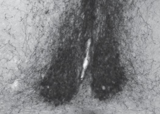
73 A Figure 2.3: Suprachiasmatic nucleus 100 μm B C 59


74 A Figure 2.4: Accessory nuclei B f 100 μm 200 μm C D 200 μm opt f 50 μm 60


75 A Figure 2.5: Other hypothalamic nuclei B f 200 μm opt C 100 μm 100 μm 61


76 A f Figure 2.6: BNST and medial amygdala B ic f 200 μm 100 μm C f ic D 200 μm 100 μm E 500 μm F opt f 100 μm G 500 μm H opt opt f 100 μm 62


77 A Figure 2.7: Small versus medium fibers 100 μm B 100 μm 63


78 Figure 2.8: Cortex and hippocampus A 200 μm B ec 100 μm fmi C 200 μm D CA1 500 μm cc E 500 μm F 200 μm G 500 μm H 100 μm 64


79 A Figure 2.9: Basal ganglia and ventral forebrain LV ac AcbC AcbSh ICjM VS VDB VP 200 μm B LV ac VP VS SI MPA HDB 200 μm 65


80 Figure 2.10: Septal complex A LV cc 500 μm B LV 500 μm LSI MS LSV ac ac C LV 500 μm D LV LSD 500 μm SFO SFi f E ac 200 μm F 100 μm ac 66


81 Figure 2.11: Bed nuclei of the stria terminalis A STMA 100 μm B f D3V ac ac STMV 100 μm STMV C STMPM 100 μm ic st f D3V STMPL ac STLV STMV D sm 100 μm E sm 100 μm SM f f 3V 3V 67


82 A Figure 2.12: Amygdala B sm f GP EA 500 μm AA opt 200 μm PLH C D sm f HDB LOT 500 μm opt 200 μm ACo E F opt 100 μm 100 μm G opt H BLP opt 500 μm 200 μm 68


83 A MnPO Figure 2.13: Preoptic area B ac ac 500 μm HDB VOLT MPA 200 μm C STMV D MPOM PS AVPe 100 μm 100 μm E ac 3V StHy LPO MPOM AVPe 500 μm 69


84 Figure 2.14: Anterior hypothalamus A AHA C 3V f 3V f 500 μm 500 μm B f 3V AHC LA opt 500 μm 70

 200 μm")
85 D Figure 2.14: Anterior hypothalamus (Continued) 200 μm Rostral E Caudal SPa SCN 200 μm och 71


86 A DM Figure 2.15: Dorsomedial hypothalamus 500 μm B DM 500 μm 3V 3V f f VMH Arc VMH TuLH MTu ME C D 100 μm 3V 3V 100 μm 72


87 A Figure 2.16: Lateral hypothalamus and mammillary bodies 100 μm B 200 μm f f C opt mt PH DTM PeLH f 3V PMV 500 μm D mt 500 μm E pm RMM MB 3V VTM 500 μm MB MRe 73


88 A D3V Figure 2.17: Thalamus 500 μm B sm D3V 500 μm sm MD PT PVA PC C f 3V sm f CM 500 μm PV MD CM Rh D sm f D3V 200 μm E 200 μm Re LHb IMD Xi 74


89 F Figure 2.17: Thalamus (Continued) G D3V LHb fr H IMD fr LHb MD sm D3V mt ac I J LHb D3V ZI fr PV sm 75


90 A Figure 2.18: Tegmentum 200 μm fr IF PBP VTA SNR IPF cp MB B RMC 200 μm vtgx IF 3n PIF ml IPR cp 76


91 C Figure 2.18: Tegmentum (Continued) mrt SubB RRF SNR D ml E 200 μm mlf scp CLi 200 μm 100 μm 77


92 A Figure 2.19: Periaqueductal gray and dorsal raphe nuclei 500 μm B DMPAG csc 500 μm pc fr Aq C 100 μm D 500 μm Aq 4N mlf DRL E 500 μm Aq VLPAG 4n DRD mlf 78


 G")


93 F Figure 2.19: Periaqueductal gray and dorsal raphe nuclei (Continued) G DRL 4N 100 μm 100 μm H DMPAG 500 μm I 2Cb VLPAG DRC 4n 100 μm J pc 500 μm PAG xscp IF IPR 79

94 Figure 2.20: Caudal mesencephalic nuclei 200 μm vsc LPB scp LC Bar LDTg 80


95 A Figure 2.21: Metencephalon and myelencephalon B xscp ts ATg 200 μm 100 μm C PnC IRt 7n D Sol LSO SPO 500 μm 200 μm E Sol PnC RMg 500 μm Py 81


96 F Figure 2.21: Metencephalon and myelencephalon (Continued) Sol 12N mlf ROb RMg IO py G 500 μm H RPa RMg 200 μm 200 μm RPa I sp5 J LRt Sp5C sp5 500 μm 200 μm Rostral Caudal 82
97 CHAPTER 3 PROJECTIONS OF THE BNST AND MEDIAL AMYGDALA 3.1 Introduction Vasopressin is produced in a number of nuclei within the diencephalon and telencephalon of the mouse brain and is found in neuronal fibers and terminals in various brain regions throughout the rostral to caudal extent of the brain (Castel and Morris, 1988; Abrahamson and Moore, 2001). In order to truly understand the function of the various vasopressin cell groups, it is important to know the origin of each projection site. This task has been approached with some success in the rat. Retrograde tracing and electrolytic lesions implicated the bed nuclei of the stria terminalis (BNST) as a major source of innervation to areas such as the lateral septum, lateral habenula, periaqueductal gray, and raphe nuclei (De Vries and Buijs, 1983). Similar studies implicated the suprachiasmatic nucleus (SCN) in the innervation of the paraventricular thalamus, dorsomedial hypothalamus, preoptic area, and subparaventricular zone, and the medial amygdala (MeA) in the innervation of the lateral septum and ventral hippocampus (Hoorneman and Buijs, 1982; Caffe et al., 1987). Lesions studies have also implicted the paraventricular nucleus of the hypothalamus (PVN) in vasopressin-ir projections to the hindbrain (De Vries et al., 1983). In the mouse, the site of origin of vasopressin-ir fibers has yet to be examined, although up until this point it has been almost universally assumed to be similar to the rat. Interestingly, vasopressin-immunoreactive (-ir) fibers and terminals have qualitative differences in physical characteristics such as fiber size and terminal 83
98 distribution (Castel and Morris, 1988; Abrahamson and Moore, 2001), which is one clue to identifying the different vasopressinergic afferent pathways. These characteristically different projections innervate specific and often mutually exclusive regions of the brain. Early descriptions of vasopressin anatomy attempted to use fiber characteristics to identify site of origin (Buijs, 1978; Castel and Morris, 1988; Abrahamson and Moore, 2001). In general, small fibers were assumed to derive from the SCN and medium or large fibers form the PVN. More extensive work in rats, as mentioned above, has corroborated that same observation in regard to the PVN. However, these studies also indicate that the BNST and MeA are major sources of small fibers in addition to the SCN (De Vries and Buijs, 1983; Caffe et al., 1987). Thus, merely identifying projections by physical characteristics is not sufficient to identify site of origin and other methods must be employed. The projections of the BNST and MeA are the focus of this study. Vasopressin produced by these brain regions has been directly and indirectly implicated in thermoregulation during fever, social recognition, and aggression in the rat and pairbonding, parental behavior, and partner preference in the prairie vole (Ferris et al., 1984; Banet and Wieland, 1985; Naylor et al., 1985; Dantzer et al., 1987; Koolhaas et al., 1990; Insel et al., 1994; Wang et al., 1994; Lim et al., 2004). In addition, recent studies have suggested that altering expression of the V1a receptor, the most abundantly expressed vasopressin receptor in the brain, can affect social behavior. For example, mice with a prairie vole V1a receptor transgene have increased affiliative behavior (e.g., increased olfactory investigation and grooming of stimulus animal; (Young et al., 1999), simlar to that of prairie voles. In addition, V1a knockout mice have impaired social recognition 84
99 (Bielsky et al., 2004; Bielsky et al., 2005), although a different V1a knockout mouse shows no such deficits (Wersinger et al., 2007). One of the unique features of the BNST and MeA vasopressin system is its dependence on circulating gonadal steroids. In the absence of testoserone or estradiol, the BNST and MeA appear to stop producing vasopressin, leading to an eventual depletion of vasopressin in their numerous projection sites in rats and mice (de Vries et al., 1984; Mayes et al., 1988; Miller et al., 1992; De Vries et al., 1994; Wang and De Vries, 1995). In addition, vasopressin expression in the BNST and MeA is organized by testosterone perinatally resulting in a sexual dimorphism (De Vries et al., 1983; Wang et al., 1993; Han and De Vries, 2003); males typically have more cells than females in the BNST and MeA and a greater density of fibers in known projection sites such as the lateral septum and lateral habenula (de Vries et al., 1981; Miller et al., 1989; De Vries and al-shamma, 1990; Wang and De Vries, 1995; Rood et al., 2008). The function of sex differences in the vasopressin system is not well understood. In some cases, the female vasopressin system does not respond as the male system does. For example, social recognition cannot be blocked by vasopressin antagonists in females as it is in males (Bluthe and Dantzer, 1990), and vasopressin is not released and V1a antagonists do not delay defervescence in females during fever as occurs in males (Landgraf et al., 1990; Chen et al., 1997). Currently, there are few hypotheses explaining the function of sex differences in vasopressin; one interesting hypothesis is that sex differences in vasopressin may actually serve to compensate for other sex differences in behavior or physiology resulting in more equivalent behavior in males and females (De Vries, 2004). Finally, the sex difference in the vasopressin system originating in the BNST and MeA 85
100 has been described as one of the most consistently observed sex differences among vertebrates (De Vries and Panzica, 2006). For example, in mice, a sex difference in cell number in the BNST and MeA has been observed (Rood et al., 2008). In addition, male mice have a greater vasopressin-immunoractive-ir fiber density than females in a number of brain regions, such as the lateral septum, mediodorsal thalamus, lateral habenula, and medial amygdala (De Vries et al., 2002; Bakker et al., 2006; Gatewood et al., 2006; Rood et al., 2008). However, the vast vasopressin innervation of the mouse brain remains largely unexplored. Therefore in addition to understanding which fibers derive from the BNST and MeA, the occurrence of sex differences is also a focus of this study. In the current study, we used a simple strategy to determine the location of projections from the BNST and MeA: gonadectomy. As described above the expression of vasopressin is dependent on the presence of circulating gonadal steroids, and loss of these hormones leads to a loss of vasopressin expression in the BNST and MeA, but not in other vasopressin producing sites in rats and mice (de Vries et al., 1984; Mayes et al., 1988; Miller et al., 1992). Using this principle we examined vasopressin immunoreactivity in the brains of male and female mice that had been gonadectomized or sham operated to confirm that only BNST and MeA vasopressin production depends on circulating gonadal steroids and to test the following hypotheses: 1) a subset of small vasopressin-ir fibers and terminals in the forebrain, midbrain, and hindbrain derive from the BNST and MeA and 2) vasopressin-ir projections that originate in the BNST or MeA and not in other vasopressin producing areas are sexually dimorphic with males having a greater vasopressin-ir fiber density than females. 86
101 3.2 Methods Animals Male and female C57BL6 mice were studied in this experiment. Animals were derived from a breeding colony at the University of Massachusetts. Following weaning, animals were group-housed (5 or less per cage) with same-sex siblings under a 14:10 light cycle with lights on at 6:00 am. Animals had ad libitum access to food and water. All procedures conformed to NIH guidelines and were in accordance with a protocol approved by the University of Massachusetts. Brains from three gonadectomized male mice were used for qualitative analyses. Corroboration of qualitative results and quantitative analyses of gonadal hormone dependence and sex differences were examined in male and female mice that were either gonadectomized (12 males and 12 females) or sham operated (12 males and 10 females). Vasopressin immunoreactivity in certain projections dissipates over a number of weeks following gonadectomy and is virtually undetectable around 12 to 15 weeks following surgery (Mayes et al., 1988). We therefore chose a 15 week survival time following surgery. During this period one male sham animal and three female sham animals died for unknown reasons. Furthermore, after decoding and analyzing the data one "gonadectomized" female was found to have vasopressin immunoreactivity indistinguishable from intact females suggesting that at least one ovary was left intact. Because the quantitative measures of this subject were well within the range of intact females this subject was included as a "sham female." Thus our quantitative measures for the sham groups include results from only 11 males and 8 females. 87
102 3.2.2 Gonadectomy surgeries At approximately 60 days of age, all animals underwent bilateral gonadectomy under isoflurane anesthesia (2% in oxygen by inhalation; Halocarbon Laboratories, River Edge, NJ). Male gonadectomy was carried out through an incision at the midline of the scrotum. Testes were exposed, the blood supply and vas deferens were ligated with suture, and testes were excised. Sham animals received incisions to exposes the testes, but the blood supply and testes were left intact. Wounds were closed with surgical staples. Female gonadectomy was carried out through incisions on each flank located between the rib cage and pelvis. Ovaries were exposed, the blood supply and fallopian tubes were ligated with suture, and ovaries were excised. Sham females underwent an identical procedure except the blood supply and ovaries were left intact. The muscle wall of the body cavity was sutured closed, and the skin was closed with surgical staples. All animals were given a single injection of buprenorphine (0.05 mg/kg; Bedford Laboratories, Bedford, OH) immediately following surgery Tissue collection and processing Fifteen weeks following gonadectomy, animals were euthanized using CO 2 asphyxiation followed by rapid decapitation. Brains were removed rapidly and placed in 5% acrolein in 0.1 M phosphate buffer. Fixation lasted for four hours at which time brains were transferred to 30% sucrose in 0.1M phosphate buffer and stored at 4 C until sectioning. For gonadectomized animals used in qualitative analyses, each brain was sectioned at 30 m in a different plane: coronal, sagittal, or horizontal. For each brain, every other section was collected for vasopressin immunohistochemistry. Alternate sections were mounted on slides and Nissl stained. Brains used for quantitative analyses 88
103 were sectioned in the coronal plane and every third section was collected for vasopressin immunocytochemistry. All brain sections were stored in cryoprotectant at -20 C until staining Immunohistochemistry Immunohistochemical methods used in this experiment are identical to those described in CHAPTER Qualitative and quantitative fiber density analysis Qualitative descriptions were carried out using a bright field microscope. Quantitative analyses were carried out on images of selected representative areas illustrated in Figure 3.1 captured using a SPOT camera and software driver on a brightfield microscope equipped with a 20x objective. Fibers were analyzed in each picture using ImageJ (NIH). Briefly, each picture was modified using the "sharpen" tool. Then gray-level thresholding was carried out to determine the number of pixels covered by staining. Thresholding was done by eye by a trained experimenter to ensure the most accurate measurement of fibers in all sections. All slides were coded prior to analysis, so that the experimenter was blind to treatment groups Statistics Quantitative results were analyzed with two-factor ANOVAs. The factors were sex (male or female) and surgery (sham or gonadectomy). Fisher's least squared difference tests were used to determine group differences where appropriate. A p-value of 0.05 or less was considered to be significant throughout. Statistical values for all quantitative measures are listed in Table
104 3.3 Results Steroid-sensitive vasopressin-ir projections were defined as regions in which vasopressin-ir fibers and terminals decreased or disappeared altogether following gonadectomy. In some regions gonadectomy resulted in a complete loss of immunoreactiviy. It is not known whether or not there is an actual loss of axons and terminal boutons themselves. Decreases in medium or large fibers were not observed in any brain region, but small fibers and terminals disappeared in numerous nuclei throughout the brain. Further, quantitative analyses were made for 84 distinct regions of the brain. In general brain regions were chosen because they had a quantifiable vasopressin innervation (i.e., at least scattered fibers) and could be accurately located in almost all subjects using a variety of anatomical criteria. Many of these areas contained small fibers. Areas in which medium fibers predominate are often too sparsely and unpredictably innervated for analysis. However, a number of the more heavily innervated medium fiber sites are included. Strikingly, gonadectomy did not cause an increase in vasopressin-ir fibers in any area where a change was observed (N = 60). Please see Table 3.1 for all statistical values associated with figures below Telencephalon Steroid-sensitive vasopressin projections Vasopressin immunoreactivity appeared to decrease following castration in 32 of 55 brain regions throughout the telencephalon that have previously been shown to contain vasopressin-ir fibers. Qualitative results for the telencephalon are summarized in Table
105 In the cortex, gonadectomy reduced vasopressin-ir fiber density in the dorsolateral entorhinal cortex and ventral endopririform claustrum. In the hippocampus, the oriens and molecular layers of CA1 contain vasopressin fibers that disappeared following gonadectomy, although some dense tangles of medium to large fibers remain in the caudal molecular layer of CA1 (see figure 2.8H). In the basal ganglia, the ventral pallidum lost vasopressin immunoreactivity following gonadectomy, but the nucleus accumbens did not. Other basal ganglia nuclei do not contain vasopressin. The ventral forebrain lost immunoreactivity in the navicular postolfactory cortex located near the rostral midline, the substantia innominta, and the ventral septal area. The septal complex has one of the densest vasopressin-ir innervations in the brain, and gonadectomy eliminated all small fiber immunoreactivity in the lateral septum, septofimbrial nucleus, and septohypothalamic nucleus leaving only the sparse medium fibers. Innervation of the dorsal division of the lateral septum, the most rostral parts of the lateral septum, triangular nucleus, and subfornical organ did not change. The bed nuclei of the stria terminalis have a complex array of fibers of various types. In the anterior nuclei, only the anteromedial (STMA) and lateroventral (STLV) nuclei had a decrease in vasopressin-ir fibers; these nuclei are also the only ones that contained small fibers in intact animals. Laterodorsal nuclei had virtually no fibers to begin with and medioventral nuclei did not have a qualitative reduction in the medium fibers found there. It is notable that despite the decrease in small fibers in the anteromedial nucleus and others, a number of medium fibers remained. In the posteromedial BNST, all three regions (i.e., medial, intermediate, and lateral) lost vasopressin-immunoreactivity. However, some medium fibers were still observed in 91
106 each. Further, vasopressin immunoreactivity in small fibers persisted in the dorsal part of the medial posteromedial BNST (STMPM), whereas immunoreactivity in fibers in the ventral portion of the STMPM disappeared following gonadectomy. The nucleus of the stria medullaris also had decreased immunoreactivity following gonadectomy. Every vasopressin innervated nucleus within the amygdala lost vasopressin immunoreactivity following gonadectomy with the exception of the central nucleus, in which medium fibers remained. Some stray medium fibers also remained in the posteromedial and basomedial nuclei and in anteromedial regions adjacent to the supraoptic nucleus in gonadectomized animals Sex differences and interactions As mentioned above, all areas with small fibers examined in the telencephalon had decreased vasopressin immunoreactivity following gonadectomy. In addition, a number of these regions were also sexually dimorphic with more vasopressin-ir fibers observed in males. The decrease in vasopressin immunoreactivity was generally seen in both sexes; however, in many cases, larger decreases in males led to significant sex-bysurgery interactions. Exceptions to these general trends are noted below. Of the 24 areas examined in the telencephalon sex differences were found in 15. All 15 areas had decreased vasopressin immunoreactivity following gonadectomy and significant sex-by-surgery interactions due to a larger decrease in immunoreactivity in males than females with one exception, the rostral medioventral BNST (STMV), for which details are described below. An additional eight areas were steroid sensitive, but not sexually dimorphic. Finally, one region was neither steroid sensitive nor sexually dimorphic 92
107 (see Table 3.1 for statistics, Figures 3.1 for the location of regions analyzed, and Figures 3.2 to 3.4 for data). In the lateral septum (Figure 3.2), the densely innervated ventral part and the caudal part where the fibers of the fimbria begin to enter were strongly sexually dimorphic. On the other hand, the intermediate part in a more rostral area was not. In the anterior BNST (Figure 3.3A), only the STMA and STLV nuclei contain small fibers. There was a small but significant decrease in immunoreactivity in the STMA of males and females, but in the STLV a decrease was observed only in males. Interestingly, intact females did not differ from gonadectomized animals in the STLV. Surprisingly, in the rostral medioventral (STMV) BNST, females had a significantly greater vasopressin-ir fiber density than males, and this difference was not present in castrated animals. Typically sex differences in vasopressin-ir fiber density are in the male greater than female direction as was observed here, but, as described below, a number of regions in the mouse brain show a similar "atypical" sex difference as seen in the rostral STMV. The posteromedial BNST nuclei are innervated primarily by small fibers, and this innervation is sexually dimorphic and steroid dependent (Figure 3.3B). Males had more vasopressin-ir fibers than females in the medial and lateral parts of the posteromedial BNST and in the nucleus of the stria medullaris, and fibers decreased drastically in both sexes following gonadectomy. All regions quantified in the amygdala, including multiple rostral to caudal levels through some regions, contained almost exclusively small fibers. Although all regions responded to gondectomy with a massive decrease in vasopressin immunoreactivity, sex 93
108 differences were only observed in a subset of regions. Specifically, the rostral and caudal extended amygdala (Figure 3.4A), caudal anterodorsal medial amygdala (Figure 3.4C), posteroventral medial amygdala (Figure 3.4D), posterior basolateral amygdala (Figure 3.4E), and all levels of the posterodorsal medial amygdala (MePD; Figure 3.4F) all had significantly more vasopressin-ir fibers in intact males than females. Notably, in the caudal MePD females did not differ from gonadectomized animals. In contrast, no sex differences were observed in the rostral or caudal anterior cortical amygdala (Figure 3.4B), rostral anterodorsal medial amygdala (Figure 3.4C), anteroventral medial amygdala (Figure 3.4D), basomedial amygdala, or ventral basolateral amygdala (Figure 3.4E) Diencephalon Hypothalamus Steroid-sensitive vasopressin projections The hypothalamus contains most of the vasopressin-producing nuclei in the brain, and accordingly has by far the densest and most complex patterns of innervation of the entire brain. A decrease in small fiber immunoreactivity was observed in 15 out of 39 brain regions throughout the hypothalamus following gonadectomy. Notably and in contrast to the telencephalon, some regions actually retained small fibers following gonadectomy. Qualitative results for the hypothalamus are summarized in Table 3.3 and are described below. In the preoptic region, small fibers disappeared from the lateral preoptic area, medial preoptic area, parastrial nucleus, medial part of the medial preoptic nucleus, and to a very small extent the median preoptic nucleus. Many regions retained medium 94
109 fibers. In addition a number of regions also had some small fibers especially near the midline in the anteroventral periventricular nucleus and periventricular region. In the medial regions of the hypothalamus at the levels of the anterior and dorsomedial hypothalamic nuclei, there are very dense vasopressin-ir projections, which can be seen exiting the PVN. However, with the exception of a small terminal field in the most rostral part of the anterior hypothalamus, none of these regions lose vasopressin immunoreactivity following gonadectomy. In contrast, the peduncular lateral hypothalamus, which is often referred to as just lateral hypothalamus, contains small fibers and terminals that disappear following gonadectomy at all rostral to caudal levels. This innervation is most pronounced in the rostral lateral hypthalamus and decreases caudally, until almost no small fibers can be seen. However, at the level of the mammillary bodies, a dense small fiber plexus is observed in the perifornical lateral hypothalamus. In addition to the lateral hypothalamic regions, small fibers immunoreactivity disappears following gonadectomy from nuclei closer to the midline at the level of the mammillary bodies. In particular, the dorsal tuberomammillary nucleus near the third ventricle and the ventral tuberomammillary region located ventral to the mammillary bodies lose immunoreactivity following gonadectomy. The same is true for the posterior hypothalamus, ventral premammillary nucleus, and retromammillary nucleus also have a sparse to scattered innervation. Each of these areas also contains a modest innervation be medium fibers that persist following gonadectomy. 95
110 Sex differences and interactions Unlike the telencephalon, there are many regions in the hypothalamus with medium to large fibers, and a number of these regions are included in quantitative measures of vasopressin immunoreactivity. Of the 27 regions examined in the hypothalamus, 13 regions were sexually dimorphic. Five of these were atypical sex differences (i.e., female greater than male) and were not affected by gonadectomy. Other details regarding sex differences are described below. Five other regions had reduced vasopressin immunoreactivity following goandectomy, but no sex differences. Nine areas were not affected by gonadectomy and were not sexually dimorphic (see Table 3.1 for statistics, Figure 3.1 for the location of regions analyzed, and Figure 3.5 for data). In the preoptic region, the medial preoptic area and the middle and caudal parts of the medial preoptic nucleus (MPOM) were sexually dimorphic (Figure 3.5A and B); intact males had a greater fiber density than females. In each of these areas, which contain other gonadal-steroid independent vasopressin fibers, the female did not differ from gonadectomized animals. Other regions such as the median preoptic nucleus, vascular organ of the lamina terminalis, and the rostral part of the MPOM were not sexually dimorphic or responsive to gonadal hormones. Despite the decrease of small fibers in males in the MPOM some small fibers remained. In the anterior hypothalamus (AH; Figure 3.5C), there were no sex differences or effect of gonadectomy in the central part of the AH through which fibers of the PVN pass. However, in the anterior part of the AH, where small fibers are noted, a small decrease was observed in males following gonadectomy suggesting and confirming the presence of a small BNST or MeA input. 96
111 Similar to results in the central AH, medium fibers in the tuberal lateral hypothalamus and medial tuberal hypothalamus were not sexually dimorphic or affected by gonadal steroids (Figure 3.5F). In contrast nuclei closer to the midline such as the dorsomedial hypothalamus (Figure 3.5G), periventricular area (Figure 3.5E), and retrochiasmatic area (Figure 3.5H) were sexually dimorphic although in the "atypical" fashion: females greater than males. None of these regions were affected by gonadectomy, and each appears to receive both small fiber and medium fiber innervation. On the other hand the subparaventricular zone, which contains mostly small fibers was neither sexually dimorphic nor affected by gonadectomy (Figure 3.5E). In the peduncular lateral hypothalamus (PLH; Figure 3.5D), as well as the caudal perifornical region (Figure 3.5H), there were effects of gonadectomy in all but the caudal PLH, but only the dense terminal field in the rostral part of the PLH has a typical sex difference: males had more fibers than females (Figure 3.5D). Interestingly, the caudal PLH, where very few small fibers are observed, an atypical sex differences was found: females greater than males. At the level of the mammillary bodies, gonadectomy reduced small fiber immunoreactivity in the ventral tuberomammillary nucleus and ventral premammillary nucleus (PMV); Figure 3.5H), as well as in the retromammillary nucleus (RMM), which was examined at three levels (Figure 3.5I). The only sex difference was found in the caudal RMM. At this level, males had more fibers than any other group, and females did not differ from gonadectomized animals. Although there were no sex differences in intact animals in the PMV and rostral RMM; a significant interaction between sex and 97
112 hormonal status was present because gonadectomized males had significantly less vasopressin-ir fibers than other groups (Figure 3.5H and I) Thalamus Steroid-sensitive vasopressin projections The majority of vasopressin immunoreactivity in the thalamus is found in small fibers and terminals. Invariably, however, some medium fibers find their way into both lateral and medial nuclei. Only the most caudal parts of the midline thalamus rostral to the periaqueductal gray (PAG) and the reticular thalamus contain medium fibers in the absence of small fibers. Of the 14 areas containing vasopressin-immunoreactive fibers in the thalamus, eight appeared to have decreased immunoreactivity following gonadectomy. Qualitative results for the thalamus are summarized in Table 3.4. Gonadectomy caused a near total elimination of vasopressin-ir fibers in the thalamus, including the central medial, interanterodorsal, interanteromedial, mediodorsal, paracentral, rhomboid, paratenial, and lateral habenular nuclei. No qualitative changes were observed in the paraventricular, reuniens, xiphoid, or reticular thalamic nuclei suggesting a site of origin other than the BNST or MeA. However, as described below quantitative results suggest a small decrease in the rostral paraventricular nucleus following gonadectomy Sex differences and interactions Quantitative data show that areas where fiber immunoreactivity disappeared in the thalamus were generally sexually dimorphic as well: male greater than female (see Table 3.1 for statistics, Figure 3.1 for the location of regions analyzed, and Figure 3.6 for data). This was true for the multiple rostral to caudal levels through the mediodorsal (Figure 98
113 3.6C), central medial (Figure 3.6E), and lateral habenular nuclei (Figure 3.6F), except the caudal lateral habenula where there was an effect of gonadectomy, but not sex. The paratenial and rostral paraventricular nucleus were affected modestly by gonadectomy, but fiber-immunoreactivity was not sexually dimorphic in these areas (Figure 3.6A and B). In the dorsal part of the anterior paraventricular thalamus at the level where the paratenial nucleus emerges there was a decrease in vasopressin-ir fiber density only in males following castration (Figure 3.6A). An interesting feature of this area is the emergence of a dense plexus of gonadal steroid-dependent fibers adjacent to the stria medullaris. This plexus may have contributed to gonadal status effects in the anterior paraventricular and paratenial nuclei. Further caudal in the paraventricular nucleus (Figure 3.6D), there were no effects of gonadal status or sex Mesencephalon Steroid-sensitive vasopressin projections Unlike the thalamus, which is innervated almost entirely by small fibers, the mesencephalon contains regions of small and medium fiber innervation. Eighteen of the 31 brain regions containing vasopressin-ir fibers in the mesencephalon appeared to have a decrease in vasopressin immunorecativity following gonadectomy. Qualitative results for the thalamus are summarized in Table 3.5. In the most rostral part of the PAG a number of small and medium fibers can be seen ascending toward the aqueduct into the PAG. Most of these remain following castration and fibers can be seen in gonadectomized males throughout the PAG. Most of the fibers seen throughout the PAG of gonadectomized animals are short segments suggesting that they are fibers travelling somewhat rostral-to-caudal (i.e., orthogonal to 99
114 the plane of the section). Rostrally, small fibers, which appear to arrive via the ascending projection through the rostral PAG, end in the dorsomedial PAG and at the midline in the commissure of the superior colliculus. These small fibers and terminals lose vasopressin immunoreactivity following gonadectomy. Caudally at the level of the trochlear nucleus, a dense plexus of small fibers and terminals begins to form in the dorsal raphe nuclei. At the same level medium axons curve laterally into the plane of section in the ventrolateral PAG (VLPAG) and then pass into the lateral parabrachial nucleus (LPB). The small fibers in the dorsal raphe nuclei (e.g., dorsolateral, dorsal, and caudal) lose immunoreactivity following gonadectomy. Medium fibers in the VLPAG and LPB do not lose immunoreactivity. These fibers appear continuous with fibers further caudal in the locus coeruleus and Barrington's nucleus that also retain immunoreactivity following gonadectomy. From this point on some fibers continue orthogonal to the section adjacent to the fourth ventricle in both sham and castrated animals. Ventrally in the tegmentum, a number of brain regions are innervated by small fibers from the BNST and MeA. Gonadectomy reduces vasopressin immunoreactivity in the ventral tegmental area (VTA), parabrachial pigmented nucleus of the VTA, interfascicular, and parainterfascicular nuclei in more rostral and medial parts of the midbrain. Laterally, small fiber immunoreactivity disappears from the ventromedial tip of the substantia nigra pars reticulata, the caudal substantia nigra pars compacta, and the retrorubral field. The small fibers that pass through the VTA into the interfascular nucleus appear to continue on through the caudal linear raphe into the dorsal raphe nuclei. These fibers are lost following gonadectomy. There is also a group of medium 100
115 fibers that travel through the VTA, interfascicular nucleus, and caudal linear raphe in a similar path. In addition, there is a lateral path of medium fibers that are not hormonedependent and travel dorsal to the substantia nigra into the midbrain reticular formation. As these fibers pass the substantia nigra there is an increase in branching and boutons. This fiber pathway appears to curve through the medial parabrachial nucleus into the solitary nucleus in the medulla Sex differences and interactions All of the regions with small fibers in the mesencephalon that were analyzed showed a decrease following gonadectomy (N = 11). Eight of these 11 were also sexually dimorphic, although one was only sexually dimorphic in gonadectomized animals. Five additional areas had atypical sex difference, but were not affected by gonadectomy. Finally, one region was not sexually dimorphic or steroid-sensitive (see Table 3.1 for statistics, Figure 3.1 for the location of regions analyzed, and Figure 3.7 for data). The small fibers of the mediodorsal PAG and dorsal and caudal aspects of the dorsal raphe (DRC) were sexually dimorphic: males greater than females (Figure 3.7A and C). The dorsolateral dorsal raphe showed no sex differences although fiber density was decreased in gonadectomized animals (Figure 3.7C). In the DRC intact females were not different from gonadectomized males or females. In the ventrolateral PAG, there was a decrease in fibers at the most rostral level, but no sex differences. The level of vasopressin immunoreactivity in the middle VLPAG was similar in all groups. However, caudally, there were atypical sex differences in the caudal VLPAG, LPB, and medial parabrachial nuclei: females greater than males (Figure 3.7B and F). 101
116 In ventral regions, males had greater fiber densities than females in the VTA (Figure 3.7D), interfascicular nuclei (Figure 3.7E), and parainterfascicular nuclei (Figure 3.7D). There was no sex difference in the substantia nigra pars reticulata even though this area was affected by gonadectomy (Figure 3.7D). The sex difference in the caudal interfascicular nucleus was found only in gonadectomized animals because males had less vasopressin-ir fibers than females following gonadectomy (Figure 3.7E). Finally, an atypical sex difference was observed in the midbrain reticular formation at the top of the substantia nigra where the medium fibers begin to increase in number (Figure 3.7F) Metencephalon and myelencephalon Steroid-sensitive vasopressin projections Qualitative results for the metencephalon and myelencephalon are summarized in Table 3.6. There are very few innervated regions in the metencephalon. However, small fibers are found in the median raphe nucleus and the anterior tegmental nucleus, which are found between the tectospinal tract and pontine reticular formation. Both of these regions have no fibers at all following gonadectomy. A few medium fibers are seen dorsally just below the fourth ventricle and laterally in the subcoeruleus and intermediate reticular formation. These medium fibers seem to pass through the metencephalon on their way to innervate a number of regions throughout the medulla. In the medulla itself, medium fibers are observed in the solitary nucleus through its entire rostal to caudal extent, especially in caudal regions (noted in sagittal sections; Figure 2.21F). Vasopressin-ir fibers are found, ventrally, in the medullary raphe nuclei (e.g., raphe magnus, raphe pallidus, raphe obscurus, and raphe interpositus); laterally, in the alpha part and ventral part of the gigantocellular reticular nuclei, and caudally in the 102
117 lateral reticular formation and the most caudal parts of the spinal trigeminal nucleus. Fibers are observed in all of these areas following gonadectomy with no apparent decreases Sex differences and interactions Quantitative data were collected for the anterior tegmental nucleus and median raphe (see Table 3.1 for statistics, Figure 3.1 for the location of regions analyzed, and Figure 3.8 for data). Sex differences were observed in both areas: males greater than females (Figure 3.8). No region of the medulla appeared to receive steroid-sensitive vasopressin innervation. Quantitative analyses were not carried out for any area in the myelencephalon. 3.4 Discussion In rats, vasopressin cells in the bed nuclei of the stria terminalis (BNST) and medial amygdala (MeA) require gonadal hormones, in particular estrogens (De Vries et al., 1994), for vasopressin production. Similar to rats, gonadectomy decreases vasopressin immunoreactivity in a number of brain regions (DeVries et al., 1985) some of which, at least in the rat, are known to derive their vasopressin innervation from the BNST or MeA (De Vries and Buijs, 1983; Caffe et al., 1987). Given that some steroidsensitive vasopressin-ir projections in the rat are known to come from the BNST or MeA and the gross similarities between rat and mouse vasopressin systems, we believe that the loss of vasopressin immunoreactivity in a given brain area following gonadectomy in the mouse strongly suggests that that area receives vasopressin innervation from the BNST or MeA. Conversely, areas that retain vasopressin immunoreactivity following gonadectomy presumably receive their innervation from other nuclei, most likely the 103
118 various hypothalamic nuclei such as the suprachiasmatic, supraoptic, pareventricular, or various accessory nuclei, which are the only other nuclei that produce vasopressin. In the current study, brain regions throughout the telenencephalon, diencephalon, mesencephalon, and metencephalon lost vasopressin immunoreactivity following gonadectomy. This subset of brain regions as detailed above comprises the likely output of the BNST and MeA. Furthermore, many of these brain regions (25 of the 39 quantified) have a sexually dimorphic vasopressin input. In these cases males have significantly more vasopressin-ir fibers and terminals than females in the given brain region. Interestingly and quite unexpectedly, vasopressin-ir fibers from other sources (i.e., fibers did not decrease following gonadectomy) were also sexually dimorphic. However, this sex difference was atypical in that females had a greater vasopressin-ir fiber density (i.e., more fibers) than males. Before drawing too many conclusions from the results of this study, it is important to keep a few limitations in mind. First and foremost is the quantity of data. While a somewhat exhaustive approach is beneficial in providing a great many things to think about, it also greatly amplifies the opportunity for statistical error. While this is of theoretical concern, in this particular situation the presence or absence of small fibers in a region is almost always able to corroborate the statistical data. Areas where no small fibers were observed were not affected by gonadectomy as shown when you compare the qualitative and quantitative data. Furthermore, sex differences in the expected direction only occurred in areas with small fibers, which fits with the fact that the BNST and MeA are sexually dimorphic and the source of small fibers in many brain regions. Perhaps the most interesting statistical question is the presence of atypical sex differences. A couple 104
119 of areas in which this occurred could be expected based on statistical error, but there were 10 different brain region in which females had a greater vasopressin-ir fiber density than males. Furthermore, these 10 areas were all comparable in that they contained almost exclusively medium fibers. Finally, not one region that changed following gonadectomy had an increase in vasopressin-ir fibers, which would be expected by chance given the number of observations made. Aside from the number of tests, it is important to note that within each brain region the data can be quite variable. Individual data points are illustrated in Figure 4.2 for the caudal lateral septum of intact males. The subject with the lowest value has a value that is only 18% of the highest animal (a 5-fold difference). However, despite this variability we saw sex differences in many brain regions where one would expect to find a sex difference based on data from other species (De Vries and Panzica, 2006). Finally, as with any study, we must be careful to not generalize too far. We have thoroughly examined sex differences in the vasopressin system of only one mouse strain. It is possible that other strains may have differences in the amount of fibers projecting to different brain regions and that sex differences in these other strains could be bigger, small, or non-existent. However, our data seem to fit well with what is known about the rat and sex differences in vasopressin expression observed in other species. Furthermore the quantitative results presented here corroborate results found in mice previously (Rood et al., 2008). Despite the limitations of this study, we have presented a significant pool of data that provide the foundation for hypotheses regarding the function and possible sexual dimorphisms in function of the vasopressin system. 105
120 Unfortunately, an exhaustive functional review of each brain region is beyond the scope and purpose of the current project; however, a brief overview of a few functional systems highlights the widespread potential impact of vasopressinergic innervation deriving from the bed nuclei of the stria terminalis. Systems involved in arousal and awareness, behavioral state, and emotional control and cognition all contain vasopressin, often at multiple levels of the circuits involved. Below, I introduce some of the various functional circuits that contain vasopressin innervation, which presumably comes from the BNST or MeA. Interestingly, the collection of areas discussed below suggests that vasopressin may be involved in regulating the behavioral state of an animal in addition to or instead of regulating individual behaviors. One such region involved in controlling behavioral state hat contains vasopressin in the mouse is the midbrain raphe: dorsal and median raphe nuclei. These regions produce serotonin. Drugs that lead to an increase in serotonin neurotransmission have an anxiolytic effect. Conversely, low levels of serotonin lead to an increase in anxiety. Low serotonin levels are also associated with higher levels of aggression in humans (Berman et al., 1997). The influence of serotonin on aggression has been reviewed elsewhere (Veenema and Neumann, 2007), but briefly research indicates that various forms of aggression can be correlated with both low and high levels of serotonin in different rat and mouse strains. In the brain, serotonin is produced primarily in hind brain nuclei including the dorsal and median raphe nuclei (Michelsen et al., 2007). Each of these brain regions in the mouse contains sexually-dimorphic vasopressin-ir fibers from the BNST and MeA. The presence of vasopressin in the raphe, especially the very dense innervation of the dorsal raphe, suggests a functional link between vasopressin release 106
121 and serotonin neurons, although it must be noted that no direct evidence to this effect has yet been reported. In addition to direct projections to the serotonin system, a dense plexus of vasopressin-ir fibers is found in the lateral habenular nucleus. This innervation is also sexually dimorphic except possibly in more caudal regions of the nucleus. The lateral habenular nucleus plays a key role in information flow through the "dorsal diencephalic conduction system (Sutherland, 1982)." This title refers to a pathway from the forebrain to the hindbrain that passes through the stria medullaris into the habenular complex, the neurons of which send axons through the fasciculus retroflexus to the hindbrain (Wang and Aghajanian, 1977; Sutherland, 1982). In fact, the neurons in the lateral habenula project directly to the dorsal raphe nucleus and inhibit serotonergic neurons (Wang and Aghajanian, 1977; Reisine et al., 1982). The purpose and function of vasopressin at each of these levels (i.e., the dorsal raphe and lateral habenular nucleus) is currently unknown. In addition to the dorsal raphe, the lateral habenular nucleus has projections to the ventral tegmental area (VTA) and substantia nigra pars compacta (Christoph et al., 1986; Hikosaka et al., 2008). Similar to the effect on the raphe, stimulation of the lateral habenula inhibits dopamine neurons in both the VTA and substantia nigra (Reisine et al., 1982; Christoph et al., 1986). Dopaminergic neurons in the VTA and substantia nigra project extensively to the basal ganglia (de Olmos and Heimer, 1999). These connections, in particular those from VTA, suggest that vasopressin is anatomically linked to another important regulatory system, namely, the "ventral striatopallidal system (de Olmos and Heimer, 1999)." Structures in this pathway are also referred to as the "limbic loop of the basal ganglia." Briefly, the VTA projects to the ventral striatum (i.e., 107
122 nucleus accumbens), which then projects to the ventral pallidum or substantia innominata. Neurons in the ventral pallidum project to the mediodorsal thalamic nucleus, which then projects to specific brain regions, which in turn project to the ventral striatum among other regions. Interestingly, vasopressin-ir fibers from the BNST and MeA intersect this "limbic loop" at multiple levels including the VTA, ventral pallidum, mediodorsal thalamus, and interfascicular nucleus, which also contains dopaminergic neurons with forebrain projections. A functional link has yet to be made between vasopressin and most of these brain region; however, one region, the ventral pallidum, has been implicated in affilitive behavior and pair-bonding in the monogamous prairie vole. Overexpression of the V1a receptor, the primary vasopressin receptor in the brain, in the ventral forebrain including the ventral pallidum increases affiliative behavior and partner preference in both socially monogamous (i.e., prairie vole) and promiscuous species (i.e., meadow voles; (Pitkow et al., 2001; Lim et al., 2004). Further, Fos expression increases in the ventral pallidum, mediodorsal thalamus, and medial amygdala following mating stimuli, and injections of a V1a receptor antagonist into the ventral pallidum, but not the lateral ventricle, medial amygdala, or mediodorsal thalamus, inhibits partner preference formation (Lim and Young, 2004). While in mice the densest BNST / MeA projections within the thalamus innervate the mediodorsal thalamus ("limbic loop") and the lateral habenula ("dorsal diencephalic conduction system"), vasopressin-ir fibers are also found in a number of midline and intralaminar nuclei, including the central medial, paracentral, and interanterodorsal nuclei. The afferent and efferent projections of these nuclei are reviewed in (Van der Werf et al., 2002). In general, the interanterodorsal nucleus appears to be connected to 108
123 the amygdala and limbic frontal cortex. The central medial nucleus receives inputs from a number of regions including reticular formation, serotonin neurons, superior colliculus, and hindbrain cholinergic nuclei. The paracentral nucleus receives multimodal sensory information from the auditory and visual systems. These nuclei with their diverse inputs and outputs appear to play a role in attention and awareness. Based on the patterns of inputs, it appears likely that these nuclei integrate sensory and limbic system information with midbain and hindbrain activational systems, such as the serotonergic and cholinergic nuclei. Perhaps the most commonly studied area in terms of vasopressin function is the lateral septum. This brain region receives massive topographic projections from the hippocampus, and projects to regions throughout the brain in a region specific manner (Swanson and Cowan, 1979; Risold and Swanson, 1997). The ventral lateral septum and caudal lateral septum, which contain one of the largest and densest vasopressin terminal fields, project to the medial preoptic region (ventral lateral septum) and the lateral preoptic area and lateral hypothalamus (caudal lateral septum). These areas themselves have vasopressin innervation from the BNST or MeA and are thought to be involved in a host of behaviors, including reproductive and aggressive behaviors, both of which require social interaction. Vasopressin in the lateral septum has been studied in regard to social memory, aggression, parental behavior, partner preference, thermoregulation, and anxiety (Bernardini et al., 1983; Naylor et al., 1985; Dantzer et al., 1988; Insel et al., 1994; Wang et al., 1994; Everts and Koolhaas, 1999; Bielsky et al., 2005). Currently, functional evidence most strongly supports a role for vasopressin in social recognition memory, particularly in male rats (Dantzer et al., 1987; Bluthe et al., 1990). Vasopressin receptor 109
124 (V1a) antagonists, antisense, and null mutations all disrupt social recognition memory (Dantzer et al., 1987; Dantzer et al., 1988; Landgraf et al., 1995; Bielsky et al., 2004). In V1a knock-out animals, rescue of V1a receptors in the lateral septum alone with the use of viral vectors restored social recognition memory (Bielsky et al., 2005). In addition, V1a agonists injected into the lateral septum facilitate social recognition memory (Dantzer et al., 1987; Dantzer et al., 1988). Clearly, given the anatomical location of vasopressin in the mouse brain, this peptide is likely to affect a number of brain pathways that mediate particular aspects of a behavioral response to stimuli. However, surprisingly few data have been generated to this effect. If links between brain areas and functions are summarized, our knowledge would look like this: social recognition = lateral septum; pair-bonding = ventral pallidum; thermoregulation in response to fever = ventral septal area; flank-marking = anterior hypothalamus; and aggression = lateral septum / raphe nuclei / anterior hypothalamus (Ferris et al., 1984; Naylor et al., 1985; Dantzer et al., 1988; Lim and Young, 2004; Bielsky et al., 2005; Beiderbeck et al., 2007). However, as outlined above, a number of brain regions that constitute major modulatory systems are innervated by the vasopressin producing cells of the BNST and MeA. I hypothesize that stimulation of the BNST / MeA vasopressin system initiates a brain wide mobilization of systems that guide the animal toward making ethologically relevant and "correct" behavioral responses to specific behavioral stimuli. For example, when presented with a reproductively receptive female, a male mouse should most likely respond with affiliative and mating behavior rather than a display of offensive aggression. Examining such a hypothesis will require a more global approach to thinking about vasopressin function compared to the relatively 110
125 narrow conclusions drawn thus far. Nevertheless, the pieces of the puzzle that we do have give credence to the idea that research into vasopressin function will greatly increase our understanding of animal and perhaps human social behavior. 111
126 Table 3.1: Statistics for quantitative analyses Statistical values are given below for all quantitative comparisons made throughout CHAPTER 3. Data are grouped by brain region and overall effect and alphabetized within each section. Telencephalon Sex Surgery Interaction Brain Region F p F p F p Sex and surgery difference Anterodorsal medial amygdala, (1, 35) (1, 35) <0.001 (1, 35) caudal (MeAD) Extended amygdala, rostral (EA) (1, 38) (1, 38) <0.001 (1, 38) Extended amygdala, caudal (EA) (1, 36) (1, 36) <0.001 (1, 36) Lateral amygdala, caudal (LA) (1, 27) (1, 27) <0.001 (1, 27) Lateral septal nucleus, ventral (1, 33) (1, 33) <0.001 (1, 33) <0.001 part (LSV) Lateral septal nucleus, ventral & (1, 36) (1, 36) <0.001 (1, 36) intermediate part (LSV&I) Lateroventral BNST (STLV) (1, 35) (1, 35) <0.001 (1, 35) Nucleus of the stria medullaris (1, 38) (1, 38) <0.001 (1, 38) (SM) Posterolateral medial BNST (1, 38) (1, 38) <0.001 (1, 38) (STMPL) Posteromedial BNST (STMPM) (1, 34) <0.001 (1, 34) <0.001 (1, 34) < Posterodorsal medial amygdala, (1, 34) (1, 34) <0.001 (1, 34) rostral (MePD) Posterodorsal medial amygdala, (1, 33) <0.001 (1, 33) <0.001 (1, 33) <0.001 middle (MePD) Posterodorsal medial amygdala, (1, 27) <0.001 (1, 27) <0.001 (1, 27) caudal (MePD) Posteroventral medial amygdala (MePV) Surgery difference only Anterior cortical amygdala, rostral (ACo) (1, 33) (1, 35) Anterior cortical amygdala, (1, 34) caudal (ACo) Anterodorsal medial amygdala, (1, 34) rostral (MeAD) Anteromedial BNST (STMA) (1, 36) Anteroventral medial amygdala (1, 34) (MeAV) Basolateral Amygdala, Ventral (1, 34) (BLV) Basomedial amygdala (BMA) (1, 34) Lateral septal nucleus, (1, 35) intermediate part (LSI) (1, 33) (1, 35) (1, 34) (1, 34) (1, 36) (1, 34) (1, 34) (1, 34) (1, 35) <0.001 (1, 33) <0.001 (1, 35) <0.001 (1, 34) <0.001 (1, 34) (1, 36) (1, 34) <0.001 (1, 34) <0.001 (1, 34) <0.001 (1,35) <
127 Table 3.1: Statistics for quantitative analyses (continued) Telencephalon (Cont.) Sex Surgery Interaction Brain Region F p F p F p Female > Male sex difference Medioventral BNST, rostral (STMV) No differences Medioventral BNST, caudal (STMV) (1, 36) (1, 34) (1, 36) (1, 34) (1, 36) (1, 34) Diencephalon Sex Surgery Interaction Brain Region F p F p F p Sex and surgery difference Anterior hypothalamus, anterior (AHA) Anterior paraventricular thalamus, caudal and dorsal (PVA) (1, 36) (1, 37) 3.5 Central medial n., rostral (CM) (1, 37) Central medial n., middle (CM) (1, 37) Central medial n., caudal (CM) (1, 38) Lateral habenula, rostral (LHb) (1, 38) Lateral habenula, middle (LHb) (1, 36) Medial preoptic area (MPA) (1, 26) 0.48 Medial part of medial preoptic (1, 32) nucleus, middle (MPOM) Medial part of medial preoptic (1, 33) nucleus, caudal (MPOM) Medial retromammillary (1, 31) nucleus, rostral (RMM) Medial retromammillary (1, 21) nucleus, caudal (RMM) Mediodorsal thalamus, rostral (1, 38) (MD) Mediodorsal thalamus, middle (1, 37) (MD) 7.2 Mediodorsal thalamus, caudal (1, 38) (MD) Peduncular part of the lateral (1, 36) hypothalamus, rostral (PLH) Premammillary nucleus, ventral (1, 28) (PMV) Surgery difference only Anterior paraventricular (1, 37) thalamus, rostral (PVA) Lateral habenula, caudal (LHb) (1, 36) (1, 36) (1, 37) (1, 37) (1, 37) (1, 38) (1, 38) <0.001 (1, 36) (1, 26) (1, 32) (1, 33) (1, 31) (1, 21) (1, 38) (1, 37) (1, 38) (1, 36) (1, 28) (1, 37) (1, 36) (1, 36) (1, 37) <0.001 (1, 37) <0.001 (1, 37) <0.001 (1, 38) <0.001 (1, 38) <0.001 (1, 36) (1, 26) (1, 32) <0.001 (1, 33) (1, 31) <0.001 (1, 21) <0.001 (1, 38) <0.001 (1, 37) <0.001 (1, 38) <0.001 (1, 36) (1, 28) (1, 37) <0.001 (1, 36) < < < <
128 Table 3.1: Statistics for quantitative analyses (continued) Diencephalon (Cont.) Sex Surgery Interaction Brain Region F p F p F p Surgery difference only Medial retromammillary (1, 32) (1, 32) (1, 32) nucleus, middle (RMM) Paratenial Nucleus (PT) (1, 38) (1, 38) <0.001 (1, 38) Peduncular part of the lateral (1, 35) (1, 35) <0.001 (1, 35) hypothalamus, middle1 (PLH) Peduncular part of the lateral (1, 37) (1, 37) 0.11 (1, 37) hypothalamus, middle2 (PLH) Perifornical lateral (1, 30) (1, 30) <0.001 (1, 30) hypothalamus (PeFLH) Ventral Tuberomammillary Nucleus (VTM) Female > Male sex difference Dorsomedial Hypothalamus (DM), rostral (1, 32) (1, 36) Dorsomedial Hypothalamus (1, 37) (DM), caudal Peduncular part of the lateral (1, 37) hypothalamus, caudal (PLH) Periventricular Hypothalamic (1, 36) Nucleus (Pe) Retrochiasmatic area (RCh) (1, 35) No differences Anterior hypothalamus, central, (1, 35) rostral (AHC) Anterior hypothalamus, central, caudal (AHC) Anterior paraventricular thalamus, caudal and ventral (PVA) (1, 37) (1, 38) Medial part of medial preoptic nucleus, rostral (MPOM) (1, 35) Medial tuberal hypothalamus (1, 37) (MTu) Median preoptic nucleus (1, 31) (MnPO) Paraventricular thalamus, rostral (1, 36) (PV) Paraventricular thalamus, caudal (1, 36) (PV) Subparaventricular zone (SPa) (1, 33) Tuberal region of hypothalamus (1, 37) (TuLH), rostral Tuberal region of hypothalamus (1, 38) (TuLH), caudal Vascular organ of the lamina (1, 31) terminalis (VOLT) (1, 32) (1, 36) <0.001 (1, 37) (1, 37) (1, 36) (1, 35) (1, 35) (1, 37) (1, 38) (1, 35) (1, 37) (1, 31) (1, 36) (1, 36) (1, 33) (1, 37) (1, 38) (1, 31) <0.001 (1, 32) (1, 36) (1, 37) (1, 37) (1, 36) (1, 35) (1, 35) (1, 37) (1, 38) (1, 35) (1, 37) (1, 31) (1, 36) (1, 36) (1, 33) (1, 37) < (1, 38) (1, 31)
129 Table 3.1: Statistics for quantitative analyses (continued) Mesencephalon Sex Surgery Interaction Brain Region F p F p F p Sex and surgery difference Dorsomedial PAG (DMPAG) (1, 37) <0.001 (1, 37) <0.001 (1, 37) < Dorsal raphe, dorsal (DRD) (1, 36) (1, 36) <0.001 (1, 36) < Dorsal raphe, caudal (DRC) (1, 37) (1, 37) <0.001 (1, 37) Interfascicular nucleus (IF), (1, 33) <0.001 (1, 33) <0.001 (1, 33) <0.001 rostral Interfascicular nucleus (IF), (1, 38) (1, 38) <0.001 (1, 38) middle Interfascicular nucleus (IF), (1, 38) (1, 38) <0.001 (1, 38) caudal parainterfascicular nucleus (PIF) (1, 37) (1, 37) <0.001 (1, 37) Ventral tegmental area (VTA) (1, 34) Surgery difference only Dorsal raphe, dorsolateral (1, 37) (DRDL) Substantia nigra pars reticulata (SNR) Ventrolateral PAG (VLPAG), rostral Female > Male sex difference Lateral parabrachial nucleus (LPB) Medial parabrachial nucleus (MPB) Midbrain reticular formation (mrt) Ventrolateral PAG (VLPAG), caudal No differences Ventrolateral PAG (VLPAG), middle (1, 34) (1, 37) (1, 36) (1, 36) (1, 38) (1, 35) (1, 36) (1, 34) (1, 37) (1, 34) (1, 37) (1, 36) (1, 36) (1, 38) (1, 35) (1, 36) <0.001 (1, 34) <0.001 (1, 37) <0.001 (1, 34) <0.001 (1, 37) < (1, 36) (1, 36) (1, 38) (1, 35) (1, 36) <0.001 Metencephalon Sex Surgery Interaction Brain Region F p F p F p Sex and surgery difference Median raphe nucleus (MnR) (1, 36) Anterior tegmental nucleus (ATg) (1, 37) (1, 36) (1, 37) <0.001 (1, 36) <0.001 (1, 37)
130 Table 3.2: Vasopressin-ir fibers in the telencephalon Summary of vasopressin-ir fibers throughout the telencephalon. Data are grouped by region and then by sensitivity to steroids. Vasopressin immunoreactivity either decreased or did not change following gonadectomy (GDX). A qualitative increase in vasopressin-ir fibers was not observed in any area. In the intact and GDX columns, letters indicate the relative density of staining: F, fibers; T, terminals; sp, sparse; sc, scattered; m, moderate; d, dense; -0-, no fibers observed. Cortex Intact GDX Steroid-Sensitive: Decrease in Vasopressin Immunoreactivity Dorsolateral entorhinal cortex F(sp-sc) F(sp) Ventral endopiriform claustrum (VEn) F(sp-sc) F(sp) No Change in Vasopressin Immunoreactivity Cingulate Cortex F(sp) F(sp) Dorsal endopiriform nucleus (DEn) F(sp) F(sp) Dorsal peduncular cortex (DP). F(sp) F(sp) Ectorhinal cortex. F(sp) F(sp) Frontal cortex F(sp) F(sp) Infralimbic (IL) F(sp) F(sp) Insular Cortex F(sp) F(sp) Medial orbital cortex (MO) F(sp) F(sp) Piriform cortex (Pir) F(sp) F(sp) Prelimbic cortex (PrL) F(sp) F(sp) Hippocampus Intact GDX Steroid-Sensitive: Decrease in Vasopressin Immunoreactivity Lacunosum moleculare (LMol) F(sp-sc) F(sp) Oriens layer hippocampus F(sc) -0- Ventral Subiculum F(sp-sc) -0- No Change in Vasopressin Immunoreactivity Fimbria F(sp) F(sp) 116
131 Table 3.2: Vasopressin-ir fibers in the telencephalon (Continued) Basal Ganglia Intact GDX Steroid-Sensitive: Decrease in Vasopressin Immunoreactivity Ventral Pallidum (VP) F(sp-d) F(sp) No Change in Vasopressin Immunoreactivity Nucleus Accumbens F(sp-sc) F(sp-sc) Septal Complex Intact GDX Steroid-Sensitive: Decrease in Vasopressin Immunoreactivity Lateral septal nucleus, intermediate part (LSI) F(sc-d) T(sc-d) F(sp-sc) Lateral septal nucleus, ventral part (LSV) Septofimbrial nucleus (SFi) Septohypothalamic nucleus (SHy) F(sc-d) T(sc-d) F(sp-sc) F(sc-m) T(sc-m) F(sp-sc) F(sp) F(sp-sc) No Change in Vasopressin Immunoreactivity Lateral septal nucleus, dorsal part (LSD) F(sp) F(sp) Rostral lateral septum (LSD, I, & V) F(sp-sc) F(sp-sc) Subfornical organ (SFO) F(sp) F(sp) Triangular septal nucleus F(sp) F(sp) (TS) Ventral Forebrain Intact GDX Steroid-Sensitive: Decrease in Vasopressin Immunoreactivity Navicular postolfactory cortex (Nv) F(sp-sc) F(sp) Substantia Innominata (SI) F(sp-d) F(sp) Ventral Septal Area F(m-d) F(sp) No Change in Vasopressin Immunoreactivity Dorsal tenia tecta F(sp-sc) F(sp-sc) 117
132 Table 3.2: Vasopressin-ir fibers in the telencephalon (continued) BNST Intact GDX Steroid-Sensitive: Decrease in Vasopressin Immunoreactivity Anteromedial BNST (STMA) F(m) T(m) F(sc) Lateroventral BNST (STLV) Posterointermediate BNST (STMPI) Nucleus of the stria medullaris (SM) Posterolateral medial BNST (STMPL) Posteromedial BNST (STMPM) F(sp-sc) T(sc) F(sc-m) T(sc-m) F(sc) F(sc-m) T(sc-m) F(m-d) T(m-d) F(sp-sc) F(sp) F(sp) F(sp-sc) F(sc) T(sc) No Change in Vasopressin Immunoreactivity IPAC F(sp) F(sp) Juxtacapsular BNST (STLJ) F(sp) F(sp) Laterodorsal BNST (STLD) Lateroposterior BNST (STLP) Medioventral BNST (STMV) F(sp) F(sp) F(sc-m) F(sp) F(sp) F(sc-m) 118
133 Table 3.2: Vasopressin-ir fibers in the telencephalon (continued) Amygdala Intact GDX Steroid-Sensitive: Decrease in Vasopressin Immunoreactivity Anterior amygdaloid area (AA) F(sp-sc) F(sp) Anterior cortical amygdala (ACo) Anterodorsal medial amygdala (MeAD) Anterolateral amygdalohippocampal area (AHiAL) Anteroventral medial amygdala (MeAV) Basomedial amygdala (BMA) Basolateral, posterior (BLP) Extended amygdala (EA) Intraamygdalar division of stria terminalis (STIA) F(sc-m) T(sc-m) F(m-d) T(m-d) F(m) T(m) F(sc) T(sc) F(sc) T(sc) F(sp-sc) T(sp-sc) F(m) T(m) F(sp) T(sp) F(sp) F(sp) -0- F(sp) F(sp) -0- F(sp) F(sp) IPACM / IPACL F(sp) T(sp) F(sp) Posterodorsal medial F(m-d) F(sp) amygdala (MePD) T(m-d) Posterolateral cortical amygdala (PLCo) Posteroventral medial amygdala (MePV) F(sc) T(sc) F(sc-m) T(sc-m) F(sp) F(sp) No Change in Vasopressin Immunoreactivity Central amygdala (CeA) F(sp) F(sp) 119
134 Table 3.3: Vasopressin-ir fibers in the diencephalon: Hypothalamus Summary of vasopressin-ir fibers throughout the diencephalon: hypothalamus. Data are grouped by region and then by sensitivity to steroids. Vasopressin immunoreactivity either decreased or did not change following gonadectomy (GDX). A qualitative increase in vasopressin-ir fibers was not observed in any area. In the intact and GDX columns, letters indicate the relative density of staining: F, fibers; T, terminals; sp, sparse; sc, scattered; m, moderate; d, dense; -0-, no fibers observed. Hypothalamus Intact GDX Steroid-Sensitive: Decrease in Vasopressin Immunoreactivity Preoptic Region Lateral preoptic area (LPO) Medial part of medial preoptic nucleus (MPOM) Medial preoptic area (MPA) Median preoptic nucleus (MnPO) Parastrial nucleus (PS) F(sp-m) T(sp-m) F(sc-m) T(sc-m) F(sc-m) T(sc-m) F(m) T(m) F(d) T(d) F(sp) F(sp-sc) T(sp) F(sp) F(sp-m) F(sp-sc) Anterior Hypothalamic Region Anterior hypothalamus, anterior (AHA) F(sp-d) T(sp-m) F(sp-sc) Dorsal tuberomammillary nucleus (DTM) Medial retromammillary nucleus (RMM) Perifornical nucleus (PeF) Posterior hypothalmaus (PH) Ventral premammillary nucleus (PMV) Ventral tuberomammillary nucleus (VTM) Mammillary Body Region F(d) T(d) F(sc) T(sc) F(m-d) T(m-d) F(sp-sc) F (sp-sc) T (sp) F(d) T(d) F(sc) T(sp) F(sp-sc) F(sc-m) F(sp) F(sp) F(sp-sc) 120
135 Table3.3: Vasopressin-ir fibers in the diencephalon: Hypothalamus (Continued) Hypothalamus Cont. Intact GDX Lateral Hypothamalic Region Lateral hypothalamus (LH) Peduncular part of the lateral hypothalamus (PLH) Perifornical lateral hypothalamus (PeFLH) F(sc-d) T(sc-d) F(m-d) T(m-d) F(m-d) T(m-d) F(sp-sc) F(sp-d) F(sp-sc) No Change in Vasopressin Immunoreactivity Preoptic Region Anteroventral periventricular Area (AVPe) Periventricular hypothalamic nucleus (Pe) Striohypothalamic nucleus (StHy). Vascular organ of the lamina terminalis (VOLT) Ventrolateral preoptic area (VLPO) Ventromedial preoptic nucleus (VMPO) F(sc-d) T(sc-d) F(sc-d) T(m-d) F(sc) T(sc) F(sc) F(sc) T(sc) F(sc) F(sc-d) T(sc-d) F(sc-d) T(m-d) F(sc) T(sc) F(sc) F(sc) T(sc) F(sc) Anterior Hypothalamic Region Anterior hypothalamus, central (AHC) F(m-d) F(m-d) Lateroanterior hypothalamus (LA) Paraventricular nucleus (PVN) Parvicellular paraventricular hypothalamus (PaAP) Periventricular region (Pe) Retrochiasmatic area (RCh) Subparaventricular zone (SPa). Suprachiasmatic nucleus (SCN) F(sp-sc) F(m-d) T(sc-m) F(d) T(d) F(d) T(d) F(sc-d) T(sc-d) F(d) T(d) F(m-d) T(m-d) F(sp-sc) F(m-d) T(sc-m) F(d) T(d) F(d) T(d) F(sc-d) T(sc-d) F(d) T(d) F(m-d) T(m-d) 121
136 Table 3.3: Vasopressin-ir fibers in the diencephalon: Hypothalamus (Continued) Hypothalamus Cont. Intact GDX Supraoptic commissure (sox) F(sc) F(sc) Tuberal lateral hypothalamus (TuLH) F(m-d) F(m-d) Dorsomedial Hypothalamic Nucleus Arcuate nucleus (Arc) F(sp-sc) T(sp-sc) F(sp-sc) T(sp-sc) Dorsomedial hypothalamus (DMH) Medial tuberal hypothalamus (MTu) Ventromedial hypothalamus (VMH) F(sc-d) T(sc-d) F(sp-d) F(sp) F(sc-d) T(sc-d) F(sp-d) F(sp) Zona incerta (ZI) F(sp) F(sp) Mammillary Body Region Dorsal hypothalamic area (DA) F(sp) F(sp) Lateral retromammillary nucleus (RML) Endopeduncular nucleus (EP) F(sp-sc) F(sp-sc) F(sp-sc) F(sp-sc) 122
137 Table 3.4: Vasopressin-ir fibers in the diencephalon: Thalamus Summary of vasopressin-ir fibers throughout the diencephalon: thalamus. Data are grouped by sensitivity to steroids. Vasopressin immunoreactivity either decreased or did not change following gonadectomy (GDX). A qualitative increase in vasopressin-ir fibers was not observed in any area. In the intact and GDX columns, letters indicate the relative density of staining: F, fibers; T, terminals; sp, sparse; sc, scattered; m, moderate; d, dense; -0-, no fibers observed. Thalamus Intact GDX Steroid-Sensitive: Decrease in Vasopressin Immunoreactivity Central medial thalamic nucleus (CM) F(sc-m) T(sc-m) F(sp) Interanterodorsal thalamic nucleus (IAD) Interanteromedial thalamic nucleus (IAM) Lateral habenular nucleus (LHb) Mediodorsal thalamic nucleus (MD) Paracentral thalamus (PC) Paratenial thalamus (PT) Rhomboid thalamic nucleus (Rh) F(m) T(m) F(m) T(m) F(sc-d) T(sc-d) F(d) T(d) F(m) T(m) F(sp-sc) T(sp-sc) F(sp-sc) T(sp-sc) F(sp) T(sp) -0- F(sp) T(sp) F(sp) No Change in Vasopressin Immunoreactivity Paraventricular thalamic nucleus, anterior (PVA) F(sc-d) T(sc-d) F(sc-d) T(sc-d) Paraventricular thalamic nucleus, middle (PV) Paraventricular thalamic nucleus, posterior (PVP) F(sc-m) T(sc-m) F(sp-sc) T(sp-sc) F(sc-m) T(sc-m) F(sp-sc) T(sp-sc) Reticular nucleus (Rt) F(sp) F(sp) Reuniens thalamic nucleus (Re) F(sp-sc) T(sp) F(sp-sc) T(sp) Xiphoid nucleus (Xi) F(sc-m) T(sc-m) F(sc-m) T(sc-m) 123
138 Table 3.5: Vasopressin-ir fibers in the mesencephalon Summary of vasopressin-ir fibers throughout the mesencephalon. Data are grouped by region and then by sensitivity to steroids. Vasopressin immunoreactivity either decreased or did not change following gonadectomy (GDX). A qualitative increase in vasopressin-ir fibers was not observed in any area. In the intact and GDX columns, letters indicate the relative density of staining: F, fibers; T, terminals; sp, sparse; sc, scattered; m, moderate; d, dense; -0-, no fibers observed. PAG and dorsal raphe Intact GDX Steroid-Sensitive: Decrease in Vasopressin Immunoreactivity Commissure of the superior colliculus (csc) F(sp-sc) T(sp-sc) F(sp) Dorsal raphe, caudal (DRC) Dorsal raphe, dorsal (DRD) Dorsolateral PAG (DLPAG) Dorsomedial PAG (DMPAG) Periaqueductal gray, rostral (PAG) Ventrolateral dorsal raphe (DRVL) Ventrolateral PAG (VLPAG) F(sp-m) T(sp-m) F(sp-d) T(sp-d) F(sp-sc) T(sp-sc) F(m-d) T(m-d) F(sp-sc) T(sp) F(m-d) T(m-d) F(sc-d) T(sp-sc) F(sp-m) F(sp-sc) F(sp-sc) F(sp-sc) F(sp-sc) F(sp-sc) F(sc-d) No Change in Vasopressin Immunoreactivity Dorsal raphe, interfascicular (DRI) F(sp-sc) F(sp-sc) Dorsal raphe, ventral (DRV) F(sp-sc) F(sp-sc) Lateral PAG (LPAG) F(sp-sc) F(sp-sc) Tegmentum Intact GDX Steroid-Sensitive: Decrease in Vasopressin Immunoreactivity Caudal linear raphe (CLi) (Fsp-m) F(sp-sc) Interfascicular nucleus (IF) F(sc-d) T(sc-d) F(sp) Medial retromammillary nucleus (RMM) F(sp-sc) T(sp-sc) F(sp) 124
139 Table 3.5: Vasopressin-ir fibers in the mesencephalon (continued) Tegmentum Cont. Intact GDX Parabrachial pigmented nucleus of the VTA (PBP) F(sp-sc) T(sp) F(sp-sc) Parainterfascicular nucleus (PIF) Retrorubral field (RRF) Substantia nigra pars compacta (SNC) Substantia nigra, lateral part (SNL) Substantia nigra pars reticulata (SNR) - ventromedial tip Ventral tegmental area (VTA) F(sp-d) T(sp-d) F(sp-m) T(sp-m) F(sc-m) T(sp-m) F(sc-m) T(sp-m) F(sp-sc) T(sp-sc) F(sc-m) T(sc-m) F(sp-sc) F(sp-sc) F(sp-sc) F(sp-sc) F(sp) F(sp-sc) No Change in Vasopressin Immunoreactivity Barrington's nucleus (Bar) F(sc-m) F(sc-m) Lateral parabrachial (LPB) F(sc-m) F(sc-m) Locus coeruleus (LC) F(sc-m) F(sc-m) Medial parabrachial F(sp-sc) F(sp-sc) (MPB) Midbrain reticular formation (mrt) Parasubthalamic nucleus (PSTh) Peripeduncular nucleus (PP) F(sp-sc) F(sp-sc) F(sp) F(sp-sc) F(sp-sc) F(sp) Rostral linear raphe (RLi) F(sp) F(sp) Subthalamic nucleus F(sp) F(sp) (STh) Zona incerta (ZI) F(sc) F(sc) 125
140 Table 3.6: Vasopressin-ir fibers in the metencephalon and myelencephalon Summary of vasopressin-ir fibers throughout the metencephalon and Myelencephalon. Data are grouped by region and then by sensitivity to steroids. Vasopressin immunoreactivity either decreased or did not change following gonadectomy (GDX). A qualitative increase in vasopressin-ir fibers was not observed in any area. In the intact and GDX columns, letters indicate the relative density of staining: F, fibers; T, terminals; sp, sparse; sc, scattered; m, moderate; d, dense; -0-, no fibers observed. Metencephalon Intact GDX Steroid-Sensitive: Decrease in Vasopressin Immunoreactivity Anterior tegmental nucleus (ATg) F(sp-sc) T(sp-sc) -0- Median raphe (MnR) F(sp-sc) T(sp-sc) -0- Myelencephalon Intact GDX No Change in Vasopressin Immunoreactivity Gigantocellular reticular nucleus, alpha part (GiA) F(sp-sc) F(sp-sc) Gigantocellular reticular n., ventral part (GiV) Intermediate reticular formation (IRt) F(sp-sc) F(sp-sc) F(sp-sc) F(sp-sc) Lateral reticular n. (LRt) F(sp-m) F(sp-m) Parvicellular reticular n., F(sp) F(sp) alpha part (PCRtA) Raphe interpositus nucleus (RIP) Raphe magnus nucleus (RMg) Raphe obscurus nucleus (ROb) Raphe pallidus nucleus (RPa) Spinal trigeminal n., caudal part (Sp5C) F(sp) F(sp-sc) F(sp) F(sp-sc) F(sp-m) F(sp) F(sp-sc) F(sp) F(sp-sc) F(sp-m) Solitary nucleus (Sol) F(sc-d) F(sc-d) Subcoeruleus nuclei F(sp) F(sp) (SubC) Supermedullary vellum (SMV) F(sp) F(sp) 126
141 3.5 Figure legends Figure 3.1: Regions chosen for quantitative analysis All regions analyzed were chosen based on characteristic corresponding to atlas images images from The Mouse Brain (Franklin and Paxinos, 2008). Modified images from that atlas are used to illustrate regions analyzed. Location associated figure (graph) is given for each area. 2Cb, lobule 2 of the cerebellar vermis; 3V, 3rd ventricle; 4V, 4th ventricle; ac, anterior commissure; Aq, aqueduct; cp, cerebral peduncle; D3V, dorsal 3rd ventricle; f, fornix; fr, fasciculus retroflexus; ic, internal capsule; IPF, interpeduncular fossa; LV, lateral ventricle; mcp, middle cerebellar peduncle; ml, medial lemniscus; mlf, medial longitudinal fasciculus; MRe, mammillary recess of the 3rd ventricle; mt, mammillothalamic tract; opt, optic tract; pm, principal mammillary tract; scp, superior cerebellar peduncle; sm, stria medullaris; SNR, substantia nigra, reticular part; ts, tectospinal tract; xscp, decussation of the superior cerebellar peduncle A) i. medioventral BNST...3.3A ii. median preoptic nucleus...3.5a iii. vascular organ of the lamina terminalis...3.5a iv. medial preoptic area...3.5a B) i. lateral septum, intermediate ii. lateral septum, ventral iii. anteromedial BNST...3.3A C) i. medioventral BNST...3.3A ii. medial part of medial preoptic nucleus...3.5b D) i. lateral septum, caudal E) i. lateroventral BNST...3.3A ii. medial part of medial preoptic nucleus...3.5b F) i. medial part of medial preoptic nucleus...3.5b G) i. medial posteromedial BNST...3.3B H) i. medial posterolateral BNST...3.3B ii. anterior paraventricular nucleus...3.6a I) i. anterior paraventricular nucleus, dorsal...3.6a ii. mediodorsal thalamus, rostral...3.6c iii. paratenial thalamus...3.6b iv. anterior paraventricular thalamus. ventral...3.6a J) i. nucleus of the stria medullaris...3.3b ii. extended amygdala, rostral...3.4a iii. peduncular lateral hypothalamaus...3.5d iv. anterior hypothalamus, anterior...3.5c J) i. peduncular lateral hypothalamus...3.5d ii. anterior hypothalamus, central...3.5c iii. subparaventricular zone E K) i. extended amygdala...3.4a ii. anterodorsal medial amygdala...3.4c iii. anterior cortical amygdala...3.4b 127
142 L) i. paraventricular thalamus...3.6d ii. mediodorsal thalamus...3.6c iii. central medial thalamus E M) i. mediodorsal thalamus...3.6c ii. paraventricular thalamus...3.6d iii. central medial thalamus E N) i. peduncular lateral hypothalamus...3.5d ii. anterior hypothalamus, central...3.5c O) i. mediodorsal thalamus...3.6c ii. paraventricular thalamus...3.6d iii. central medial thalamus E P) i. tuberal lateral hypothalamus F ii. periventricular nucleus E iii. retrochiasmatic nucleus...3.5h Q) i. basomedial amygdala E ii. anterodorsal medial amygdala...3.4c iii. anterior cortical amygdala...3.4b iv. anteroventral medial amygdala...3.4d R) i. tuberal lateral hypothalamus F S) i. lateral habenula F ii. interanterodorsal thalamus E T) i. dorsomedial hypothalamus...3.5g U) i. ventral basolateral amygdala E ii. posterodorsal medial amygdala F iii. posteroventral medial amygdala...3.4d V) i. lateral habenula F W) i. peduncular lateral hypothalamus...3.5d ii. dorsomedial hypothalamus...3.5g iii. tuberal lateral hypothalamus F X) i. posterodorsal medial amygdala F Y) i. lateral habenula F Z) i. posterior basolateral amygdala E ii. posterodorsal medial amygdala F AA) i. medial retromammillary nucleus I ii. ventral premammillary nucleus...3.5h iii. perifornical lateral hypothalamus...3.5h BB) i. medial retromammillary nucleus I ii. ventral tuberomammillary nucleus...3.5h CC) i. substantia nigra pars reticulata...3.7d ii. ventral tegmental area...3.7d iii. interfascicular nucleus E iv. medial retromammillary nucleus I DD) i. interfascicular nucleus E ii. parainterfascicular nucleus...3.7d EE) i. dorsomedial periaqueductal gray...3.7a FF) i. midbrain reticular formation F 128
143 GG) i. interfascicular nucleus E HH) i. anterior tegmental nucleus II) i. ventrolateral periaqueductal gray...3.7b ii. dorsolateral raphe nucleus...3.7c JJ) i. median raphe nucleus KK) i. ventrolateral periagueductal gray...3.7b ii. dorsal raphe, dorsal part...3.7c LL) i. ventrolateral periaqueductal gray...3.7b ii. dorsal raphe, caudal...3.7c iii. lateral parabrachial nucleus F iv. medial parabrachial nucleus F Figure 3.2: Quantitative results: Lateral septum Significant main effects are shown with labeled bars. In cases where post-hoc tests were carried out, letters are used to illustrate significant differences between groups (p < 0.05). Different letters indicate group differences. Figure 3.4: Quantitative results: Amygdala Significant main effects are shown with labeled bars. In cases where post-hoc tests were carried out, letters are used to illustrate significant differences between groups (p < 0.05). Different letters indicate group differences. Figure 3.5: Quantitative results: Hypothalamus Significant main effects are shown with labeled bars. In cases where post-hoc tests were carried out, letters are used to illustrate significant differences between groups (p < 0.05). Different letters indicate group differences. Figure 3.6: Quantitative results: Thalamus Significant main effects are shown with labeled bars. In cases where post-hoc tests were carried out, letters are used to illustrate significant differences between groups (p < 0.05). Different letters indicate group differences. Figure 3.7: Quantitative results: Mesencephalon Significant main effects are shown with labeled bars. In cases where post-hoc tests were carried out, letters are used to illustrate significant differences between groups (p < 0.05). Different letters indicate group differences. Figure 3.8: Quantitative results: Metencephalon Significant main effects are shown with labeled bars. In cases where post-hoc tests were carried out, letters are used to illustrate significant differences between groups (p < 0.05). Different letters indicate group differences. 129


144 A Bregma 0.50 C i Figure 3.1: Regions chosen for quantitative analysis LV ac i ii ii iii iv iii ac LV ac B Bregma 0.50 D i Bregma 0.14 ii 3V i f Bregma 0.14 E i ac ac ac F Bregma 0.02 G Bregma LV i f ii D3V ac 3V 3V i i D3V sm f 3V ii 3V Bregma Bregma H 130

145 I Bregma Figure 3.1: Regions chosen for quantitative analysis (Continued) ii i ic i f sm ii iii iii iv iv f J Bregma K f i L Bregma i ii iii 3V ii Bregma iii M Bregma O Bregma sm ii sm i i iii D3V ii iii D3V ic ic i i f f ii ii 3V 3V N D3V Bregma Bregma P iii 131

146 Q Figure 3.1: Regions chosen for quantitative analysis (Continued) R Bregma i iii iv ii i Bregma S i T Bregma U Bregma i ii ii iii i i Bregma Bregma V W X Bregma i iii ii i Bregma
147 Y Bregma Figure 3.1: Regions chosen for quantitative analysis (Continued) D3V LV opt i i ii fr Z Bregma AA BB Bregma iii mt ii i 3V ii i pm MRe Bregma CC DD Bregma EE Bregma i ii i iii IPF iv Aq fr ii i i Bregma Bregma FF SNR 133
148 GG Figure 3.1: Regions chosen for quantitative analysis (Continued) mlf HH Bregma cp ml i ts i xscp Bregma II Aq scp mlf JJ Bregma KK Bregma i i ii Aq ii mlf mlf ts mcp iii iv scp i i 2Cb 4V ii Bregma Bregma LL 134
149 Figure 3.2: Quantitative results - Lateral septum 135
150 A Figure 3.3: Quantitative results - BNST B 136
151 A Figure 3.4: Quantitative results - Amygdala B C 137
152 D Figure 3.4: Quantitative results - Amygdala (Continued) E F 138
153 A Figure 3.5: Quantitative results - Hypothalamus B C 139
154 D Figure 3.5: Quantitative results - Hypothalamus (Continued) E F 140
155 G Figure 3.5: Quantitative results - Hypothalamus (Continued) H I 141
156 A Figure 3.6: Quantitative results - Thalamus B C 142
157 D Figure 3.6: Quantitative results - Thalamus (Continued) E F 143
158 A Figure 3.7: Quantitative results - Mesencephalon B C 144
159 D Figure 3.7: Quantitative results - Mesencephalon (Continued) E E 145
160 Figure 3.8: Quantitative results - Metencephalon 146
161 CHAPTER 4 INTRACRANIAL STEROID IMPLANTS AS AN ANATOMICAL TOOL 4.1 Introduction Vasopressin neurons in the BNST and MeA project to numerous distinct areas throughout the brain. Many of these projections are sexually dimorphic and project to brain regions implicated in the control or modulation of social behavior (reviewed briefly in CHAPTER 3). Using the steroid-dependent nature of neurons in the BNST and MeA, we have identified a subset of brain regions innervated by these two nuclei, creating a more well-defined picture of the telencephalic vasopressin system as a whole. However, delineation of the independent projections of the BNST and MeA remains to be elucidated. In the rat, the BNST and MeA are both sensitive to circulating gonadal steroid hormones, and gonadectomy results in a loss of immunoreactivity in their projections (de Vries et al., 1984; De Vries and al-shamma, 1990; De Vries et al., 1994). A similar gonadal steroid dependence is likely for the BNST and MeA of the mouse. Thus other approaches must be taken to isolate each region's independent subset of projection sites. One such approach used with some success historically is brain lesions. In particular, large unilateral electrolytic lesions of the medial amygdala eliminated vasopressin immunoreactivity in the ventral hippocampus and decreased immunoreactivity in the lateral septum (Caffe et al., 1987). In addition, these lesions did not appear to impact vasopressin immunoreactivity in the lateral habenula, mediodorsal thalamus, ventral tegmental area, central gray, dorsal raphe nuclei, or locus coeruleus (Caffe et al., 1987). 147
162 Unilateral electrolytic lesions of the BNST in rats led to a decrease in vasopressin immunoreactivity in the lateral septum, ventral diagonal band, anterior amygdala, lateral habenula, periventricular gray and locus coeruleus (De Vries and Buijs, 1983). One caveat with electrolytic lesions is the destruction of fibers of passage. This is especially poignant when discussing the BNST and MeA, which share extensive bidirectional projections (Simerly, 2002; Cooke and Simerly, 2005). Thus vasopressin-ir fibers passing through either of these areas would be damaged during lesioning and site of origin could be misinterpreted. This caveat could be avoided by using excitoxic lesions, which spare fibers of passage. However, vasopressin cells in the BNST, especially, are spread across a large region. In order to kill the correct cells, a large region must be damaged, and the potential for spread of neurotoxin would be high. Given the potential problems with using lesions to determine BNST and MeA projections, we returned to the idea that the steroid sensitivity of BNST or MeA neurons can be manipulated. Even though gross hormone manipulations, such as gonadectomy, would not be effective for sorting projections of the medial amygdala and BNST, we reasoned that hormones delivered locally could rescue vasopressin immunoreactivity in one nucleus (i.e., BNST or MeA) without affecting the other in a gonadectomized animal. Previous research showed that testosterone melted into a cannula aimed at the BNST is able to restore vasopressin gene expression unilaterally in the BNST as measured by vasopressin mrna expression (Brot et al., 1993). In another study carried out in musk shrews, local steroid implants made of steroid hormone, bone wax, and graphite were placed in specific brain regions in order to examine regional effects of hormones on sex 148
163 behavior. In this experiment, spread of hormone was demonstrated to be local and unilateral at one week following implantation (Veney and Rissman, 2000). In the current study, we used steroid hormone implants similar to those used by Veney and Rissman (2000) in an attempt to selectively restore vasopressin expression to the BNST following long-term gonadectomy. We predicted that steroid hormones delivered directly to the BNST would result in a restoration of vasopressin immunoreactivity in a subset of fibers known to be innervated by the BNST or MeA. Surprisingly testosterone proprionate implants did not restore vasopressin immunoreactivity after a survival time of four weeks post-implantation. This unforeseen result forced us to reevaluate our implant strategy. Below we chronicle the work carried out to optimize the implantation of steroid hormones in the BNST. 4.2 Methods Animals Male C57BL6 mice were studied in this experiment. Animals were derived from a breeding colony at the University of Massachusetts. Following weaning, animals were group-housed (5 or less per cage) with same-sex siblings under a 14:10 light cycle with lights on at 6:00 am. Animals had ad libitum access to food and water. All procedures conformed to NIH guidelines and were in accordance with a protocol approved by the University of Massachusetts Gonadectomy surgeries At approximately 60 days of age, animals underwent bilateral gonadectomy. Surgical procedures were similar to those described above (see CHAPTER 3.2.2). 149
164 4.2.3 Intracranial implants Animals were anesthetized with 2% isoflurane (Halocarbon Laboratories, River Edge, NJ) prior to being placed in a Kopf stereotaxic apparatus and received 2% isoflurane for the duration of the surgery. After exposing the skull, a hole was drilled at 0.1 mm to the right of the midline and 0.03mm caudal to bregma (Experiment 1) or 0.09 mm to the right and 0.05mm (Experiments 3a-3c). A 26 gauge stainless steel cannula that projected 4 mm below a plastic pedestal (Plastics One, Roanoke, VA) was used to deliver implants. Implants were a mix of bone wax (Lukens, Reading, PA), graphite, and steroid hormone (implant specifications are given below with experimental designs). To place the implant, the cannula was lowered until the pedestal touched the skull, and a 33 gauge inner cannula, which projected 1mm past the end of the guide, was used to dislodge the implant from the guide. Immediately after placing the implant, the guide and inner cannula were removed, the hole in the skull was patched with bone wax, and the wound was sutured closed. Animals received an injection of buprenorphine (0.05 mg/kg; Bedford Laboratories, Bedford, OH) immediately following surgery Subcutaneous implants Animals were anesthetized with 2% isoflurane throughout the procedure. Briefly, a small incision was made at the nape of the neck and a small silicone implant containing either testosterone or estradiol (implant specifications are given below with experimental designs) was placed under the skin. The wound was closed with a surgical staple. Animals received an injection of buprenorphine (0.05 mg/kg ) immediately following surgery. 150
165 4.2.5 Tissue collection and processing One to four weeks following implant surgery, animals were sacrificed using CO 2 asphyxiation followed by rapid decapitation. For immunohistochemistry, brains were removed rapidly and placed in 5% acrolein in 0.1 M phosphate buffer. Fixation lasted for four hours at which time brains were transferred to 30% sucrose in 0.1M phosphate buffer and stored at 4 C until sectioning. Brains were sectioned in the coronal plane at 30 m using a freezing microtome, and every third section was collected and stored in cryoprotectant at -20 C prior to vasopressin immunohistochemistry. In addition, an alternate series of sections were mounted on gel coated slides, allowed to dry at least overnight, and Nissl stained. For in situ hybridization (Experiment 3c), brains were removed rapidly and placed in 2-methyl butane, which had been stored on dry ice, for approximately 10 sec and then stored at -80 C until sectioning. Brains were sectioned in the coronal plane at 20 m using a cryostat. Every third section was collected and stored at -80 C until processing for vasopressin in situ hybridization Nissl staining Nissl staining methods are identical to those described in CHAPTER Immunohistochemistry Immunohistochemical methods used in this experiment are identical to those described in CHAPTER except the antibody used was from a new lot and a concentration of 1:8000 was used In situ hybridization Prior to hybridization, slides were soaked in the following rinses under gentle agitation: 1) 4% paraformaldehyde in 0.1M sodium phosphate buffer at 4 C for 15 min; 151
166 2) 0.1M phosphate buffered saline at 4 C for 2 min; 3) 0.1M triethanolamine buffer (ph 8.0) for 1 min at room temperature (remainder of rinses were done at room temperature also); 4) 0.1M triethanolamine buffer (ph 8.0) with 2.5ul / ml acetic anhydride for 10 min; 5) 2x standard sodium citrate buffer (SSC, 0.3 M sodium chloride and 0.03 M sodium citrate in distilled water, ph 7.0) for 1 min; 6) 70% ethanol for 2 min; 7) 100% ethanol for 2 min; 8) chloroform for 5 min; 9) 100% ethanol for 2 min Hybridization was carried out by adding 60 l of [ 35 S] labeled probe in hybridization buffer (50% formamide, 10% dextran sulfate, 0.3M NaCl, 10mM Tris, 1mM EDTA, 1x Denhardt s solution, 10 mm dithiothreitol) to each slide, covering the mixture with parafilm, and incubating it at 37 C for 18 hours. All solutions for pre-hybridization steps were made with DEPC-treated water. Following hybridization, slides were dipped in 1x SSC to remove parafilm and hybridization buffer. Sections were rinsed in 1x SSC for 4 x 15 min at 55 C. Subsequent rinses were done at room temperature in the following order: 1) 1x SSC for 3 x 35 min; 2) 70% ethanol for 2 min; 3) 95% ethanol for 2 min; 4) 100% ethanol for 2 min. After verifying the hybridization by placing test slides against film, slides were dipped in emulsion (NTB2, Kodak, Rochester, NY) and preheated for 1 hour in a 42 C water bath. Once slides were dry, they were stored at 4 C in light tight boxes containing desiccant capsules. Twenty-four days later, slides were developed in the following solutions at 14 C: 1) Dektol-19 developer (Kodak, Rochester, NY) for 2 min; 2) distilled water for 30 seconds; 3) fixer (Kodak, Rochester, NY) for 5 min; 4) distilled water for 5 min. Brain sections were then counterstained with methyl green: 1) slides were rinsed under running water for 3 min; 2) 2% methyl green for 30 seconds; 3) running water for 1 152
167 min; and 4) 2 rapid dips into 50% ethanol. Finally, slides were coverslipped using Cytoseal (Richard Allen Scientific, Kalamazoo, MI) Experimental designs Experiment 1 Between 15 and 18 weeks following gonadectomy, animals received either testosterone proprionate (TP; Sigma-Aldrich, St. Louis, MO) or control intracranial implants. TP implants were made from bone wax, graphite, and TP (3:1:1 by weight). Control implants contained bone wax and graphite (3:1 by weight). After mixing, the wax mixture was pressed thin between two glass slides. Implant pellets were formed by tapping a cannula into the wax five times. Animals were euthanized at either two (n=4 TP and n=2 control) or four weeks (n=4 TP and n=2 control) following implant surgery. Brains were collected and processed for vasopressin immunohistochemistry as described above Experiment 2 Fifteen weeks following gonadectomy, animals received subcutaneous implants of either testosterone (T) or estradiol (E 2). T implants were made from silicone tubing (0.062 ID / OD, Helix Medical, Carpinteria, CA), totaled 7 mm in length, were loaded with 3mm of T (Sigma-Aldrich, St. Louis, MO), and sealed with medical grade silicone adhesive (Dow Corning, Midland, MI). E 2 implants were made from Silastic tubing (1.57mm ID / 2.41mm OD, Dow Corning, Midland, MI), totaled 9 mm in length, were packed with 5 mm of a 1:1 mixture of 17- estradiol and cholesterol, and sealed with medical grade silicone adhesive (Dow Corning, Midland, MI). 153
168 Animals were euthanized at either one (n=2 T and n=2 E 2 ), two (n=2 T and n=2 E 2), or four weeks (n=2 T and n=2 E 2 ) following implant surgery. Brains were collected and processed for vasopressin immunohistochemistry as described above Experiment 3a Twenty weeks following gonadectomy, animals received either E 2 or control intracranial implants. E 2 implants were made from bone wax, graphite, and E 2 (3:2:1 by weight). Control implants contained bone wax and graphite (3:2 by weight). After mixing, the wax mixture was pressed thin between two glass slides. Implant pellets were formed by tapping a cannula into the wax five times. Animals were euthanized four weeks following implant surgery (n=6 E 2 and n=2 control). Brains were collected and processed for vasopressin immunohistochemistry as described above Experiment 3b Twenty weeks following gonadectomy, animals received E 2 intracranial implants. E 2 implants were made as described in experiment 3a except pellets were formed by tapping a cannula a single time into the wax mixture. Animals were euthanized one week following implant surgery (n=3 E 2 ). Brains were collected and processed for vasopressin immunohistochemistry as described above Experiment 3c Twelve months following gonadectomy, animals received either concentrated or dilute E 2 intracranial implants. E 2 implants were made from bone wax, graphite, and E 2 (3:2:1 by weight for concentrated pellets and 3:2:0.1 by weight for dilute implants). Implant pellets were formed by tapping a cannula into the wax mixture a single time. 154
169 Animals were euthanized two weeks following implant surgery (n=3 E 2 ). Brains were collected and processed for either vasopressin immunohistochemistry (n=1 concentrated ( 3:2:1) E 2 and n=2 dilute (3:2:0.1) E 2 ) or vasopressin in situ hybridization (n=2 concentrated ( 3:2:1) E 2 and n=4 dilute (3:2:0.1) E 2 ) as described above. 4.3 Results and Conclusions Experiment 1: Intracranial TP implants As expected animals receiving control implants did not have any small caliber fibers in the lateral septum, mediodorsal thalamus, or lateral habenula. In each case implants were located near the BNST (Figure 4.1). TP was expected to cause an increase in small vasopressin-ir fibers in the lateral septum, mediodorsal thalamus, and lateral habenula; however, only a tiny fraction of normal immunoreactivity was restored. All TP implants were located directly adjacent to or near the BNST (Figure 4.1). Animals killed at both 2 weeks and 4 weeks had a few small faint fibers in the three areas examined. However, as quantitative measures of vasopressin-ir fiber density for the caudal lateral septum show, none of the males had a fiber density that reached the minimum for intact males (see Figure 4.2 for data and photomicrographs). Data for intact males were obtained from experiments described in CHAPTER 3. The most impressive subject had only lightly stained fibers in the lateral septum (Figure 4.2C), lateral habenula, and mediodorsal thalamus. Although a small amount of immunoreactivity was observed following implant surgery at both two and four weeks, it was not enough to provide conclusive evidence of BNST derived projections throughout the brain, which was the intended goal. This limited effect raised the question, "Why did the TP not adequately increase vasopressin 155
170 production?" Several explanations could have led to our results. It may not be possible to completely reestablish vasopressin production after long-term castration in mice. Alternatively, the release of steroid from the pellet may have been inadequate or too fast (i.e., removed from the system too quickly). Finally, it is possible that TP was not efficiently converted to estrogens in long-term gonadectomized animals, which is required for vasopressin production (De Vries et al., 1994). In subsequent experiments, we attempted to address some of these possibilities. First, we used subcutaneous hormone implants to see if we could actually reinstate adequate levels of vasopressin production after long-term castration. Second we attempted to stimulate vasopressin production with E 2 implants placed directly in the BNST. The results of these experiments are given below Experiment 2: Subcutaneous T or E 2 implants Both T and E 2 rescued vasopressin immunoreactivity at 1, 2, and 4 weeks following subcutaneous capsule implantation (Figure 4.3). However, the amount of immunoreactivity was quite low at 1 week. In fact, vasopressin-ir fiber density analysis of the caudal lateral septum indicates that immunoreactivity at one week was lower than the lowest intact male in a previous study (Figure 4.3G). T treated animals appeared to be within a normal range by 2 weeks following implantation. E 2 animals had less immunoreactivity at 2 weeks, but were similar to T treated animals by 4 weeks. Based on these results, we concluded that both T and E 2 have the ability to rescue vasopressin immunoreactivity in fibers of the lateral septum two weeks following steroid exposure. Therefore we know that long term gonadectomy does not eliminate the capability of vasopressin neurons to make protein in response to hormones. 156
171 However, we still cannot conclude whether or not TP would have acted differently than T, and we still cannot determine whether intracranial implants would be as effective as a subcutaneous implant. However, because E 2 was also able to reinstate fiber immunoreactivity and is the hormone most directly responsible for vasopressin expression (De Vries et al., 1994), we decided to test the effectiveness of intracranial E 2 implants in order to resolve the question about the efficacy of intracranial implants Experiment 3a: Large E 2 implants in the BNST All animals that received E 2 implants had vasopressin fibers in the lateral septum and mediodorsal thalamus four weeks following implantation (Figure 4.6B). The two control animals did not have any vasopressin-immunoreactivity in the septum or mediodorsal thalamus (Figure 4.5A). A few of the implants were located in the thalamus at the caudal end of the PVN (Figure 4.4). Because these subjects still had immunoreactivity in the LS, the implants likely supplied high doses of E 2 that had a large diffusion range. In experiment 3b, we implanted subjects with a 1-tap pellet as opposed to the 5-tap pellet used in the current experiment Experiment 3b: Small E 2 implants in the BNST All three subjects had E 2 implants that were located in or adjacent to the BNST (Figure 4.4). Despite the short one week survival period all subjects had vasopressin-ir fibers in the lateral septum and mediodorsal thalamus (Figure 4.6C). However no qualitative difference was observed between the side ipsilateral and contralateral to the implant. Therefore, we believe that the spread of E 2 was still too large. In experiment 3c, we implanted subjects with a 10-fold lower dose of E 2 in a 1-tap implant. 157
172 4.3.5 Experiment 3c: Dilute, small E 2 implants in the BNST Three animals were examined using vasopressin immunohistochemistry. Six other animals were examined using in situ hybridization to examine vasopressin mrna levels. These subjects had a two week survival period following steroid implantation. Subject DB68 received a 3:2:1 implant similar to those in experiment 3b. This subject had vasopressin fibers in the lateral septum, mediodorsal thalamus, medial amygdala, and amygdalohippocampal zone. The other two subjects (DB67 and 71) received 3:2:0.1 E 2 implants. These animals had qualitatively lighter staining in the lateral septum. In fact the level of vasopressin-ir fiber density was below the lowest intact male from CHAPTER 3. It should be noted that for DB67, the implant was located caudally at the level of the dorsomedial hypothalamus. The fact that there were fibers in the septum at all suggests that either the amygdala, which occurs at this level, was actually being stimulated, or the spread of hormone was still enough to reach the BNST, which was about the same rostral distance from the implant as the amygdala was lateral. It is possible that a better placed implant would be more efficacious. However, DB68 had an implant that, while caudal to the BNST, was still much closer than DB67 and still vasopressin immunoreactivity was low in the lateral septum. None of the four subjects receiving 3:2:0.1 implants or the two subjects receiving 3:2:1 implants had silver grains in parvocellular cells of the BNST. Cells were observed in other nuclei that are not dependent on gonadal steroids such as the PVN, SCN, and accessory nuclei. This result suggests that at two weeks following implantation, the implants are no longer providing adequate stimulation to increase vasopressin expression in the BNST. Subsequent studies would be required to examine the time course of 158
173 estradiol stimulation of vasopressin expression in the BNST. This result does not conflict with immunohistochemical results, as it is known that peptide immunoreactivity is present often long after hormones have been removed in the mouse, whereas vasopressin mrna levels decline within one day following hormone removal, gonadectomy (Mayes et al., 1988; Miller et al., 1992). 4.4 Discussion In the current study, we attempted to reinstate vasopressin specifically in the BNST by implanting steroid containing micro-implants into the BNST. In our first attempt, testosterone propionate implants failed to stimulate adequate vasopressin immunoreactivity at 2 and 4 weeks post-implantation. Consequently, we implanted animals with subcutaneous implants of estradiol or testosterone to show that we could actually reinstate vasopresisn immunoreactivity. In fact, animals had a substantial increase, and vasopressin immunoreactivity was similar to intact animals at 2-4 weeks post implantation, although immunoreactivity was low at one week. Knowing that we could reinstate vasopressin immunoreactivity, we then implanted estradiol implants. Estradiol implants were able to raise vasospressin immunoreactivity to intact levels at 4 weeks post-implantation. However, the large concentrated (3:2:1) E 2 implants led to increases in vasopressin immunoreactivity in BNST and medial amygdala cells both ipsilateral and contralateral to the implant site. We thought that perhaps smaller and less concentrated implants would prevent hormone from reaching the medial amygdala. Small concentrated pellets were also able to increase vasopressin immunoreactivity even at one week and two weeks. However small dilute implants did not raise vasopressin immunoreactivity to intact male levels at two weeks. In addition, neither dilute nor 159
174 concentrated small pellets appeared to stimulate vasopressin expression for two full weeks as no mrna positive cells were observed in the BNST after two weeks. Further investigation would be required to determine the actual timecourse of E 2 action on mrna expression. Our original goal was to identify vasopressin projections of the BNST versus the medial amygdala by selectively stimulating expression of the BNST, but not the medial amygdala and eventually the amygdala, but not the BNST. However, given the extensive diffusion of concentrated estradiol implants and the inadequate vasopressin immunoreactivity with smaller, less concentrated implants, it appears that this approach is unlikely to effectively induce BNST, but not amygdala vasopressin production for our purposes. However, the fact that estradiol implants (even the less concentrated dose) was able to affect vasopressin production at all suggests that these types of implants may be useful for studies examining functional actions of gonadal hormones on the BNST, but not the medial amygdala. The small implant did increase vasopressin production as seen by a small increase in vasopressin immunoreactivity in the lateral septum, but it did not increase staining in the hippocampus, which in rats receives input from the amygdala (Caffe et al., 1987). Interestingly, at two weeks following intracranial estradiol implantation vasopressin immunoreactivity was apparent. However, vasopressin mrna expression visualized by situ hybridization was not evident in the BNST, although cells were observed in hypothalamic nuclei. This suggests that the steroid hormone estradiol maintained expression for only a short time (i.e., less than two weeks). Further work would be required to characterize the time course of estradiol release from the implants. 160
175 This could be done using vasopressin mrna expression as a bioassay for steroid availability. In theory, the vasopressin cells in the BNST will produce as long as estradiol is present. As stated above, estradiol implants of the type used here are not likely to provide reliable data on projections of the BNST or MeA, although others have had success in delivering steroids to just the BNST with steroid packed canullae in rats (Brot et al., 1993). Other methods may be more effective in pursuing the goal of identifying the individual projections of the BNST and MeA. In particular, classical techniques such as tract tracing and brain lesions could be useful despite their caveats. In particular, large lesions are required to get all of the vasopressin cells ruling out excitotoxic strategies, bilateral BNST lesions are fatal (De Vries and Buijs, 1983), and electrolytic lesions damage reciprocal pathways from the BNST to the MeA (Simerly, 2002; Cooke and Simerly, 2005). Tracing studies also present some unique challenges. Typically, adequate staining of vasopressin cells in the BNST and medial amygdala cannot be achieved without colchicine injections making a dual-label tracingimmunohistochemistry strategy difficult although not impossible (Caffe et al., 1987). Despite these caveats lesions have been used successfully in rats (De Vries and Buijs, 1983; Caffe et al., 1987). One of the rationales for studying the anatomy of the mouse vasopressin system is the importance of the mouse model in the use of molecular transgenic and gene targeting approaches. It may be that some of these more molecular tricks will actually allow us to completely understand the anatomy. One molecular tool currently available is a chimeric gene that codes for a glucocorticoid receptor / estrogen receptor. This chimera contains 161
176 the DNA binding domain of the estrogen receptor and the ligand binding of the glucocorticoid receptor (Kaufer et al., 2004). In rats, this chimeric gene has been transfected into brains using viral vectors. When transfected into the hippocampus, the effects of high levels of glucocorticoids fail to cause spatial memory impairment and neuronal injury causes less hippocampal damage (Kaufer et al., 2004; Ferguson et al., 2008). Presumably, glucocorticoids are acting on protective estrogenic mechanisms due to the chimeric protein. Because vasopressin gene expression in the BNST and MeA is most likely controlled by the estrogen receptor (De Vries et al., 1994), it is possible that we could use a similar approach with the goal of using ubiquitous glucocorticoids to drive expression of vasopressin mrna in the BNST in long-term gonadectomized animals. Other future alternatives could include vasopressin cell specific targeting systems. Recently, viral vector systems have been created that combine the vasopressin gene promoter with a reporter gene in an effort to track region specific gene expression dependent upon the vasopressin promoter. However, at present expression of the reporter gene has only been shown in cells of the PVN and SON (Dr. Valery Grinevich, personal communications). Cell-type specific transgenic animals are also a possibility. However, cell-type specific approaches, while interesting, are not currently available or or refined enough to answer our questions about BNST versus MeA projections. In summary, intracranial estradiol implants were able to restore vasopressin immunoreactivity; however, implants capable of adequately restoring fibers for anatomical investigations affected both the BNST and MeA as opposed to just the BNST. This suggests that intracranial estradiol implants at present are not a viable method for examining the unique projections of the BNST or MeA. In addition, testosterone 162
177 propionate implants were unable to adequately restore vasopressin immunoreactivity bringing into question the effectiveness of this modified steroid in the brain. Future studies with different techniques, driving vasopressin expression with a chimeric GR / ER receptor for example, will be required to delineate the separate projections of these brain regions. 163
178 4.5 Figure legends Figure 4.1: Experiment 1 - Implant locations Implant locations for experiment 1 are shown above. All implants were located adjacent to or near the bed nuclei of the stria terminalis. Bregma coordinates are listed in the top left corner of each panel. Control implants are marked with a "C". The photomicrograph in the bottom right corner shows the implant location for animal DB1, which received a control implant. Diagrams are modified from Franklin and Paxinos (2008). ac, anterior commissure; f, fornix; LV, lateral ventricle; sm, stria medullaris Figure 4.2: Experiment 1 - Data and photomicrographs TP implants resulted in only a minimal increase in vasopressin immunoreactivity. Photomicrographs above show vasopressin-immunoreactive (-ir) fibers in the caudal lateral septum in animals that received control implants and were killed at 2 (A) or 4 (B) weeks following implantation. These animals, as expected, did not have an increase in vasopressin fibers. In addition, some animals received TP implants and were killed at 2 (C) or 4 (D) weeks following implantation. Vasopressin-ir fibers were present, but not in large quantities. A scatter plot (E) shows the measured vasopressin-ir fiber density in the caudal lateral septum for subjects under each condition. Each dot represents a single subject. The "Mean" and "Min." lines indicate the mean and minimum values for vasopressin-ir fiber density in intact male mice for the same region; these data are from animals studied in CHAPTER 3 (see Figure 3.2). Figure 4.3: Experiment 2 - Data and photomicrographs Both estradiol (E 2 ) and testosterone (T) subcutaneous implants restored vasopressin immunoreactivity to the lateral septum. However the response was much lower at 1 week than at 2 or 4 weeks. Photomicrographs above show vasopressinimmunoreactive (-ir) fibers in male mice treated with E 2 and killed at 1 (A), 2 (B), or 4 (C) weeks following implantation and those treated with T and killed at 1 (D), 2 (E), or 4 (F) weeks following implantation. A scatter plot (G) shows the measured vasopressin-ir fiber density in the caudal lateral septum for subjects under each condition. Each dot represents a single subject. The "Mean" and "Min." lines indicate the mean and minimum values for vasopressin-ir fiber density in intact male mice for the same region; these data are from animals studied in CHAPTER 3 (see Figure 3.2). At 1 week, vasopressin-ir fibers were fewer than in intact male mice, but by 2 and 4 weeks postimplantation values were within the normal range. 164
179 Figure 4.4: Experiment 3 - Implant locations Implant locations for experiment 3 are shown above. Implants were not as consistently located as in experiment 1. Some implants were located near the caudal BNST (Bregma -0.34), but others were as far caudal as the dorsomedial hypothalamus (Bregma -1.34). Bregma coordinates are listed in the top left corner of each panel. The particular experiment that each animal belonged to is demarcated by the letters "a-c" except for numbered implants, which correspond to particular animals for which photomicrographs of the caudal lateral septum are shown in Figures 4.5A-D. Diagrams are modified from Franklin and Paxinos (2008). 3V, third ventricle; DM, dorsomedial hypothalamus; EP, entopeduncular nucleus; f, fornix; ic, internal capsule; LV, lateral ventricle; PVN, paraventricular hypothalamus; sm, stria medullaris Figure 4.5: Experiment 3 - Data and photomicrographs Large, concentrated estradiol implants (B) restored vasopressin immunoreactivity, while control implants (A) had no effect. Small, concentrated implants (C) were also able to increase vasopressin immunoreactivity in a surprisingly short time period (1 week). Finally, small, dilute implants (D) were only able to marginally increase immunoreactivity, but not to levels of intact males. Placement of small, dilute implants may be a factor as they were well caudal to the bed nuclei, which was their intended target. A scatter plot (E) shows the measured vasopressin-ir fiber density in the caudal lateral septum for subjects under each condition. Each dot represents a single subject. The "Mean" and "Min." lines indicate the mean and minimum values for vasopressin-ir fiber density in intact male mice for the same region; these data are from animals studied in CHAPTER 3 (see Figure 3.2). Only animals with the large, concentrated implants approached the mean value of intact males. 165


180 Figure 4.1: Experiment 1 - Implant locations 0.50 LV 0.38 LV ac ac 0.26 LV LV C ac C C f ac LV LV C sm f f ac 500 μm 166


181 A Figure 4.2: Experiment 1 - Data and photomicrographs 2wk control B 4wk control 200 μm 200 μm C 2wk TP D 4wk TP 200 μm 200 μm E Vasopressin-ir fiber density (pixels x 10 3 ) Mean Min. Survival Time Intact 2 wk 2 wk 4 wk 4 wk Males 5-tap 5-tap 5-tap 5-tap Treatment Control TP Control TP 167


182 A Figure 4.3: Experiment 2 - Data and photomicrographs 1wk - E 2 D 1wk - T 200 μm 200 μm B 2wk - E 2 E 2wk - T 200 μm 200 μm C 4wk - E 2 F 4wk - T 200 μm 200 μm 168
183 -0.34 LV Figure 4.4: Experiment 3 - Implant locations sm a b sm f ic f b PVN 3V sm ic 46 c PVN ic a a c a PVN f 3V f 3V ic a PVN EP 67 a f 3V f DM 3V 169


184 A Figure 4.5: Experiment 3 - Data and photomicrographs 4wk 5-tap Control B 4wk 5-tap 1.0 E μm μm 45 C 1wk 1-tap 1.0 E 2 D 2wk 1-tap 0.1 E μm μm 67 E Vasopressin-ir fiber density (pixels x 10 3 ) Mean Min. Survival Time Intact 4 wk 4 wk 1 wk 2 wk 2 wk Males Treatment 5-tap 5-tap 1-tap 1-tap 1-tap Control 1.0 E E E E 2 Experiment 3A 3A 3B 3C 3C 170
185 CHAPTER 5 PROJECTIONS OF THE SUPRACHIASMATIC NUCLEUS 5.1 Introduction The suprachiasmatic nucleus (SCN) contains a dense cluster of parvocellular neurons that stain darkly for vasopressin immunohistochemistry. In the mouse, as in the rat (Buijs, 1978; Abrahamson and Moore, 2001), vasopressin cells are found in the outer shell of the suprachiasmatic nucleus (see Figure 2.3). These cells are surrounded by a dense cloud of fibers and terminals. Within the coronal plane, many fibers can be seen ascending into the subparaventricular zone and some appear to extend farther into the paraventricular nucleus (PVN) and thalamus. Prior to the discovery of vasopressin expression in the bed nuclei of the stria terminalis (BNST) and medial amygdala (MeA), the SCN was thought to be the site of origin for all small caliber vasopressin-immunoreactive (-ir) fibers and associated terminal fields (Buijs, 1978; Sofroniew and Weindl, 1978b; de Vries et al., 1981). However, lesions of the SCN in the rat resulted in a loss of vasopressin immunoreactivity in only a subset of brain regions including the vascular organ of the lamina terminalis, dorsomedial hypothalamus, and paraventricular nucleus of the thalamus (Hoorneman and Buijs, 1982). Other regions such as the lateral septum, lateral habenula, and dorsal raphe retained their vasopressin innervation. The subparaventricular zone, which is adjacent to the SCN and contains dense terminal fields and small vasopressin-ir fibers was not mentioned, however, photomicrographs suggest that this region was damaged in the lesion process (Hoorneman and Buijs, 1982). 171
186 Retrograde tracing of projections to the lateral septum implicated the BNST even though vasopressin expression had not yet been described in this area (De Vries and Buijs, 1983). The SCN and paraventricular nucleus did not appear to project to the lateral septum at all suggesting the existence of other vasopressin producing sites (Hoorneman and Buijs, 1982; De Vries and Buijs, 1983). The discovery of parvocellular vasopressin-ir cells in the bed nuclei of the stria terminalis and the medial amygdala was actually reported around the same time as the tracing and lesion studies (Caffe and van Leeuwen, 1983; van Leeuwen and Caffe, 1983). Lesions of these sites has suggested that in fact many of the regions throughout the brain with small caliber fibers and terminal fields, including the lateral septum, receive their innervation from the BNST and MeA (De Vries and Buijs, 1983; DeVries et al., 1985). However, even when these sources of vasopressin are removed by removing circulating gonadal steroid hormones (e.g., through gonadectomy), a diverse set of central vasopressin-immunoreactive (-ir) projections remain (DeVries et al., 1985). Presumably, many of these derive from the SCN. In the current study, we used electrolytic lesions of the SCN to eliminate fibers derived from this nucleus. Lesions were carried out on gonadectomized animals. Gonadectomy eliminates immunoreactivity in projections from the BNST and MeA, which by decreasing the complexity of the overall vasopressin distribution increases the resolution of SCN small caliber fibers. This made our results much easier to interpret. We predicted that lesions to the SCN would eliminate the small fibers and terminal fields after remaining after gonadectomy leaving behind only medium to large caliber fibers of the PVN, SON and accessory nuclei. 172
187 5.2 Methods Animals Male C57BL6 mice were studied in this experiment. Animals were derived from a breeding colony at the University of Massachusetts. Following weaning, animals were group-housed (5 or less per cage) with same-sex siblings under a 14:10 light cycle with lights on at 6:00 am. Animals had ad libitum access to food and water. All procedures conformed to NIH guidelines and were in accordance with a protocol approved by the University of Massachusetts. Fourteen male mice were used in this experiment. All 14 were gonadectomized. After a number of months, 7 animals received lesions of the SCN, and 7 received sham lesions Gonadectomy surgeries At approximately 60 days of age, animals underwent bilateral gonadectomy. Surgical procedures were similar to those described above (see section 3.2.2) Suprachiasmatic nucleus lesions Approximately 8 months following gonadectomy, male mice underwent stereotaxic surgery and received either a lesion of the SCN or a sham lesion. Mice were anesthetized with isoflurane (2% isoflurane, Halocarbon Laboratories, River Edge, NJ) and positioned in a stereotaxic apparatus. A hole was drilled at bregma, and a plastic coated tungsten electrode was lowered at the midline and 0.1 mm rostral to bregma to a depth of 5.5 mm below the dura. An electrolytic lesion was made by applying a 0.5 ma current to the electrode for 20 seconds; no current was applied during sham lesions. The electrode was removed immediately following lesion-making, the hole in the skull was 173
188 filled with bone wax, and the wound was sutured closed. All animals were given a single injection of buprenorphine (0.05 mg/kg; Bedford Laboratories, Bedford, OH) immediately following surgery Tissue collection and processing Three weeks following stereotaxic surgery, animals were sacrificed using CO 2 asphyxiation followed by rapid decapitation. Brains were removed rapidly and placed in 5% acrolein in 0.1 M phosphate buffer. Fixation lasted for four hours at which time brains were transferred to 30% sucrose in 0.1M phosphate buffer and stored at 4 C until sectioning. Brains were sectioned in the coronal plane at 30 m using a freezing microtome, and every third section was collected and stored in cryoprotectant at -20 C prior to vasopressin immunohistochemistry. In addition, every third section was mounted on gel coated slides, allowed to dry at least overnight, and Nissl stained Nissl staining Nissl staining methods are identical to those described in CHAPTER Immunohistochemistry Immunohistochemical methods used in this experiment are identical to those described in CHAPTER except the antibody used was from a new lot a concentration of 1:8000 was used Analysis The small number of subjects made a quantitative analysis impractical. Therefore visual inspection was made of each of the animals receiving large SCN lesions as well as several control animals. Qualitative observations are detailed below. 174
189 5.3 Results Location of SCN lesions Seven gonadectomized males received lesions of the SCN. In two animals the lesions were too far caudal resulting in an incomplete lesion of the SCN. In another animal the lesion was quite small damaging vasopressin-ir neurons only at the medial part of the SCN. These three animals were excluded from the study. Lesions of the remaining subjects and one of the sham subjects are depicted in Figure 5.1. Subjects DD33 and DD35, had extensive unilateral lesions with some contralateral damage. Subjects DD37 and DD44 had extensive bilateral damage to the SCN Vasopressin immunoreactivity following SCN lesions Sham lesioned animals had vasopressin-ir fibers in similar areas as compared to gonadectomized mice described in CHAPTER 3. The four subjects with large lesions of the SCN showed reduction in vasopressin-ir fibers in similar areas. However, the fiber loss in subjects 37 and 44 was more complete. The remaining fibers in subjects DD33 and DD35 were mostly unilateral suggesting a more complete lesion on one side than the other. In all cases where immunoreactivity disappears, it is likely there is actually a loss of fibers as opposed to just vasopressin. The lesion of the SCN would destroy cell bodies that support the vasopressin containing fibers. Qualitative observations are summarized in Table 5.1. SCN lesions affected only small fibers in the diencephalon and the medial posteromedial bed nuclei of the stria terminalis (STMPM; Figure 5.2). This part of the BNST is very close to the rostra-most anterior paraventricular thalamus and some fibers could make their way into the STMPM from here. 175
190 The small fiber innervation of the diencephalon is most extensive among midline nuclei in the thalamus and periventricular nuclei in the hypothalamus. In cross sections, the dense cloud of fibers surrounding the SCN does not appear to spread very far laterally and many fibers can be seen extending dorsally (see Figure 2.14). In sagittal sections fibers of the SCN appear to spread out in multiple directions including major rostral, dorsal, and caudal projections (see figure 2.14D). Following SCN lesion a decrease in small fibers in the diencephalon was observed. In fact, a virtual elimination of all small fibers and terminal fields throughout the brain was observed in animals that were gonadectomized with SCN lesions. In the rostral hypothalamus (i.e., preoptic region), vasopressin immunoreactivity disappears from the anteroventricular periventricular region, ventromedial preoptic area, striohypothalamic nucleus, and the medial preoptic nucleus (Figure 5.3). All of these nuclei are found adjacent to the third ventricle except the striohypothalamic nucleus, which lies slightly more lateral ventral to the fornix. No qualitative changed was observed in regions further forward such as the vascular organ of the lamina terminalis. The SCN is situated at the transition of the anterior hypothalamus and preoptic area, and the densest regions of SCN derived vasopressin are found here. The subparaventricular zone (SPa) is easily the densest region as almost all vasopresin-ir fibers emanating from the SCN pass through it. These fibers are eliminated following SCN lesions (Figure 5.4); a number of them are destroyed directly by the lesion as well. As the fibers ascend through the SPa some appear to stray laterally into the more medial parts of the anterolateral hypothalamus and anterior hypothalamus. Indeed these these regions showed a decrease in vasopressin-ir staining following SCN lesioning as well 176
191 (Figure 5.4). Most of these ascending fibers appear to enter the PVN and some continue into the thalamus. One of the largest projections of the SCN is the ascending projection that leaves the SCN in an anterodorsal path (see Figure 2.14D) and enters the anterior paraventricular thalamus (PV; Figure 5.5). Many fibers can be seen passing through the striohypothalamic area and parvicellular paraventricular hypothalamus between the third ventricle and the fornix into the thalamus. There are also a number of fibers that appear to cross the midline at this location. This large ascending projection is mostly confined to the anterior PV, although there is some infiltration into the most rostral parts of the mediodorsal and paratenial nuclei. Fibers in all of these locations lose immunoreactivity following SCN lesion. Caudally, as the mediodorsal nucleus becomes prominent there is almost no crossover of medial fibers into more lateral regions and all dorsal small vasopressin-ir fibers in gonadectomized animals are found in the PV (Figure 5.6). A few small fibers make their way back into the posterior PV as well. In the ventral thalamus, there are small fibers in the xiphoid and reuniens nuclei (Figure 5.7). These nuclei lie dorsal to the PVN, and the fibers that enter them appear to enter via the PVN below. As in other areas, these small fibers disappear following SCN lesions. Finally, some projections of the SCN pass caudally again following the midline. Following SCN lesions, fiber immunoreactivity disappears from the arcuate nucleus, periventricular area, retrochiasmatic area (Figure 5.8), and the dorsomedial nucleus (DM; Figure 5.9). Some fibers appear to continue through the DM until the level of the mammillary bodies as fibers in the dorsal tuberomammillary nucleus disappear as well, although this innervation appears slight. Interestingly, areas that lose vasopressin 177
192 immunoreactivity often have medium caliber fibers that remain. This is especially true for the DM, which has a moderate innervation of medium fibers even after SCN lesions. 5.4 Discussion In the current study, we compared gonadectomized animals with or without a lesion of the SCN in order to identify the projection sites of this nucleus. Gonadectomy eliminates vasopressin expression in the BNST and MeA causing a subsequent decrease in vasopressin immunoreactivity within the projections of those nuclei (de Vries et al., 1984; DeVries et al., 1985; Mayes et al., 1988; Miller et al., 1992). This eliminates much of the clutter of fibers throughout the brain presumably leaving behind only small caliber fibers of the SCN. Cells from other nuclei have larger caliber fibers. Thus following gonadectomy all remaining small caliber vasopressin-ir fibers should derive from the SCN and be eliminated by SCN lesions. This was the case. Interestingly, all of the nuclei that lost vasopressin-ir fibers were located at or near the midline. The only projections ascribed to the SCN with a modest lateral spread of small fibers were the medial preoptic nucleus and dorsomedial hypothalamus. Even the projections to the thalamus maintain a strictly midline path innervating the PV and xiphoid nuclei most strongly. By far the densest terminal field that is eliminated by SCN lesions is the SPa, which is located just dorsal to the SCN itself. One of the limitations of this study is the small number of subjects. Only two animals had complete lesions of the SCN and even these appeared to have at least a few cells that escaped lesion. This was likely due to a rather modest lesion size as most of the lesions hit close enough to destroy part of the SCN on both sides. In a previous study in rats, lesions were much larger extending well into the SPa almost to the PVN. However, 178
193 the patterns of fiber loss were similar in each of the four animals examined and likely are representative of the population of C57BL6 mice in our colony. Gonadectomized controls that did not have lesions of the SCN had small caliber fibers in only a subset of brain regions following gonadectomy. This subset of brain regions matches well with areas where vasopressin was lost in the lesioned animals. Our results mostly corroborate observations made in other species. Lesions of the SCN in rats lead to a decrease in fibers in the paraventricular and dorsomedial hypothalmaus as well as the vascular organ of the lamina terminalis (VOLT; (Hoorneman and Buijs, 1982)). In the current study, no or extremely few small fibers made it to the OVLT, although fibers do track rostrally in the periventricular region including the anteroventral periventricular area and the ventromedial preoptic area. We describe a number of regions in the mouse brain that were not mentioned studies using rats such as the two just mentioned, the xiphoid nucleus, the arcuate nucleus, the PVN, and the subparaventricular zone. SCN lesions in golden hamsters also affect vasopressin immunoreactivity similar to the current study (Delville et al., 1998). Fibers were present in intact hamsters and disappeared in lesioned hamsters from all the same nuclei as reported here. This includes the VOLT, which also contained medium fibers that did not disappear following SCN lesions in mice or in hamsters (Delville et al., 1998). Finally, our results contradict the previous descriptions of vasopressin-ir projections of the SCN for mice (Castel and Morris, 1988; Abrahamson and Moore, 2001). Through the use of castration followed by SCN lesions, we have unequivocally determined which vasopressin-ir fibers derive from the SCN, whereas in the aforementioned study, the authors based the site of origin solely on fiber size and morphology and consequently 179
194 misjudged the site of origin for a number of brain regions including the lateral septum and lateral habenula. Work in this manuscript and others clearly demonstrate the BNST or MeA as site of origin (Castel and Morris, 1988; Abrahamson and Moore, 2001). The suprachiasmatic nucleus has been known as the master pacemaker of the biological clock since the early 1970's (Stephan and Zucker, 1972), and the presence of vasopressin in the SCN has been known for almost as long (Vandesande et al., 1975). The function of vasopressin in the SCN has only been examined in a small number of studies as the focus of circadian research has long been the mechanism of the biological clock (Bittman, 2009). The anatomical data from the mouse suggest that vasopressin fibers are well placed to affect neuroendocrine rhythms given the dense innervation of the periventricular region. Projections to the paraventricular thalamus may be involved in rhythms in arousal and behavioral state as this area receives multiple other inputs from various monoaminergic cell groups involved in these functions (Van der Werf et al., 2002). Projections to the subparaventricular zone and dorsomedial hypothalamus are likely involved in regulating a large number of behavioral processes including sleep, food intake, and locomotor activity (Chou et al., 2003; Saper et al., 2005). Lesions of the dorsomedial hypothalamus lead to decreased circadian rhythms of wakefulness, food intake, locomotor activity, and glucocorticoid levels (Chou et al., 2003; Kalsbeek et al., 2008). A recent study provides the first solid evidence for a vasopressin function in circadian rhythms. V1a receptor mrna was shown to be expressed under circadian control in the SCN even under constant darkness (Jin et al., 1999; Li et al., 2009). Further, in a V1a receptor knock-out but not wild type mice lose periodicity in home cage activity levels and wheel running during constant darkness. The rhythmic regulation of 180
195 the protein prokineticin 2 was also disrupted in V1a knock-outs (Li et al., 2009). These data suggest a role of vasopressin in the regulation of circadian rhythms, although the actual circuitry and circuit level mechanism of action have yet to be identified. Given the similarity to other species of the projections from the SCN to midline regions of the diencephalon, it is possible that the function of these vasopressin projections is rather well conserved across species. In general, the vasopressin fibers of the SCN project to neuroendocrine, arousal, and behavioral control centers all of which mediate functions with distinct biological rhythms. One final point should be made in regard to vasopressin-ir projections. In this study, we eliminated vasopressin from the bed nuclei of the stria terminalis, medial amygdala, and SCN by using a combination of gonadecotmy and SCN lesions. The fibers that are left after this joint approach presumably constitute the innervation from the paraventricular hypothalamus, supraoptic nucleus, and a number of accessory nuclei located throughout the mouse hypothalamus as these are the only other areas that show appreciable levels of vasopressin production. Another somewhat daunting task is to isolate the projections of these different cell groups in order to understand their individual functions. However, as discussed in CHAPTER 4, certain viral vector and transgenic targeting approaches may be useful. In particular, injections of a viral vector encoding the vasopressin promoter and a reporter gene have been shown to be expressed only in neurons of the PVN or SON (Valery Grinevich, personal communications). Thus, while not useful for separating BNST versus MeA projections, this technique may prove extremely useful for delineating certain hypothalamic projections. 181
196 Table 5.1: Vasopressin-ir fibers following SCN lesions Summary of vasopressin-ir changes in response to suprachiasmatic nucleus lesions of gonadectomized (GDX) male mice. A detailed summary is given for the diencephalon as this was the primary region in which fibers were lost following lesioning. Areas are grouped by effect of SCN lesion and then by region. A qualitative increase in vasopressin-ir fibers was not observed in any area. In the "GDX / Sham lesion" and "GDX / SCN lesion" columns, letters indicate the relative density of staining: F, fibers; T, terminals; sp, sparse; sc, scattered; m, moderate; d, dense; -0-, no fibers observed. Telencephalon GDX / Sham lesion Medial posteromedial F(sc) BNST (STMPM) GDX / SCN Lesion F(sp) Diencephalon: Hypothalamus GDX / Sham lesion GDX / SCN lesion Decreased following SCN Lesion Preoptic Region Anteroventral periventricular Area (AVPe) F(sc-d) T(sc-d) F(sp-sc) Medial part of medial preoptic nucleus (MPOM) Striohypothalamic nucleus (StHy). Ventromedial preoptic nucleus (VMPO) F(sp-sc) T(sp) F(sc) T(sc) F(sc) F(sp) F(sc) F(sp) Anterior Hypothalamic Region Anterior hypothalamus, anterior (AHA) F(sp-sc) T(sc) F(sp-sc) Anterior hypothalamus, central (AHC) Lateroanterior hypothalamus (LA) Paraventricular nucleus (PVN) Parvicellular paraventricular hypothalamus (PaAP) Periventricular region (Pe) Retrochiasmatic area (RCh) F(m-d) T(sc) F(sp-sc) T(sc) F(m-d) T(sc-m) F(d) T(d) F(d) T(d) F(sc-d) T(sc-d) F(m-d) F(sp-sc) F(sc) F(sp-sc) F(sc) F(sc) 182
197 Table 5.1: Vasopressin-ir fibers following SCN lesions (continued) Diencephalon: Hypothalamus GDX / Sham lesion GDX / SCN lesion Decreased following SCN lesion Anterior Hypothalamic Region Subparaventricular zone (SPa). F(d) T(d) F(sp-sc) Suprachiasmatic nucleus (SCN) F(m-d) T(m-d) Lesioned Mammillary Body Region Dorsal tuberomammillary nucleus (DTM) F(sc) T(sp) F(sp) Dorsomedial Hypothalamus Arcuate nucleus (Arc) F(sp-sc) T(sp-sc) F(sp) Dorsomedial hypothalamus (DMH) F(sc-d) T(sc-d) F(sp-m) No change following SCN lesion Preoptic Region Lateral preoptic area (LPO) F(sp) F(sp) Medial preoptic area (MPA) Median preoptic nucleus (MnPO). F(sp) F(sp-m) F(sp) F(sp-m) Parastrial nucleus (PS). F(sp-sc) F(sp-sc) Periventricular F(sc-d) F(sp) hypothalamic nucleus (Pe) T(m-d) Vascular organ of the lamina terminalis (VOLT) F(sc) F(sc) Ventrolateral preoptic area (VLPO) F(sc) T(sc) F(sc) Anterior Hypothalamic Region Supraoptic commissure (sox) F(sc) F(sc) Tuberal lateral hypothalamus (TuLH) F(m-d) F(m-d) Lateral Hypothalamus Endopeduncular nucleus (EP) F(sp-sc) F(sp-sc) Lateral hypothalamus (LH) F(sp-sc) F(sp-sc) 183
198 Table 5.1: Vasopressin-ir fibers following SCN lesions (continued) Diencephalon: Hypothalamus GDX / Sham lesion GDX / SCN lesion No change following SCN lesion Lateral Hypothalamus Peduncular part of the F(sp-d) F(sp-d) lateral hypothalamus (PLH) Perifornical lateral hypothalamus (PeFLH) F(sp-sc) F(sp-sc) Mammillary Body Region Dorsal hypothalamic area (DA) F(sp) F(sp) Lateral retromammillary nucleus (RML) Medial retromammillary nucleus (RMM) Perifornical nucleus (PeF) Posterior hypothalmaus (PH) Ventral premammillary nucleus (PMV) F(sp-sc) F(sp-sc) F(sc-m) F(sp) F(sp) F(sp-sc) F(sp-sc) F(sc-m) F(sp) F(sp) Ventral tuberomammillary nucleus (VTM) F(sp-sc) F(sp-sc) Dorsomedial Hypothalamus Medial tuberal hypothalamus (MTu) F(sp-d) F(sp-d) Ventromedial hypothalamus (VMH) F(sp) F(sp) Zona incerta (ZI) F(sp) F(sp) 184
199 Table 5.1: Vasopressin-ir fibers following SCN lesions (continued) Diencephalon: Thalamus GDX / Sham lesion GDX / SCN lesion Decreased following SCN lesion Mediodorsal thalamic nucleus (MD) F(sp) T(sp) -0- Paratenial thalamus (PT) Paraventricular thalamic nucleus, anterior (PVA) Paraventricular thalamic nucleus, middle (PV) Paraventricular thalamic nucleus, posterior (PVP) Reuniens thalamic nucleus (Re) Xiphoid nucleus (Xi) F(sp) T(sp) F(sc-d) T(sc-d) F(sc-m) T(sc-m) F(sp-sc) T(sp-sc) F(sp-sc) T(sp) F(sc-m) T(sc-m) F(sp) F(sp) F(sp) F(sp) F(sp) F(sp) No change following SCN lesion Central medial thalamic nucleus (CM) F(sp) F(sp) Interanterodorsal thalamic nucleus (IAD) Interanteromedial thalamic nucleus (IAM) Lateral habenular nucleus (LHb) Paracentral thalamus (PC) Reticular nucleus (Rt) F(sp) F(sp) Rhomboid thalamic nucleus (Rh) F(sp) Mesencephalon GDX / Sham lesion Metencephalon GDX / Sham lesion Myelencephlon GDX / Sham lesion F(sp) GDX / SCN lesion GDX / SCN lesion GDX / SCN lesion 185
200 5.5 Figure legends Figure 5.1: Location of SCN lesions Each row corresponds to one subject. The top row shows the SCN from a representative sham lesioned animal. Lesions are shown for the four subjects used for descriptive analyses in the bottom four rows. The columns depict lesions at rostral to caudal levels of the SCN anatomically matched to the sham lesioned animal in the top row. Figure 5.2: Vasopressin -ir fibers in the STMPM Vasopressin-ir fibers in the STMPM of gonadectomized mice with either a sham lesion (A) or SCN lesion (B). Note the lack of small fibers in the dorsal STMPM (indicated by arrows). ac, anterior commissure; f, fornix; ic, internal capsule; SCN, suprachiasmatic nucleus; st, stria terminalis; STMPM, medial posteromedial bed nucleus of the stria terminalis Figure 5.3: Vasopressin -ir fibers in the preoptic area Vasopressin-ir fibers in the preoptic area of gonadectomized mice with either a sham lesion (A) or SCN lesion (B). Note the lack of small fibers in the AVPe and VMPO (indicated by arrows) and the MPOM. 3V, 3rd ventricle; AVPe, anteroventral periventricular nucleus; MPOM, medial preoptic nucleus, medial part; SCN, suprachiasmatic nucleus; VMPO, ventromedial preoptic nucleus Figure 5.4: Vasopressin -ir fibers in the subparaventricular zone Vasopressin-ir fibers in the SPa of gonadectomized mice with either a sham lesion (A) or SCN lesion (B). Note the lack of small fibers in the SPa (indicated by arrows). 3V, 3rd ventricle; AH, anterior hypothalamus; LA, lateroanterior hypothalamic nucleus; SCN, suprachiasmatic nucleus; SPa, subparaventricular zone Figure 5.5: Vasopressin -ir fibers in the anterior paraventricular thalamus Vasopressin-ir fibers in the PVA of gonadectomized mice with either a sham lesion (A) or SCN lesion (B). Note the lack of small fibers entering the PVA adjacent to the third ventricle (indicated by arrows). 3;, 3rd ventricle, PVA, anterior paraventricular thalamus; SCN, suprachiasmatic nucleus Figure 5.6: Vasopressin -ir fibers in the paraventricular thalamus Vasopressin-ir fibers in the PV of gonadectomized mice with either a sham lesion (A) or SCN lesion (B). Note the lack of small fibers especially near the dorsal 3rd ventricle. D3V, dorsal 3rd ventricle; PV, paraventricular thalamus; SCN, suprachiasmatic nucleus Figure 5.7: Vasopressin -ir fibers in the xiphoid nucleus Vasopressin-ir fibers in the Xi of gonadectomized mice with either a sham lesion (A) or SCN lesion (B). Note the lack of small fibers in and around the PVN adjacent to the 3rd ventricle (indicated by arrows). 3V, 3rd ventricle; PVN; paraventricular hypothalamus; Re, reuniens nucleus; SCN, suprachiasmatic nucleus; Xi, xiphoid nucleus 186
201 Figure 5.8: Vasopressin -ir fibers in the retrochiasmatic area Vasopressin-ir fibers in the RCh of gonadectomized mice with either a sham lesion (A) or SCN lesion (B). Note the lack of small fibers around the 3rd ventricle in the Pe and ventral in the RCh (indicated by arrows). 3V, 3rd ventricle; Pe, periventricular hypothalamic nucleus; RCh, retrochiasmatic area; SCN, suprachiasmatic nucleus; VMH, ventromedial hypothalamic nucleus Figure 5.9: Vasopressin -ir fibers in the dorsomedial hypothalamus Vasopressin-ir fibers in the DM of gonadectomized mice with either a sham lesion (A) or SCN lesion (B). SCN lesioned animal had a unilateral lesion. Note the lack of small fibers adjacent to the 3rd ventricle only on the right side (see arrows). 3V, 3rd ventricle; DM, dorsomedial hypothalamus; SCN, suprachiasmatic nucleus; VMH, ventromedial hypothalamic nucleus 187
202 Figure 5.1: Location of SCN lesions DD2 Sham DD33 DD35 DD37 DD μm 188


203 A Figure 5.2: Vasopressin -ir fibers in the STMPM Sham lesion st ic f ac 200 μm B SCN Lesion st ic f 200 μm ac 189


204 A Figure 5.3: Vasopressin -ir fibers in the preoptic area Sham lesion 3V MPOM AVPe VMPO 200 μm B SCN Lesion MPOM AVPe 3V VMPO 200 μm 190


205 A Figure 5.4: Vasopressin -ir fibers in the subparaventricular zone Sham lesion AH 3V SPa B LA 200 μm AH 3V SCN SCN Lesion SPa LA 200 μm 191


206 A Figure 5.5: Vasopressin -ir fibers in the anterior paraventricular thalamus Sham lesion PVA 100 μm 3V B SCN Lesion PVA 100 μm 3V 192


207 A Figure 5.6: Vasopressin -ir fibers in the paraventricular thalamus Sham lesion D3V PV 100 μm B D3V SCN Lesion PV 100 μm 193


208 A Figure 5.7: Vasopressin -ir fibers in the xiphoid nucleus Sham lesion 100 μm Re Xi 3V B SCN Lesion 100 μm Re Xi 3V 194


209 A Figure 5.8: Vasopressin -ir fibers in the retrochiasmatic area Sham lesion 200 μm VMH Pe 3V RCh B Pe SCN Lesion 200 μm VMH 3V RCh 195


210 A Figure 5.9: Vasopressin -ir fibers in the dorsomedial hypothalamus Sham lesion 200 μm DM 3V VMH B SCN Lesion 200 μm DM 3V VMH 196
Hypothalamus. To learn how the brain regulates neuroendocrine secretions NTA Ch 14, pgs Key Figs: 14-3; 14-4,
 Hypothalamus Objectives To learn the general organization of the hypothalamus and the functions of the major nuclei NTA Ch 14, pgs. 419-422 Key Figs: 14-2, 14-3 To learn how the brain regulates neuroendocrine
Biological Bases of Behavior. 3: Structure of the Nervous System
 Biological Bases of Behavior 3: Structure of the Nervous System Neuroanatomy Terms The neuraxis is an imaginary line drawn through the spinal cord up to the front of the brain Anatomical directions are
Chemical Control of Behavior and Brain 1 of 9
 Chemical Control of Behavior and Brain 1 of 9 I) INTRO A) Nervous system discussed so far 1) Specific 2) Fast B) Other systems extended in space and time 1) Nonspecific 2) Slow C) Three components that
Hypothalamus. lies below the hypothalamic sulcus. includes the following ventral surface structures:
 Hypothalamus I. Overview The Hypothalamus is a division of the diencephalon. lies within the floor and ventral part of the walls of the third ventricle, functions primarily in the maintenance of homeostasis.
Systems Neuroscience Dan Kiper. Today: Wolfger von der Behrens
 Systems Neuroscience Dan Kiper Today: Wolfger von der Behrens wolfger@ini.ethz.ch 18.9.2018 Neurons Pyramidal neuron by Santiago Ramón y Cajal (1852-1934, Nobel prize with Camillo Golgi in 1906) Neurons
For more information about how to cite these materials visit
 Attribution: Department of Neurology, 2009 License: Unless otherwise noted, this material is made available under the terms of the Creative Commons Attribution Non-commercial Share Alike 3.0 License: http://creativecommons.org/licenses/by-nc-sa/3.0/
Brain Mechanisms of Emotion 1 of 6
 Brain Mechanisms of Emotion 1 of 6 I. WHAT IS AN EMOTION? A. Three components (Oately & Jenkins, 1996) 1. caused by conscious or unconscious evaluation of an event as relevant to a goal that is important
Chapter 3. Structure and Function of the Nervous System. Copyright (c) Allyn and Bacon 2004
 Allyn and Bacon 2004") Chapter 3 Structure and Function of the Nervous System 1 Basic Features of the Nervous System Neuraxis: An imaginary line drawn through the center of the length of the central nervous system, from the
Chapter 3 Structure and Function of the Nervous System 1 Basic Features of the Nervous System Neuraxis: An imaginary line drawn through the center of the length of the central nervous system, from the
9.14 Class 32 Review. Limbic system
 9.14 Class 32 Review Limbic system 1 Lateral view Medial view Brainstem, sagittal section Sensory- Perceptual Motor Behavior Major functional modules of the CNS Motivation Courtesy of MIT Press. Used with
Hypothalamus is related to most area of the brain especially the subcortical and limbic system.
 بسم هللا الرحمن الر حيم Hypothalamus &Sleep: Homeostasis: keeping the internal environment within the normal range and this is the main function of the hypothalamus. Two regulatory systems that work on
Oxytocin and Early Experience. Sue Carter The Brain Body Center Department of Psychiatry University of Illinois at Chicago
 Oxytocin and Early Experience Sue Carter The Brain Body Center Department of Psychiatry University of Illinois at Chicago At the center of the neuroendocrine mechanisms for parental behavior (both sexes)
Hypothalamus. Small, central, & essential.
 Hypothalamus Small, central, & essential. Summary: You can t live without a hypothalamus. Located at the junction between the brain stem and the forebrain Medial hypothalamus: interface between the brain
Introduction to Systems Neuroscience. Nov. 28, The limbic system. Daniel C. Kiper
 Introduction to Systems Neuroscience Nov. 28, 2017 The limbic system Daniel C. Kiper kiper@ini.phys.ethz.ch http: www.ini.unizh.ch/~kiper/system_neurosci.html LIMBIC SYSTEM The term limbic system mean
nucleus accumbens septi hier-259 Nucleus+Accumbens birnlex_727
 Nucleus accumbens From Wikipedia, the free encyclopedia Brain: Nucleus accumbens Nucleus accumbens visible in red. Latin NeuroNames MeSH NeuroLex ID nucleus accumbens septi hier-259 Nucleus+Accumbens birnlex_727
Medical Neuroscience Tutorial
 Pain Pathways Medical Neuroscience Tutorial Pain Pathways MAP TO NEUROSCIENCE CORE CONCEPTS 1 NCC1. The brain is the body's most complex organ. NCC3. Genetically determined circuits are the foundation
For more information about how to cite these materials visit
 Author(s): Peter Hitchcock, PH.D., 2009 License: Unless otherwise noted, this material is made available under the terms of the Creative Commons Attribution Non-commercial Share Alike 3.0 License: http://creativecommons.org/licenses/by-nc-sa3.0/
Study Guide Unit 2 Psych 2022, Fall 2003
 Study Guide Unit 2 Psych 2022, Fall 2003 Subcortical Anatomy 1. Be able to locate the following structures and be able to indicate whether they are located in the forebrain, diencephalon, midbrain, pons,
SAMPLE EXAMINATION QUESTIONS
 SAMPLE EXAMINATION QUESTIONS PLEASE NOTE, THE QUESTIONS BELOW SAMPLE THE ENTIRE LECTURE COURSE AND THEREORE INCLUDE QUESTIONS ABOUT TOPICS THAT WE HAVE NOT YET COVERED IN CLASS. 1. Which of the following
Exam 2 PSYC Fall (2 points) Match a brain structure that is located closest to the following portions of the ventricular system
 Match a brain structure that is located closest to the following portions of the ventricular system") Exam 2 PSYC 2022 Fall 1998 (2 points) What 2 nuclei are collectively called the striatum? (2 points) Match a brain structure that is located closest to the following portions of the ventricular system
Exam 2 PSYC 2022 Fall 1998 (2 points) What 2 nuclei are collectively called the striatum? (2 points) Match a brain structure that is located closest to the following portions of the ventricular system
Neurotransmitter Systems I Identification and Distribution. Reading: BCP Chapter 6
 Neurotransmitter Systems I Identification and Distribution Reading: BCP Chapter 6 Neurotransmitter Systems Normal function of the human brain requires an orderly set of chemical reactions. Some of the
Neurobiology of Aggression and Violence: Systems, Intervention, and Impact
 Neurobiology of Aggression and Violence: Systems, Intervention, and Impact Neal G. Simon, Ph. D. Professor Dept. of Biological Sciences Lehigh University Lecture Outline 1. Overview 2. Regulatory Systems
Vasopressin innervation of sexually dimorphic structures of the gerbil forebrain under various hormonal conditions.
 University of Massachusetts Amherst ScholarWorks@UMass Amherst Masters Theses 1911 - February 2014 1990 Vasopressin innervation of sexually dimorphic structures of the gerbil forebrain under various hormonal
LIMBIC SYSTEM. Dr. Amani A. Elfaki Associate Professor Department of Anatomy
 LIMBIC SYSTEM Dr. Amani A. Elfaki Associate Professor Department of Anatomy Learning Objectives Define the limbic system Identify the parts of the limbic system Describe the circulation of the limbic system
Behavioral and Motivational mechanisms of Brain. Limbic system and the Hypothalamus
 Behavioral and Motivational mechanisms of Brain Limbic system and the Hypothalamus 1 General functions 1. Control of behavior 2. Control level of activities in different parts of brain 3. Motivational
M555 Medical Neuroscience Lab 1: Gross Anatomy of Brain, Crainal Nerves and Cerebral Blood Vessels
 M555 Medical Neuroscience Lab 1: Gross Anatomy of Brain, Crainal Nerves and Cerebral Blood Vessels Anatomical Directions Terms like dorsal, ventral, and posterior provide a means of locating structures
Neurobiology of Aggression and Violence: Systems, Intervention, and Impact
 Neurobiology of Aggression and Violence: Systems, Intervention, and Impact Neal G. Simon, Ph. D. Professor Dept. of Biological Sciences Lehigh University Outline: Goals 1. Overview 2. Regulatory Systems
Parts of the Brain. Hindbrain. Controls autonomic functions Breathing, Heartbeat, Blood pressure, Swallowing, Vomiting, etc. Upper part of hindbrain
 Parts of the Brain The human brain is made up of three main parts: 1) Hindbrain (or brainstem) Which is made up of: Myelencephalon Metencephalon 2) Midbrain Which is made up of: Mesencephalon 3) Forebrain
Receptors and Neurotransmitters: It Sounds Greek to Me. Agenda. What We Know About Pain 9/7/2012
 Receptors and Neurotransmitters: It Sounds Greek to Me Cathy Carlson, PhD, RN Northern Illinois University Agenda We will be going through this lecture on basic pain physiology using analogies, mnemonics,
Curricular Requirement 3: Biological Bases of Behavior
 Curricular Requirement 3: Biological Bases of Behavior Name: Period: Due Key Terms for CR 3: Biological Bases of Behavior Key Term Definition Application Acetylcholine (Ach) Action potential Adrenal glands
Page 1 L 58. The University of Connecticut Schools of Medicine and Dental Medicine Humans Systems: Organ Systems /2013 RETICULAR FORMATION
 Page 1 L 58 Douglas L. Oliver, Ph.D. The University of Connecticut Schools of Medicine and Dental Medicine Humans Systems: Organ Systems 1 2012/2013 RETICULAR FORMATION Lecture Lecture: Douglas Oliver
Cephalization. Nervous Systems Chapter 49 11/10/2013. Nervous systems consist of circuits of neurons and supporting cells
 Nervous Systems Chapter 49 Cephalization Nervous systems consist of circuits of neurons and supporting cells Nervous system organization usually correlates with lifestyle Organization of the vertebrate
Nervous System: Part IV The Central Nervous System The Brain
 Nervous System: Part IV The Central Nervous System The Brain Can you survive when part of your brain is destroyed? 2 Essential Knowledge 3.D.2 2. Cells communicate with each other through direct contact
Acetylcholine (ACh) Action potential. Agonists. Drugs that enhance the actions of neurotransmitters.
 Action potential. Agonists. Drugs that enhance the actions of neurotransmitters.") Acetylcholine (ACh) The neurotransmitter responsible for motor control at the junction between nerves and muscles; also involved in mental processes such as learning, memory, sleeping, and dreaming. (See
Acetylcholine (ACh) The neurotransmitter responsible for motor control at the junction between nerves and muscles; also involved in mental processes such as learning, memory, sleeping, and dreaming. (See
CHAPTER 48: NERVOUS SYSTEMS
 CHAPTER 48: NERVOUS SYSTEMS Name I. AN OVERVIEW OF NERVOUS SYSTEMS A. Nervous systems perform the three overlapping functions of sensory input, integration, and motor output B. Networks of neurons with
S. DEURVEILHER AND K. SEMBA* Neuroscience 130 (2005)
") Neuroscience 130 (2005) 165 183 INDIRECT PROJECTIONS FROM THE SUPRACHIASMATIC NUCLEUS TO MAJOR AROUSAL-PROMOTING CELL GROUPS IN RAT: IMPLICATIONS FOR THE CIRCADIAN CONTROL OF BEHAVIOURAL STATE S. DEURVEILHER
Neuroscience 130 (2005) 165 183 INDIRECT PROJECTIONS FROM THE SUPRACHIASMATIC NUCLEUS TO MAJOR AROUSAL-PROMOTING CELL GROUPS IN RAT: IMPLICATIONS FOR THE CIRCADIAN CONTROL OF BEHAVIOURAL STATE S. DEURVEILHER
Medical Neuroscience Tutorial Notes
 Medical Neuroscience Tutorial Notes Blood Supply to the Brain MAP TO NEUROSCIENCE CORE CONCEPTS 1 NCC1. The brain is the body's most complex organ. LEARNING OBJECTIVES After study of the assigned learning
9.01 Introduction to Neuroscience Fall 2007
 MIT OpenCourseWare http://ocw.mit.edu 9.01 Introduction to Neuroscience Fall 2007 For information about citing these materials or our Terms of Use, visit: http://ocw.mit.edu/terms. 9.01 Recitation (R02)
biological psychology, p. 40 The study of the nervous system, especially the brain. neuroscience, p. 40
 biological psychology, p. 40 The specialized branch of psychology that studies the relationship between behavior and bodily processes and system; also called biopsychology or psychobiology. neuroscience,
Autonomic Nervous System and Hypothalamus
 Lu Chen LSA room 201 Phone: (510) 643-8163 Email: luchen@berkeley.edu Office hours: M,W,F, 10-11 am 1 Autonomic Nervous System and Hypothalamus Lu Chen, Ph.D. MCB, UC Berkeley 2 Hypothalamus Brain stem
The Endocrine System
 Collin College BIOL 2402 Anatomy/Physiology 2 Chapter 18 The Endocrine System 1 Pituitary Gland or Hypophysis The Pituitary Gland Also called hypophysis Lies within sella turcica Hangs inferior to hypothalamus
THE ORGANIZATION OF AMYGDALOPETAL PROJECTIONS FROM THE LATERAL HYPOTHALAMUS AND PREOPTIC AREA IN THE RAT
 ACTA NEUROBIOL. EXP. 1977, 37: 247-252 THE ORGANIZATION OF AMYGDALOPETAL PROJECTIONS FROM THE LATERAL HYPOTHALAMUS AND PREOPTIC AREA IN THE RAT Liliana NITECKA, Olgierd NARKIEWICZ and Czeslaw JAKIEL Department
Detailed protocol Only dissected human brain samples are stored. The microdissection is performed on frozen brains and the samples are kept on -70 C.
 2008 Detailed protocol Only dissected human brain samples are stored. The microdissection is performed on frozen brains and the samples are kept on -70 C. BrainNet Europe II Project Co-ordinator: Prof.
THE CENTRAL NERVOUS SYSTE M
 THE CENTRAL NERVOUS SYSTE M Structure and Functio n THIRD EDITIO N PER BRODAL A Brief Survey, x i Studying the Structures and Function of the Nervous System, xii i Animal Experiments Crucial for Progress,
Neural Communication. Central Nervous System Peripheral Nervous System. Communication in the Nervous System. 4 Common Components of a Neuron
 Neural Communication Overview of CNS / PNS Electrical Signaling Chemical Signaling Central Nervous System Peripheral Nervous System Somatic = sensory & motor Autonomic = arousal state Parasympathetic =
1. The basic anatomy of the Central Nervous System (CNS)
") Psyc 311A, fall 2008 Conference week 1 Sept 9 th to 11 th TA: Jürgen Germann; e-mail: jurgen.germann@mcgill.ca Overview: 1. The basic anatomy of the Central Nervous System (CNS) 2. Cells of the CNS 3.
Psyc 311A, fall 2008 Conference week 1 Sept 9 th to 11 th TA: Jürgen Germann; e-mail: jurgen.germann@mcgill.ca Overview: 1. The basic anatomy of the Central Nervous System (CNS) 2. Cells of the CNS 3.
DOWNLOAD PDF HYPOTHALAMIC PEPTIDE HORMONES AND PITUITARY REGULATION
 Chapter 1 : Hormones of the Hypothalamus Library of Congress Cataloging in Publication Data Workshop on Peptide-Releasing Hormones, National Institutes of Health, Hypothalamic peptide hormones and pituitary
14 - Central Nervous System. The Brain Taft College Human Physiology
 14 - Central Nervous System The Brain Taft College Human Physiology Development of the Brain The brain begins as a simple tube, a neural tube. The tube or chamber (ventricle) is filled with cerebrospinal
Psychology in Your Life
 Sarah Grison Todd Heatherton Michael Gazzaniga Psychology in Your Life SECOND EDITION Chapter 2 The Role of Biology in Psychology 1 2016 W. W. Norton & Company, Inc. 2.1 How Do Our Nervous Systems Affect
The Role of Oxytocin in the Stress and Anxiety Response
 The Role of Oxytocin in the Stress and Anxiety Response by Rose C. Mantella BS, Allegheny College, 1998 Submitted to the Graduate Faculty of the School of Pharmacy in partial fulfillment of the requirements
1/10/2013. What do neurons look like? Topic 14: Spinal Cord & Peripheral Nerves. How do neurons work? The nervous impulse. Specialized Neurons
 Topic 4: Spinal Cord & Peripheral Nerves What do neurons look like? Neurons What do they look like? How do they work? Neuronal and spinal organization What is the difference between neuron & nerve? How
Nervous System, Neuroanatomy, Neurotransmitters
 Nervous System, Neuroanatomy, Neurotransmitters Neurons Structure of neurons Soma Dendrites Spines Axon Myelin Nodes of Ranvier Neurons Structure of neurons Axon collaterals 1 Neurons Structure of neurons
Functions of hypothalamus
 The Hypothalamus Functions of hypothalamus Endocrine function Caloric balance Osmolarity balance Thermal regulation Autonomic balance Sleep Affective behavior Memory Somatic movements Anatomy of
Ch 13: Central Nervous System Part 1: The Brain p 374
 Ch 13: Central Nervous System Part 1: The Brain p 374 Discuss the organization of the brain, including the major structures and how they relate to one another! Review the meninges of the spinal cord and
Development of Brain Stem, Cerebellum and Cerebrum
 Development of Brain Stem, Cerebellum and Cerebrum The neural tube cranial to the 4th pair of somites develop into the brain. 3 dilatations and 2 flexures form at the cephalic end of the neural tube during
Introduction to the Central Nervous System: Internal Structure
 Introduction to the Central Nervous System: Internal Structure Objective To understand, in general terms, the internal organization of the brain and spinal cord. To understand the 3-dimensional organization
E) A, B, and C Answer: D Topic: Concept 49.1 Skill: Knowledge/Comprehension
 A, B, and C Answer: D Topic: Concept 49.1 Skill: Knowledge/Comprehension") Chapter 49 Nervous Systems Multiple-Choice Questions 1) Which of the following is (are) characteristic of a simple nervous system? A) a nerve net such as is found in cnidarians B) nerve cell ganglia C)
Chapter 49 Nervous Systems Multiple-Choice Questions 1) Which of the following is (are) characteristic of a simple nervous system? A) a nerve net such as is found in cnidarians B) nerve cell ganglia C)
Art labeling Activity: Figure 16.1
 ANP 1105D Winter 2013 Assignment 6 part I: The Endocrine Sy... Assignment 6 part I: The Endocrine System, Chapter 16 Due: 11:59pm on Monday, March 4, 2013 Note: To understand how points are awarded, read
Nsci 2100: Human Neuroanatomy 2017 Examination 3
 Name KEY Lab Section Nsci 2100: Human Neuroanatomy 2017 Examination 3 On this page, write your name and lab section. On your bubble answer sheet, enter your name (last name, space, first name), internet
Developmental sequence of brain
 Cerebellum Developmental sequence of brain Fourth week Fifth week Location of cerebellum Lies above and behind the medullar and pons and occupies posterior cranial fossa Location of cerebellum External
Course Booklet. We have felt the pain that Neuroscience is giving you.
 Exams Stressing You Out? Take Action! Course Booklet NEUR 1202 Carleton University* *TranscendFinals is not affiliated with the university We have felt the pain that Neuroscience is giving you. Our mission
Prof. Saeed Abuel Makarem & Dr.Sanaa Alshaarawy
 Prof. Saeed Abuel Makarem & Dr.Sanaa Alshaarawy 1 Objectives By the end of the lecture, you should be able to: Describe the anatomy and main functions of the thalamus. Name and identify different nuclei
Central nervous system (CNS): brain and spinal cord Collections of cell body and dendrites (grey matter) are called nuclei/nucleus Nucleus can also
: brain and spinal cord Collections of cell body and dendrites (grey matter) are called nuclei/nucleus Nucleus can also") Chapter 3 Part 1 Orientation Directions in the nervous system are described relatively to the neuraxis An imaginary line drawn through the center of the length of the central nervous system, from the bottom
Chapter 3 Part 1 Orientation Directions in the nervous system are described relatively to the neuraxis An imaginary line drawn through the center of the length of the central nervous system, from the bottom
The neurvous system senses, interprets, and responds to changes in the environment. Two types of cells makes this possible:
 NERVOUS SYSTEM The neurvous system senses, interprets, and responds to changes in the environment. Two types of cells makes this possible: the neuron and the supporting cells ("glial cells"). Neuron Neurons
A Quantitative Comparative Study
 THE JOURNAL OF COMPARATIVE NEUROLOGY 366~726-737 ( 1996) Immunoreactivity of Central Vasopressin and Oxytocin Pathways in Microtine Rodents: A Quantitative Comparative Study ZUOXIN WANG, LEI ZHOU, TERRENCE
Chapter 16: Endocrine System 1
 Ch 16 Endocrine System Bi 233 Endocrine system Endocrine System: Overview Body s second great controlling system Influences metabolic activities of cells by means of hormones Slow signaling Endocrine glands
b. The groove between the two crests is called 2. The neural folds move toward each other & the fuse to create a
 Chapter 13: Brain and Cranial Nerves I. Development of the CNS A. The CNS begins as a flat plate called the B. The process proceeds as: 1. The lateral sides of the become elevated as waves called a. The
PSY 302: CHAPTER 3 NOTES THE BRAIN (PART II) - 9/5/17. By: Joseline
 - 9/5/17. By: Joseline") PSY 302: CHAPTER 3 NOTES THE BRAIN (PART II) - 9/5/17 By: Joseline Left 3 MAJOR FISSURES : 2HEMISPHERES Right Lateral Ventricle Central Fissure Third Ventricle Sulcus Lateral Fissure Gyros Fissure- Fissures
PSY 302: CHAPTER 3 NOTES THE BRAIN (PART II) - 9/5/17 By: Joseline Left 3 MAJOR FISSURES : 2HEMISPHERES Right Lateral Ventricle Central Fissure Third Ventricle Sulcus Lateral Fissure Gyros Fissure- Fissures
NROSCI/BIOSC 1070 and MSNBIO 2070 September 11, 2017 Control Mechanisms 2: Endocrine Control
 NROSCI/BIOSC 1070 and MSNBIO 2070 September 11, 2017 Control Mechanisms 2: Endocrine Control Hormones are chemical messengers that are secreted into the blood by endocrine cells or specialized neurons.
CEREBRUM & CEREBRAL CORTEX
 CEREBRUM & CEREBRAL CORTEX Seonghan Kim Dept. of Anatomy Inje University, College of Medicine THE BRAIN ANATOMICAL REGIONS A. Cerebrum B. Diencephalon Thalamus Hypothalamus C. Brain Stem Midbrain Pons
Anatomy and Physiology (Bio 220) The Brain Chapter 14 and select portions of Chapter 16
 The Brain Chapter 14 and select portions of Chapter 16") Anatomy and Physiology (Bio 220) The Brain Chapter 14 and select portions of Chapter 16 I. Introduction A. Appearance 1. physical 2. weight 3. relative weight B. Major parts of the brain 1. cerebrum 2.
Anatomy and Physiology (Bio 220) The Brain Chapter 14 and select portions of Chapter 16 I. Introduction A. Appearance 1. physical 2. weight 3. relative weight B. Major parts of the brain 1. cerebrum 2.
Internal Organisation of the Brainstem
 Internal Organisation of the Brainstem Major tracts and nuclei of the brainstem (Notes) The brainstem is the major pathway for tracts and houses major nuclei, that contain sensory, motor and autonomics
The Nervous System. Biological School. Neuroanatomy. How does a Neuron fire? Acetylcholine (ACH) TYPES OF NEUROTRANSMITTERS
 TYPES OF NEUROTRANSMITTERS") Biological School The Nervous System It is all about the body!!!! It starts with an individual nerve cell called a NEURON. Synapse Neuroanatomy Neurotransmitters (chemicals held in terminal buttons that
Biological School The Nervous System It is all about the body!!!! It starts with an individual nerve cell called a NEURON. Synapse Neuroanatomy Neurotransmitters (chemicals held in terminal buttons that
Overview of the Nervous System (some basic concepts) Steven McLoon Department of Neuroscience University of Minnesota
 Steven McLoon Department of Neuroscience University of Minnesota") Overview of the Nervous System (some basic concepts) Steven McLoon Department of Neuroscience University of Minnesota 1 Coffee Hour Tuesday (Sept 11) 10:00-11:00am Friday (Sept 14) 8:30-9:30am Surdyk s
Overview of the Nervous System (some basic concepts) Steven McLoon Department of Neuroscience University of Minnesota 1 Coffee Hour Tuesday (Sept 11) 10:00-11:00am Friday (Sept 14) 8:30-9:30am Surdyk s
Limbic system. Lecture 29, November 10, 2017
 Limbic system Lecture 29, November 10, 2017 Circadian rhythms (Latin, approximately a day ) Regulation of our daily rhythm Eating Sleeping Defecating Periods of activity Suprachiasmatic n. http://slideplayer.com/slide/6351731/
The Nervous System. Functions of the Nervous System input gathering To monitor occurring inside and outside the body Changes =
 The Nervous System Functions of the Nervous System input gathering To monitor occurring inside and outside the body Changes = To process and sensory input and decide if is needed output A response to integrated
PHYSIOLOGY of LIMBIC SYSTEM
 PHYSIOLOGY of LIMBIC SYSTEM By Dr. Mudassar Ali Roomi (MBBS, M.Phil.) Assistant Professor Physiology Limbic system: (shown in dark pink) Limbic = border Definition: limbic system means the entire neuronal
Brainstem: Midbrain. 1. Midbrain gross external anatomy 2. Internal structure of the midbrain:
 Brainstem: Midbrain 1. Midbrain gross external anatomy 2. Internal structure of the midbrain: cerebral peduncles tegmentum tectum (guadrigeminal plate) Midbrain Midbrain general features location between
Neural Basis of Motor Control
 Neural Basis of Motor Control Central Nervous System Skeletal muscles are controlled by the CNS which consists of the brain and spinal cord. Determines which muscles will contract When How fast To what
processes in the central nervous system (CNS), affecting many of the during the course of ethanol treatment. Ethanol stimulates the release of
, affecting many of the during the course of ethanol treatment. Ethanol stimulates the release of") INTRODUCTION INTRODUCTION Neuroscience research is essential for understanding the biological basis of ethanol-related brain alterations and for identifying the molecular targets for therapeutic compounds
INTRODUCTION INTRODUCTION Neuroscience research is essential for understanding the biological basis of ethanol-related brain alterations and for identifying the molecular targets for therapeutic compounds
VASOPRESSIN: from VNTRs to cellular mechanisms to emotion the fraught nonapeptide
 VASOPRESSIN: from VNTRs to cellular mechanisms to emotion the fraught nonapeptide We first examined the arginine vasopressin 1a receptor (AVPR1a) promoter region microsatellites (allele frequencies
Stanley Pruisinger 1980's
 Neuroanatomy Prion disease cerebellum chapter b/c cerebellar ataxia here as a warning for obvious reasons. Creutzfeldt - Jakob Disease (CJD) "Spongiform" (brain turns to sponge) Jews in Lybia who ate
By Dr. Saeed Vohra & Dr. Sanaa Alshaarawy
 By Dr. Saeed Vohra & Dr. Sanaa Alshaarawy 1 By the end of the lecture, students will be able to : Distinguish the internal structure of the components of the brain stem in different levels and the specific
The Endocrine System. PowerPoint Lecture Presentations prepared by Jason LaPres. Lone Star College North Harris
 18 The Endocrine System PowerPoint Lecture Presentations prepared by Jason LaPres Lone Star College North Harris NOTE: Presentations extensively modified for use in MCB 244 & 246 at the University of Illinois
Neuro-Physiology Kamal Mohammed Lecturer Of Physiology LECTURE NO (-) Hypothalamus. Faculty Of Medicine Dept.Of Physiology
 Hypothalamus. Faculty Of Medicine Dept.Of Physiology") LECTURE NO (-) Neuro-Physiology Kamal Mohammed Lecturer Of Physiology Hypothalamus Faculty Of Medicine Dept.Of Physiology Hypothalamus Less than 1% of the brain mass Many connect the hypothalamus to the
LECTURE NO (-) Neuro-Physiology Kamal Mohammed Lecturer Of Physiology Hypothalamus Faculty Of Medicine Dept.Of Physiology Hypothalamus Less than 1% of the brain mass Many connect the hypothalamus to the
Unit 3: The Biological Bases of Behaviour
 Unit 3: The Biological Bases of Behaviour Section 1: Communication in the Nervous System Section 2: Organization in the Nervous System Section 3: Researching the Brain Section 4: The Brain Section 5: Cerebral
THE CENTRAL NERVOUS SYSTEM. The Brain & Spinal Cord
 THE CENTRAL NERVOUS SYSTEM The Brain & Spinal Cord Review: Nervous System Parallel Distributed Processing Composition of the CNS Nuclei: Clusters of neurons in the CNS ( neighborhoods ) Fiber Tracts/Pathways:
BEHAVIORAL AND NEURAL EFFECTS IN QUAIL EXPOSED TO XENOESTROGENS DURING EMBRYONIC DEVELOPMENT
 BEHAVIORAL AND NEURAL EFFECTS IN QUAIL EXPOSED TO XENOESTROGENS DURING EMBRYONIC DEVELOPMENT Panzica G.C.*, Mura E., Viglietti-Panzica C.* UNIVERSITY OF TORINO, ITALY Dept. Anatomy, Pharmacology and Forensic
Immunohistochemical Localization of Corticotropin-Releasing Factor, [Arginine 8 ]- Vasopressin and Oxytocin Neurons in the Goat Hypothalamus
![Immunohistochemical Localization of Corticotropin-Releasing Factor, [Arginine 8 ]- Vasopressin and Oxytocin Neurons in the Goat Hypothalamus](/thumbs/93/111071470.jpg "Immunohistochemical Localization of Corticotropin-Releasing Factor, [Arginine 8 ]- Vasopressin and Oxytocin Neurons in the Goat Hypothalamus") Immunohistochemical Localization of Corticotropin-Releasing Factor, [Arginine 8 ]- Vasopressin and Oxytocin Neurons in the Goat Hypothalamus Takefumi KIKUSUI*, Yukari TAKEUCHI, and Yuji MORI** Laboratory
Immunohistochemical Localization of Corticotropin-Releasing Factor, [Arginine 8 ]- Vasopressin and Oxytocin Neurons in the Goat Hypothalamus Takefumi KIKUSUI*, Yukari TAKEUCHI, and Yuji MORI** Laboratory
PSY 215 Lecture #5 (01/26/2011) (Anatomy of the Brain) Dr. Achtman PSY 215. Lecture 5 Anatomy of the Brain Chapter 4, pages 86-96
 (Anatomy of the Brain) Dr. Achtman PSY 215. Lecture 5 Anatomy of the Brain Chapter 4, pages 86-96") Corrections: none needed PSY 215 Lecture 5 Anatomy of the Brain Chapter 4, pages 86-96 Announcements: Reminder: The first midterm is in one week! Everyone is encouraged to start studying (recommend 30/night
Corrections: none needed PSY 215 Lecture 5 Anatomy of the Brain Chapter 4, pages 86-96 Announcements: Reminder: The first midterm is in one week! Everyone is encouraged to start studying (recommend 30/night
Topic/Objective: Identify the structures and functions of
 Cornell Notes Textbook Diagrams Video notes Topic/Objective: Identify the structures and functions of the human nervous system. Name: Class/Period: Date: Essential Question: How does the nervous system
 Spinal Cord Tracts DESCENDING SPINAL TRACTS: Are concerned with somatic motor function, modification of ms. tone, visceral innervation, segmental reflexes. Main tracts arise form cerebral cortex and others
Nervous System. Student Learning Objectives:
 Nervous System Student Learning Objectives: Identify the primary parts of the neuron Identify the major structures of the central nervous system Identify the major structures of the peripheral nervous
The Central Nervous System I. Chapter 12
 The Central Nervous System I Chapter 12 The Central Nervous System The Brain and Spinal Cord Contained within the Axial Skeleton Brain Regions and Organization Medical Scheme (4 regions) 1. Cerebral Hemispheres
Cortical Control of Movement
 Strick Lecture 2 March 24, 2006 Page 1 Cortical Control of Movement Four parts of this lecture: I) Anatomical Framework, II) Physiological Framework, III) Primary Motor Cortex Function and IV) Premotor
A sketch of the central nervous system and its origins. MIT 9.14 Classes 31
 A sketch of the central nervous system and its origins G. E. Schneider 2014 Part 9: Hypothalamus & Limbic System MIT 9.14 Classes 31 The vertebrate medial pallium; in mammals: the hippocampal formation
Vasopressin (V 1a ) Receptor Binding, mrna Expression and Transcriptional Regulation by Androgen in the Syrian Hamster Brain
 Receptor Binding, mrna Expression and Transcriptional Regulation by Androgen in the Syrian Hamster Brain") Journal of Neuroendocrinology, 2000, Vol. 12, 1179±1185 Vasopressin (V 1a ) Receptor Binding, mrna Expression and Transcriptional Regulation by Androgen in the Syrian Hamster Brain L. J. Young,*.Z. Wang,².T.
Journal of Neuroendocrinology, 2000, Vol. 12, 1179±1185 Vasopressin (V 1a ) Receptor Binding, mrna Expression and Transcriptional Regulation by Androgen in the Syrian Hamster Brain L. J. Young,*.Z. Wang,².T.
8/26/13. Announcements
 Announcements THM questions will start for points on Wednesday. Make sure you are registered correctly! Problems registering for BioPortal? Make sure you are using the link from the syllabus or FAQ. 30
 BRAIN AND ITS VITAL FUNCTIONS 1 Brain and Its Vital Functions Student s Name Institution Name Professor s Name Course Title BRAIN AND ITS VITAL FUNCTIONS 2 The brain is the integral organism and all its
DISSECTION OF THE SHEEP'S BRAIN
 Sheep Brain Dissection Guide Page 1 DISSECTION OF THE SHEEP'S BRAIN Introduction The purpose of the sheep brain dissection is to familiarize you with the threedimensional structure of the brain and teach
Neurotransmitter Systems III Neurochemistry. Reading: BCP Chapter 6
 Neurotransmitter Systems III Neurochemistry Reading: BCP Chapter 6 Neurotransmitter Systems Normal function of the human brain requires an orderly set of chemical reactions. Some of the most important