REGULATION AND SYNCHRONIZATION OF THE MASTER CIRCADIAN CLOCK BY PURINERGIC SIGNALING FROM SUPRACHIASMATIC NUCLEUS ASTROCYTES
|
|
|
- Candice Shepherd
- 5 years ago
- Views:
Transcription
1 REGULATION AND SYNCHRONIZATION OF THE MASTER CIRCADIAN CLOCK BY PURINERGIC SIGNALING FROM SUPRACHIASMATIC NUCLEUS ASTROCYTES A Dissertation by ALISA DIANE WOMAC Submitted to the Office of Graduate Studies of Texas A&M University in partial fulfillment of the requirements for the degree of DOCTOR OF PHILOSOPHY August 2012 Major Subject: Biology
2 Regulation and Synchronization of the Master Circadian Clock by Purinergic Signaling from Suprachiasmatic Nucleus Astrocytes Copyright 2012 Alisa Diane Womac
3 REGULATION AND SYNCHRONIZATION OF THE MASTER CIRCADIAN CLOCK BY PURINERGIC SIGNALING FROM SUPRACHIASMATIC NUCLEUS ASTROCYTES A Dissertation by ALISA DIANE WOMAC Submitted to the Office of Graduate Studies of Texas A&M University in partial fulfillment of the requirements for the degree of DOCTOR OF PHILOSOPHY Approved by: Chair of Committee, Committee Members, Head of Department, Mark Zoran Gladys Ko David Earnest Paul Hardin E.D. Harris U.J. McMahan August 2012 Major Subject: Biology
4 iii ABSTRACT Regulation and Synchronization of the Master Circadian Clock by Purinergic Signaling from Suprachiasmatic Nucleus Astrocytes. (August 2012) Alisa Diane Womac, B.S., Texas A&M University Chair of Advisory Committee: Dr. Mark Zoran Molecular, cellular, and physiological processes within an organism are set to occur at specific times throughout the day. The timing of these processes is under control of a biological clock. Nearly all organisms on Earth have biological clocks, ranging from unicellular bacteria and fungi to multicellular plants, insects, reptiles, fish, birds, and mammals. The biological clock is an endogenous time-keeping mechanism that generates the onset of many processes and coordinates the phases of processes over 24 hours. While the biological clock allows these organisms to maintain roughly 24-hour, or circadian, timing in daily processes, many organisms have the ability to set their clocks, or entrain them, to changes in light. In mammals, the suprachiasmatic nucleus (SCN) is the master biological clock that entrains daily physiological and behavioral rhythms to the appropriate times of day and night.
5 iv The SCN is located in the hypothalamus and contains thousands of neurons and glia that function in coordinating system-level physiological rhythms that are entrained to environmental light cues. Many of these neurons and glia are individual circadian oscillators, and the cellular mechanisms that couple them into ensemble oscillations are emerging. Adenosine triphosphate (ATP) is a transmitter involved in local communication among astrocytes and between astrocytes and neurons. ATP released from astrocytes may play a role in SCN cellular communication and synchrony. Extracellular ATP accumulated rhythmically in the rat SCN in vivo, and ATP released from rat SCN astrocytes in vitro was rhythmic, with a periodicity near 24 hours. ATP released from mouse SCN astrocytes was circadian, and disruption of the molecular clock abolished rhythmic extracellular ATP accumulation. SCN astrocyte cultures with disrupted molecular clocks also had marked reductions in total ATP accumulation compared to SCN astrocyte cultures with functional biological clocks. Furthermore, ATP-induced calcium transients were rhythmic, and this rhythmic purinergic sensitivity was abolished in clock mutant astrocytes. Pharmacological blockade of purinergic signaling, with antagonists of both the P2X7 and P2Y1 receptors, led to a gradual reduction in the amplitude of coordinated ATP accumulation over three days. These purinergic receptor antagonists, as expected, led to a reduction in calcium responses of SCN astrocytes to ATP and led to a dampening of clock gene expression rhythms as determined by PER2::LUC bioluminescence reporting in SCN astrocytes.
6 v These data demonstrate that astrocytes of the mammalian SCN rhythmically release ATP and are rhythmically sensitive to ATP in a manner dependent on their intrinsic molecular clock. Ensemble rhythmicity of SCN astrocytes is, in turn, dependent on that rhythmic purinergic signaling via both P2X and P2Y classes of ATP receptors. These results are indicative of a functional role for ATP accumulation within the SCN, with astrocytes releasing ATP every 24 hours for continual signaling onto astrocytes and neurons to maintain daily coordinated synchrony of the clocks in these cells.
7 vi Mom, Dad, Ryan, Janie & AJ Thank you for your unending love, encouragement & optimism. It made this all possible!
8 vii ACKNOWLEDGEMENTS I would like to thank my mentor, Dr. Mark Zoran, for everything over the past several years. You have been continually encouraging during my time as your student, and I so much appreciate and value your dedication to my research training and progress. Your enthusiasm for your research, for the investigation of novel ideas, and for experimental approaches has motivated me as a scientist. I am so thankful to have you as my advisor, and working with you has prepared me for my future in research. Thank you for all that you have done! My committee members, Dr. Gladys Ko, Dr. Dave Earnest, Dr. Paul Hardin, and Dr. E.D. Harris, have contributed to the progression of my research. I am very grateful for your commitment, instruction, recommendations, and interest in my development as a scientist. Thank you for your advice on many topics, as I will keep it close in mind, and thank you for your time given towards shaping the direction of my research. I owe so many thanks to my awesome friends in the lab: Zane Lybrand, Jarret Richardson, and Jeff Burkeen. I cannot imagine what my time would have been like here without you guys. I have loved every moment that we have spent together, from bonfires and meaties at Bahia and our trips to Washington, D.C., to our love for Roever. I am so very grateful for you guys and for our friendship.
9 viii To my family, you all have been my greatest supporters. I could not have accomplished all that I have without you. Mom and Dad, your guidance, love, and encouragement have been constant, and I cherish it more than I can express. I love you and all that you have done to get me here. Ry, Tills, and A-man, thank you for the support you have given me from the start. We go through everything together, and I am so lucky to have the greatest brothers and sister to share in this experience. I love you guys, and I am so appreciative of you and so proud of everything that you do. I am so fortunate and blessed to have the best fam, and I love you so much!
10 ix NOMENCLATURE AMP ATP BBG BMAL1 Adenosine-5'-monophosphate Adenosine-5 -triphosphate Brilliant Blue G, specific antagonist to P2X7R Brain and Muscle Arnt-Like protein1, mammalian canonical clock protein Bmal1 Brain and muscle arnt-like protein1, mammalian canonical clock gene Ca 2+ camp CLOCK Calcium ion 3'-5'-cyclic adenosine monophosphate Circadian Locomotor Output Cycles Kaput, mammalian canonical clock protein Clock Circadian locomotor output cycles kaput, mammalian canonical clock gene CRY Cry CRYPTOCHROME, mammalian canonical clock protein Cryptochrome, mammalian canonical clock gene
11 x CT DD E-box GTP K + LD MRS Circadian time Constant darkness conditions Enhancer box, binding site within promoter region Guanosine-5'-triphosphate Potassium ion Light-dark conditions 2'-Deoxy-N 6 -methyladenosine 3',5'-bisphosphate tetrasodium salt (MRS-2179), specific antagonist to P2Y1R Na + PER Per P2XR P2YR SCN SCN2.2 ZT Sodium ion PERIOD, mammalian canonical clock protein Period, mammalian canonical clock gene Purinergic Receptor, ligand-gated ionotropic receptor Purinergic Receptor, G-protein coupled metabotropic receptor Suprachiasmatic Nucleus Suprachiasmatic Nucleus immortalized cell line Zeitgeber time
12 xi TABLE OF CONTENTS Page ABSTRACT... DEDICATION... ACKNOWLEDGEMENTS... NOMENCLATURE... TABLE OF CONTENTS... LIST OF FIGURES... LIST OF TABLES... iii vi vii ix xi xiii xv CHAPTER I INTRODUCTION... 1 Mammalian Circadian Clock... 2 Intercellular Communication... 8 II CIRCADIAN RHYTHMS OF EXTRACELLULAR ATP ACCUMULATION IN SUPRACHIASMATIC NUCLEUS CELLS AND CULTURED ASTROCYTES Introduction Materials and Methods Results Discussion III CLOCK-CONTROLLED PURINERGIC SIGNALING MEDIATES SYNCHRONIZATION AMONG SUPRACHIASMATIC NUCLEUS ASTROCYTES Introduction Materials and Methods Results... 49
13 xii Discussion IV DISCUSSION AND CONCLUSIONS The Mammalian Master Circadian Pacemaker Circadian Regulation of ATP Signaling Circadian Clock Synchrony Regulated by Purinergic Signaling Calcium Signaling and ATP Accumulation Role of ATP Signaling in Peripheral Clocks Circadian ATP Signaling and Sleep Homeostasis Conclusion REFERENCES VITA
14 xiii LIST OF FIGURES Page Figure 1 Figure 2 Figure 3 Figure 4 Figure 5 Figure 6 Figure 7 Hierarchical organization the mammalian circadian system... 4 Comparison of ATP levels in the medium of SCN2.2 cultures Circadian regulation of ATP levels in living SCN2.2 cultures Circadian rhythms of extracellular ATP accumulation in representative SCN2.2 cultures Diurnal and circadian ATP rhythms in the rat SCN in vivo Circadian rhythms of extracellular ATP accumulation in primary cultures of cortical astrocytes Rhythms of extracellular ATP accumulation did not persist in mouse SCN cells containing targeted disruption of Per1 and Per Figure 8 ATP-evoked calcium responses were rhythmic Figure 9 Figure 10 Figure 11 Figure 12 Purinergic receptor proteins were expressed in SCN2.2 astrocytes ATP-evoked calcium responses were disrupted by purinergic receptor inhibition Purinergic receptor antagonists diminished ATP accumulation rhythm amplitude PER2::LUC reporter rhythms were disrupted by purinergic receptor antagonism... 64
15 xiv Page Figure 13 Figure 14 Figure 15 Astrocyte purinergic signaling regulates synchronous function of the SCN Intracellular calcium rhythms and ATP accumulation Circadian periodicities of ATP accumulation in peripheral cellular oscillators... 86
16 xv LIST OF TABLES Page Table 1 Table 2 Table 3 Periodicities determined by Fourier transform analysis of ATP-dependent chemiluminescence over 72 hours Purinergic receptor antagonist modulation of SCN2.2 ATP accumulation rhythm Purinergic receptor antagonist modulation of PER2::LUC bioluminescence amplitude... 65
17 1 CHAPTER I INTRODUCTION Nearly all life on Earth has the inherent ability to tell time. This ability is crucial for the survival of organisms, as almost every aspect of their life cycle relies on timing. The time-keeping mechanism that regulates daily, monthly, seasonal, and annual fluctuations in physiological and behavioral processes occurring within the organism is referred to as the biological clock. The biological clock controls the timing of many processes, such as gene transcription, hormone and body temperature cycles, metabolism, and periods of activity and rest. Fluctuations in these events occur at roughly the same time every 24 hours, producing a circadian rhythm. The internal time-keeping mechanism maintains the timing and phase of circadian rhythms, but it must be able to set these rhythms to the surrounding environment so that they are occurring at the proper times during the day or night. In order to set the timing every day, the biological clock must have some way of perceiving light from the environment. This connection of the internal biological clock to the external light-dark environment allows the organism to entrain its internal timing to the light, as well as anticipate the changing light cycle over 24 hours. Furthermore, it This dissertation follows the style of The Journal of Neuroscience.
18 2 allows an organism to adjust the timing of its clock each day to match the light cycle, a process known as photoentrainment. This resetting of the clock is necessary since the internal clock does not keep precise 24-hour timing. MAMMALIAN CIRCADIAN CLOCK The biological clock establishes circadian rhythmicity and synchrony among different physiological and behavioral processes within an organism to the appropriate time of day or night. In mammals, the biological clock capable of synchronizing daily rhythms to the time of day is the suprachiasmatic nucleus (SCN). The SCN is a paired structure in the hypothalamus that contains roughly 20,000 neurons and an abundance of astrocytes. Many of these neurons contain cell-autonomous clocks, meaning they have the capability to produce periodic oscillations in gene transcription, translation, and electrical activity at nearly 24-hour periods independent of input from other cells (Gillette and Reppert, 1987; Welsh et al, 1995). The coupling mechanisms that coordinate these individual cellular oscillators within the SCN are not fully identified. Nonetheless, the coordination of SCN oscillations produces and maintains synchronized rhythmicities that influence behavioral, biochemical and physiological processes.
19 3 The SCN serves as the master circadian pacemaker in mammals and imposes circadian rhythmicity upon peripheral cellular oscillators in the organism through neuronal and hormonal influences, thereby coordinating those oscillations to overt behavioral and physiological rhythms (Silver et al., 1996; Ueyama et al., 1999). Light is perceived by the retina and photic information is conveyed via the optic nerve, composed of the axons from retinal ganglion cells (RGCs), to different areas of the brain for processing. A small subset of RGCs projects to the SCN in the hypothalamus in a pathway called the retinohypothalamic tract (RHT) (Moore and Lenn, 1972; Moore et al., 1995). This subset of RGCs contains a photosensitive pigment called melanopsin, which allows these RGCs to be directly sensitive to light (Hattar et al., 2002). The SCN is therefore a brain area composed of individual neuronal oscillators that function as an ensemble tissue oscillator. The SCN receives light (input) cues from the environment through the RHT pathway and then communicates this photic information as coordinated (output) rhythms to other body tissues throughout the organism, thereby driving circadian rhythms in physiology and behavior (Figure 1).
20 4 INPUT CENTRAL PACEMAKER OUTPUT Activity/Rest Metabolism Alertness Figure 1. Hierarchical organization the mammalian circadian system. The centralized pacemaker, the suprachiasmatic nucleus, is located in the brain and is entrained to the appropriate time of day by input signals. Entrainment of the central pacemaker maintains coordination of peripheral tissue oscillators and physiological output rhythms.
21 5 The SCN is located directly above the optic chiasm in the anterior hypothalamus. It is organized into two compartments: the ventrolateral core and the dorsomedial shell. Pacemaker cells that reside in the core are entrained by light stimulus from direct retinal inputs and communicate this synchronizing cue to neighboring core neurons, to clockcontaining cellular oscillators located in the SCN shell, or to other target regions in the brain via synchronous firing rhythms (Hastings and Herzog, 2004). Molecular clocks control circadian rhythms in intracellular processes found within individual cells. The clock is composed of several genes and gene products that participate in transcriptional-translational feedback loops that activate and inhibit their own gene expression and expression of numerous clock-controlled genes. The timing of the transcriptional-translational feedback loop, of the activation and inhibition of clock gene expression, takes nearly 24 hours. This 24-hour feedback loop drives rhythms in individual oscillators, and the coupling of oscillators throughout the SCN produces a coordinated, ensemble rhythm.
22 6 Several genes are core components of the mammalian canonical clock machinery: Period (Per) 1, Per2, Per3, Cryptochrome (Cry) 1, Cry2, Bmal1, Clock, & Rev-erbα. The canonical clock has persistent transcriptional-translational feedback loops that allow for accurate timing of the circadian oscillation (Hardin, 2004). At the start of the oscillation occurring at early subjective day, CLOCK and BMAL1 proteins form heterodimers and bind to the E-box promoter sequences of the Per, Cry and Rev-erbα genes to activate their transcription (Gekakis et al., 1998; Hastings and Herzog, 2004). Transcription of Per and Cry genes continues until sufficient amounts of PER and CRY proteins have accumulated in the cytoplasm. As these proteins accumulate, their stability is affected by casein kinase 1ε (CK1ε), which uses ATP to either phosphorylate PER and mark it for degradation or phosphorylate the PER/CRY complex, thus increasing its stabilization and inducing the nuclear translocation of the heterodimer (Takano et al., 2000; Lee et al., 2001; Akashi et al., 2002; Takano et al., 2004). Once inside the nucleus, CRY of the PER/CRY complex binds directly to the CLOCK/BMAL1 complex on the promoter to inactivate transcription of Cry and Per genes, among others, thus creating a negative-feedback loop, which occurs around early subjective night. As REV-ERBα accumulates, it binds to the ROR response element (RORE) binding sites within the Bmal1 promoter to repress transcription. CRY also inactivates transcription of Rev-erbα, and with the lack of REV-ERBα inhibition at the RORE binding sites and the competitive binding of the transcriptional activating protein Retinoid-related Orphan Receptor (ROR) at the RORE sequence, Bmal1 transcription is able to resume and maintain the positive feedback loop (Preitner et al., 2002; Ueda et al., 2002). Like most
23 7 molecular clock mechanisms in diverse organisms (Bell-Pedersen et al., 2005), this process takes nearly 24 hours to complete and is tightly regulated to maintain this circadian timing. The central model system of this research is an immortalized rat SCN cell line that is composed of neuronal and glial cell types. The SCN2.2 cell line, characterized by Dr. David Earnest, was created from the presumptive anlage of the rat SCN and immortalized by infection with a retroviral vector encoding the adenovirus 12S E1A gene (Earnest et al., 1999). This cell line retains endogenous circadian properties that make it a beneficial system for investigating mechanisms of clock-controlled neural physiology. SCN2.2 cells produce rhythmic expression of neurotrophins and neuropeptides found in the SCN in vivo as well as canonical clock and clock-controlled genes, and they have the ability to restore overall rhythmicity once transplanted into SCN-lesioned rats (Earnest et al., 1999; Allen et al., 2001). Therefore, the SCN2.2 cell line exhibits fundamental properties of the mammalian circadian clock in vitro.
24 8 INTERCELLULAR COMMUNICATION In the brain, electrical activity is rapidly communicated between neurons via chemical synapses. These synapses are formed between presynaptic axon termini and postsynaptic cells. Also found at this site of communication are glial cells. Historically, glial cells have largely been defined as supporting cells; however, research has shown that these cells, particularly astrocytes, play a significant role in modulating synaptic transmission, as their numerous processes are in contact with thousands of synapses (Araque, et al., 1999; Bacci, et al., 1999). At the synapse, the astrocytic process surrounds the axon terminal, synaptic cleft, and postsynaptic dendritic spine to provide maximum neuronalglial interaction (Tamada et al., 1998). Astrocytes remove excess extracellular levels of the neurotransmitter glutamate from the synaptic cleft to avoid neuronal excitotoxicity, as well as accumulated potassium ions (Rothstein et al., 1996; Newman, 2003). Astrocytes provide nutrients such as glucose, lactate, glutamine, and glutamate to the neurons (Hertz et al., 1999; Magistretti et al., 1999). Astrocytes mediate synaptic transmission by releasing gliotransmitters that act on pre- and postsynaptic neurons, indicating that they have a significant role in modulating electrical communication in the brain (Parpura and Haydon, 2000; Pascual et al., 2005). Based on these findings, the tripartite synapse, comprised of presynaptic and postsynaptic neurons and the astrocytic process that surrounds the synapse, is thought to regulate brain neurophysiology (Araque et al., 1999; Haydon, 2001; Newman, 2003).
25 9 Astrocytes have been found to communicate via electrical synapses, or gap junctions (Welsh and Reppert, 1996; Blomstrand et al., 1999; Pascual et al., 2005). Communication across a span of cells occurs with the release of calcium ions from intracellular stores (van den Pol et al., 1992). This cytosolic increase in calcium is propagated through a large number of glial cells, and one mechanism that regulates this propagation involves gap junctions. The other mode of calcium wave propagation involves cells not coupled by gap junctions, but rather, whose intracellular calcium levels can be elevated by purinergic receptor-binding of ATP released from neighboring astrocytes. Extracellular ATP diffusion and gap junctional coupling are major mediators of intercellular calcium signaling. Elevation of cytosolic calcium arises from activation of the inositol-1,4,5-triphosphate (IP 3 ) pathway. When an astrocyte is excited through purinergic receptor-binding or through gap-junctional signaling, phosphatidylinositol- 4,5-bisphosphate is cleaved into IP 3 and diacylglycerol (DAG) by phospholipase C. IP 3 binds to IP 3 receptors located on the endoplasmic reticulum (ER) to trigger release of intracellular Ca 2+ stores into the cytosol. Because gap junctional pores can pass small molecules up to 1kDa in size, IP 3 and Ca 2+ can travel to coupled astrocytes, causing increases in intracellular calcium levels by activating IP 3 receptors in these adjacent cells. As astrocytes are excited, they can release their own gliotransmitters. ATP, released from astrocytes through exocytotic, hemichannel-mediated, or other mechanisms, can diffuse through the extracellular space to reach adjacent astrocytes. Activation of purinergic receptors on adjacent astrocytes leads to elevated cytosolic calcium via influx of ions and the generation of IP 3, and the release of ATP, thus
26 10 perpetuating the spread of the calcium wave. Purinergic receptors are divided into two major categories: ligand-gated ionotropic receptors (P2X) and G-protein-coupled metabotropic receptors (P2Y) (Abbracchio and Burnstock, 1994; Fields and Burnstock, 2006) and are expressed in neurons and glia. One interest of ours is to investigate the cellular source of factors that synchronize SCN cells. Because many synapses have astrocytic contacts, diffusible ATP released from astrocytes at a coordinated time may assist in synchronizing neurons to each other. Daily oscillations in clock gene expression are coordinated among many cells in the SCN. Within the SCN, synchronization among the pacemaker cells is of vital importance if rhythmicity of biological processes throughout the organism is to occur. Oscillators in the SCN must be coordinated to each other; however, the mechanism of inter-oscillator coupling is not fully understood. Roles for interastrocytic signaling by diffusible molecules have been proposed (Guthrie et al., 1999; Colwell, 2000; Parpura and Haydon, 2000; Fellin et al., 2004; Maywood et al., 2006). Glutamate and PACAP (pituitary adenylate cyclase-activating polypeptide) are the phase-resetting neurotransmitters released from retinal ganglion cell afferents and are responsible for entraining the SCN to the light-dark cycle (Hannibal et al., 2000). The majority of neurons in the SCN contain the neurotransmitter γ-aminobutyric acid (GABA) and GABA receptors, and for this reason, it has been deemed the principal neurotransmitter of the SCN and a potential synchronizing molecule among the SCN oscillators (Decavel and van den Pol, 1990; Moore and Speh, 1993; Liu and Reppert, 2000). Several
27 11 neuropeptides are expressed and released by SCN neurons in a circadian manner. Vasoactive intestinal polypeptide (VIP) may have a role in the photic entrainment pathway by synchronizing arginine vasopressin (AVP)-containing shell neurons to the light-entrained period of the core neurons, creating a synchronously coupled oscillator (Reed et al., 2001). Gastrin-releasing peptide (GRP) is rhythmically expressed in core cells that synapse with RGCs and possibly has a role in the entrainment of SCN cells to light (Tanaka et al., 1997; McArthur et al., 2000). While several factors are required for maintaining circadian rhythmicity and may be synchronizing cues between cells in the SCN, ATP released from astrocytes may play a role in SCN cellular communication and synchrony. In these studies, we examine the individual circadian oscillators of the SCN and the cellular mechanisms that couple their oscillations into ensemble rhythms. Adenosine triphosphate (ATP) is a transmitter involved in local communication among astrocytes and between astrocytes and neurons, and its potential contribution in SCN cellular communication and synchrony was investigated. The data presented here implicate ATP as a synchronizing agent among clock oscillators and highlight its influence on circadian regulation of daily rhythms.
28 12 CHAPTER II CIRCADIAN RHYTHMS OF EXTRACELLULAR ATP ACCUMULATION IN SUPRACHIASMATIC NUCLEUS CELLS AND CULTURED ASTROCYTES* INTRODUCTION In mammals, the suprachiasmatic nuclei (SCN) of the hypothalamus function as the master pacemaker, orchestrating circadian rhythmicity in the brain and peripheral tissues. The SCN also generate circadian oscillations that persist in the absence of external input. SCN cells intrinsically produce circadian rhythms of neuropeptide secretion, cellular metabolism, electrical activity, and gene expression in vivo and in vitro (Lee et al., 2001). These circadian oscillations are not only an ensemble property of the SCN, but are autonomously generated by individual SCN neurons (Welsh et al., 1995; Hastings & Herzog, 2004). For example, rhythmic GFP-fluorescence driven by *Reprinted with permission from Circadian Rhythms of Extracellular ATP Accumulation in Suprachiasmatic Nucleus Cells and Cultured Astrocytes by Alisa Womac, Jeff Burkeen, Niki Neuendorff, David Earnest, and Mark Zoran, The European Journal of Neuroscience, Volume 30, Pages , Copyright 2009 by Wiley-Blackwell Publishing.
29 13 the clock gene Per1 is a composite of the autonomous oscillations (Quintero et al., 2003). Other neural loci also contain cell-autonomous clocks similar to those found in the SCN. Individual olfactory bulb neurons exhibit circadian oscillations of Per1 transcription and firing rate in vitro (Granados-Fuentes et al., 2004). Identification of the genes and signal molecules responsible for the coordination of oscillations among multiple cellular clocks within the SCN (Bell-Pedersen et al., 2005) and other brain regions is therefore of critical importance for understanding how SCN clock cells are coupled and how extra-scn neural oscillators maintain local time for indigenous processes. ATP, besides providing a critical energy source for driving cellular chemical reactions, is a signaling molecule involved in intercellular communication between astrocytes and neurons (Haydon, 2001; Scemes & Giaume, 2006). ATP can bind to a class of receptors, the purinergic P2 receptors, on astrocytes and neurons to elicit cellular responses. In addition, once ATP is released from astrocytes, it can be hydrolyzed and accumulate as extracellular adenosine in the brain and can regulate synaptic transmission and neural integration (Pascual et al., 2005; Fellin et al., 2006). Furthermore, gliotransmission is thought to regulate aspects of brain metabolism (Bernardinelli et al., 2004; Magistretti, 2006). Therefore, ATP is a good candidate for a signal that mediates the local coordination of individual circadian clocks in the SCN and perhaps in other brain regions.
30 14 Because the expression of genes involved in the regulation of ATP oscillates in the SCN (Menger et al., 2005), we first examined ATP production by SCN cells for evidence of rhythmic fluctuations in levels of this gliotransmitter in vitro and in vivo. Immortalized rat SCN cells (SCN2.2) were used for our in vitro analysis because these cells retain the endogenous rhythm-generating and pacemaker properties of the SCN in situ (Allen et al., 2004). The cellular composition of the SCN2.2 line is similar to the rat SCN, consisting of a heterogeneous population of neural cells that includes large numbers of astrocytes, which provide ATP as an important signal in intercellular communication. In addition, in vivo microdialysis methods were used to determine whether the rat SCN is marked by diurnal and circadian oscillations in ATP levels. Because circadian oscillations and the underlying clockworks are common to extra-scn neural cells (Granados-Fuentes et al., 2004; Guilding & Piggins, 2007), we next determined whether cortical astrocytes express circadian patterns of extracellular ATP accumulation in vitro. Evidence for the circadian regulation of extracellular ATP levels in SCN cells and in other neural oscillators suggests that ATP may be an important circadian output of the clockworks in the SCN and some neural oscillators in other brain regions.
31 15 MATERIALS AND METHODS Cell culture conditions. SCN2.2 cells were cultured on laminin-coated dishes (60mm; Corning, Corning, NY, USA) and maintained at 37 C and 5% CO 2 in Minimum Essential Medium (MEM; Invitrogen, Carlsbad, CA, USA), supplemented with 10% fetal bovine serum (FBS), glucose (3000µg/ml), L-glutamine (292µg/ml), and 1% penicillin-streptomycin-neomycin (PSN) mixture (Invitrogen). Primary cortical astrocytes were obtained from the forebrains of Sprague-Dawley rat pups on postnatal day 2 using a differential detachment method (Li et al., 2008) and cultured under similar conditions. During cell propagation, the medium was changed at 48-hour intervals, and cultures were split every 2-3 days. Experiment 1: Temporal profile of ATP production in SCN2.2 cultures. In order to determine if ATP levels fluctuated over 24 hours, ATP levels were examined for evidence of rhythmic variation in living cultures of SCN2.2 cells that were derived from a single passage. Prior to experimental analysis, cells were propagated as described above, seeded onto a 24-well plate in culture medium with a decreased FBS concentration of 5% and 24 hours later subjected to medium replacement with serumfree neurobasal medium (supplemented with glucose, L-glutamine, and 1X B-27 serumfree supplement; Invitrogen). Pairs of individual cultures in a 24-well plate were used as replicates of 12 specific time points, and the paired wells were exposed for 2 hours to serum-free medium containing either dimethylsulfoxide (DMSO; Sigma, St. Louis, MO,
32 16 USA) vehicle (0.1%) or 15µM forskolin (FSK; Calbiochem, La Jolla, CA, USA). Exposure to DMSO or FSK began 24 hours after cells were plated, at the time of serumfree medium replacement. This procedure was repeated every 2 hours for the remaining pairs of cultures to provide for image analysis of 12 consecutive time points over a complete 24-hour cycle on one plate. FSK, an adenylate cyclase agonist that increases cyclic AMP (camp) levels, has been used to coordinate rhythms of clock gene expression and glucose uptake by acting as a synchronizing agent across SCN2.2 cell cultures (Allen et al., 2001). Immediately after treatment, cells were rinsed and maintained thereafter in serum-free neurobasal medium. Chemiluminescent imaging of ATP levels was performed 24hr later on living cultures incubated in fresh serum-free neurobasal medium (1ml) containing 10µl luciferase (3mg/ml; Sigma) and 20µl luciferin (3mg/ml; Invitrogen) for 30 minutes prior to analysis. Experiment 2: Temporal profile of extracellular ATP accumulation in SCN2.2 cultures. To examine extracellular accumulation of ATP and its potential contribution to the profiles observed in the preceding experiment, ATP levels in triple replicates were analyzed in serial samples of the medium from SCN2.2 cultures (N=13). SCN2.2 cells were treated as described in Experiment 1 except that cultures were maintained in 60mm dishes throughout this analysis. After lowering the serum concentration to 5%, SCN2.2 cultures were exposed to either DMSO (N=6) or 15µM FSK (N=5) for 2 hours; controls (N=2) were untreated. The medium was replaced with serum-free neurobasal medium (3ml) containing the aforementioned supplements, and 2 hours later experimental
33 17 analysis was initiated by collecting and replacing medium (1ml) from all cultures at 2- hour intervals for 72 hours. To determine if sampling procedures influenced extracellular ATP accumulation, some experiments were performed to increase the time for total volume exchange from 6 hours to 16 hours by collecting/replacing smaller sample volumes (500µl) every 2 hours from cultures incubated in 4ml of medium. Media samples were frozen, stored at -20 C, and later analyzed for ATP accumulation using a luciferin/luciferase (luc/luc) chemiluminescence assay. Experiment 3: Temporal profile of ATP levels in the rat SCN. In vivo microdialysis methods were used to determine whether extracellular ATP levels fluctuated rhythmically in the rat SCN. Experimental subjects were eight Sprague-Dawley rats ( gm). These animals were born and reared in the vivarium at the Texas A&M University System Health Science Center under a standard 12-hour light:12-hour dark photoperiod (LD 12:12; lights-on at 0600 hours). Prior to experimental analysis, animals were housed 2-3 per cage. Access to food and water was provided ad libitum and periodic animal care was performed at random times. The procedures used in this study were approved by the University Laboratory Animal Care Committee at Texas A&M University (AUP # ). Chronic placement of a guide cannula for the microdialysis probe (CMA11, CMA Microdialysis, North Chelmsford, MA, USA) in the SCN region was accomplished using empirical stereotaxic techniques (Earnest et al., 1999: Liang et al., 2000). During the light phase of the LD 12:12 cycle, animals were anesthetized (xylazine 65mg/kg; ketamine 87mg/kg) and stereotaxic coordinates (0.9mm posterior to
34 18 Bregma; 0.4mm lateral to midline; 5.8mm ventral to the dura) were used to place the cannula assembly (guide with dummy stylet) along the lateral margin of the SCN. Three small screws were inserted in the skull (one anterior and two in posterior-lateral locations) and the exposed portion of the guide cannula was secured in place to these anchors with dental acrylic resin. After a recovery period of 24-30hr, animals were anesthetized with isofluorane (VEDCO Inc., St. Joseph, MO, USA) and following removal of the dummy stylet, a microdialysis probe (CMA 11, CMA Microdialysis; 240µm diameter; cuprophane membrane with 6kD cut-off) was inserted into the guide cannula. The microdialysis probes were designed to provide for extension of the probe tip ~1mm beyond the guide cannula and for limited perfusion (~50µm) of the surrounding parenchyma. Probes were attached to micro-bore tubing traveling through a microdialysis swivel and head tether assembly (Instech, Plymouth Meeting, PA, USA) that allowed animal movement around the cage. Artificial cerebrospinal fluid (acsf) was delivered to the probe via a KDS220 Infusion Pump (KD Scientific, Holliston, MA, USA) at a rate of 2µl/min and beginning at zeitgeber time (ZT) or circadian time (CT) 12, samples (~120µl) were collected in a cooled (8 C) fraction collector (820 Microsampler, SciPro Inc., Sanborn, NY, USA) at 2-hour intervals for 24 hours. During this analysis, animals were either maintained under LD 12:12 conditions (N=5) or exposed to constant darkness (DD) (N=3). Microdialysis samples were frozen and stored at -80 C until later assay of ATP levels. At the conclusion of microdialysis sampling procedures, animals were anesthetized (sodium pentobarbital 3mg/kg) and sacrificed by transcardiac perfusion with 50ml of 0.1M phosphate buffer (ph=7.3) containing heparin
35 19 followed by ml of 4% paraformaldehyde. Immediately after perfusion, the brains were removed, post-fixed for 1-2 hours at 4 C, and stored overnight in cryoprotectant solution (15% sucrose in 0.15M phosphate buffer). The tissue was then frozen and sectioned in the coronal plane at 30µm using a sliding microtome. Coronal sections containing the SCN were mounted on glass slides, air-dried overnight, stained with Cresyl violet, and coverslipped with Permount. Probe placement in relation to the SCN was determined by localization of the ventral extent of the cannula tract in mounted sections using brightfield microscopy. Experiment 4: Temporal profiles of ATP accumulation in the culture medium from other neural cell types. For each of two biological replicates, cultures of primary cortical astrocytes were propagated on 60mm dishes and analyzed for evidence of rhythmic ATP accumulation in the medium. Similar to the analysis in Experiment 2, the serum concentration was reduced to 5% and then all astrocyte cultures were either untreated (N=2) or exposed to DMSO (N=2) or 15µM FSK (N=2) for 2 hours followed by sampling of culture medium (1ml) at 2-hour intervals for 72 hours.
36 20 Chemiluminescence assays for analysis of ATP levels. To analyze ATP levels in living cultures (Experiment 1), chemiluminescent imaging was performed on SCN2.2 cells that were maintained in a humidified incubator at 37 C and 5% CO 2 equipped with a liquid nitrogen-cooled CCD camera (Versarray, Photometrics, Tucson, AZ, USA). The CCD was cooled to -110 C and images were captured using 5min exposures in total darkness. Chemiluminescence images were captured in three consecutive exposures, and intensities of luminescence from the collected images were analyzed using MetaMorph4.6 imaging software (Universal Imaging Corporation, Downingtown, PA, USA). Cell-free, chemiluminescence assays of extracellular ATP levels were performed by incubating aliquots (100µl) of media samples (Experiment 2 and 4) or aliquots (20µl) of microdialysis samples (Experiment 3) with 1µl of luciferase and 2µl of luciferin in wells of a black, 96-well plate (Thermo, Milford, MA, USA). ATP-dependent chemiluminescent activity produced by media or microdialysis samples was measured in constant darkness using a multiplate Packard TopCount scintillation counter (Meriden, CT, USA). Based on the repeated analysis of the same samples across multiple assays, interassay variation in the determination of ATP levels was less than 10%.
37 21 To approximate ATP levels in living SCN2.2 cultures (Experiment 1) and in conditioned culture medium (Experiment 2 and 4), standard curves were generated for both the CCD-based imaging assay and the TopCount (TC)-based photomultiplier assay using known concentrations of ATP (Figure 2A). Chemiluminescence derived from culture media samples (Experiment 2 and 4) was calibrated relative to assay standards ranging from 1pM to 100nM ATP in unconditioned medium. For microdialysis samples (Experiment 3), chemiluminescent activity was calibrated to ATP standards ranging from 1-10nM. Comparison of the standard curves revealed that the sensitivity of ATP detection is similar between the imaging and photomultiplier assays. Internal controls consisting of unconditioned medium (Experiment 2 and 4) without ATP standard, luciferase, or luciferin were included on all analyzed plates. An important consideration in the implemented design of Experiments 1, 2 and 4 (i.e., the use of serum- free medium) was based on methodological analysis indicating that the luciferase reaction was dramatically disrupted by the presence of serum in the culture medium. In this analysis, ATP standards containing FBS exhibited a dose-dependent suppression of chemiluminescent signal and media samples from SCN2.2 cultures containing 10% FBS consistently produced lower signal intensities than those obtained from cultures maintained in serum-free medium (data not shown).
 camera and TopCount (TC) photomultiplier assays were used to compare ATP levels.")
38 22 A B Figure 2. Comparison of ATP levels in the medium of SCN2.2 cultures. Standard concentrations of ATP generated with both chargecoupled device (CCD) camera and TopCount (TC) photomultiplier assays were used to compare ATP levels. A) Two standard curves were generated using known concentrations ranging from 1pM to 100nM ATP (CCD assay, solid circles; TC assay, open circles) and compared to experimentally determined averages for peak (P) and trough (T) levels of SCN2.2 rhythms in extracellular ATP accumulation. The estimated range of rhythmic ATP levels in living SCN2.2 cultures (dashed lines) and in media samples from SCN2.2 cultures (dotted lines) was between 10pM (trough) and 10nM (peak) ATP. Comparable levels of ATP were estimated from medium images (N=4 cultures) and media samples (N=4 cultures). B) Chemiluminescent activity in the medium from SCN2.2 cultures is dependent on ATP. Bars denote determinations of ATP levels in media samples collected from SCN2.2 cultures treated with vehicle (CON) or apyrase (APY), an enzyme that degrades ATP. Chemiluminescent signal was significantly reduced in APYtreated SCN2.2 cultures (p<0.05; N=4) relative to that of control cultures (N=18).
39 23 Technical analysis was also performed to confirm that chemiluminescent activity was dependent on ATP in the culture medium. Treatment with apyrase (50U/ml), an enzyme that degrades ATP, abolished detectable chemiluminescence in the medium from SCN2.2 cultures (Figure 2B), demonstrating that ATP is necessary to drive the Luc/Luc reaction in this assay. Statistical analysis. Raw chemiluminescence data (photons/sec) were normalized in relation to the maximum for each culture, which was arbitrarily set at 100. The normalized data from Experiment 2 and 4 was subjected to a Lomb-Scargle Fourier Transform analysis using AutoSignal software (Systat Software Inc., Point Richmond, CA, USA). A least-square fitting of the data was applied with a sinusoidal parametric function. Through regression analysis at various frequencies, the period ( ) of recurrent oscillations was extracted from the time series data, with significant periods ranging from 22 to 26 hours. For analysis of extracellular ATP accumulation in SCN2.2 cultures treated with apyrase and in the SCN in vivo (Experiment 3), paired and pooled t-tests were performed to determine if peak levels were significantly different from trough values. The value was set at 0.05 for all statistical analyses.
40 24 RESULTS Experiment 1: Temporal profile of ATP production in SCN2.2 cultures. To determine whether ATP produced in living cultures of SCN2.2 cells oscillate in a rhythmic fashion, cells were bathed in luciferin/luciferase-containing medium and images of ATPdependent chemiluminescence were captured. Rhythmic fluctuations in ATP levels were observed in each of 10 independent SCN2.2 cultures. ATP levels that were measured were presumably from the medium; however, detection of intracellular ATP levels may have contributed to observed chemiluminescence. Dimethylsulfoxide- (DMSO; N=5) and forskolin-treated (FSK; N=5) cultures were similar with regard to the expression of these ATP rhythms (Figure 3) and peak chemiluminescence ranging from raw pixel intensity values, without significant differences in chemi-luminescence values between DMSO- and FSK-treated cultures. Rhythms in ATP levels were marked by robust differences of greater than 17-fold between peak and trough values. Based on comparisons with standard curve determinations using known concentrations of ATP (Figure 2A), ATP levels were approximated at 10nM for the peak and at 10pM for the trough of the ATP rhythms in SCN2.2 cultures. The rhythmic peak in SCN2.2 ATP levels typically persisted for 2-8 hours before declining to basal levels. In most of the cultures (9/10), the temporal profiles of ATP-dependent chemi-luminescence exhibited a single peak over the 24-hour time course. One DMSO- treated culture exhibited a bimodal pattern in which the primary peak in ATP levels was followed 10 hours later by a secondary, low-amplitude peak. The phase of the ATP rhythms in SCN2.2 cultures
41 25 Figure 3. Circadian regulation of ATP levels in living SCN2.2 cultures. Images (top) and corresponding temporal patterns (bottom) of ATP-dependent chemiluminescence captured from representative SCN2.2 cultures exposed to DMSO (left panels, closed square) or FSK (right panels, closed circle) for 2 hours immediately prior to this analysis. Symbols denote determinations of signal intensity at 2-hour intervals by image-based analysis of luminescence produced by the luciferin/luciferase reaction. The plotted values correspond to chemiluminescent signal measurements that were normalized in relation to the maximum for each culture, which was arbitrarily set at 100.
42 26 was variable within and between the DMSO and FSK treatment groups. This variability in rhythm phase is presumably related to comparisons founded on three experiments using cultures derived from separate passages. However, DMSO- and FSK-treated SCN2.2 cultures within a given experiment generated ATP rhythms in which peak levels were either concurrent or 12 hours out of phase. ATP-dependent luminescence from cultures lacking luciferin, luciferase, or both reagents in the assay medium was equivalent to background levels in blank wells with no evidence of rhythmicity. Experiment 2: Temporal profile of extracellular ATP accumulation in SCN2.2 cultures. Because ATP accumulation in the medium presumably contributed to the profiles observed in the preceding image-based analysis of living cells, we next conducted chemiluminescence assays on cell-free samples of conditioned medium from SCN2.2 cultures to distinguish the extent and temporal pattern of extracellular ATP accumulation. Consistent with oscillations in ATP levels observed in chemiluminescent imaging of living cells (Experiment 1), untreated (N=2), DMSO- (N=6) and FSK-treated (N=5) SCN2.2 cultures exhibited rhythmic profiles of ATP accumulation in the medium with recurrent peaks at circadian intervals (Figure 4). Similar to the values established for living SCN2.2 cultures in Experiment 1, standard curve estimates of ATP levels (Figure 2A) were about 10nM for the peak and 10pM for the trough of SCN2.2 rhythms in extracellular ATP accumulation. These circadian rhythms in extracellular ATP levels persisted for 3 cycles in all SCN2.2 cultures (N=13) with peak-to-peak intervals of typically hours (10/13) and 4-57 fold differences between peak and trough levels
43 27 Figure 4. Circadian rhythms of extracellular ATP accumulation in representative SCN2.2 cultures. SCN2.2 cultures were untreated (top), or exposed to DMSO (middle) or FSK (bottom) for 2 hours immediately prior to this analysis. For comparison, the extracellular ATP rhythms are depicted for an untreated SCN2.2 culture in which sampling procedures were modified so as to increase the time for total volume exchange (top panel;, dashed line) and for a DMSO-treated SCN2.2 culture rinsed with Neurobasal medium (middle panel;, dashed line) rather than CMF buffer prior to the initiation of sampling. Symbols denote normalized values for photomultiplier tube-based determinations of ATP-dependent chemiluminescence in the medium from individual SCN2.2 cultures at 2- hour intervals for 72 hours. Dotted lines demarcate 24-hour intervals.
44 28 of chemiluminescence. In the three remaining cultures, the extracellular ATP rhythms were distinguished by peak-to-peak intervals of 16 hours (2 DMSO-treated) or 28 hours (1 DMSO-treated). The phase of the ATP rhythms in SCN2.2 cultures differed across treatment groups and even exhibited a degree of variability among individual cultures exposed to the same treatment presumably because the data are derived from three biological replicates. Phase differences across treatment groups were especially evident during the first cycle such that the timing of initial ATP peaks in individual SCN2.2 cultures ranged from 2-20 hours after the onset of analysis. Based on Fourier transform analysis, circadian frequencies were predominant in the temporal profiles of extracellular ATP accumulation for 11 of 13 independent cultures and the mean (±SEM) period ( ) for these SCN2.2 rhythms was 23.7 ± 0.8 hours. It is noteworthy that when different sampling procedures were used to collect a smaller volume of media at 2-hour intervals and increase the time for total volume exchange from 6 to 16 hours, the rhythms of extracellular ATP accumulation and their underlying properties in SCN2.2 cultures (N=2) were similar to those observed using the standard protocol (Figure 4). To examine the possible influence of low calcium treatment associated with exposure to CMF buffer at the onset of analysis, additional experiments were performed using Neurobasal medium to rinse cultures prior to the initiation of sampling. In SCN2.2 cultures rinsed with Neurobasal medium (N=5), the initial elevation in extracellular ATP levels was greatly diminished, but the amplitude of the extracellular ATP rhythms was similar relative to that found in CMF-exposed cells (Figure 4). Collectively, these observations indicate that the rhythms in extracellular ATP accumulation are not a product of either
45 29 sampling procedures or FSK- and low calcium-induced increases in ATP levels, but instead are endogenous to SCN2.2 cells. Experiment 3: Temporal profile of ATP levels in the rat SCN. Post-experimental histological analysis confirmed probe placement in seven out of eight animals along the lateral margin of the left SCN and dorsal to the chiasm with no evidence of damage to the SCN. Similar to the rhythmic pattern expressed by SCN2.2 cells in vitro, extracellular ATP accumulation in the SCN of these rats showed overt signs of rhythmicity under both LD 12:12 (N=4) and DD (N=3) conditions. During exposure to LD 12:12, SCN levels of ATP remained low throughout the daytime and the first half of the night, rapidly increased reaching peak values near the middle of the dark phase, and then declined over the remainder of the night returning to basal values just prior to the onset of the light phase (Figure 5A). Peak levels of ATP in the SCN during the night were significantly (P<0.05) and about fold greater than those observed throughout the daytime. The SCN rhythms in extracellular ATP levels persisted during exposure to DD with comparable amplitudes (i.e., peak-to-trough differences of fold) and peak levels occurring during the middle of the subjective night (Figure 5B). This crest in the circadian regulation of ATP accumulation is coincident with the rhythmic peak in
46 30 A B Figure 5. Diurnal and circadian ATP rhythms in the rat SCN in vivo. A) Top panel depicts the representative temporal pattern of extracellular ATP accumulation in the rat SCN during entrainment to LD 12:12. Bottom panel represents the temporal profile of extracellular ATP levels in the anterior hypothalamic area (AHA) about µm caudal to the SCN. The bar at the top signifies the timing of the light (open) and dark (closed) phase in the LD 12:12 cycle. B) Representative temporal profile of ATP accumulation in the rat SCN during exposure to constant darkness (DD). Symbols denote the raw data for photomultiplier tube-based determinations of ATP-dependent chemiluminescence in individual sets of microdialysis samples collected at 2-hour intervals for 24 hours.
47 31 SCN cellular content of ATP that occurs during the middle of the subjective night (Yamazaki et al., 1994). In the remaining animal where the probe was located in the anterior hypothalamic area (AHA) about µm caudal to the SCN, ATP levels were consistently low and exhibited no evidence of diurnal, circadian or even regular rhythmic, fluctuations (Figure 5). In contrast to this finding, Yamazaki and co-workers (1994) reported that ATP content fluctuates on a circadian basis in the AHA with peak levels occurring during the middle of the subjective day. It is unclear why the AHA is distinguished by the circadian regulation of cellular content, but not extracellular accumulation, of ATP. Cellular content presumably reflects ATP levels found in both neurons and glia within a given brain region whereas extracellular accumulation is derived from astrocytes. Thus, a potential explanation for the low ATP levels and lack of rhythmicity in the AHA is that astrocytes are much more prevalent within the SCN than the AHA and other regions of the hypothalamus (Morin et al., 1989). Nevertheless, the observed regional differences in extracellular ATP levels suggest that our microdialysis probes and analysis provide a good reflection of regional ATP levels to distinguish SCN profiles from those in surrounding areas.
48 32 Experiment 4: Temporal profiles of ATP accumulation in the medium from cultured cortical astrocytes. The SCN2.2 rhythm in ATP accumulation may reflect the functional activity of astrocytes, which represent a prominent component of this multipotent cell line. Therefore, we next examined extracellular ATP levels in astrocyte cultures derived from another brain region. Specifically, primary cultures of cortical astrocytes were analyzed for evidence of circadian fluctuations in extracellular ATP accumulation in vitro. Similar to the rhythmic patterns observed in SCN2.2 cells, ATP levels in the medium from primary cultures of untreated (N=2), DMSO- (N=7) and FSK-treated (N=7) rat cortical astrocytes oscillated with recurrent peaks at intervals of hours (Figure 6). In all treatment groups, the amplitude of these circadian oscillations in extracellular ATP accumulation was robust, with 9-92 fold differences between peak and trough levels over 3-4 cycles. Fourier transform analysis of the temporal patterns of extracellular ATP accumulation revealed that predominant frequencies were circadian in all cortical astrocyte cultures and the mean (±SEM) for these astrocyte rhythms was 23.1 ± 0.2 hours.
49 33 Figure 6. Circadian rhythms of extracellular ATP accumulation in primary cultures of cortical astrocytes. Astrocyte cultures were untreated (top), or exposed to DMSO (middle) or FSK (bottom) for 2 hours immediately prior to this analysis. Symbols denote normalized values for photomultiplier tube-based determinations of ATP-dependent chemiluminescence in the medium from astrocyte cultures at 2-hour intervals for 72 hours.
50 34 DISCUSSION Chemiluminescence-based analysis of ATP levels revealed that SCN2.2 cells generate circadian oscillations in the production and extracellular accumulation of this gliotransmitter in vitro. Moreover, the rat SCN was similarly characterized by daily and circadian fluctuations in extracellular ATP levels in vivo. The rhythmic regulation of ATP levels was anticipated in SCN2.2 cultures and in the SCN in vivo for several reasons. First, transcriptional profiling studies indicate that the expression of some genes in ATP signaling and metabolic pathways is similarly clock-controlled in SCN2.2 cells and the SCN in vivo (Panda et al., 2002; Menger et al., 2005). The circadian clock in SCN2.2 cells influences mitochondrial energy transduction through the rhythmic expression of mitochondrial ATP synthase 8 (mt-atp8) and Ca 2+ transporting ATPase (Atp2a3), and impacts upon glucose metabolism by regulating oscillations in the expression of malic enzyme 1 (Me1), hexokinase 2 (Hk2), and glyoxylate reductase/hydroxypyruvate reductase, an enzyme that mediates the conversion of serine to glucose. Second, cellular content of both ATP and camp fluctuate on a circadian basis in the rat SCN. The cellular content of ATP in extracted SCN tissue reaches peak levels during the middle of the subjective night (Yamazaki et al., 1994) and SCN content of camp in vitro is marked by bimodal peaks during the late subjective day and late subjective night (Prosser & Gillette, 1991). Finally, circadian oscillations are a hallmark property of SCN metabolism. Both SCN2.2 cells and the rat SCN are distinguished by circadian regulation of 2-deoxyglucose (2DG) utilization (Allen et al., 2001; Schwartz,
51 ) as well as rhythmic expression of Glut-1 (Slc2a1), the primary facilitative transporter of D-glucose, and Mct1 (Slc16a1), a major transporter of ketone bodies and lactate in glial cells (Menger et al., 2005). ATP levels in the medium also oscillated with a periodicity of approximately 24 hours in primary cultures of cortical astrocytes. It is interesting that circadian oscillations in extracellular ATP accumulation were similarly observed in astrocytes even when cultures were untreated or vehicle-treated because non-scn cells are typically unable to sustain circadian rhythmicity as an ensemble in vitro in the absence of SCN pacemaking cues, serum shock, or activation of various signal transduction pathways (Allen et al., 2001). Thus, the circadian oscillations in ATP accumulation reported here may represent a pervasive physiological output of the mammalian cellular clock in SCN cells and astrocytes from at least some brain regions. The mechanism responsible for generating these ATP oscillations in SCN cells and cortical astrocytes is unknown. Although our data have limited implications in this regard, it seems likely that ATP release, uptake, and degradation may individually or even collectively contribute to the observed circadian rhythms in extracellular ATP accumulation. The differential prevalence of circadian oscillations among genes regulating glucose metabolism, mitochondrial energy transduction, and metabolite transporters in SCN cells (Rutter et al., 2002; Menger et al., 2005) raises the possibility that the oscillations in extracellular ATP accumulation may represent a byproduct of
52 36 rhythms in SCN cellular metabolism. However, this explanation is incompatible with the phase differences between the oscillation in ATP levels and these metabolic or cellular rhythms in the SCN. For example, the ATP oscillations in the rat SCN reach their apex during the night (Figure 5) in advance of the daytime peaks in SCN neural activity and glucose utilization (Inouye & Kawamura, 1982; Schwartz, 1991). Alternatively, cell lysis, calcium influx via voltage-dependent calcium channels (VDCCs), neuron-like exocytotic release, or membrane passage via channels or transporters have been linked to the regulation of ATP release from cells (Pascual et al., 2005; Scemes & Giaume, 2006). In relation to our investigation, studies of cortical and hippocampal astrocytes are noteworthy in suggesting that calcium entry through VDCCs or release from intracellular stores may mediate a calcium-regulated exocytosis of ATP-containing vesicles (Queiroz et al., 1999; Pascual et al., 2005). Thus, these calcium-dependent mechanisms may play a role in regulating extracellular ATP accumulation and its circadian profile in our astrocyte cultures and even in SCN2.2 cells because VDCCs are rhythmically expressed and inhibition of VDCCs disrupts clock gene oscillations in these cells (Nahm et al., 2005). The functional implications of extracellular ATP rhythms are similarly equivocal, but the present findings raise the possibility that this nucleotide may play a role in intercellular signaling between circadian oscillators in the SCN and other brain regions. ATP released from astrocytes acts as an autocrine or paracrine messenger that regulates intercellular calcium waves (Scemes & Giaume, 2006) and intercellular communication
53 37 via gliotransmission among astrocytes and neurons (Haydon, 2001). In turn, intercellular gliotransmission is thought to regulate brain metabolism (Bernardinelli et al., 2004; Magistretti, 2006). After its release, extracellular ATP is degraded by ectonucleotidases and its primary metabolites, adenosine and 5 -AMP, are involved in regulating hypothalamic mechanisms of sleep (Scammell et al., 2001) and metabolic processes in the liver (Zhang et al., 2006), respectively. Interestingly, the collective observations from other studies of camp-dependent signaling indicate that camp content in the SCN similarly fluctuates on a circadian basis with peak levels during the subjective day and that this SCN oscillation is accompanied by circadian regulation of camp response element (CRE) activity (Murakami & Takahashi, 1983; Obrietan et al., 1999; O Neill et al., 2008). Thus, the SCN rhythm in extracellular ATP accumulation may represent a local signal that is involved in clock control of gliotransmission and synchronizing the rhythmic behavior of individual cellular oscillators. ATP may influence intercellular communication between autonomous SCN oscillators via a direct action or through the regulation of purinergic signaling by its metabolite, adenosine. The latter mechanism is compatible with electrophysiological evidence for adenosine A1 and A2 receptors in the SCN (Chen & van den Pol, 1997). It is also noteworthy that during astrogliosis the activation of P2X purinergic receptors by elevated levels of extracellular ATP is coupled with increases in glial fibrillary acidic protein (GFAP) expression and process elongation (Neary et al., 1994, 1996). Consistent with this relationship between extracellular ATP accumulation and astrocytic properties,
54 38 the observed peak of the ATP rhythm during the night precedes the time when both GFAP distribution and astrocytic process elongation in the SCN are at their maxima during the day (Lavialle & Serviere, 1993). ATP signaling and its rhythmic regulation in SCN oscillators may thus be important in the activation of neuroglial endfeet networks so as to modulate ion buffering, transmitter uptake, and energy transfer. Although further analysis will be necessary to determine the specific functions of extracellular ATP rhythms, their prevalence in the mammalian SCN and differential expression in cortical astrocytes suggest that ATP may represent an important signaling molecule for circadian timekeeping among astrocytes and between astrocytes and neurons in the SCN and some other brain regions.
55 39 CHAPTER III CLOCK-CONTROLLED PURINERGIC SIGNALING MEDIATES SYNCHRONIZATION AMONG SUPRACHIASMATIC NUCLEUS ASTROCYTES INTRODUCTION Circadian rhythms in physiological and behavioral events are coordinated such that their temporal patterns are maintained in appropriate phase with the animal's external environment. These 24-hour rhythms are detectable in virtually all animals and are the direct outputs of an internal time-keeping mechanism that establishes rhythmicity and synchronizes the phase of disparate biological processes within the organism (Bell- Pedersen et al., 2005). In mammals, a pair of clustered neurons in the hypothalamus, the suprachiasmatic nuclei (SCN), function as the master circadian pacemaker, which drives system-level physiological rhythms and entrains them to environmental light cues via photic inputs from the eyes (Hattar et al., 2002). The SCN itself is composed of thousands of individual neuronal oscillators, and the cellular mechanisms that couple them into ensemble oscillations capable of driving downstream neural outputs are emerging (Prosser and Gillette, 1989; Silver et al., 1996; Welsh et al., 2010). Clearly, a
56 40 number of chemical signals transmitted at SCN synaptic connections, including glutamate, vasoactive intestinal peptide (VIP) and gamma-aminobutyric acid (GABA), are key players. Along with multiple types of neurons, the SCN contains an abundance of neuroglial cells, specifically astrocytes (van den Pol, 1980; Morin et al., 1989), which express receptors for a range of neuromodulators such as melatonin (Peters et al., 2005), serotonin (Deecher et al., 1993), testosterone (Karatsoreos et al., 2011), glutamate (Bowman and Kimelberg, 1984) and ATP (King et al., 1996). SCN neurons generate sustained ensemble circadian oscillations via synchronizing intercellular signals (Liu et al., 2007; Webb et al., 2009). Similarly, cultured astrocytes exhibit circadian oscillations that can be sustained by SCN explants (Prolo et al., 2005) and entrained by vasoactive intestinal polypeptide (VIP) (Marpegan et al., 2009). SCN astrocytes release ATP rhythmically and this gliotransmitter accumulates in rat SCN extracellular fluid and in the medium of rat SCN cell cultures with a periodicity near 24 hours (Womac et al., 2009). These rhythms are dependent on intracellular and mitochondrial calcium signaling (Burkeen et al., 2011), but the intercellular signaling mechanisms regulating ensemble rhythms in SCN astrocyte physiology remain unknown. Recently, Marpegan et al. (2011) demonstrated that ATP is rhythmically released from primary cultures of mouse cortical astrocytes and that these rhythms are disrupted by mutations in clock
57 41 genes, such as Clock, Bmal1, Period (Per1 and Per2) and Cryptochrome (Cry1 and Cry2), whose expression is required for the generation of behavioral rhythms in mammals. Therefore, we have hypothesized that rhythmic release of ATP from SCN astrocytes is also regulated by this canonical clock mechanism, and that clock-controlled ATP release itself is necessary to maintain ensemble synchronization of astrocytic rhythms in gene expression and physiological outputs. To test this hypothesis, we used mouse SCN cell lines generated from multiple genotypes, including mper2 Luc transgenic mice and Per1 ldc Per2 ldc double mutant mice, to monitor clock gene rhythms and disrupt molecular clock function in SCN astrocytes, respectively. Additionally, we identified purinergic receptors expressed on astrocytes of rat SCN cell cultures and employed receptor-specific pharmacological antagonists to disrupt astrocytic ATP signaling. Disruption of both the intracellular molecular clock mechanism and intercellular ATP signaling mechanisms abolished ensemble rhythms in ATP release from SCN astrocytes.
58 42 MATERIALS AND METHODS Cell culture conditions. SCN2.2 cell cultures were derived from fetal progenitors of the rat SCN (embryonic day 15) immortalized with the adenovirus E1A gene (Earnest et al., 1999). SCN2.2 cells were cultured on laminin-coated dishes (60mm; Corning, Corning, NY, USA) and maintained at 37 C and 5% CO 2 in Minimum Essential Medium (MEM; Invitrogen, Carlsbad, CA, USA), supplemented with 10% fetal bovine serum (FBS), glucose (3000µg/ml), L-glutamine (292µg/ml), and 1% penicillin-streptomycinneomycin (PSN) mixture (Invitrogen). mper2 Luc and Per1 ldc Per2 ldc immortalized SCN cells were provided by David Earnest (Texas A&M Health Science Center, College Station, TX, USA), and establishment of immortalized cell lines was described previously (Farnell et al., 2011). Cells were cultured under the same conditions as SCN2.2 cells. During cell propagation, the medium was changed at 48-hour intervals, and cultures were split every 2-3 days.
59 43 Temporal profile of extracellular ATP accumulation. To examine extracellular accumulation over 72 hours, two biological replicates were performed in which ATP levels were analyzed in serial samples of the medium from mper2 Luc cultures (N=10), Per1 ldc Per2 ldc cultures (N=10), SCN2.2 cultures (N=9) and SCN2.2 cultures treated with Brilliant Blue G (BBG; n=9), an antagonist of the P2X7 receptor, or 2'-Deoxy-N 6 - methyladenosine 3',5'-bisphosphate tetrasodium salt (MRS-2179; N=9), an antagonist of the P2Y1 receptor. Cells derived from a single passage were propagated and treated as described above in cell culture conditions. Following the reduction in serum concentration of MEM medium to 5% FBS 24 hours after plating then to serum-free neurobasal medium (supplemented with glucose, L-glutamine, and 1X B-27 serum-free supplement; Invitrogen) 48 hours after plating (T0), experimental analysis was initiated by collecting and replacing medium (500 µl) from all cultures at 2-hour intervals for 72 hours. Media samples were frozen, stored at -20 C and later analyzed for ATP accumulation using a chemiluminescence assay. Drug treatments with 10 nm MRS-2179 (Tocris Bioscience, Bristol, UK) or BBG (Sigma-Aldrich, St. Louis, MO, USA) were initiated at T0 and were included in the replacement medium every 2 hours.
60 44 Extracellular ATP accumulation assay. Extracellular ATP levels in media samples were quantified using a TopCount luminometer-based assay similar to that described previously (Womac et al., 2009). Media samples were snap-frozen in liquid nitrogen and stored at -80 C until subsequent analysis of ATP accumulation. Cell-free, chemiluminescence assays of extracellular ATP levels were performed by incubating aliquots (100 µl) of thawed media samples with 1 µl of luciferase (Sigma-Aldrich) and 2 µl of luciferin (Invitrogen) in wells of a black, 96-well plate (Thermo, Milford, MA, USA). ATP-dependent chemiluminescent activity produced by media samples was measured in constant darkness using a multiplate Packard TopCount scintillation counter (Meriden, CT, USA). ATP-dependent chemiluminescence was calibrated relative to assay standards ranging from 1 pm to 100 nm ATP. Internal controls consisting of medium without ATP, luciferase, or luciferin were included on all analyzed plates. For analysis and representation of rhythmicity between the trough and peak time points of the extracellular ATP accumulation rhythm, a chemiluminescence peak-to-trough ratio (C P/T ) was calculated as the chemiluminescence intensity at the ATP peak time point divided by the intensity at the ATP trough time point. Estimated ATP concentrations were determined by setting average trough chemiluminescence values to 10 pm. Fold differences in amplitude were calculated by dividing the peak chemiluminescence value by the subsequent trough chemiluminescence value for each 24-hour cycle.
61 45 Calcium imaging. SCN2.2, mper2 Luc, and Per1 ldc Per2 ldc cultures were sub-cultured onto poly-d-lysine and laminin-coated, 2-well Nunc glass chamber slides (Thermo Fisher Scientific, Rochester, NY, USA) in 5% FBS medium for Ca 2+ imaging. Time point 0 (T0) was established after cells were washed and placed in 5% FBS medium. Drug treatments were initiated at T0, with 10 nm MRS-2179 or BBG added to neurobasal medium. At T24, the 5% FBS medium was washed out and replaced with neurobasal medium. Prior to Ca 2+ imaging at T40 and T50 time points, extracellular ATP accumulation was determined from media samples using the chemiluminescence assay. Cultures were loaded at T40 and T50 with a cell-permeant acetoxymethyl ester (AM) of cytosolic Ca 2+ sensitive dye, 4 μm FLUO-4 AM (Molecular Probes, Eugene, OR, USA), in neurobasal medium for 1 hour at 37 C in 5% CO 2. Transient increases in resting cytoplasmic Ca 2+ were elicited by bath application (15 µl) of 1 µm ATP in neurobasal medium. Control applications of neurobasal medium were also performed. Calcium transients in cells were monitored by FLUO-4 AM imaging with an Olympus IX70 inverted microscope (20X objective), with images acquired every 2 seconds for ~3 minutes. Images were acquired using a CoolSnapHQ2 camera (Actimetrics, Wilmette, IL, USA) and analyzed using SimplePCI 6.0 imaging software (Compix, Inc., Cranberry Township, PA, USA). For monochromatic imaging with FLUO-4 AM, an excitation wavelength of 490 nm was used and single fluorescence images were captured. For each captured image, four regions of interest (ROI), each approximately 200 pixels x 300 pixels in size, were randomly chosen, background was subtracted, and fluorescence intensity levels for all cells within the ROI were measured. Transients were calculated as
62 46 percent changes in fluorescence by dividing fluorescence ~10 seconds after ATP application by fluorescence ~10 prior to ATP application. The mean percentages of cells responding to ATP application with detectable large transients (greater than 50% increases in fluorescence) were determined from the analysis. Immunocytochemistry. For analysis of P2X7 and P2Y1 receptor immunoreactivity, SCN2.2 cells plated to glass chamber slides were treated to a serum-reduction protocol then fixed for 30 min with 4% paraformaldehyde. Cells were washed with phosphatebuffered saline (PBS; Invitrogen), PBS containing 0.4% Triton-X, and 10% blocking solution before being incubated for 24 hours in blocking solution containing rabbit anti- P2X7 (Alamone Labs, Jerusalem, Israel) diluted 1:300 or rabbit anti-p2y1(alamone Labs) diluted 1:300. Cells were washed then treated with goat anti-rabbit Alexa Fluor 488 IgG (Invitrogen) diluted 1:1000 in blocking solution for 4 hours prior to imaging. Images were captured with a confocal microscope using 100x oil immersion objective and analyzed using Image J software. Images of the cell membrane that were closest to the coverslip were selected for analysis (N=6 for each receptor at each time point). Threshold was adjusted to remove background noise, and all changes to settings were identical across images. Protein isolation and western blotting. To assay levels of P2X7R and P2Y1R, protein was extracted with extraction buffer containing 20mM Tris ph 7.5, 137mM NaCl, 1% Triton X-100, 10% Glycerol, 10 mm NaF, 10mM glyceroβ-phosphate, 2mM EDTA 1
63 47 mm PMSF, 1 mm orthovaudale, 1X HALT Protease Inhibitor Cocktail (Thermo Scientific, Waltham MA). Protein concentration was determined using NanoDrop spectroscopy (A 280 of 1 = 1 mg/ml protein), and 50 µg of protein were boiled for 5 minutes in 4 Laemmli sample buffer. Samples were run on 10% SDS/PAGE gels and blotted to an Immobilon-P nitrocellulose membrane (Millipore, Billerica MA) according to standard methods. Total P2X7R and P2Y1R protein were detected by western blot using rabbit anti-p2x7 (Alamone Labs, Jerusalem, Israel) or rabbit anti-p2y1(alamone Labs) with goat anti-rabbit HRP secondary (BioRad, Hercules, CA, USA) antibodies, and actin loading control was detected by actin primary (BD Biosciences, San Jose, California, USA) with goat anti-mouse HRP secondary (BioRad). Immuno-reactivity was visualized on X-ray film (Phenix, Candler, NC, USA) with Super Signal West Pico chemi-luminescence Detection (Thermo Scientific, Waltham, MA, USA). Real-time analysis of mper2::luc bioluminescence. Bioluminescence analysis was performed according to Farnell et al., Briefly, mper2 Luc cultures on 35mm dishes (Corning) were placed in DMEM recording media containing 10 mm HEPES, 0.03% NaHCO 3, g/l glucose, 25 units/ml penicillin, 25 μg/ml streptomycin (Sigma- Aldrich), 1x N2 supplement, and 0.1 mm luciferin. Dishes were airtight-sealed with sterile glass coverslips (VWR, Radnor, PA, USA) and sterile silicon grease (Dow Corning, Midland, Michigan, USA). Bioluminescence was continuously recorded for ~70 s at 10 min intervals for 5 days using an automated 32-channel luminometer (LumiCycle; Actimetrics, Wilmette, IL, USA) that was maintained within a standard cell
64 48 culture incubator at 32 C. At 130 hours, media was changed and MRS (10nM) and BBG (10nM) drug treatments were added, with control dishes treated with vehicle control. Bioluminescence recordings continued for another 5 days, with drug washout and media change occurring at 250 hours. Recordings continued for the washout phase for another 5 days. The first 12 hours following a media change was excluded from data analysis due to transient induction of bioluminescence. Bioluminescence data were analyzed using the LumiCycle Analysis program (Actimetrics). For each raw data set, baseline drift was removed by fitting a polynomial curve with an order equal to one less than the number of recorded cycles. Circadian frequencies in the data were detected by Fourier transform analysis from AutoSignal software (Systat Software Inc., Point Richmond, CA, USA). Statistical analysis. Raw chemiluminescence data (photons/sec) were normalized in relation to the maximum for each culture, which was arbitrarily set at 100 %. The normalized data was subjected to a Lomb-Scargle Fourier transform analysis using AutoSignal software. A least-square fitting of the data was applied with a sinusoidal parametric function. Through regression analysis at various frequencies, the period ( ) of recurrent oscillations was extracted from the time series data, with significant periods ranging from 22 to 26 hours. In most cases, paired and pooled t-tests were performed to determine if changes in fluorescence, responding cells, or ATP levels were significantly different between peak and trough times. The value was set at 0.05 for all statistical analyses.
65 49 RESULTS The SCN, which regulates behavioral rhythmicity in mammals, is potentially modulated by a growing number of neurotransmitters and neuromodulators, including the gliotransmitter ATP. ATP is released rhythmically from astrocytes of the hypothalamic SCN and cortex (Womac et al., 2009; Burkeen et al., 2011) and mutations in clock genes disrupt rhythmic ATP release from cortical astrocytes (Marpegan et al., 2011). Similarly, ATP rhythms were abolished in SCN astrocytes derived from mice with targeted disruption of Per1 and Per2 (Per1 ldc Per2 ldc ; N=10; Figure 7). In contrast, ATP accumulation in parallel cultures of SCN astrocytes derived from mper2 Luc mice (N=10) with functional molecular clocks was rhythmic, as determined by Fourier transform analysis (Table 1). High amplitude fluctuations of extracellular ATP accumulation were not discernable after the first 30 hours of analysis in the Per1 ldc Per2 ldc SCN cultures. Mean chemiluminescence in these cultures was significantly lower than mper2 Luc SCN cultures at their subjective peak and subjective trough of ATP accumulation (p<0.05; Table 1), as determined from peak and trough times of ATP accumulation in the mper2 Luc SCN cultures. Estimated basal ATP concentration for mper2 Luc SCN cultures (i.e., trough ATP levels) was greater than 10 pm compared to an estimated basal level of less than 1 pm for Per1 ldc Per2 ldc cultures and this difference in basal release level was statistically significant (p<0.05; Figure 7B). Thus, molecular clock disruption caused both arrhythmicity in SCN astrocytic ATP release and significantly lowered basal ATP accumulation.
 Representative traces of ATP-dependent chemiluminescence for SCN cell cultures illustrate patterns of ATP release.")
.")
66 50 A B Figure 7. Rhythms of extracellular ATP accumulation did not persist in mouse SCN cells containing targeted disruption of Per1 and Per2. A) Representative traces of ATP-dependent chemiluminescence for SCN cell cultures illustrate patterns of ATP release. Immortalized mouse SCN cell cultures containing a PER2::LUC fusion protein (mper2 Luc ; solid circles) exhibited circadian rhythmicity in the temporal pattern in the levels of extracellular ATP accumulation. ATP release rhythms were abolished in SCN astrocytes derived from mice with targeted disruption of Per1 and Per2 (Per1 ldc /Per2 ldc ; open circles). B) Estimated levels of extracellular ATP at trough times for mper2 Luc (Per2:L SCN; N=10) and at subjective trough times for Per1 ldc /Per2 ldc (PerDM SCN; N=10) cultures. These basal levels of ATP accumulation were significantly lower in SCN astrocytes with dysfunctional biological clocks (*, p<0.05). Subjective trough times are determined by the timing of troughs in control cultures, since these cultures are arrhythmic.

67 Table 1. Periodicities determined by Fourier transform analysis of ATP-dependent chemiluminescence over 72 hours. 51
68 52 The binding of ATP to purinergic receptors on astrocytes activates calcium influx and release of Ca 2+ from intracellular stores (Suadicani et al., 2006). We tested the efficacy of exogenous ATP to raise intracellular calcium levels in SCN astrocytes derived from mice with disrupted and functional clocks, as monitored with the Ca 2+ -sensitive dye FLUO-4 AM. ATP application (1µM) evoked calcium transients in both mper2 Luc SCN (Figure 8A) and Per1 ldc Per2 ldc SCN astrocytes (Figure 8B), whereas neurobasal medium alone had no effect. ATP-activated Ca 2+ responses varied between peak and trough time points in mper2 Luc SCN glial cells (N=5), with significantly larger responses at the trough time point (low extracellular ATP), compared to those at the peak (high extracellular ATP; Figure 8C; p<0.001). Furthermore, a greater percentage of mper2 Luc SCN cells responded to ATP application when extracellular ATP accumulation was low (p<0.005; Figure 8D). In contrast, the amplitude of evoked calcium transients and the number of cells responding in Per1 ldc Per2 ldc SCN astrocytes (Figure 8C-D) were not different between subjective peak and trough ATP accumulation time points.
69 53 Figure 8. ATP-evoked calcium responses were rhythmic. A) ATP-induced calcium transients in individual mper2 Luc SCN cells were analyzed as percent change in fluorescence over 60 seconds. Each trace represents fluorescence changes in a cell at either the ATP trough or ATP peak time. Individual mper2 Luc cells were imaged for fluorescence changes when extracellular ATP was low (black traces) and when ATP was high (gray traces). Brief fluorescence decreases immediately preceding increases were artifacts of the method of ATP bath application. B) Fluorescence changes in individual Per1 ldc /Per2 ldc cells were similar when extracellular ATP was at its subjective trough (black traces) or at its subjective peak (gray traces). Subjective trough and subjective peak times were determined by the timing of the trough and peak in control mper2 Luc SCN cultures, since ATP accumulation in Per1 ldc /Per2 ldc SCN cultures was arrhythmic. C) Mean percent changes in fluorescence of mper2 Luc SCN and Per1 ldc /Per2 ldc SCN cultures in response to ATP at times of high and low extracellular ATP. Calcium transients were significantly larger at trough time points in controls (*, p<0.001; C P/T = 4.68 ± 0.48; N=5), as compared to the double mutant astrocytes (N=5) with identical responses at both time points. D) The percentages of cells responding with ATP-induced calcium transients in mper2 Luc SCN and Per1 ldc /Per2 ldc SCN cultures at times of high and low extracellular ATP. Again, controls were significantly different (*, p<0.005), but mutants were not. Transients were calculated by dividing fluorescence at ~10 seconds after ATP application by fluorescence at ~10 seconds prior to ATP application.


70 54 A B C D Figure 8. Continued.
71 55 ATP released from astrocytes binds to multiple types of purinergic receptors in glial cells (Burnstock and Knight, 2004), and ATP receptor genes are rhythmically expressed in the rat SCN and SCN2.2 cell line (Menger et al., 2005). Therefore, we conducted both immunocytochemistry and Western blot analyses to test for the presence of purinergic receptor proteins in SCN2.2 astrocytes. Immunoreactivity was detected for the P2X7R, an ionotropic ATP receptor, and the P2Y1R, a metabotropic ATP receptor (Figure 9). To determine if these purinergic receptors elicited Ca 2+ responses to ATP, calcium transients were measured in SCN2.2 cells following exogenous ATP application in the presence of BBG, an antagonist of P2X7R, and MRS, an antagonist of P2Y1R. Bath application of 1 µm ATP elicited Ca 2+ transients in control SCN2.2 astrocytes (Figure 10A). The largest Ca 2+ responses, as indicated by the percent change in fluorescence, were detected when extracellular ATP was low and these responses were significantly different than those elicited when ATP accumulation was high (p<0.001; N=5; Figure 10B). In addition, when extracellular ATP levels were high, the percentage of cells responding to ATP

 Immunoreactivity for a P2X7 receptor antibody")

 P2X7R and P2Y1R protein was detected with")
72 56 A P2X7 B P2Y1 C P2R β-actin X7 Y1 P2Y1 Figure 9. Purinergic receptor proteins were expressed in SCN2.2 astrocytes. A) Immunoreactivity for a P2X7 receptor antibody visualized with confocal microscopy. Scale bar is 20 µm. B) Immunoreactivity for the P2Y1 receptor antibody visualized with confocal microscopy. Scale bar is 20 µm. C) P2X7R and P2Y1R protein was detected with antibodies in Western blot analyses of SCN2.2 cell cultures.
73 57 Figure 10. ATP-evoked calcium responses were disrupted by purinergic receptor inhibition. A) ATP-induced calcium transients in individual cells were analyzed as percent change in fluorescence over 60 seconds. Brief fluorescence decreases immediately preceding increases were artifacts of the method of ATP bath application. Individual SCN2.2 cells were imaged for fluorescence changes when extracellular ATP was low (>50% ΔF) and when ATP was high (0% ΔF). Each trace represented fluorescence change over 60 seconds in an individual cell at either an ATP trough or ATP peak time. B) Mean percent changes in fluorescence (>50% ΔF) of control, MRS- and BBG-treated cultures in response to ATP at times of high and low extracellular ATP (N=5). BBGtreated cultures had significantly lower calcium responses compared to controls, as indicated by lower percent fluorescence changes (p<0.01). C) The percentages of cells responding with ATP-induced calcium transients in control, MRS- and BBG-treated SCN2.2 cultures at times of high and low extracellular ATP (p<0.05, MRS; p<0.001, BBG). Transients were calculated by dividing fluorescence ~10 seconds after ATP application by fluorescence ~10 seconds prior to ATP application.

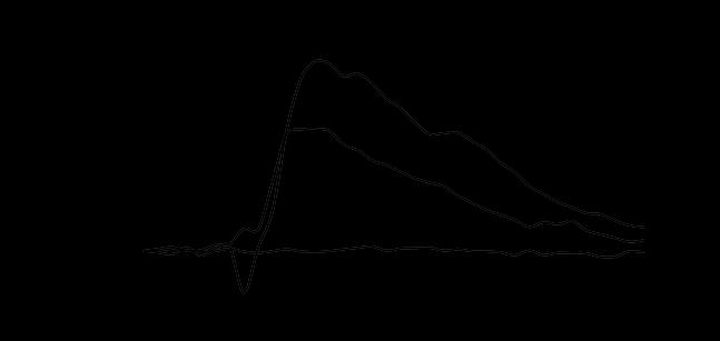
74 58 A B C Figure 10. Continued.
75 59 application was low and increased significantly when ATP accumulation was lower (p<0.001; Figure 10C). In the presence of pharmacological antagonism, the P2X7 receptor antagonist, BBG, led to a significantly diminished Ca 2+ response at the ATP trough (p<0.01), compared to controls (Figure 10B). Although the P2Y1 receptor antagonist, MRS, decreased the Ca 2+ response at this time point, the decrease was not significantly different from untreated controls (Figure 10B). Furthermore, when extracellular ATP accumulation was low (trough), MRS- and BBG-treated cultures had significantly fewer cells responding to exogenous ATP with large calcium transients compared to untreated control cultures (p<0.05, MRS; p<0.001, BBG; Figure 10C). ATP-dependent chemiluminescence ratios (C P/T ) were calculated between peak and trough time points for control, MRS-treated and BBG-treated cultures prior to antagonist exposure and C P/T ratios of 3.31 ±.60, 2.67 ±.43, and 2.32 ±.48, respectively, were not significantly different. Thus, SCN2.2 astrocyte sensitivity to ATP was rhythmic and purinergic receptor pharmacology supported the existence and function of P2X7 and P2Y1 receptors in SCN2.2 astrocytes.
76 60 Extracellular ATP accumulation is rhythmic in SCN2.2 cell cultures (Womac et al., 2009) and, consequently, purinergic signaling might contribute to the synchronization of SCN astrocyte rhythms. To test the role of purinergic signaling in the mediation of clock-controlled outputs, we disrupted P2X7R- and P2Y1R-mediated ATP signaling. Media samples were collected over 72 hours from control cultures (N=9), MRS-treated cultures (N=9) and BBG-treated cultures (N=9) and extracellular ATP levels were measured (Figure 11A). Initial peaks in ATP accumulation were potentiated by P2Y1R antagonism in MRS-treated cultures, and the estimated concentration of basal ATP was greatest in these MRS-treated cultures (p<0.05; Figure 11B, Table 2), as compared to control basal levels. BBG treatment lowered ATP levels at both peak and trough time points (Table 2). Furthermore, while control cultures maintained robust peaks in ATP chemiluminescence over 72 hours, the amplitude of ATP peaks diminished over this timeframe in MRS-treated cultures and was all but abolished in BBG-treated cultures. Amplitude differences of peak-trough accumulation by the 3 rd cycle (48-72 hours) were significantly less in MRS- and BBG-treated cultures compared to controls (p<0.005; Figure 11C). Thus, treatment with purinergic receptor antagonists altered the amount of ATP released from SCN2.2 cells and caused the damping of the ATP accumulation rhythm.
. MRS- and BBG-treated SCN2.")

 Estimated levels of extracellular ATP at trough times for control, MRS-treated, and BBG-treated cultures illustrate that basal ATP accumulation was")
77 61 A B C Figure 11. Purinergic receptor antagonists diminished ATP accumulation rhythm amplitude. A) SCN2.2 cultures exhibited circadian rhythmicity in the temporal pattern of extracellular ATP accumulation levels (N=9). MRS- and BBG-treated SCN2.2 cultures exhibited altered rhythms in extracellular ATP accumulation (N=9). Representative traces of ATP-dependent chemiluminescence from individual cultures illustrate that rhythms in ATP accumulation were dampened in MRS- and BBG-treated cultures over 72 hours. B) Estimated levels of extracellular ATP at trough times for control, MRS-treated, and BBG-treated cultures illustrate that basal ATP accumulation was enhanced by P2Y1 antagonism (*, p<0.05). C) Fold differences in amplitude measured between 48 and 72 hours revealed persistent and robust rhythms in ATP accumulation in control cultures, but both MRS-treated and BBG-treated cultures had diminished rhythm amplitudes (*, p<0.005).

78 Table 2. Purinergic receptor antagonist modulation of SCN2.2 ATP accumulation rhythm. 62
79 63 Inhibiting P2X7 and P2Y1 receptors with specific antagonists decreased rhythms in accumulation of ATP, an output of the circadian clock. This may suggest that ATP signaling involving these receptors contributes to the maintenance of robust circadian outputs. To test this possibility, we treated mper2 Luc SCN luciferase reporter cells with MRS, BBG or vehicle control and recorded bioluminescence for 120 hours before, during, and after drug treatment. All cultures maintained robust circadian rhythms in PER2::LUC bioluminescence prior to the drug treatment phase (Figure 12). Predominant frequencies, as detected by Fourier transform analysis over 4 cycles, were circadian in all treated and control cultures, and each treatment group had a mean period near 22 hours (N=6 per group; Table 3). Periods did not change between pre-treatment and drug treatment phases; however, the amplitude of PER2::LUC bioluminescence declined following MRS and BBG treatment (Figure 12; Table 3). Although bioluminescence rhythms dampened during the MRS and BBG-treatment phase compared to the pretreatment phase, only BBG treatment caused significant damping of PER2::LUC rhythmic bioluminescence throughout the entire treatment and post-treatment phases (p<0.005; Figure 12; Table 3). PER2::LUC bioluminescence amplitudes during the posttreatment (washout) phase in MRS- and BBG-treated cultures did not recover to pretreatment levels, whereas PER2::LUC bioluminescence in controls maintained rhythm amplitudes similar pre-treatment phase levels throughout the experiment (Figure 12; Table 3).
 illustrate the robustness of the mper2::luc expression profiles.")
 caused a reduction in mper2::luc bioluminescence amplitude, damping of PER2::LUC oscillations, and a diminution in the number of cycles that persisted in the")
80 64 Figure 12. PER2::LUC reporter rhythms were disrupted by purinergic receptor antagonism. Representative traces of mper2::luc bioluminescence from control, MRS- and BBG-treated mper2 Luc SCN cultures (N=6) illustrate the robustness of the mper2::luc expression profiles. Bioluminescence was recorded for 5 days for pre-treatment, drug treatment, and drug washout experimental phases. Treatment of BBG (bottom records) caused a reduction in mper2::luc bioluminescence amplitude, damping of PER2::LUC oscillations, and a diminution in the number of cycles that persisted in the PER2::LUC rhythm. MRS treatment (middle records) caused similar, but not as severe, alterations in mper2::luc bioluminescence. Media changes occurred at the times of drug treatment and washout.

81 Table 3. Purinergic receptor antagonist modulation of PER2::LUC bioluminescence amplitude. 65
82 66 DISCUSSION Organisms possess a circadian clock mechanism for entraining the internal timing of molecular and physiological events to the external environment. These circadian entrainment and pacemaker mechanisms in mammals are localized to the suprachiasmatic nuclei (SCN) of the hypothalamus, which synchronize peripheral oscillations to 24-hour rhythms in appropriate phase with the local photoperiod (Silver et al., 1996; Ueyama et al., 1999; Schibler and Sassone-Corsi, 2002; Dibner et al., 2010; Welsh et al., 2010). The SCN contain nearly 20,000 neurons (Moore, 1996; Abrahamson and Moore, 2001) and an abundance of astrocytes (Morin et al., 1989). Daily oscillations in clock gene expression are coordinated among SCN neurons (Reppert and Weaver, 2001; Welsh et al., 2010) and this coordination is necessary to produce robust circadian rhythms in animal behavior. However, the mechanism of inter-oscillator coupling within the SCN is not well understood (Hastings and Herzog, 2004). We hypothesized that ATP release was an output of the molecular clock in astrocytes, with one of its functions being the regulation of intercellular coupling among SCN glial cells. SCN astrocytes exhibit circadian oscillations in various physiological processes (Prolo et al., 2005; Becquet et al., 2007; Womac et al., 2009; Burkeen et al., 2011), can be entrained by VIP (Marpegan et al., 2009), and can affect circadian neuronal activity (Prosser et al., 1994). Still, the mechanisms that couple individual SCN astrocytic oscillators into ensemble oscillators remain undefined. In the present studies, we have demonstrated that clockcontrolled ATP release from SCN astrocytes and purinergic signaling through P2
83 67 receptors contribute to ensemble synchronization of rhythms in clock gene expression, ATP-evoked calcium signaling, and circadian rhythms in ATP release itself. ATP release from astrocytes and its rhythmic extracellular accumulation is under circadian clock control (Marpegan et al., 2011; Figure 7). Here, we have demonstrated that clock-controlled ATP accumulation affects clock output rhythms and the robustness of the ensemble SCN astrocytic clock. Communication via ATP signaling mechanisms varies over 24 hours in SCN astrocytes, where intrinsic oscillations exist in calcium responses to ATP application (Figures 8 and 10). This rhythmicity in inherent sensitivity to ATP may provide a mechanism of phase resetting, a mechanism by which organisms adjust their clock each day to correspond with daily light-dark cycles (Albrecht et al., 2001). Rhythmic ATP accumulation was affected by inhibiting ATP signaling via purinergic receptor antagonism (Figure 11), suggesting that, as high levels of ATP accumulate extracellularly every 24 hours, this purinergic signal feeds back upon the astrocytes, perhaps triggering physiological adjustments influencing the clock through calcium-dependent signaling mechanisms. BBG, a P2X7R antagonist, abolished peak levels of ATP in most cultures and significantly reduced the basal level of ATP accumulation; whereas, MRS, a P2Y1R antagonist, raised the basal level of ATP accumulation and enhanced the initial amplitude of ATP release peaks. These differences in antagonistic effects were surprising and may be explained by examining the calcium signaling mechanisms evoked by receptor activation. For the P2X7R, ATP activation induces an influx of Ca 2+ ions that raises cytosolic calcium concentration and
84 68 can influence the activation of clock gene expression. Following P2Y1R activation, the phospholipase C (PLC) signaling pathway initiates an increase in cytosolic calcium by release from intracellular stores and that can influence transcription, as well. P2Y receptors are classified by which G-proteins they are associated with, and P2Y1 receptors are generally coupled to G q /G 11 GTPases (Verkhratsky, 2005; Abbracchio et al., 2009). Interestingly, P2Y receptors can activate other G-protein signaling pathways and can form hetero-oligodimers with other P2Y receptors to activate varying responses (White et al., 2003; White and Burnstock, 2006; Ecke et al., 2009). P2Y stimulation of G s -dependent adenylate cyclase activation leads to increased conversion of intracellular ATP to camp and subsequent activation of camp signaling pathways. Whereas intracellular content of ATP (Yamazaki et al., 1994) and extracellular levels of ATP (Womac et al., 2009) both peak in the rodent SCN at night, rhythmic SCN cellular content of camp (Prosser & Gillette, 1991) peaks during the day and is accompanied by rhythmic regulation of camp response element (CRE) activity (O Neill et al., 2008). In the present studies, P2Y1R inhibition of SCN astrocytes may have disrupted a P2Y/G s - dependent camp signaling, thereby explaining the elevated basal levels of extracellular ATP in the MRS-treated cultures.
85 69 The hypothesis that ATP signaling mediates circadian rhythms among SCN astrocytes was further studied using PER2::LUC bioluminescence reporting of mper2 gene expression following treatment with purinergic receptor antagonists. Analysis of PER2 reporter rhythms revealed less robust rhythms in ensemble bioluminescence in the presence of purinergic antagonists (Figure 12). Disruption of ATP signaling caused a decrease in bioluminescence reporting of ensemble Per2 gene expression, suggesting that ATP regulates, in some manner, synchrony between astrocytic oscillators of the ensemble SCN clock. We conclude that the decline in PER2::LUC bioluminescence is due to uncoupling of synchronized oscillators within the culture. A direct damping effect on the molecular clock mechanism within individual oscillating cells seemingly is not occurring as a result of purinergic receptor inhibition. In rats, manipulation of SCN input and output pathways abolishes behavioral rhythmicity, but the molecular clock remains functional (Schwartz et al., 1987), demonstrating that arrhythmicity is not due to an affected molecular clock. Here, robust amplitudes of PER2::LUC bioluminescence indicated strong inter-oscillator coupling and robust coordination of rhythmic gene expression. Our results imply that ATP signaling, via both P2X and P2Y classes of purinergic receptors, synchronizes SCN astrocytic oscillators leading to coordinated ensemble rhythms in clock gene expression and ATP production, release, and accumulation. Future studies must determine if individual SCN astrocytes, as is the case with SCN neurons (Welsh et al., 1995; Hastings & Herzog, 2004), are actually individual oscillators functioning in ensemble physiological synchrony and whether ATP signaling disruption causes uncoupling of these individual oscillators. Taken together,
86 70 the current data are indicative of a functional role for clock-controlled ATP signaling by astrocytes within the mammalian SCN. Rhythmic extracellular ATP accumulation has been established as an output of the circadian clock; however, the function of that purine accumulation is unknown. Astrocytes play a role in modulating synaptic transmission, as their numerous processes contact vast numbers of synapses within the mammalian brain (Araque et al., 1999; Bacci et al., 1999). The tripartite synapse, consisting of presynaptic and postsynaptic elements and the astrocytic end-feet that surround them, is thought to regulate many aspects of brain neurophysiology (Araque et al., 1999; Haydon, 2001; Newman, 2003). In Drosophila, the glia-specific gene ebony mediates circadian rhythms in locomotor behavior, demonstrating that glial cells can modulate complex neuronal networks (Suh and Jackson, 2007; Ng et al., 2011). In rats, circadian neuronal firing patterns from SCN slices are altered when glial coupling and metabolism are disrupted, signifying glial involvement in maintaining rhythmicity in the SCN (Prosser et al., 1994). ATP is released from astrocytes as a gliotransmitter that can modulate synaptic communication (Cotrina et al., 1998; Guthrie et al., 1999; Haydon, 2001; Scemes & Giaume, 2006) and has been implicated in hippocampal plasticity (Pascual et al., 2005) and sleep (Halassa et al., 2009). ATP mediates neuronal-glial signaling by binding to purinergic receptors, either ionotropic P2X receptors or G-protein-coupled P2Y receptors, to initiate elevations in intracellular Ca 2+. Disrupting signaling through these receptors affects both neuronal and astrocyte physiology, in part by alterations in adenosine signaling
87 71 following ATP hydrolysis (Edwards et al., 1992; Evans et al., 1992; Fields and Stevens, 2000; Fumagalli et al., 2003; Fields and Burnstock, 2006). Interestingly, electrophysiological evidence exists for presence of adenosine A1 and A2 receptors in the SCN (Chen & van den Pol, 1997), suggesting potential glia-neuron interactions originating from clock-controlled ATP release rhythms from SCN astrocytes. Blockade of purinergic signaling with P2X7 and P2Y1 antagonists disrupted multiple aspects of clock-controlled ATP signaling in SCN astrocytes, suggesting that purinergic signaling pathways may have broad influence on the coordination and synchrony of circadian rhythms within the SCN. Furthermore, these results suggest that clock-controlled ATP release throughout the mammalian brain could have important influences over circadian rhythms in brain function.
88 72 CHAPTER IV DISCUSSION AND CONCLUSIONS THE MAMMALIAN MASTER CIRCADIAN PACEMAKER In mammals, biological clocks are found in many different cell types. The retina, astrocytes, pituitary gland, heart, lungs, liver, and kidneys are among some of the tissues that are composed of cellular oscillators containing biological clocks (Yamazaki et al., 2000; Yoo et al., 2004; Prolo et al., 2005; Ko et al., 2007). Because many organs in mammals have biological clocks that can all keep time locally within the organ, it is proposed that these tissues are coordinated by a pacemaking oscillator that can synchronize the timing of clocks in each organ to the appropriate time of day. This structure is the suprachiasmatic nucleus (SCN) and represents the master circadian pacemaker in mammals. The SCN is entrained by daylight and then coordinates the phases of peripheral biological clocks to this timing (Silver, 1996; Ueyama, 1999; Schibler and Sassone- Corsi, 2002; Dibner, et al., 2010; Welsh et al., 2010). Individual cellular oscillators in the SCN rhythmically express genes that have been identified as core components of the
89 73 mammalian canonical clock machinery: Clock, Bmal1, Period (Per1 and Per2),Cryptochrome (Cry1 and Cry2), and Rev-erbα. The self-sustained rhythms of these molecular clock components are driven by 24-hour transcriptional and posttranslational feedback loops and are found in all oscillators, including peripheral clock cells. However, it is the ability for the oscillators within the SCN to coordinate the phases of peripheral clocks that sets them apart. The SCN contains retinorecipient pacemaking neurons that communicate light information received from retinal inputs to other neuronal and astrocytic oscillators in the SCN. The oscillators within the SCN itself are coupled to one another, and this coordination of SCN oscillations imposes 24- hour rhythmicity upon peripheral cellular oscillators in an organism. This coupling mechanism within the SCN produces and maintains synchronized rhythmicities that influence behavioral, biochemical and physiological processes. We hypothesize that one function of ATP signaling is to contribute to this mechanism of coupling SCN oscillators.
90 74 CIRCADIAN REGULATION OF ATP SIGNALING These studies present a novel finding, that ATP is released from the SCN in vivo and from SCN cell cultures and accumulates extracellularly every 24 hours. It is not surprising that rhythms in ATP accumulation persist from these cells, as several genes involved in the regulation of ATP oscillate in the SCN (Menger et al., 2005). Intracellular ATP content oscillates, with peak levels detected during mid-subjective night (Yamazaki et al., 1994). In addition, ATP is released from neurons and astrocytes to modulate neuronal-glial synaptic communication, so its extracellular detection is expected. ATP accumulation is identified here as a 24-hour circadian rhythm. This rhythmic accumulation is characterized as an output of the clock, one of the many processes controlled by endogenous molecular clock oscillations. A non-functional circadian clock, unable to express Per1 and Per2 properly, abolishes rhythmic accumulation, demonstrating its classification as a circadian rhythm and the necessity of the clock to produce this rhythm. The purpose of SCN astrocytes releasing ATP every 24 hours is of interest. Astrocytes play a significant role in modulating synaptic transmission, as their numerous processes contact thousands of synapses (Araque, et al., 1999; Bacci et al., 1999). The tripartite synapse has since been established as presynaptic and postsynaptic neurons that
91 75 communicate with astrocytes to regulate brain neurophysiology (Araque, et al., 1999; Haydon, 2001; Newman, 2003). ATP is released from astrocytes as a gliotransmitter that can modulate communication at the site of the synapse (Cotrina et al., 1998; Guthrie et al., 1999; Haydon, 2001). ATP mediates neuronal-glial signaling by binding to its receptors, either ionotropic P2X receptors or metabotropic P2Y receptors, on astrocytes and neurons to initiate elevations in intracellular Ca 2+. Disrupting signaling through these receptors affects neuronal and astrocytic excitability (Edwards et al., 1992; Evans et al., 1992; Fields and Stevens, 2000; Fumagalli et al., 2003; Fields and Burnstock, 2006) and highlights the importance of ATP in modulating intercellular communication (Jackson, 2011). The present studies support these findings, in which ATP signaling mediates astrocytic communication, and a disruption in this signaling pathway has a broader influence on the coordination of communication and synchrony within the SCN. Communication via ATP signaling mechanisms between astrocytes and neurons may vary over 24 hours, as our results suggest that SCN cells contain an innate oscillation in cellular response to ATP. Activation of purinergic receptors with exogenous ATP varies at different levels of ATP accumulation (peak vs. trough), with less receptor activation and smaller cellular responses measured when extracellular ATP accumulation is high. Large cellular responses, recorded as large increases in intracellular Ca 2+, and receptor
92 76 activation are seen when extracellular ATP is low. Different physiological responses to ATP over 24 hours indicate that the clock may be regulating not only the output rhythm of ATP accumulation but also cellular sensitivity to transmitter signaling. Receptor availability and turnover may be under circadian control, and if so, cellular responsiveness is directly affected by the clock. This inherent sensitivity to respond to ATP signaling may also provide a method of phase resetting, a mechanism by which organisms can adjust clock timing slightly each day to correspond with daily light-dark cycles (Albrecht et al., 2001). Animals can respond to light pulses by either advancing or delaying their activity, depending on when during the night the pulse is given. This demonstrates the ability of the entrainable SCN clock to reset its phase, indicating its responsiveness to stimuli. Thus, cellular oscillators must be sensitive to a stimulus in order to be reset. Here, that sensitivity could be modulated by receptor function and availability, characteristics that may also be under clock control.
93 77 CIRCADIAN CLOCK SYNCHRONY REGULATED BY PURINERGIC SIGNALING This study reveals that ATP accumulates from SCN astrocytes to synchronize the many oscillators within the SCN. ATP acts in this manner through its purinergic receptors that are found on astrocytes and neurons. ATP can initiate Ca 2+ waves among populations of astrocytes by activating the IP 3 signaling pathway that leads to the increase in cytosolic Ca 2+. This rise in Ca 2+ triggers the release of gliotransmitters, such as ATP, that participate in the propagation of the Ca 2+ wave by binding to purinergic receptors on adjacent astrocytes. IP 3 signaling molecules can also pass through gap junctions, which couple astrocytes to one another, to raise intracellular Ca 2+. The coordinated release of ATP from SCN astrocytes has the ability to signal to adjacent neuronal and astrocytic oscillators and cause a simultaneous rise in Ca 2+ among these oscillators. This synchronized elevation of cytosolic Ca 2+ may trigger clock gene transcriptional activation, leading to initiation of the molecular transcriptional-translational feedback loop. SCN oscillators will then have the timing of their molecular clocks set to the same phase, as they have been exposed to phase-setting ATP signals that activate transcription via signaling pathways that elevate cytosolic Ca 2+. As intracellular Ca 2+ rises, it binds to CaMKII, a protein kinase that activates CREB. CREB is a transcription factor that binds
94 78 to the camp response element (CRE) on the promoter sequence of genes to activate transcription. The only gene of the canonical clock components to contain CRE sequences is Period. Because of this, Per induction can occur rapidly in response to a Ca 2+ -elevating stimulus. CLOCK and BMAL1 are continuously bound to the Per promoter and can readily initiate Per transcription if the stimulus is applied at a specific time during the feedback loop. The data presented here suggest that ATP is a Ca 2+ - elevating stimulus, eliciting cytosolic Ca 2+ increases via purinergic receptor signaling. As ATP accumulates every 24 hours and signals to adjacent SCN astrocytes and neurons, it may cause the activation of Per transcription through elevated intracellular Ca 2+ signaling in these oscillators. Activation of Per expression by ATP signaling coordinates the transcriptional feedback loop in the oscillators to the same phase. In this manner, ATP could be a synchronizing cue among SCN oscillators, resetting the timing of clock gene expression to the same phase every day so that SCN cells, as a whole, produce one, coordinated 24-hour rhythm that can be imposed upon peripheral clocks. We investigated this possible function of accumulated extracellular ATP as a synchronizing molecule by disrupting ATP signaling mechanisms in SCN astrocytes. Using pharmacological agents, we antagonized the P2X7 and P2Y1 ATP receptors and studied the effects on ATP accumulation and synchrony among SCN cells in culture. Disrupting the function of these receptors implicates ATP as a modulator of synchrony
95 79 among SCN cells. The potential role of ATP as a circadian signaling molecule contributing to the maintenance of synchronized phasing between oscillators is revealed in these studies when receptor antagonism altered ATP accumulation output rhythms and dampened bioluminescence reporting of Per2 clock gene expression. Rhythmic ATP accumulation was affected by inhibiting ATP from binding several of its receptors. This may suggest that, as high levels of ATP accumulate extracellularly every 24 hours, this signaling molecule acts on its receptors across many neurons and astrocytes within the SCN with the function to maintain proper coordination of the clock and its output rhythms. Observed changes to ensemble PER2::LUC rhythmicity when treated with purinergic receptor antagonists support this hypothesis. Analysis of PER2::LUC bioluminescence rhythms revealed less robust rhythms in ensemble bioluminescence among the treated cells in culture. Taken together, these data are indicative of a functional role for ATP accumulation within the SCN, whereas astrocytes release ATP every 24 hours for continual signaling onto astrocytes and neurons to maintain daily coordinated synchrony of the clocks in these cells (Figure 13).

96 80 A B C Figure 13. Astrocyte purinergic signaling regulates synchronous function of the SCN. A) ATP is rhythmically released from SCN astrocytes and accumulates over 24 hours. Rhythmic ATP accumulation is an output of the circadian clock, and the timing of its release and action on purinergic receptors are synchronized among astrocytes. B) Disruption of signaling mechanisms via purinergic receptors eliminates ATP as a synchronizing cue among SCN oscillators. C) This disruption causes coordinated timing of cellular clocks to become unsynchronized, affecting their clock output rhythms and coordinated reception of synchronizing cues.
97 81 CALCIUM SIGNALING AND ATP ACCUMULATION Previous studies conducted by our lab demonstrated a circadian rhythm in cytosolic calcium levels in SCN2.2 cell cultures. An antiphase relationship between intracellular Ca 2+ and extracellular ATP was observed, as assays for both molecules were simultaneously conducted at 1-hour intervals. The peaks and troughs in Ca 2+ fluorescence had an inverse phase in relation to rhythmic ATP-dependent chemiluminescence (Figure 14A), demonstrating an antiphasic relationship between rhythmic extracellular ATP accumulation and cytosolic Ca 2+ concentration. Rhythms in extracellular ATP accumulation and cytosolic Ca 2+ in SCN2.2 astrocytes are 12 hours out of phase, suggesting that peak levels of astrocytic Ca 2+ coincide with the daytime peak in SCN neuronal Ca 2+ (Colwell, 2000; Ikeda et al, 2003; Irwin and Allen, 2009). Determining the timing of peak astrocytic Ca 2+ elevations is based on the timing of extracellular ATP accumulation in the rat SCN. We found that extracellular ATP accumulation in the rat SCN peaks at late night (Figure 5; Womac et al., 2009); therefore, cytosolic Ca 2+ peaks roughly 12 hours after that, during the day.
 The time course of ATP-dependent chemiluminescence (blue line) and cytosolic calcium fluorescence as detected with Fluo-4-AM (green line)")
 occur in phase with extracellular ATP accumulation.")


98 82 A B Rhod-2 MitoTracker Merge Figure 14. Intracellular calcium rhythms and ATP accumulation. (A) The time course of ATP-dependent chemiluminescence (blue line) and cytosolic calcium fluorescence as detected with Fluo-4-AM (green line) illustrates the anti-phase and inverse relationship between these two cellular rhythms. Mitochondrial calcium elevations as detected by Rhod-2-AM (red dotted line) occur in phase with extracellular ATP accumulation. (B) Fluorescence imaging was performed with MitoTracker Green, a mitochondrial marker, and Rhod-2-AM, a mitochondrial Ca 2+ marker in SCN2.2 cells. Rhod-2 fluorescence colocalized with MitoTracker Green fluorescence. This fluorescent colabeling verified that that Rhod-2 was primarily an indicator of mitochondrial Ca 2+ levels in these cultures. These data from Figure 14 are reprinted with permission from Mitochondrial Calcium Signaling Mediates Rhythmic Extracellular ATP Accumulation in Suprachiasmatic Nucleus Astrocytes by Jeff Burkeen, Alisa Womac, David Earnest, and Mark Zoran, The Journal of Neuroscience, Volume 31, Pages , Copyright 2011 by The Society for Neuroscience.
99 83 Mitochondrial calcium levels fluctuated in phase with extracellular ATP accumulation and 12 hours out of phase with fluctuating cytosolic Ca 2+ levels (Figure 14A, B). Calcium signaling in the mitochondria regulates its function, including the production of ATP (Santo-Domingo and Demurex, 2010; McCormack et al., 1990). Blocking calcium from entering the mitochondria disrupts the extracellular ATP accumulation rhythm, and presumably ATP production. Ru360, a specific mitochondrial calcium uniporter inhibitor, significantly reduced extracellular ATP accumulation in treated SCN2.2 cultures as compared to untreated cultures at both trough and peak ATP accumulation time points. Our data illustrates that rhythmic ATP accumulation might lag slightly behind ATP production, since cellular ATP content peaks at mid subjective night (Yamazaki et al., 1994). The antiphase relationship between extracellular ATP accumulation and cytosolic Ca 2+ may be contributed to the utilization of ATP and the movement of ions by ATPdependent transport proteins. Several P-type ATPases, including the sodium/potassium pump (Na + /K + ATPase) and the plasma membrane Ca 2+ ATPase (PMCA), hydrolyze ATP to transport ions either into or out of the cell. The Na + /K + ATPase maintains proper sodium and potassium ion concentrations inside neurons and astrocytes by pumping Na + ions out of the cell and K + ions into the cell, and it uses ATP to sustain this balance. In the SCN, neuronal firing is greatest during the day, and likewise, Na + /K + ATPase activity is higher during this same time to reestablish resting membrane potentials (Wang and Huang, 2004; Ko et al., 2009). This rise in Na + /K + ATPase activity utilizes
100 84 large quantities of ATP at times of increased neuronal firing, and it could be occurring in both neurons and astrocytes, as astrocytes function to clear excess potassium from the synaptic cleft during neuronal activity. Therefore, Na + /K + ATPases would hydrolyze ATP in abundance within astrocytes during the day, accounting for low levels of accumulated ATP. However, when neuronal activity and Na + /K + ATPase activity decrease during the night, ATP is available to accumulate extracellularly as it is not being hydrolyzed at a higher rate. The plasma membrane Ca 2+ ATPase functions to transport elevated concentrations of Ca 2+ ions out of the cell by hydrolyzing ATP. We see that Ca 2+ levels in astrocytes oscillate over 24 hours, most likely peaking during the day, as the antiphase peak in ATP accumulation occurs during late night in the SCN. As we hypothesize that intracellular Ca 2+ oscillations are peaking during the day, plasma membrane Ca 2+ ATPase activity may be coinciding with neuronal activity. During neuronal firing, astrocytes are activated by neurotransmitters, leading to elevated intracellular calcium levels. Plasma membrane Ca 2+ ATPases transport calcium to the extracellular space, hydrolyzing intracellular ATP in the process. Lower levels of ATP are available for accumulation as they are being utilized in this transport process. As elevated Ca 2+ levels drop, presumably concurring with decreased neuronal activity, plasma membrane Ca 2+ ATPase activity may decrease. This would indicate that nonhydrolyzed intracellular ATP would be available to accumulate extracellularly. The timing of this process would account for the 12-hour antiphase relationship between extracellular ATP accumulation and intracellular Ca 2+ oscillations.
101 85 ROLE OF ATP SIGNALING IN PERIPHERAL CLOCKS The focus of these studies has been on SCN astrocytes and their ability to affect clock synchrony through purinergic signaling. What we understand from these results is that ATP accumulation is an output of the clock. We base this on evidence obtained from these studies, in which accumulation is driven by the clock and is abolished when the molecular clockwork mechanism is disrupted. As it is known, clocks are found in many peripheral tissues aside from the master circadian pacemaker. We observed this novel rhythm of ATP accumulation in several peripheral clock oscillators: fibroblasts, hepatocytes, and cortical astrocytes. Each of these oscillators exhibits rhythmic ATP accumulation with periods ranging from ~20 hours to ~25 hours (Figure 15). The range in period length among these different cell types is indicative of local time-keeping among peripheral oscillators in the absence of SCN timing. The SCN induces a 24-hour rhythm on peripheral clocks, and when these clocks are left to oscillate without SCN influence, they manage to keep time with a period that is endogenous to each peripheral clock. Here, each of these cultures maintains the rhythmic clock output of ATP accumulation. As we propose, ATP may function as a synchronizing signal among oscillators within the SCN. Additionally, ATP may have the same function in maintaining synchrony among peripheral clocks in each tissue. We revealed that ATP
 and Lomb-Scargle periodicities (τ ± SEM). SCN2.2 cultures (N=11) had τ: 23.68 ± 0.72 hours.")
102 86 Figure 15. Circadian periodicities of ATP accumulation in peripheral cellular oscillators. Representative traces from individual cultures illustrate temporal pattern of ATP accumulation. All cultures underwent Fourier transform analysis to determine the number of rhythmic cultures (N) and Lomb-Scargle periodicities (τ ± SEM). SCN2.2 cultures (N=11) had τ: ± 0.72 hours. Primary cortical astrocyte cultures (N=16) had τ: ± 0.20 hours. Hepa1c1c7 hepatocyte cultures (N=3) had τ: ± 0.51 hours. NIH/3T3 fibroblast cultures (N=6) had τ: ± 1.63 hours.
Sleep-Wake Cycle I Brain Rhythms. Reading: BCP Chapter 19
 Sleep-Wake Cycle I Brain Rhythms Reading: BCP Chapter 19 Brain Rhythms and Sleep Earth has a rhythmic environment. For example, day and night cycle back and forth, tides ebb and flow and temperature varies
The Success of Decomposition
 11/21/11 Mechanism and Levels of Organization: Recomposing and Situating Circadian Clocks The Success of Decomposition Moving beyond per, researchers in the 1990s and early 2000s identified many clock
Biological Clocks. Lu Chen, Ph.D. MCB, UC Berkeley. What is biological clock?
 Biological Clocks Lu Chen, Ph.D. MCB, UC Berkeley 1 What is biological clock? All eukaryotes and some prokaryotes display changes in gene activity, biochemistry, physiology, and behavior that wax and wane
BIOLOGY. Cell Communication CAMPBELL. Reece Urry Cain Wasserman Minorsky Jackson. Lecture Presentation by Nicole Tunbridge and Kathleen Fitzpatrick
 CAMPBELL BIOLOGY TENTH EDITION Reece Urry Cain Wasserman Minorsky Jackson 11 Cell Communication Lecture Presentation by Nicole Tunbridge and Kathleen Fitzpatrick Cellular Messaging Cells can signal to
Biological Clocks. Lu Chen, Ph.D. MCB, UC Berkeley. Why Does Melatonin Now Outsell Vitamin C??
 Biological Clocks Lu Chen, Ph.D. MCB, UC Berkeley 1 Why Does Melatonin Now Outsell Vitamin C?? Wake / sleep complaints are extremely prevalent. Much melatonin is consumed in an attempt to overcome the
Transcription Regulation And Gene Expression in Eukaryotes (Cycle G2 # )
") Transcription Regulation And Gene Expression in Eukaryotes (Cycle G2 #13709-01) CIRCADIAN SIGNALING NETWORKS RG. Clerc May 19. 2010 www.fmi.ch/training/teaching Circadian rythms : most physiological processes
Transcription Regulation And Gene Expression in Eukaryotes (Cycle G2 #13709-01) CIRCADIAN SIGNALING NETWORKS RG. Clerc May 19. 2010 www.fmi.ch/training/teaching Circadian rythms : most physiological processes
Chapter 11 Introduction to the Nervous System and Nervous Tissue Chapter Outline
 Chapter 11 Introduction to the Nervous System and Nervous Tissue Chapter Outline Module 11.1 Overview of the Nervous System (Figures 11.1-11.3) A. The nervous system controls our perception and experience
CIRCADIAN SIGNALING NETWORKS
 Transcription Regulation And Gene Expression in Eukaryotes Cycle G2 (lecture 13709) FS 2014 P. Matthias and RG Clerc Roger G. Clerc 07.05.2014 CIRCADIAN SIGNALING NETWORKS Master pacemaker SCN «Slave clocks»
REVIEWS. Linking neural activity and molecular oscillations in the SCN
 Linking neural activity and molecular oscillations in the SCN Christopher S. Colwell Abstract Neurons in the suprachiasmatic nucleus (SCN) function as part of a central timing circuit that drives daily
Chapter 2: Cellular Mechanisms and Cognition
 Chapter 2: Cellular Mechanisms and Cognition MULTIPLE CHOICE 1. Two principles about neurons were defined by Ramón y Cajal. The principle of connectional specificity states that, whereas the principle
Cell Communication. Chapter 11. PowerPoint Lectures for Biology, Seventh Edition. Lectures by Chris Romero. Neil Campbell and Jane Reece
 Chapter 11 Cell Communication PowerPoint Lectures for Biology, Seventh Edition Neil Campbell and Jane Reece Lectures by Chris Romero Overview: The Cellular Internet Cell-to-cell communication Is absolutely
Neurotransmitter Systems II Receptors. Reading: BCP Chapter 6
 Neurotransmitter Systems II Receptors Reading: BCP Chapter 6 Neurotransmitter Systems Normal function of the human brain requires an orderly set of chemical reactions. Some of the most important chemical
Principles of Anatomy and Physiology
 Principles of Anatomy and Physiology 14 th Edition CHAPTER 12 Nervous Tissue Introduction The purpose of the chapter is to: 1. Understand how the nervous system helps to keep controlled conditions within
Chapter 11. Cell Communication
 Chapter 11 Cell Communication Overview: The Cellular Internet Cell-to-cell communication Is absolutely essential for multicellular organisms Concept 11.1: External signals are converted into responses
Cell Communication. Chapter 11. Biology Eighth Edition Neil Campbell and Jane Reece. PowerPoint Lecture Presentations for
 Chapter 11 Cell Communication PowerPoint Lecture Presentations for Biology Eighth Edition Neil Campbell and Jane Reece Lectures by Chris Romero, updated by Erin Barley with contributions from Joan Sharp
Transcription Regulation And Gene Expression in Eukaryotes FS 2016 Graduate Course G2
 Transcription Regulation And Gene Expression in Eukaryotes FS 2016 Graduate Course G2 P. Matthias and RG Clerc Pharmazentrum Hörsaal 2 16h15-18h00 CIRCADIAN SIGNALING NETWORKS Master pacemaker SCN «slave
Receptors Families. Assistant Prof. Dr. Najlaa Saadi PhD Pharmacology Faculty of Pharmacy University of Philadelphia
 Receptors Families Assistant Prof. Dr. Najlaa Saadi PhD Pharmacology Faculty of Pharmacy University of Philadelphia Receptor Families 1. Ligand-gated ion channels 2. G protein coupled receptors 3. Enzyme-linked
MOLECULAR AND CELLULAR NEUROSCIENCE
 MOLECULAR AND CELLULAR NEUROSCIENCE BMP-218 November 4, 2014 DIVISIONS OF THE NERVOUS SYSTEM The nervous system is composed of two primary divisions: 1. CNS - Central Nervous System (Brain + Spinal Cord)
PSYCH 260 Exam 2. March 2, Answer the questions using the Scantron form. Name:
 PSYCH 260 Exam 2 March 2, 2017 Answer the questions using the Scantron form. Name: 1 1 Main Please put in their proper order the steps that lead to synaptic communication between neurons. Begin with the
SUPPLEMENTARY INFORMATION
 Supplementary Figure 1. Normal AMPAR-mediated fepsp input-output curve in CA3-Psen cdko mice. Input-output curves, which are plotted initial slopes of the evoked fepsp as function of the amplitude of the
Parathyroid Hormone, But Not Melatonin, Resets The Bone Circadian Clock
 Parathyroid Hormone, But Not Melatonin, Resets The Bone Circadian Clock Naoki Okubo 1,2,3, Yoichi Minami 1,3, Hiroyoshi Fujiwara 2, Tatsuya Kunimoto 1,2,3, Toshihiro Hosokawa 1,2,3, Ryo Oda 2, Toshikazu
Modeling Rhythms on Differents Levels: Cells, Tissues, and Organisms
 Modeling Rhythms on Differents Levels: Cells, Tissues, and Organisms Hanspeter Herzel Institute for Theoretical Biology (ITB) Charité and Humboldt University Berlin Molecular Chronobiology SCN-neuron nucleus
Lecture 14. Insect nerve system (II)
") Lecture 14. Insect nerve system (II) Structures (Anatomy) Cells Anatomy How NS functions Signal transduction Signal transmission Overview More on neurons: ions, ion channel, ligand receptor Signal transduction:
Lecture 14. Insect nerve system (II) Structures (Anatomy) Cells Anatomy How NS functions Signal transduction Signal transmission Overview More on neurons: ions, ion channel, ligand receptor Signal transduction:
Psych 181: Dr. Anagnostaras
 Psych 181: Dr. Anagnostaras Lecture 5 Synaptic Transmission Introduction to synaptic transmission Synapses (Gk., to clasp or join) Site of action of most psychoactive drugs 6.5 1 Synapses Know basic terminology:
Chapter 2. The Cellular and Molecular Basis of Cognition Cognitive Neuroscience: The Biology of the Mind, 2 nd Ed.,
 Chapter 2. The Cellular and Molecular Basis of Cognition Cognitive Neuroscience: The Biology of the Mind, 2 nd Ed., M. S. Gazzaniga, R. B. Ivry, and G. R. Mangun, Norton, 2002. Summarized by B.-W. Ku,
3) Most of the organelles in a neuron are located in the A) dendritic region. B) axon hillock. C) axon. D) cell body. E) axon terminals.
 Most of the organelles in a neuron are located in the A) dendritic region. B) axon hillock. C) axon. D) cell body. E) axon terminals.") Chapter 48 Neurons, Synapses, and Signaling Multiple-Choice Questions 1) A simple nervous system A) must include chemical senses, mechanoreception, and vision. B) includes a minimum of 12 ganglia. C) has
Chapter 48 Neurons, Synapses, and Signaling Multiple-Choice Questions 1) A simple nervous system A) must include chemical senses, mechanoreception, and vision. B) includes a minimum of 12 ganglia. C) has
SYNAPTIC COMMUNICATION
 BASICS OF NEUROBIOLOGY SYNAPTIC COMMUNICATION ZSOLT LIPOSITS 1 NERVE ENDINGS II. Interneuronal communication 2 INTERNEURONAL COMMUNICATION I. ELECTRONIC SYNAPSE GAP JUNCTION II. CHEMICAL SYNAPSE SYNAPSES
PHYSIOLOGY AND MAINTENANCE Vol. V - Biological Rhythms - Tarja Porkka-Heiskanen, Jarmo T. Laitinen
 BIOLOGICAL RHYTHMS Tarja Porkka-Heiskanen, Institute of Biomedicine, University of Helsinki, Finland Jarmo T. Laitinen Department of Physiology, University of Kuopio, Finland Keywords: Light, melatonin,
Clicker Question. The Need to Decompose. Mechanism and Reduction: Decomposing Circadian Clocks
 Mechanism and Reduction: Decomposing Circadian Clocks Clicker Question On the Deductive-Nomological (DN) model of reduction, which of the following does not figure in providing the explanation (i.e., is
BIPN 140 Problem Set 6
 BIPN 140 Problem Set 6 1) The hippocampus is a cortical structure in the medial portion of the temporal lobe (medial temporal lobe in primates. a) What is the main function of the hippocampus? The hippocampus
Communication Between
 Communication Between Neurons Bởi: OpenStaxCollege The electrical changes taking place within a neuron, as described in the previous section, are similar to a light switch being turned on. A stimulus starts
Cell Communication. Chapter 11. Biology Eighth Edition Neil Campbell and Jane Reece. PowerPoint Lecture Presentations for
 Chapter 11 Cell Communication PowerPoint Lecture Presentations for Biology Eighth Edition Neil Campbell and Jane Reece Lectures by Chris Romero, updated by Erin Barley with contributions from Joan Sharp
BIPN 140 Problem Set 6
 BIPN 140 Problem Set 6 1) Hippocampus is a cortical structure in the medial portion of the temporal lobe (medial temporal lobe in primates. a) What is the main function of the hippocampus? The hippocampus
Communication within a Neuron
 Neuronal Communication, Ph.D. Communication within a Neuron Measuring Electrical Potentials of Axons The Membrane Potential The Action Potential Conduction of the Action Potential 1 The withdrawal reflex
Chapter 2. The Cellular and Molecular Basis of Cognition
 Chapter 2. The Cellular and Molecular Basis of Cognition Cognitive Neuroscience: The Biology of the Mind, 2 nd Ed., M. S. Gazzaniga,, R. B. Ivry,, and G. R. Mangun,, Norton, 2002. Summarized by B.-W. Ku,
SUPPLEMENTARY INFORMATION. Supplementary Figure 1
 SUPPLEMENTARY INFORMATION Supplementary Figure 1 The supralinear events evoked in CA3 pyramidal cells fulfill the criteria for NMDA spikes, exhibiting a threshold, sensitivity to NMDAR blockade, and all-or-none
Chapter 9. Cellular Signaling
 Chapter 9 Cellular Signaling Cellular Messaging Page 215 Cells can signal to each other and interpret the signals they receive from other cells and the environment Signals are most often chemicals The
Nervous System. Master controlling and communicating system of the body. Secrete chemicals called neurotransmitters
 Nervous System Master controlling and communicating system of the body Interacts with the endocrine system to control and coordinate the body s responses to changes in its environment, as well as growth,
The Nervous System. Functions of the Nervous System input gathering To monitor occurring inside and outside the body Changes =
 The Nervous System Functions of the Nervous System input gathering To monitor occurring inside and outside the body Changes = To process and sensory input and decide if is needed output A response to integrated
QUIZ/TEST REVIEW NOTES SECTION 7 NEUROPHYSIOLOGY [THE SYNAPSE AND PHARMACOLOGY]
![QUIZ/TEST REVIEW NOTES SECTION 7 NEUROPHYSIOLOGY [THE SYNAPSE AND PHARMACOLOGY]](/thumbs/86/94924826.jpg "QUIZ/TEST REVIEW NOTES SECTION 7 NEUROPHYSIOLOGY [THE SYNAPSE AND PHARMACOLOGY]") QUIZ/TEST REVIEW NOTES SECTION 7 NEUROPHYSIOLOGY [THE SYNAPSE AND PHARMACOLOGY] Learning Objectives: Explain how neurons communicate stimulus intensity Explain how action potentials are conducted along
QUIZ/TEST REVIEW NOTES SECTION 7 NEUROPHYSIOLOGY [THE SYNAPSE AND PHARMACOLOGY] Learning Objectives: Explain how neurons communicate stimulus intensity Explain how action potentials are conducted along
Cell Communication. Biology Eighth Edition Neil Campbell and Jane Reece. PowerPoint Lecture Presentations for
 Chapter 11 Cell Communication PowerPoint Lecture Presentations for Biology Eighth Edition Neil Campbell and Jane Reece Lectures by Chris Romero, updated by Erin Barley with contributions from Joan Sharp
Chapter 4 Neuronal Physiology
 Chapter 4 Neuronal Physiology V edit. Pg. 99-131 VI edit. Pg. 85-113 VII edit. Pg. 87-113 Input Zone Dendrites and Cell body Nucleus Trigger Zone Axon hillock Conducting Zone Axon (may be from 1mm to more
GENERAL CHARACTERISTICS OF THE ENDOCRINE SYSTEM FIGURE 17.1
 GENERAL CHARACTERISTICS OF THE ENDOCRINE SYSTEM FIGURE 17.1 1. The endocrine system consists of glands that secrete chemical signals, called hormones, into the blood. In addition, other organs and cells
Plasma membranes. Plasmodesmata between plant cells. Gap junctions between animal cells Cell junctions. Cell-cell recognition
 Cell Communication Cell Signaling Cell-to-cell communication is essential for multicellular organisms Communicate by chemical messengers Animal and plant cells have cell junctions that directly connect
Exam 3. Short Answer/Terminology (1 pt each except where noted, 35 pts total) Version B. Zoology 250, Fall 2003
 Version B. Zoology 250, Fall 2003") Exam 3 Version B Zoology 250, Fall 2003 5 pages, 50 points total please check to see that your copy is complete. Please SIGN your name here if you would like me to post your grade by the last five digits
Exam 3 Version B Zoology 250, Fall 2003 5 pages, 50 points total please check to see that your copy is complete. Please SIGN your name here if you would like me to post your grade by the last five digits
Cell Communication. Chapter 11. Biology. Eighth Edition Neil Campbell and Jane Reece. PowerPoint Lecture Presentations for
 Chapter 11 Cell Communication PowerPoint Lecture Presentations for Biology Eighth Edition Neil Campbell and Jane Reece Lectures by Chris Romero, updated by Erin Barley with contributions from Joan Sharp
Summarized by B.-W. Ku, E. S. Lee, and B.-T. Zhang Biointelligence Laboratory, Seoul National University.
 Chapter 2. The Cellular l and Molecular Basis of Cognition Cognitive Neuroscience: The Biology of the Mind, 3 rd Ed., M. S. Gazzaniga, R. B. Ivry, and G. R. Mangun, Norton, 2008. Summarized by B.-W. Ku,
Selective Distribution of Retinal Input to Mouse SCN Revealed in Analysis of Sagittal Sections
 584058JBRXXX10.1177/0748730415584058JOURNAL OF BIOLOGICAL RHYTHMSLokshin et al. / SELECTIVE DISTRIBUTION OF RETINAL INPUT TO SCN research-article2015 LETTER Selective Distribution of Retinal Input to Mouse
Cellular Messengers. Intracellular Communication
 Cellular Messengers Intracellular Communication Most common cellular communication is done through extracellular chemical messengers: Ligands Specific in function 1. Paracrines Local messengers (neighboring
Chapter 3 subtitles Action potentials
 CELLULAR NEUROPHYSIOLOGY CONSTANCE HAMMOND Chapter 3 subtitles Action potentials Introduction (3:15) This third chapter explains the calcium current triggered by the arrival of the action potential in
Neurobiology of Circadian Rhythms
 ARC-IBRO ISN Joined Neuroscience School Behavioural Bioassays in Neuroscience: Brain and Behavior From Invertabrates To Small Mammals 4-14 December 2014 ICIPE, Nairobi KENYA Neurobiology of Circadian Rhythms
A Multicellular Model for Differential Regulation of Circadian Signals in the Core and Shell Regions of the Suprachiasmatic Nucleus
 University of Massachusetts Amherst From the SelectedWorks of Michael A Henson November 7, 2011 A Multicellular Model for Differential Regulation of Circadian Signals in the Core and Shell Regions of the
Cell Communication. Chapter 11. Biology Eighth Edition Neil Campbell and Jane Reece. PowerPoint Lecture Presentations for
 Chapter 11 Cell Communication PowerPoint Lecture Presentations for Biology Eighth Edition Neil Campbell and Jane Reece Lectures by Chris Romero, updated by Erin Barley with contributions from Joan Sharp
Neurotransmitter Systems I Identification and Distribution. Reading: BCP Chapter 6
 Neurotransmitter Systems I Identification and Distribution Reading: BCP Chapter 6 Neurotransmitter Systems Normal function of the human brain requires an orderly set of chemical reactions. Some of the
Chapter 12 Nervous Tissue. Copyright 2009 John Wiley & Sons, Inc. 1
 Chapter 12 Nervous Tissue Copyright 2009 John Wiley & Sons, Inc. 1 Terms to Know CNS PNS Afferent division Efferent division Somatic nervous system Autonomic nervous system Sympathetic nervous system Parasympathetic
Lecture 22: A little Neurobiology
 BIO 5099: Molecular Biology for Computer Scientists (et al) Lecture 22: A little Neurobiology http://compbio.uchsc.edu/hunter/bio5099 Larry.Hunter@uchsc.edu Nervous system development Part of the ectoderm
Shift 1, 8 July 2018, 09:30-13:00
 Shift 1, 8 July 2018, 09:30-13:00 CNS patterning A001-A014 Stem cells: basic biology and postnatal neurogenesis - part I Development of neural systems: Molecular and genetic characterisationa Epigenetic
The Brain & Homeostasis. The Brain & Technology. CAT, PET, and MRI Scans
 The Brain & Homeostasis Today, scientists have a lot of information about what happens in the different parts of the brain; however they are still trying to understand how the brain functions. We know
Neurotransmitter Systems III Neurochemistry. Reading: BCP Chapter 6
 Neurotransmitter Systems III Neurochemistry Reading: BCP Chapter 6 Neurotransmitter Systems Normal function of the human brain requires an orderly set of chemical reactions. Some of the most important
Drug Receptor Interactions and Pharmacodynamics
 Drug Receptor Interactions and Pharmacodynamics Dr. Raz Mohammed MSc Pharmacology School of Pharmacy 22.10.2017 Lec 6 Pharmacodynamics definition Pharmacodynamics describes the actions of a drug on the
Chapter 5 subtitles GABAergic synaptic transmission
 CELLULAR NEUROPHYSIOLOGY CONSTANCE HAMMOND Chapter 5 subtitles GABAergic synaptic transmission INTRODUCTION (2:57) In this fifth chapter, you will learn how the binding of the GABA neurotransmitter to
2402 : Anatomy/Physiology
 Dr. Chris Doumen Lecture 2 2402 : Anatomy/Physiology The Endocrine System G proteins and Adenylate Cyclase /camp TextBook Readings Pages 405 and 599 through 603. Make use of the figures in your textbook
Receptors and Drug Action. Dr. Subasini Pharmacology Department Ishik University, Erbil
 Receptors and Drug Action Dr. Subasini Pharmacology Department Ishik University, Erbil Receptors and Drug Action Receptor Receptor is defined as a macromolecule or binding site located on the surface or
Cell Communication. Local and Long Distance Signaling
 Cell Communication Cell to cell communication is essential for multicellular organisms Some universal mechanisms of cellular regulation providing more evidence for the evolutionary relatedness of all life
BIOLOGY 2050 LECTURE NOTES ANATOMY & PHYSIOLOGY I (A. IMHOLTZ) FUNDAMENTALS OF THE NERVOUS SYSTEM AND NERVOUS TISSUE P1 OF 5
 FUNDAMENTALS OF THE NERVOUS SYSTEM AND NERVOUS TISSUE P1 OF 5") P1 OF 5 The nervous system controls/coordinates the activities of cells, tissues, & organs. The endocrine system also plays a role in control/coordination. The nervous system is more dominant. Its mechanisms
P1 OF 5 The nervous system controls/coordinates the activities of cells, tissues, & organs. The endocrine system also plays a role in control/coordination. The nervous system is more dominant. Its mechanisms
ANATOMY & PHYSIOLOGY - CLUTCH CH. 6 - CELL COMMUNICATION.
 !! www.clutchprep.com CONCEPT: CELL-TO-CELL CONNECTIONS AND SIGNALING Gap and Tight Junctions: Adjacent cells communicate and hold on to each other via junctions. Two important kinds: Gap Junctions are
Lecture 9: Cell Communication I
 02.05.10 Lecture 9: Cell Communication I Multicellular organisms need to coordinate cellular functions in different tissues Cell-to-cell communication is also used by single celled organisms to signal
Biological Rhythms. Today s lecture
 Biological Rhythms (a review of general endocrinology) 35 Neuroendocrine control: homeostatic responses and biological rhythms. A role for anticipation or feed-forward mechanisms or scheduled events. Biological
Chp. 17 FUNCTIONAL ORG. Char.of the Endocrine System
 Chp. 17 FUNCTIONAL ORG. Char.of the Endocrine System Glands that secrete chemical signals (hormones) into circulatory system Hormone characteristics Produced in small quantities Secreted into intercellular
University of Groningen. Melatonin on-line Drijfhout, Willem Jan
 University of Groningen Melatonin on-line Drijfhout, Willem Jan IMPORTANT NOTE: You are advised to consult the publisher's version (publisher's PDF) if you wish to cite from it. Please check the document
Introduction to Neurobiology
 Biology 240 General Zoology Introduction to Neurobiology Nervous System functions: communication of information via nerve signals integration and processing of information control of physiological and
STEIN IN-TERM EXAM -- BIOLOGY FEBRUARY 16, PAGE
 STEIN IN-TERM EXAM -- BIOLOGY 3058 -- FEBRUARY 16, 2017 -- PAGE 1 of 9 There are 25 questions in this Biology 3058 exam. All questions are "A, B, C, D, E, F, G, H" questions worth one point each. There
Memory Systems II How Stored: Engram and LTP. Reading: BCP Chapter 25
 Memory Systems II How Stored: Engram and LTP Reading: BCP Chapter 25 Memory Systems Learning is the acquisition of new knowledge or skills. Memory is the retention of learned information. Many different
Major Structures of the Nervous System. Brain, cranial nerves, spinal cord, spinal nerves, ganglia, enteric plexuses and sensory receptors
 Major Structures of the Nervous System Brain, cranial nerves, spinal cord, spinal nerves, ganglia, enteric plexuses and sensory receptors Nervous System Divisions Central Nervous System (CNS) consists
BCOR 011 Lecture 19 Oct 12, 2005 I. Cell Communication Signal Transduction Chapter 11
 BCOR 011 Lecture 19 Oct 12, 2005 I. Cell Communication Signal Transduction Chapter 11 External signal is received and converted to another form to elicit a response 1 Lecture Outline 1. Types of intercellular
Lipids and Membranes
 Lipids and Membranes Presented by Dr. Mohammad Saadeh The requirements for the Pharmaceutical Biochemistry I Philadelphia University Faculty of pharmacy Membrane transport D. Endocytosis and Exocytosis
Chapter 17. Lecture and Animation Outline
 Chapter 17 Lecture and Animation Outline To run the animations you must be in Slideshow View. Use the buttons on the animation to play, pause, and turn audio/text on or off. Please Note: Once you have
Supplementary Figure 1. SybII and Ceb are sorted to distinct vesicle populations in astrocytes. Nature Neuroscience: doi: /nn.
 Supplementary Figure 1 SybII and Ceb are sorted to distinct vesicle populations in astrocytes. (a) Exemplary images for cultured astrocytes co-immunolabeled with SybII and Ceb antibodies. SybII accumulates
Chapter 11: Functional Organization of Nervous Tissue
 Chapter 11: Functional Organization of Nervous Tissue I. Functions of the Nervous System A. List and describe the five major nervous system functions: 1. 2. 3. 4. 5. II. Divisions of the Nervous System
General Principles of Endocrine Physiology
 General Principles of Endocrine Physiology By Dr. Isabel S.S. Hwang Department of Physiology Faculty of Medicine University of Hong Kong The major human endocrine glands Endocrine glands and hormones
Homeostasis. Endocrine System Nervous System
 Homeostasis Endocrine System Nervous System 2004-2005 Regulation Why are hormones needed? chemical messages from one body part to another communication needed to coordinate whole body homeostasis & regulation
Circadian Rhythm Disturbances: What Happens When Your Biological Clock Is In The Wrong Time Zone
 Circadian Rhythm Disturbances: What Happens When Your Biological Clock Is In The Wrong Time Zone Steven A. Thau MD Chief, Pulmonary, Sleep Department. Phelps Hospital, Northwell Health Internal Clock Examples
Cell Communication. Chapter 11. Key Concepts in Chapter 11. Cellular Messaging. Cell-to-cell communication is essential for multicellular organisms
 Chapter 11 Cell Communication Dr. Wendy Sera Houston Community College Biology 1406 Key Concepts in Chapter 11 1. External signals are converted to responses within the cell. 2. Reception: A signaling
Synaptic Transmission: Ionic and Metabotropic
 Synaptic Transmission: Ionic and Metabotropic D. Purves et al. Neuroscience (Sinauer Assoc.) Chapters 5, 6, 7. C. Koch. Biophysics of Computation (Oxford) Chapter 4. J.G. Nicholls et al. From Neuron to
Communication Between Neurons *
 OpenStax-CNX module: m46503 1 Communication Between Neurons * OpenStax This work is produced by OpenStax-CNX and licensed under the Creative Commons Attribution License 3.0 By the end of this section,
BIPN100 F15 Human Physiology 1 Lecture 3. Synaptic Transmission p. 1
 BIPN100 F15 Human Physiology 1 Lecture 3. Synaptic Transmission p. 1 Terms you should know: synapse, neuromuscular junction (NMJ), pre-synaptic, post-synaptic, synaptic cleft, acetylcholine (ACh), acetylcholine
Altered Entrainment to the Day/Night Cycle Attenuates the Daily Rise in Circulating Corticosterone in the Mouse
 University of Nebraska - Lincoln DigitalCommons@University of Nebraska - Lincoln Papers in Veterinary and Biomedical Science Veterinary and Biomedical Sciences, Department of 2014 Altered Entrainment to
Lecture: CHAPTER 13 Signal Transduction Pathways
 Lecture: 10 17 2016 CHAPTER 13 Signal Transduction Pathways Chapter 13 Outline Signal transduction cascades have many components in common: 1. Release of a primary message as a response to a physiological
Regulation of cell function by intracellular signaling
 Regulation of cell function by intracellular signaling Objectives: Regulation principle Allosteric and covalent mechanisms, Popular second messengers, Protein kinases, Kinase cascade and interaction. regulation
Chapter 45: Synapses Transmission of Nerve Impulses Between Neurons. Chad Smurthwaite & Jordan Shellmire
 Chapter 45: Synapses Transmission of Nerve Impulses Between Neurons Chad Smurthwaite & Jordan Shellmire The Chemical Synapse The most common type of synapse used for signal transmission in the central
The plasma membrane plays a key role in most cell signaling
 CONCEPT 5.6 The plasma membrane plays a key role in most cell signaling In a multicellular organism, whether a human being or an oak tree, it is cell-to-cell communication that allows the trillions of
Chapter 11: Nervous System and Nervous Tissue
 Chapter 11: Nervous System and Nervous Tissue I. Functions and divisions of the nervous system A. Sensory input: monitor changes in internal and external environment B. Integrations: make decisions about
Cell Communication CHAPTER 11
 Cell Communication CHAPTER 11 What you should know: The 3 stages of cell communication: reception, transduction, and response. How a receptor protein recognizes signal molecules and starts transduction.
10.1: Introduction. Cell types in neural tissue: Neurons Neuroglial cells (also known as neuroglia, glia, and glial cells) Dendrites.
 Dendrites.") 10.1: Introduction Copyright The McGraw-Hill Companies, Inc. Permission required for reproduction or display. Cell types in neural tissue: Neurons Neuroglial cells (also known as neuroglia, glia, and glial
10.1: Introduction Copyright The McGraw-Hill Companies, Inc. Permission required for reproduction or display. Cell types in neural tissue: Neurons Neuroglial cells (also known as neuroglia, glia, and glial
Applied Neuroscience. Conclusion of Science Honors Program Spring 2017
 Applied Neuroscience Conclusion of Science Honors Program Spring 2017 Review Circle whichever is greater, A or B. If A = B, circle both: I. A. permeability of a neuronal membrane to Na + during the rise
EE 791 Lecture 2 Jan 19, 2015
 EE 791 Lecture 2 Jan 19, 2015 Action Potential Conduction And Neural Organization EE 791-Lecture 2 1 Core-conductor model: In the core-conductor model we approximate an axon or a segment of a dendrite
Modeling Depolarization Induced Suppression of Inhibition in Pyramidal Neurons
 Modeling Depolarization Induced Suppression of Inhibition in Pyramidal Neurons Peter Osseward, Uri Magaram Department of Neuroscience University of California, San Diego La Jolla, CA 92092 possewar@ucsd.edu
Cell Communication. Chapter 11. Overview: The Cellular Internet
 Chapter 11 Cell Communication Overview: The Cellular Internet Cell-to-cell communication is essential for multicellular organisms Biologists have discovered some universal mechanisms of cellular regulation
Nature Neuroscience: doi: /nn Supplementary Figure 1
 Supplementary Figure 1 Drd1a-Cre driven ChR2 expression in the SCN. (a) Low-magnification image of a representative Drd1a-ChR2 coronal brain section (n = 2) showing endogenous tdtomato fluorescence (magenta).
Skeletal Muscle Contraction 4/11/2018 Dr. Hiwa Shafiq
 Skeletal Muscle Contraction 4/11/2018 Dr. Hiwa Shafiq Skeletal Muscle Fiber About 40 per cent of the body is skeletal muscle, and 10 per cent is smooth and cardiac muscle. Skeletal muscles are composed