Elastosonography. Prof. Massimo Midiri Direttore Istituto di Radiologia Policlinico Universitario Paolo Giaccone Diagnostica per Immagini, - Palermo
|
|
|
- Beverly Simmons
- 5 years ago
- Views:
Transcription


1 Elastosonography Prof. Massimo Midiri Direttore Istituto di Radiologia Policlinico Universitario Paolo Giaccone Diagnostica per Immagini, - Palermo
2 everybody has certainly had, at least once in a lifetime, the experience of a medical examination and therefore knows, for personal experience, the meaning of the term "clinical palpation".

3 everybody has certainly had, at least once in a lifetime, the experience of a medical examination and therefore knows, for personal experience, the meaning of the term "clinical palpation". The reason why each good medical doctor does not omit palpating the Patient is that it has been known since the ancient times that the different tissues and organs have different elasticity or stiffness and that the pathological processes generally determine a variation of suchphysical properties.


4 everybody has certainly had, at least once in a lifetime, the experience of a medical examination and therefore knows, for personal experience, the meaning of the term "clinical palpation". The reason why each good medical doctor does not omit palpating the Patient is that it has been known since the ancient times that the different tissues and organs have different elasticity or stiffness and that the pathological processes generally determine a variation of suchphysical properties. For this reason the "palpation" performed during a medical examination is of fundamental utility for the identification of a process. The different nature of the pathological processes determines variations in the elastic properties and consequently the terms "elastic" or "stiff" can be generally associated with "benign" or "malignant" (as a general rule)
5 The limitations of Clinical Palpation" made by the Doctor with his hands and so that manual are:? -the subjectivity of the feeling that by definition can not be quantified and therefore can not be reproduced -the inability to identify the different tissue components (or organs ) and that together generate the feeling of "elastic" or "hard". LIMITS CLINICAL PALPATION - SUBJECTIVE - NON RECOGNIZE THE STRUCTURES
6 The ELASTOSONOGRAPHY must be considered as an Electronic Palpation, that can overcome some limits of the clinical palpation. ELECTRONIC PALPATION -NON SUBJECTIVE - RECOGNIZE THE STRUCTURES

 -Interpretation of imaging")
7 The ELASTOSONOGRAPHY is a diagnostic method: -Non Invasive (Patients does not discomfort) Real-time Tissue Elastography -Simple and Rapid (few buttons, no adjustments, few minutes) -Not "Easy" (in its execution... as Ultrasound Exams in general; better skilled Operator) -Interpretation of imaging (is useful integration with the ultrasound data; NO alone!)

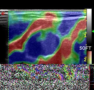
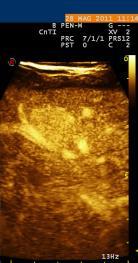
8 ELASTOSONOGRAPHY is a diagnostic method of recent introduction, which uses ultrasound and intends to obtain accurate information on the"elasticity and "stiffness" of the anatomical and pathological structures identified with US imaging.
9 GUIDELINES Elastosonography is an ultrasound technology currently used in the characterization of the focal and nodular areas of breast; nodules on scars and big lesions (more than 2 cm and anyway wider than the sampled area) must be preferentially excluded. The interpretation of the elastosonograms requires to the operator a global experience in the diagnostic imaging in senology. Unlike Color-Doppler, the Elastosonographic module does not require particular adjustments for use.

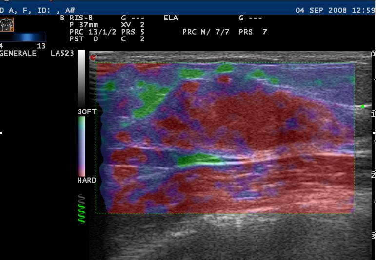
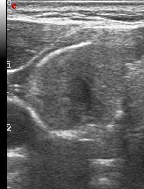
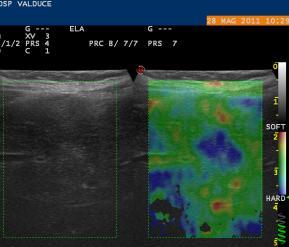
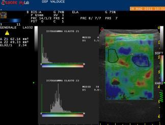
10 METHODOLOGY The nodular lesion must be centrally localized in relation to the sampled area. The dimensions of the Elastosonographic box must preferably take up almost the whole field of vision of the transducer for a correct analysis of the elasticity of the nodule and of the surrounding parenchyma. It is better to use a view in Dual Mode to obtain at the same time on the screen of the scanner both the elastosonographic image and the related C-Mode one; this trick is very useful to check in real time the position of the lesion in relation to the scanning plane.

11 The right elastosonographic scanning is based on a rhythmical compression and a release movement perpendicular to the skin surface, performed with constancy -Today it is easier (more sensitive US systems!!!!) rhythmic compression and release movement perpendicular to skin surface

12 Non Correct movement (oblique)

13 MALIGNANT BENIGN The Italian Team of Study, on the basis of its clinical experience of above 1000 cases studied, proposes a revision of the classification of Prof. Ueno. The aim is to give a graduation of scores more in accordance with the distribution of the pathologies in the normal routine of work, including therefore also scores which can favour, above all in the small lesions, the not always easy differentiation between liquid and solid.

14 Breast Carcinoma

15 RESULTS Elastosonography proved to be specific in the benign lesions, including the BI-RADS 3 ones of more critical interpretation (329 lesions, equal to 37,6%). Setting the cut-off point between the elastosonographic scores 3 and 4, the negative predictive value was 98%. BI-RADS 3 VPN = 98% BI-RADS 3 per US (American College of Radiology), a solid mass with circumscribed magins, oval shape and horizontal orientation, most likely a fibroadenoma, should have a less than 2 percent risk of malignancy. Altough additional multicentric data may confirm safety of follow-up rather than biopsy based on us findings,short-interval follow-up is currently increasing as a manegement strategy. Nonpalpable complicated cysts and clustered microcysts might also be placed in this category for short-interval follow-up.


16 What can be studied with Elastosonography? Among the other fields of employment in the study of the superficial organs, we must quote, always with the target of characterization "benign versus malignant", lymph nodes, thyroid and the muscle-articular structures. In this application, however, the results of Elastosonography are not proven as in the case of the breast


17 Thyroid benign nodules with shell "calcified.... is HARD At Thyroid Scintigraphy the nodule.. is COLD (Tumor or Cyst?)
18 What can be studied with Elastosonography? A particular case, also from a historic point of view, is represented by the study of the hepatic parenchyma. This peculiarity derives from the anatomical position of liver (which is not a superficial organ), from the specific object of study (the hepatic fibrosis, associated with cirrhosis, not the study of the nature of focal lesions) and from the kind of methodological approach, which, in the case of liver, were various. At present, thanks to probes with higher capability of penetrationand particular anatomical approaches, it is possible to obtain, with the systems commercially available, elastometric information at least for thesuperficial portions of the hepatic parenchyma.


19 FNH Object of study: the nature of focal lesions
20 Conclusions Elastosonography is, among the different methods that use Ultrasound to get diagnostic information, the technique with the most recent clinical introduction. Therefore we must not be surprised by the related lack of scientific contributions available in literature which evaluate the diagnostic performances The diagnostic potentiality of Elastosonography is linked to the operator's individual capability (it is performed by an expert Ultrasound user) and if combined with the traditional ultrasound analysis, it can give diagnostic data of considerable importance. All this in a non-invasive way and without increasing discomfort for the Patient.

21 Conclusions B-mode Color Doppler e Power Doppler FNH? Adenoma? Ultrasound Characterization Elastosonography CEUS

22 Conclusions Elastosonography Other Modality FUSION IMAGING
THYROID NODULES: THE ROLE OF ULTRASOUND
 THYROID NODULES: THE ROLE OF ULTRASOUND NOVEMBER 2017 DR. DEAN DURANT DEFINITION Thyroid nodule: Focal area within the thyroid gland with echogenicity different from surrounding parenchyma. THYROID NODULES
An improved quantification tool for breast ElastoScan : E-Breast
 An improved quantification tool for breast ElastoScan : E-Breast Volker F. Duda, MD, Christine Köhler, MD Interdisciplinary working group Senological diagnostics, University Hospital Gießen and Marburg
Imaging in breast cancer. Mammography and Ultrasound Donya Farrokh.MD Radiologist Mashhad University of Medical Since
 Imaging in breast cancer Mammography and Ultrasound Donya Farrokh.MD Radiologist Mashhad University of Medical Since A mammogram report is a key component of the breast cancer diagnostic process. A mammogram
Full ultrasound breast volumes. Faster scans. Streamlined workflow. ACUSON S2000 Automated Breast Volume Scanner. Answers for life.
 Full ultrasound breast volumes. Faster scans. Streamlined workflow. ACUSON S2000 Automated Breast Volume Scanner Answers for life. 1 ACQUIRE An automated whole breast solution. Reduced acquisition time.
LIVER IMAGING TIPS IN VARIOUS MODALITIES. M.Vlychou, MD, PhD Assoc. Professor of Radiology University of Thessaly
 LIVER IMAGING TIPS IN VARIOUS MODALITIES M.Vlychou, MD, PhD Assoc. Professor of Radiology University of Thessaly Hepatocellular carcinoma is a common malignancy for which prevention, screening, diagnosis,
Pushing the boundaries
 Samsung Medison is a global leading medical devices company. Founded in 1985, the company now sells cutting-edge medical devices including diagnostic ultrasound, digital X-ray and blood analyzer, around
Daily inspiration. Ultrasound system HS70A SAMSUNG MEDISON CO., LTD. Scan code or visit to learn more
 CT-HS70A-V1.01-GI-FTW-160415-EN Ultrasound system HS70A Scan code or visit www.samsungmedison.com/ to learn more SAMSUNG MEDISON CO., LTD. 2015-2016 Samsung Medison All Rights Reserved. Samsung Medison
Diagnostic TRUS Elastography of the Prostate
 Diagnostic TRUS Elastography of the Prostate George Zacharopoulos Department of Diagnostic Ultrasound Hygeia Hospital Athens, Greece Prostate HI-RTE Why we need Elastography Better Detection of possible
Step up to the iu22 The key reasons are now even more compelling
 Step up to the iu22 The key reasons are now even more compelling The reasons to step up to the iu22 are now more compelling than ever Reason #1 Reduce failed ultrasound exams on your technically difficult
Ultrasonic Tissue Strain Imaging
 Transcript Details This is a transcript of an educational program accessible on the ReachMD network. Details about the program and additional media formats for the program are accessible by visiting: https://reachmd.com/programs/advances-in-medical-imaging/ultrasonic-tissue-strain-imaging/3647/
Leonard M. Glassman MD
 BI-RADS The New BI-RADS Leonard M. Glassman MD FACR Former Chief of Breast Imaging American Institute for Radiologic Pathology Washington Radiology Associates, PC Breast Imaging Reporting and Data System
Does elastography change the indication to biopsy? IBDC
 Does elastography change the indication to biopsy? A LEXANDRA A THANASIOU, M D DEPARTMENT OF RADIOLOGY CURIE INSTITUTE PARIS, FRANCE IBDC Ultrasound Detected Cancers Physician-performed ultrasound increases
Liver Ultrasound - Beyond the Basics. Pamela Parker Lead Sonographer
 Liver Ultrasound - Beyond the Basics Pamela Parker Lead Sonographer Aims Review what we know about the liver Reasons for imaging Focal lesions Diffuse disease Can we do more? The Liver The Liver The Liver
Insight, Intelligence, and more
 DC-80 Diagnostic Ultrasound System Insight, Intelligence, and more P/N:ENG-DC-80-210285X12P-20170725 Overview The best patient care is your ultimate goal. To achieve this requires confident diagnosis even
Simplifying liver assessment in internal medicine
 Ultrasound Customer story Simplifying liver assessment in internal medicine Philips Affiniti ultrasound for elastography and contrast-enhanced ultrasound (CEUS) Where Sonography Institute, Uster, Switzerland
Byung Ihn Choi, M.D. Department of Radiology Seoul National University Hospital
 Byung Ihn Choi, M.D. Department of Radiology Seoul National University Hospital CEUS & US Elastography : Contents CEUS Introduction Contrast agents & imaging Clinical application US Video WS Summary US
S. Murgo, MD. Chr St-Joseph, Mons Erasme Hospital, Brussels
 S. Murgo, MD Chr St-Joseph, Mons Erasme Hospital, Brussels? Introduction Mammography reports are sometimes ambiguous and indecisive. ACR has developped the BIRADS. BIRADS consists of a lexicon in order
ORIGINAL ARTICLE EVALUATION OF BREAST LESIONS USING X-RAY MAMMOGRAM WITH HISTOPATHOLOGICAL CORRELATION
 Available online at www.journalijmrr.com INTERNATIONAL JOURNAL OF MODERN RESEARCH AND REVIEWS IJMRR ISSN: 2347-8314 Int. J. Modn. Res. Revs. Volume 3, Issue 10, pp 807-814, October, 2015 ORIGINAL ARTICLE
ACRIN 6666 IM Additional Evaluation: Additional Views/Targeted US
 Additional Evaluation: Additional Views/Targeted US For revised or corrected form check box and fax to 215-717-0936. Instructions: The form is completed based on recommendations (from ID form) for additional
ElastoScan in breast diagnostics : 10 Most Frequently Discussed Objectives
 ElastoScan in breast diagnostics : 10 Most Frequently Discussed Objectives V. Duda, MD, C. Köhler, MD, A. Stamm, MD, A. Storch, MD Interdisciplinary working group Senological diagnostics, University Hospital
MEASUREMENT OF EFFECT SOLID TUMOR EXAMPLES
 MEASUREMENT OF EFFECT SOLID TUMOR EXAMPLES Although response is not the primary endpoint of this trial, subjects with measurable disease will be assessed by standard criteria. For the purposes of this
Contrast-enhanced Breast MRI RSSA 2013
 Contrast-enhanced Breast MRI RSSA 2013 Prof. dr. Maurice van den Bosch University Medical Center Utrecht, the Netherlands Index 1) Breast cancer 2) Why MRI of the breast 3) Technique 4) Interpretation
Optimizing Breast Sonography
 Optimizing Breast Sonography Cindy Rapp BS, RDMS, FSDMS, FAIUM Denver, Colorado Breast Sonography general goal to make a more specific diagnosis than can be made with clinical and mammographic findings
Thyroid Nodules: What to do next?
 Thyroid Nodules: What to do next? Ally P. H. Prebtani Professor of Medicine Internal Medicine, Endocrinology & Metabolism McMaster University Canada Copyright 2017 by Sea Courses Inc. All rights reserved.
When You Need To Know More.
 www.siemens.com/ultrasound When You Need To Know More. ACUSON S2000 Ultrasound System Table of Contents Powerful Imaging 01 Penetrating Insight 02 03 Revealing Perspectives 04 05 Smart Workflow 06 Ergonomics
Breast Ultrasound: Improving Your Skills & Patient Care
 Breast Ultrasound: Improving Your Skills & Patient Care Objectives Discuss US techniques available for image optimization. Review & compare the US appearances of benign & malignant masses. Cherie M. Kuzmiak,
Breast Imaging Lexicon
 9//201 200 BI RADS th Edition 201 BI RADS th Edition Breast Imaging Lexicon Mammographic Pathology and Assessment Categories Deborah Thames, R.T.(R)(M)(QM) The Advanced Health Education Center Nonmember:
The Thyroid Imaging Reporting and Data System (TIRADS) for ultrasound of the thyroid : a pratical approach
 for ultrasound of the thyroid : a pratical approach") The Thyroid Imaging Reporting and Data System (TIRADS) for ultrasound of the thyroid : a pratical approach Poster No.: C-2425 Congress: ECR 2015 Type: Educational Exhibit Authors: M. Ben Lassoued, B. Souissi,
The Thyroid Imaging Reporting and Data System (TIRADS) for ultrasound of the thyroid : a pratical approach Poster No.: C-2425 Congress: ECR 2015 Type: Educational Exhibit Authors: M. Ben Lassoued, B. Souissi,
OPTO-ACOUSTIC BREAST IMAGING
 OPTO-ACOUSTIC BREAST IMAGING A Novel Fusion of Functional and Morphologic Imaging Reni S. Butler, MD A. Thomas Stavros, MD F. Lee Tucker, MD Michael J. Ulissey, MD PURPOSE 1. Explain opto-acoustic (OA)
Diagnostic Dilemmas of Breast Imaging
 Diagnostic Dilemmas of Breast Imaging Common Causes of Error in Breast Cancer Detection By: Jason Cord, M.D. Mammography: Initial Imaging The standard for detection of breast cancer Screening mammography
Imaging in Pediatric Thyroid disorders: US and Radionuclide imaging. Deepa R Biyyam, MD Attending Pediatric Radiologist
 Imaging in Pediatric Thyroid disorders: US and Radionuclide imaging Deepa R Biyyam, MD Attending Pediatric Radiologist Imaging in Pediatric Thyroid disorders: Imaging modalities Outline ACR-SNM-SPR guidelines
Accurate Guidance. Confident Outcomes.
 Accurate Guidance. Confident Outcomes. CIVCO s VirtuTRAX is a breakthrough technology that enables virtual needle tracking to expand the capabilities of ultrasound interventions. VirtuTRAX allows users
BI-RADS Update. Martha B. Mainiero, MD, FACR, FSBI Brown University Rhode Island Hospital
 BI-RADS Update Martha B. Mainiero, MD, FACR, FSBI Brown University Rhode Island Hospital No Disclosures BI-RADS History 1980s Quality Issues ACR Accreditation BI-RADS 1994 2003 4 th Edition MRI, US January
Introducing next-generation shear wave elastography for breast
 Ultrasound ElastQ Imaging Introducing next-generation shear wave elastography for breast Philips ElastQ Imaging Vijay Shamdasani, PhD, MBA Tissue stiffness has long provided important information about
3D Total Breast Ultrasound
 www.siemens.com/abvs 3D Total Breast Ultrasound ACUSON S2000 Automated Breast Volume Scanner (ABVS) and syngo.ultrasound Breast Analysis (susba) Answers for life. All Women Are Different. So why should
New Imaging Modalities for better Screening and Diagnosis
 New Imaging Modalities for better Screening and Diagnosis Miri Sklair-Levy, MD Department of Diagnostic Imaging Sheba Medical Center, Sackler School of Medicine, Tel Aviv University Department of Diagnostic
1. Clinical breast exam. 2. Imaging. 3. Biopsy ELASTOGRAPHY. Triple assessment. Overdiagnosed!?
 Budapest, 2017. Introduction The main purpose of all diagnostic methods is early breast cancer detection However, considering higher incidence of benign lesions comparing to malignant, there is a great
Armed Forces Institute of Pathology.
 Armed Forces Institute of Pathology www.radpath.com Armed Forces Institute of Pathology Breast Disease www.radpath.org Armed Forces Institute of Pathology Interpretation of Breast MRI Leonard M. Glassman
Melissa Hartman, DO Women s Health Orlando VA Medical Center
 Melissa Hartman, DO Women s Health Orlando VA Medical Center Most common non-skin cancer and Second deadliest cancer in women Majority are diagnosed by abnormal screening study An approach to breast cancer
AB MR Interpretation Overview
 AB MR Interpretation Overview Goal of AB MR interpretation is to maintain high sensitivity and specificity In order to minimize false positives and short term follow ups, it is fundamental to focus only
Prof. Dr. NAGUI M. ABDELWAHAB,M.D.; MARYSE Y. AWADALLAH, M.D. AYA M. BASSAM, Ms.C.
 Role of Whole-body Diffusion MR in Detection of Metastatic lesions Prof. Dr. NAGUI M. ABDELWAHAB,M.D.; MARYSE Y. AWADALLAH, M.D. AYA M. BASSAM, Ms.C. Cancer is a potentially life-threatening disease,
Contents. Basic Ultrasound Principles and Terminology. Ultrasound Nodule Characteristics
 Contents Basic Ultrasound Principles and Terminology Basic Ultrasound Principles... 1 Ultrasound System... 2 Linear Transducer for Superficial Images and Ultrasound-Guided FNA... 3 Scanning Planes... 4
ShearWave elastography in lymph nodes
 ShearWave elastography in lymph nodes Poster No.: B-0158 Congress: ECR 2015 Type: Authors: Keywords: DOI: Scientific Paper F. Houari, O. Lucidarme, J. Gabarre, F. Charlotte, C. Pellot- Barakat, M. Lefort,
LOGIQ S8 XDclear 2.0 Liver Procedures
 LOGIQ S8 XDclear 2.0 Liver Procedures See & quantify liver disease in a single exam Clinical Challenge Liver disease affects millions of people worldwide, and the number is growing. Clinicians need a cost-effective,
Case Scenario 1 History and Physical 3/15/13 Imaging Pathology
 Case Scenario 1 History and Physical 3/15/13 The patient is an 84 year old white female who presented with an abnormal mammogram. The patient has a five year history of refractory anemia with ringed sideroblasts
Thyroid Nodules: US Risk Stratification. Alex Tessnow, MD, FACE, ECNU University of Texas Southwestern Associate Professor of Medicine Dallas, Texas
 Thyroid Nodules: US Risk Stratification Alex Tessnow, MD, FACE, ECNU University of Texas Southwestern Associate Professor of Medicine Dallas, Texas Which of the following is true? A. All echogenic foci
Liver Tumors. Prof. Dr. Ahmed El - Samongy
 Liver Tumors Prof. Dr. Ahmed El - Samongy Objective 1. Identify the most important features of common benign liver tumors 2. Know the risk factors, diagnosis, and management of hepatocellular carcinoma
Emerging Techniques in Breast Imaging: Contrast-Enhanced Mammography and Fast MRI
 Emerging Techniques in Breast Imaging: Contrast-Enhanced Mammography and Fast MRI Lilian Wang, M.D. Breast Imaging Section Department of Radiology Northwestern Medicine Overview Rationale for new imaging
ISSN X (Print) Research Article. *Corresponding author Dr. Amlendu Nagar
 Research Article. *Corresponding author Dr. Amlendu Nagar") Scholars Journal of Applied Medical Sciences (SJAMS) Sch. J. App. Med. Sci., 2015; 3(3A):1069-1073 Scholars Academic and Scientific Publisher (An International Publisher for Academic and Scientific Resources)
Scholars Journal of Applied Medical Sciences (SJAMS) Sch. J. App. Med. Sci., 2015; 3(3A):1069-1073 Scholars Academic and Scientific Publisher (An International Publisher for Academic and Scientific Resources)
Amammography report is a key component of the breast
 Review Article Writing a Mammography Report Amammography report is a key component of the breast cancer diagnostic process. Although mammographic findings were not clearly differentiated between benign
Virtually-tracked US-guided Radiofrequency Ablation(RFA) of Benign Thyroid Nodules: Preliminary Results
 of Benign Thyroid Nodules: Preliminary Results") Virtually-tracked US-guided Radiofrequency Ablation(RFA) of Benign Thyroid Nodules: Preliminary Results e-poster: 408 Congress: ESHNR 2013 2013 Type: Scientific Poster Presentation Topic: Head and Neck
Virtually-tracked US-guided Radiofrequency Ablation(RFA) of Benign Thyroid Nodules: Preliminary Results e-poster: 408 Congress: ESHNR 2013 2013 Type: Scientific Poster Presentation Topic: Head and Neck
Thyroid in a Nutshell Dublin Catherine Kirkpatrick Consultant Sonographer ULHT
 Thyroid in a Nutshell Dublin 2017 Catherine Kirkpatrick Consultant Sonographer ULHT Acknowledgements Dr. Steve Colley Dr. Rhodri Evans Dr. Rhian Rhys Dr. Andrew McQueen Aims Anatomy & Physiology Incidence
iu22 Liver Shear Wave ElastPQ
 iu22 Liver Shear Wave ElastPQ Clinical Case Study Lucy Wang Clinical Application Specialist ASEAN Case Study: History: 58-year-old male patient, hepatitis B virus (HBV) carrier, with non clinical symptoms
China Medical Technologies, Inc.
 China Medical Technologies, Inc. China Medical Technologies, Inc. (CMT) is a high-tech enterprise, trading on Nasdaq with the ticker CMED. We currently conduct our operations principally through our wholly-owned
Ultrasound of the Breast BASICS FOR THE ORDERING CLINICIAN
 Ultrasound of the Breast BASICS FOR THE ORDERING CLINICIAN Breast Ultrasound Anatomy Skin Breast Parenchyma Pectoralis Fascia Pectoralis Breast Ultrasound Anatomy Indications for Breast Ultrasound Palpable
Detailed Program of the second BREAST IMAGING AND INTERVENTIONS PROGRAM am am : Clinician s requirements from breast imaging
 Detailed Program of the second BREAST IMAGING AND INTERVENTIONS PROGRAM 2012 Day one, 2 nd November BREAST IMAGING AND INTERVENTIONS PROGRAM 2012 9.00 AM 9.10 am Introduction 9.10 am - 9.30 am : Clinician
Radiographic Assessment of Response An Overview of RECIST v1.1
 Radiographic Assessment of Response An Overview of RECIST v1.1 Stephen Liu, MD Georgetown University May 15 th, 2015 Presentation Objectives To understand the purpose of RECIST guidelines To describe the
The latest developments - Automated Breast Volume Scanning. Dr. med. M. Golatta
 The latest developments - Automated Breast Volume Scanning Dr. med. M. Golatta Automated Breast Volume US: Why? o Mammography is limited in dense breasts: high false negative rate o Many of these tumors
Tissue Strain Analytics Virtual Touch Tissue Imaging and Quantification
 Whitepaper Tissue Strain Analytics Virtual Touch Tissue Imaging and Quantification ACUSON S2000 Ultrasound System Answers for life. Page 1 Tissue Strain Analytics: Virtual Touch Tissue Imaging and Quantification
Clinical Study B-Mode and Elastosonographic Evaluation to Determine the Reference Elastosonography Values for Cervical Lymph Nodes
 ISRN Radiology Volume 2013, Article ID 895287, 4 pages http://dx.doi.org/10.5402/2013/895287 Clinical Study B-Mode and Elastosonographic Evaluation to Determine the Reference Elastosonography Values for
Medical Review Approaches to the Diagnosis of Liver Fibrosis
 Medical Review Approaches to the Diagnosis of Liver Fibrosis Hiroko Iijima Department of Hepatobiliary and Pancreatic Disease Ultrasound Imaging Center, Hyogo College of Medicine Hiroko Iijima Department
5-6 June University of Pisa, Italy
 SCIENTIFIC PROGRAMME International School of Thyroid Ultrasonography 5-6 June 2015 - University of Pisa, Italy General information Venue This live educational course takes place at the: University of
Innovations in Ultrasound & Breast Cancer Imaging
 Innovations in Ultrasound & Breast Cancer Imaging Azra Alizad, MD Department of Radiology Mayo Clinic College of Medicine 2018 AAPM Annual meeting 2012 MFMER slide-1 Disclosure Mayo Clinic and some investigators
LOGIQ S8 XDclear 2.0. Simply Amazing
 LOGIQ S8 XDclear 2.0 Simply Amazing Available with FibroScan technology to aid in the diagnosis, staging, and monitoring of chronic liver disease Enhanced B-Flow Imaging a GE exclusive with exquisite sensitivity
Elastography. White Paper
 Elastography White Paper Strain Image in DC-8 Diagnostic Ultrasound System Shuangshuang Li, Rui Fan Relationship between the stiffness of tumor and it s malignance has far been known since the ancient
Ultrasonography of the Neck as an Adjunct to FNA. Nicole Massoll M.D.
 Ultrasonography of the Neck as an Adjunct to FNA Nicole Massoll M.D. Basic Features of Head and Neck Ultrasound and Anatomy Nicole Massoll M.D. University of Arkansas for Medical Sciences, Little Rock
Thyroid and Parathyroid Ultrasound Protocol
 Thyroid and Parathyroid Ultrasound Protocol Reviewed By: Anna Ellermeier, MD Last Reviewed: December 2017 Contact: (866) 761-4200, Option 1 **NOTE for all examinations: 1. If documenting possible flow
Evaluation of Liver Mass Lesions. American College of Gastroenterology 2013 Regional Postgraduate Course
 Evaluation of Liver Mass Lesions American College of Gastroenterology 2013 Regional Postgraduate Course Lewis R. Roberts, MB ChB, PhD Division of Gastroenterology and Hepatology Mayo Clinic College of
Title: Opto-Acoustic Breast Imaging Imaging-Pathology Correlation of Opto-Acoustic
 Title: Opto-Acoustic Breast Imaging Imaging-Pathology Correlation of Opto-Acoustic Features Respecting Malignancy Authors: R S Butler, MD; A T Stavros, MD; P T Lavin, PhD; M J Ulissey, MD; F L Tucker,
shear wave elastography
 Ultrasound ElastQ Imaging Introducing next-generation shear wave elastography Philips ElastQ Imaging Vijay Shamdasani, PhD and Hua Xie, PhD, Philips Ultrasound Tissue stiffness has long provided important
SONIMAGE HS1. Premium Portable Ultrasound
 SONIMAGE HS1 Premium Portable Ultrasound EXIT SET F2 F1 User User Gain FREEZE PREMIUM PORTABLE ULTRASOUND DESIGNED FOR YOU Sonimage HS1 2 Sonimage HS1 is Konica Minolta s Premium Portable Ultrasound designed
Strain histogram analysis for elastography in breast cancer diagnosis
 Strain histogram analysis for elastography in breast cancer diagnosis Poster No.: C-1854 Congress: ECR 2015 Type: Scientific Exhibit Authors: J. F. Carlsen, C. Ewertsen, S. Sletting, M. B. Nielsen; Copenhagen/DK
PROPOSTA DI UN NUOVO ALGORIMO PER LA DIAGNOSI ECOGRAFICA DELLE MALATTIE CRONICHE DEL FEGATO
 PROPOSTA DI UN NUOVO ALGORIMO PER LA DIAGNOSI ECOGRAFICA DELLE MALATTIE CRONICHE DEL FEGATO A. Giorgio Direttore del servizio di Ecografia Interventistica Istituto Clinico S.Rita -IRCCS -Atripalda (Avellino)
University of Pisa, Italy
 SCIENTIFIC PROGRAMME International School of Thyroid Ultrasonography University of Pisa, Italy Live educational course dates The International School of Thyroid Ultrasonography Advanced practical thyroid
BI-RADS and Breast MRI. Kathy Borovicka, M.D. Thursday February 15, 2018
 BI-RADS and Breast MRI Kathy Borovicka, M.D. Thursday February 15, 2018 Learning Objectives Be familiar with the Breast Imaging Reporting and Data System (BI-RADS) Understand the components of a breast
We evaluated the medical records of 1,015 patients who underwent surgery for primary breast cancer between February 2007 and August 2008 at St.
 Elastographic evaluation of mucinous carcinoma of the breast Miki Mori Hiroko Tsunoda Nobue Kawauchi Mari Kikuchi Satoshi Honda Koyu Suzuki Hideko Yamauchi Abstract Background Elastography is widely used
The role of US elastography in the evaluation of benign and malignant breast lesions in relation to histopathological examination
 The role of US elastography in the evaluation of benign and malignant breast lesions in relation to histopathological examination Poster No.: C-1802 Congress: ECR 2013 Type: Scientific Exhibit Authors:
LOWER EXTREMITY VENOUS COMPRESSION ULTRASOUND. CPT Stacey Good, DO Emergency Medicine Ultrasound Fellow Madigan Army Medical Center
 LOWER EXTREMITY VENOUS COMPRESSION ULTRASOUND CPT Stacey Good, DO Emergency Medicine Ultrasound Fellow Madigan Army Medical Center Learning Objectives Setup and patient positioning for optimizing success
Radiology of hepatobiliary diseases
 GI cycle - Lecture 14 436 Teams Radiology of hepatobiliary diseases Objectives 1. To Interpret plan x-ray radiograph of abdomen with common pathologies. 2. To know the common pathologies presentation.
Imaging Guided Biopsy. Edited & Presented by ; Hussien A.B ALI DINAR. Msc Lecturer,Reporting Sonographer
 Imaging Guided Biopsy Edited & Presented by ; Hussien A.B ALI DINAR. Msc Lecturer,Reporting Sonographer Objective By the End of this lessons you should : Define what biopsy Justify Aim to perform biopsy
Endocrinology and Metabolic Disorder Unit Regina Apostolorum Hospital
 Enrico Papini Endocrinology and Metabolic Disorder Unit Regina Apostolorum Hospital Albano Laziale, Italy The Following Faculty have provide no information regarding significant relationship with commercial
Current Imaging Diagnosis of the Breast Tumors
 Breast Cancer Current Imaging Diagnosis of the Breast Tumors JMAJ 45(6): 258 264, 2002 Tokiko ENDO Director of the Department of Radiology, National Nagoya Hospital Abstract: Breast masses include the
OPTO-ACOUSTIC BREAST IMAGING
 OPTO-ACOUSTIC BREAST IMAGING Imaging-Pathology Correlation of Opto-Acoustic Features in Benign and Malignant Breast Masses Reni Butler, M.D. F. Lee Tucker, M.D. Philip Lavin, Ph.D. Erin Neuschler, M.D.
Pitfalls and Limitations of Breast MRI. Susan Orel Roth, MD Professor of Radiology University of Pennsylvania
 Pitfalls and Limitations of Breast MRI Susan Orel Roth, MD Professor of Radiology University of Pennsylvania Objectives Review the etiologies of false negative breast MRI examinations Discuss the limitations
Radiology Update 2017
 Radiology Update 2017 John K. Phillips, MD Affiliated Assistant Professor of Radiology University of Tennessee Health Sciences Center Chief, Radiology and Nuclear Medicine VA Memphis Disclosures Financial:
Sonographic Features of Thyroid Nodules & Guidelines for Management
 Sonographic Features of Thyroid Nodules & Guidelines for Management Mark A. Lupo, MD, FACE, ECNU Thyroid & Endocrine Center of Florida Assistant Clinical Professor of Medicine Florida State University,
Ultrasound Physics & Doppler
 Ultrasound Physics & Doppler Endocrine University 2018 Mark Lupo, MD, FACE, ECNU Objectives Review the essential components of ultrasound physics in neck sonography Demonstrate the importance of ultrasound
Does Ultrasound Elastography Improve the Diagnostic Accuracy of Fine Needle Aspiration Cytology in Predicting Malignancy in Thyroid Nodules?
 Med. J. Cairo Univ., Vol. 82, No. 1, June: 427-437, 2014 www.medicaljournalofcairouniversity.net Does Ultrasound Elastography Improve the Diagnostic Accuracy of Fine Needle Aspiration Cytology in Predicting
Evaluation and Management of Thyroid Nodules. Nick Vernetti, MD, FACE Palm Medical Group Las Vegas, Nevada
 Evaluation and Management of Thyroid Nodules Nick Vernetti, MD, FACE Palm Medical Group Las Vegas, Nevada Disclosure Consulting Amgen Speaking Amgen Objectives Understand the significance of incidental
Breast Elastography: Our Initial Experience with Strain and Shear Wave Elastography
 Human Journals Short Communication September 2018 Vol.:10, Issue: 3 All rights are reserved by Pankaj Sharma et al. Breast Elastography: Our Initial Experience with Strain and Shear Wave Elastography Keywords:
EUS Elastography: Advances in Diagnostic EUS of the Pancreas
 Review Article http://dx.doi.org/10.3348/kjr.2012.13.s1.s12 pissn 1229-6929 eissn 2005-8330 Korean J Radiol 2012;13(S1):S12-S16 EUS Elastography: Advances in Diagnostic EUS of the Pancreas Tae Hee Lee,
AMSER Case of the Month: September 2018
 AMSER Case of the Month: September 2018 60-year-old woman with a left breast mass noted on screening mammography. Catherine McNulty, MS4 Tulane University School of Medicine Dr. Robin Sobolewski Breast
LOGIQ E9 Shear Wave Elastography
 LOGIQ E9 Shear Wave Elastography Introduction Tissue stiffness is often related to underlying disease. For millennia, physicians have used palpation as a diagnostic tool to detect various ailments such
Intuitive. Intelligent. Innovative. General Imaging
 Intuitive. Intelligent. Innovative. General Imaging CLARITY CONFIDENCE EASE OF USE 2 The perfect fit Aplio i700 helps you provide better quality of care in the shortest possible time. Combining superior
declipse SPECT Imaging Probe Worldwide first registration-free ultrasound fusion with high-resolution 3D SPECT images
 declipse SPECT Imaging Probe Worldwide first registration-free ultrasound fusion with high-resolution 3D SPECT images high-resolution 3D SPECT live ultrasound realtime fusion of SPECT / ultrasound Hybrid
GE Healthcare. LOGIQ Book XP. Innovation in compact ultrasound
 GE Healthcare LOGIQ Book XP Innovation in compact ultrasound At the leading edge of healthcare High performance compact ultrasound... anywhere Clarity Confidence Simplicity Speed For more than a century,
Pushing the boundaries
 Samsung Medison is a global leading medical devices company. Founded in 1985, the company now sells cutting-edge medical devices including diagnostic ultrasound, digital X-ray and blood analyzer, around
Advancing the Art of Breast Ultrasound Imaging
 Advancing the Art of Breast Ultrasound Imaging For the First Time Ever... An Innovation that Gives You Truly Quantifiable Tissue Elasticity Explore the Latest Technology in Ultrasound Real-Time ShearWave