Thyroid Ultrasound Physics and Doppler
|
|
|
- Garey Harrington
- 6 years ago
- Views:
Transcription


1 Thyroid Ultrasound Physics and Doppler Advanced AACE-ACE US training course 2017 Dev Abraham MD, MRCP(UK), ECNU, FACE Professor of Medicine, University of Utah No Disclosures

2 Natural Ability to see with sound
3 Sound Waves Physics of Sound Role of the propagating medium Acoustic Impedance Density, Stiffness, and Speed of Sound Reflection Occurs at Boundaries of Impedance Refraction Attenuation
4 Principles of Sound Sound is the Propagation of mechanical energy through matter. No sound in a vacuum Qualities of the transmitting medium influence sound transmission Density Stiffness Acoustic Impedance Speed of Sound (SOS)
5 Sound Waves Sound waves propagate by compression and rarefaction of molecules in space. Molecules of the medium vibrate around their resting position and transfer their energy to neighboring molecules. Waves carry energy, not matter. Frequency and period Hertz (Hz) = cycles/second Propagation speed Wavelength
6 Pulsed Waves Pulse Duration, Repetition Period and Frequency (Hz) Spatial Pulse Length (m) Determinant of axial resolution (1/2 SPL) Duty Factor (%) Clinical Correlate: Pulse duration (frequency) determines axial resolution of an ultrasound image.
7 Propagation Velocity of Common Tissues
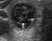
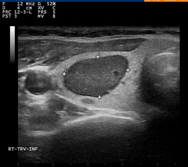
8 Q 1: One of the characteristic features of a cyst is the following: a. Echogenic texture with sharp margins b. Speckling with intra lesional sub 2 mm calcifications c. Anechoic with posterior enhancement d. Acoustic shadowing posterior to the lesion
9 Properties of Transmitting Material Acoustic Impedance Reflection of sound occurs at interfaces of Acoustic Impedance Homogeneous medium does not reflect sound Cyst Anechoic Most tissues have heterogeneous impedance Acoustic Impedance depends on the speed of sound, density and stiffness of a material
10 Reflection Reflection is the redirection of a portion of a sound beam back at the interface of tissues of unequal acoustic impedance. The greater the difference in impedance, the greater the reflection.


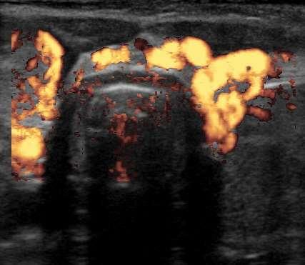
11 Benign Findings The Halo Sign Hassani, 1977 Propper, 1980
12 Suspicious Findings: Microcalcifications
13 Illustrating Physics by Understanding Artifacts Reflection Artifacts Shadowing and Enhancement Edge Artifact Reverberation Artifact Comet Tail Artifact

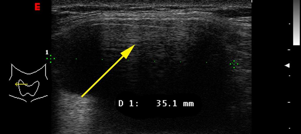
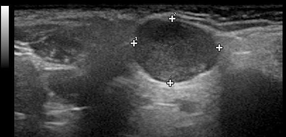
14 Shadowing and Enhancement

15 Enhancement is seen behind cysts - But can be seen behind homogeneous solid objects as well Benign hyperechoic nodule seen in Hashimoto s Thyroiditis
16 Reflection Artifacts Edge Artifact

17 Reverberation Artifact Meritt, 1998
18 Reverberation Artifact
19 Q 2: One of the following is a typical example of reverberation artifacts: a. Sub 2 mm calcifications that do not cast shadows b. Popcorn type calcification with acoustic signal drop out c. Comet tail and cats eye signs d. Mural mass within the wall of the nodule

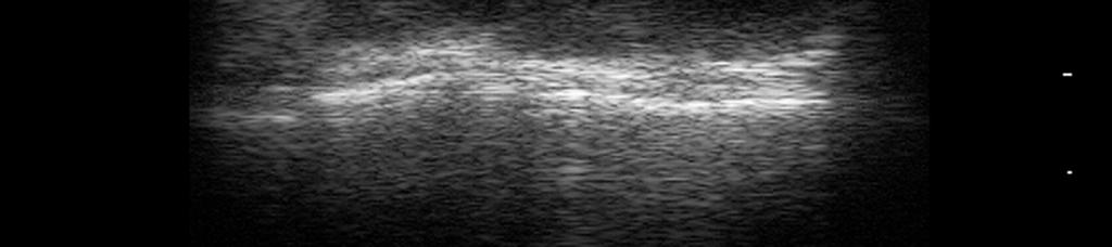
20 Comet Tail Artifact

21 Comet Tail Artifacts
22 Attenuation Attenuation of acoustic energy results from Reflection Scatter Absorption Attenuation is frequency dependent Higher frequencies have greater attenuation Less depth of imaging with higher frequencies Artifacts due to attenuation Shadowing Enhancement
23 Q 3: Which of the following is true regarding ultrasound frequency? a. Increasing frequency will increase depth of penetration. b. Increasing frequency will improve resolution. c. Increasing frequency will increase noise. d. High PRF (pulse rep frequency) is useful in the study of lymph nodes
24 Resolution AZIMUTHAL (up-down) AXIAL (distance from transducer) LATERAL (side to side)
25 Resolution Ability to discriminate two adjacent objects as separate entities. The narrower the beam, the better the lateral resolution. The higher the frequency the better the axial resolution. Poor focus Image Good focus
 determines Axial Resolution (axial resolution = 1/2 SPL) Practical Consideration - As frequency increases, axial resolution improves, but")
26 Focus and Resolution Focused beam width determines Lateral and Azimuthal Resolution Near field (Fresnel Zone) Large variations of intensity Far field (Fraunhofer Zone) Intensity more uniform Focal Zone - Area of maximal narrowing Pulse duration (frequency) determines Axial Resolution (axial resolution = 1/2 SPL) Practical Consideration - As frequency increases, axial resolution improves, but depth of imaging decreases. The depth of the focal zone is often adjustable and indicated on the display


27 Frequency and Resolution With higher frequency the resolution of the near field is improved, but the deep structures are better visualized with the lower frequency.
 that allows")
 for deep")
28 Image Optimization - Frequency Choose highest frequency (12-15 MHz) that allows adequate depth penetration. Lower frequencies (7-10 MHz) for deep structures or very obese subjects
29 Advances in Technology Signal Processing Image Enhancement Noise reduction Edge sharpening Utilization of CT and MRI reconstruction algorithms Beam Steering Spatial compounding
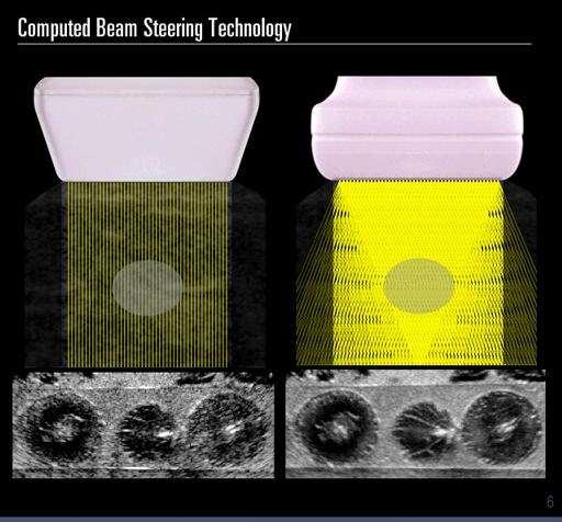
30 Signal Processing

31 Comparison of standard and processed images


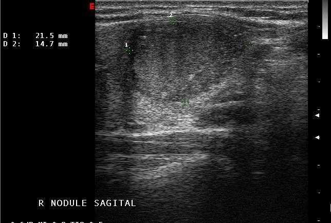
32 Effect of Compound Imaging on Artifacts Comet Tails

33 Effect of Compound imaging on Artifacts Edge Artifact and Enhancement
34 Image Optimization Create the sharpest image to allow tissue discrimination. Equipment factors: Quality of Transducer Quality of Electronics Image Enhancement and Compound Imaging User Adjustments: Depth, Gain, Frequency Focal zones Number and Location Compound Imaging Tissue Harmonic Imaging Dynamic range

35 Optimal Gain


36 Adjustment of number and position of focal zones

37 Image Optimization Dynamic range May increase conspicuity of subtle lesions

38 Doppler Shift
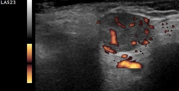
39 Color and Power Doppler Meritt, 1998

40 Color and Power Doppler Meritt, 1998
41 Color and Power Doppler Color Doppler Provides information regarding direction and velocity. More useful in vascular studies Power Doppler No information regarding velocity Less angle dependence Less noise Increased sensitivity for detection of flow

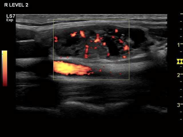
42 Color Doppler - Follicular Carcinoma Inferior right lobe - sagittal

43 Color Doppler Benign Nodule
44 Does Doppler play a role in the prediction of malignancy? (1) 494 consecutive patients with nonpalpable nodules measuring 8 to 15 millimeters. 31 malignant. 87% of cancers were solid and hypoechoic. 77% of cancers had irregular or blurred margins. Intra nodular vascular pattern was seen in 74% of cancers. Microcalcifications were seen in 29 percent of cancers. Independent risk factors for malignancy included irregular margins (RR = 16.83) intra nodular flow (RR = 14.29) microcalcifications (RR = 4.97) Recommendation for ultrasound guided fine needle aspiration if any of the independent risk factors are present, and if no risk factors are present follow-up with clinical and ultrasound evaluation. Papini E, Guglielmi R, Bianchini A, Crescenzi A, et al. Risk of malignancy in nonpalpable thyroid nodules: predictive value of ultrasound and color Doppler features. J Clin Endocrinol Metab. 2002;87(5):
45 Does Vascularity play a role in the prediction of malignancy? (2) Retrospective evaluation of 1083 nodules in 1024 patients. 814 benign. 269 malignant (97.4% PTC) Marked hypoechogenicity, noncircumscribed margins, microcalcifications and taller than wide considered suspicious by gray scale. Vascularity graded as none, peripheral or intranodular. Vascularity was frequently seen in benign nodules (31%) No vascularity was more frequent in malignant nodules (60% v 43%) (p<.001) Vascularity of nodules was NOT related to risk of malignancy. Moon HJ, et al. Can Vascularity at Power Doppler US Help Predict Thyroid Malignancy? Radiology, 2010; 255(1)
46 Why the discrepancy? What to do? Papillary versus Follicular Cancer. Power Doppler may be predictive for follicular cancer. In Moon s study 97.4% were papillary Vascularity does not seem to be predictive in papillary cancer. Consider whether features are suspicious for papillary or follicular cancer. Levine RA, Endocrine Practice 2006 Iared W, J Ultrasound Med 2010


47 Value of Doppler in the prediction of malignancy following follicular biopsy 310 follicular nodules Color Doppler flow mapping prior to surgery Grade 1: No color flow mapping inside the nodule Grade 2: Color flow mapping only in peripheral area; PI index < 1.3 Grade 3: Penetrating color flow mapping, vascularity moderate-rich Grade 4: High velocity penetrating color flow mapping, vascular rich; PI index >1.3 Sensitivity 86% Specificity 85% Fukunari N, Clinical evaluation of color Doppler imaging for the differential diagnosis of thyroid follicular lesions World J Surg, 2004; 28(12):
48 Value of Doppler in Prediction of Malignancy - Conclusions Doppler analysis does not appear useful regarding malignant potential for papillary cancer. Doppler may be of use in lesions with indeterminate (follicular) biopsies. Power Doppler has greater sensitivity and is able to detect lower degrees of internal flow. Doppler information must be interpreted along with other ultrasonographic characteristics including echogenicity, edge definition, calcifications, etc.
49 Amiodarone-induced Thyrotoxicosis Color Doppler (1) Type 1 Preexisting Thyroid abnormality Graves-like Treatment with thionamides and perchlorate Normal or increased vascularity Type 2 Preexisting Normal Thyroid Destructive thyroiditis-like Treatment with glucocorticoids Absent vascularity +/- elevated Interleukin 6 Preserved echotexture
50 Amiodarone-induced Thyrotoxicosis Color Doppler (2) Flow Absent (CFDS 0) 58% Prednisolone response rate Flow Present (CFDS 1-3) 14% Prednisolone response rate Treatment Algorithm CFDS 0: Prednisolone CFDS 1-3: Thionomides and perchlorate Combined therapy/surgery if unresponsive Wong, et. al., Intern Med J, (9-10)
51 Doppler Graves and Thyroiditis Graves Thyroid inferno Peak systolic velocity (PSV) 8-20 cm/sec Thyroiditis Micronodulation in Hashimoto s Various vascular patterns absent to hypervascular Subacute thyroiditis focally may appear as suspicious nodule Thyrotoxicosis Factitia Minimal intrathyroidal vascular flow Peak systolic velocity (PSV) 3-5 cm/sec


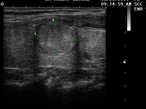
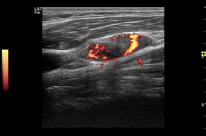
52 Graves Disease Thyroid Inferno

53 Thyroiditis Not always hypovascular
54 Graves Disease versus Destructive Thyoiditis. Multiple studies have attempted to separate Graves from thyroiditis using variety of Doppler techniques. Power Doppler Images Thyroid blood flow area Thyroid artery velocity
55 Thyroid Blood Flow Ota H et. al., Clin Endocrinol (Oxf) 67:
56 Thyroid Blood Flow Area Kurita S et al. Thyroid. 2005;15(11) 1249
57 Inferior Thyroid Artery Peak Velocity Kumar H et. al., Endocrine Practice 15(1) 6-9, 2009
58 Differential Diagnosis of Thyrotoxicosis Conclusions The distinction between Graves Disease and thyroiditis can be based on a combination of clinical features, RAI uptake, TsIg levels, and ultrasound features. RAI uptake remains the gold standard but the behavior over time is often the final determinant. Recognize the limitation of Doppler techniques and do not use in isolation.
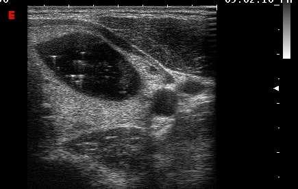
59 Clarification of Interpretation - Cyst?
60

61 Doppler Before Biopsy


62 Doppler Lymph nodes In normal nodes vessels enter centrally at the hilus, and spread along the long axis. In malignant nodes aberrant vessels enter peripherally in the node capsule. Increased (disordered) vascularity may be seen peripherally and centrally.
 CM Left")
63 0.4 (AP) by 1.0 (long axis) CM Left level 4

64 Jugular Vein
65

66 FNA TG 2067 ng/ml
67
68 Q4: One of the following US findings has high sensitivity and specificity in the prediction of metastatic lymph-nodes a) Intra lymph-nodal punctate calcification b) Cystic degeneration of lymph-nodes c) Peripheral / chaotic vascularization d) Hyperechoic texture within the lymph node e) Hypoechoic texture within the lymph node
69 US findings characteristic of metastasis Loss of hilum (100% sensitivity 29% specificity) Rounded shape short axis > 5mm (96 % specificity) Hyperechoic texture - (100% specificity) Calcification (100 % specificity) low sensitivity much less than <50% Peripheral blood flow (82% specificity) 86 % sensitivity Cystic areas 100% specificity low sensitivity Leboulleux, Giirard E, et al, JCEM, 2005 : US criteria of malignancy for cervical LN in patients with differentiated thyroid cancer
70 Doppler of Nodes Demonstration of Chaotic or peripheral vascularity in malignant nodes Can be seen in reactive nodes Normal vascularity is reassuring Power Doppler for high sensitivity Use low wall filter. Use PRF < 800. Low wall filter and low PRF both increase the sensitivity for detection of low flow.
71 Physics of Ultrasound Summary Sound transmission is dependent on the conducting medium. Sound is reflected at interfaces of acoustic impedance mismatch. Resolution is dependent on frequency and beam focal width. Artifacts such as shadowing and enhancement provide useful information. Advances in image processing, Doppler analysis, harmonic imaging, and planar reconstruction of images have improved ultrasound images. Doppler imaging of thyroid nodules does not appear to be correlated with risk of malignancy.
Endocrine University, 2016 AACE-ACE-MAYO CLINIC
 Endocrine University, 2016 AACE-ACE-MAYO CLINIC Dev Abraham MD, MRCP (UK), ECNU Professor of Medicine (clinical), Division of Endocrinology Adjunct Professor of Surgery and Pathology Medical Director,
Ultrasound Physics & Doppler
 Ultrasound Physics & Doppler Endocrine University 2018 Mark Lupo, MD, FACE, ECNU Objectives Review the essential components of ultrasound physics in neck sonography Demonstrate the importance of ultrasound
Ultrasonography of the Neck as an Adjunct to FNA. Nicole Massoll M.D.
 Ultrasonography of the Neck as an Adjunct to FNA Nicole Massoll M.D. Basic Features of Head and Neck Ultrasound and Anatomy Nicole Massoll M.D. University of Arkansas for Medical Sciences, Little Rock
Thyroid Nodules: US Risk Stratification. Alex Tessnow, MD, FACE, ECNU University of Texas Southwestern Associate Professor of Medicine Dallas, Texas
 Thyroid Nodules: US Risk Stratification Alex Tessnow, MD, FACE, ECNU University of Texas Southwestern Associate Professor of Medicine Dallas, Texas Which of the following is true? A. All echogenic foci
Thyroid Nodules: US Risk Stratification and FNA Guidelines
 Thyroid Nodules: US Risk Stratification and FNA Guidelines Mark A. Lupo, MD, FACE, ECNU Thyroid & Endocrine Center of Florida Assistant Clinical Professor of Medicine Florida State University, College
Principles of Ultrasound. Cara C. Prideaux, M.D. University of Utah PM&R Sports Medicine Fellow March 14, 2012
 Principles of Ultrasound Cara C. Prideaux, M.D. University of Utah PM&R Sports Medicine Fellow March 14, 2012 None Disclosures Outline Introduction Benefits and Limitations of US Ultrasound (US) Physics
of Thyroid Lesions Comet Tail Crystals
 2 Ultrasound Features of Thyroid Lesions There are many different features indicating a certain benign or malignant tumor type, but many of these are overlapping signs. Combining several features is considered
Contents. Basic Ultrasound Principles and Terminology. Ultrasound Nodule Characteristics
 Contents Basic Ultrasound Principles and Terminology Basic Ultrasound Principles... 1 Ultrasound System... 2 Linear Transducer for Superficial Images and Ultrasound-Guided FNA... 3 Scanning Planes... 4
Thyroid Nodule Risk Stratification and FNA Guidelines
 Thyroid Nodule Risk Stratification and FNA Guidelines Mark A. Lupo, MD, FACE, ECNU Thyroid & Endocrine Center of Florida Assistant Clinical Professor of Medicine Florida State University, College of Medicine
Endocrinology and Metabolic Disorder Unit Regina Apostolorum Hospital
 Enrico Papini Endocrinology and Metabolic Disorder Unit Regina Apostolorum Hospital Albano Laziale, Italy The Following Faculty have provide no information regarding significant relationship with commercial
Sonographic Features of Thyroid Nodules & Guidelines for Management
 Sonographic Features of Thyroid Nodules & Guidelines for Management Mark A. Lupo, MD, FACE, ECNU Thyroid & Endocrine Center of Florida Assistant Clinical Professor of Medicine Florida State University,
Ultrasound Physics & Terminology
 Ultrasound Physics & Terminology This module includes the following: Basic physics terms Basic principles of ultrasound Ultrasound terminology and terms Common artifacts seen Doppler principles Terms for
Preoperative Evaluation
 Preoperative Evaluation Lateral compartment lymph nodes are easier to detect and are amenable to FNA Central compartment lymph nodes are much more difficult to detect and FNA (Tg washout testing is compromised)
The Physics of Ultrasound. The Physics of Ultrasound. Claus G. Roehrborn. Professor and Chairman. Ultrasound Physics
 The Physics of Ultrasound Pipe Organ 10-8000 Emission Dog 452-1080 Man 85-1100 Spectrum Bat 10,000-120,000 Porpoise 7000-120,000 Claus G. Roehrborn Professor and Chairman 10 20 Cycles per second Reception
AACE/ACE Advanced Endocrine Neck Ultrasound Training Course 2016
 AACE/ACE Advanced Endocrine Neck Ultrasound Training Course 2016 This 9mm left inferior nodule should remind us all why we re here! There is no absolute number of images required for documentation
Ultrasound Principles cycle Frequency Wavelength Period Velocity
 ! Teresa S. Wu, MD, FACEP Director, EM Ultrasound Program & Fellowship Co-Director, Simulation Based Training Program & Fellowship Associate Program Director, EM Residency Program Maricopa Medical Center
Ultrasound 5/1/2017. Ultrasound in the FNA Clinic. Uses of Ultrasound Outside of. Cardiology
 Ultrasound in the FNA Clinic Martha Bishop Pitman, M.D. Director, Cytopathology Massachusetts General Hospital Professor of Pathology Harvard Medical School Boston, MA Uses of Ultrasound Outside of Radiology
Preamble (disclaimer)
") Preamble (disclaimer) PHYSICS AND PRINCIPLES OF HEAD/NECK ULTRASOUND Joseph C. Sniezek, MD FACS LTC, MC, USA Otolaryngology/H&N Surgery Tripler Army Medical Center 1. I am not a physicist 2. ACS has recommended
Preamble (disclaimer) PHYSICS AND PRINCIPLES OF HEAD/NECK ULTRASOUND Joseph C. Sniezek, MD FACS LTC, MC, USA Otolaryngology/H&N Surgery Tripler Army Medical Center 1. I am not a physicist 2. ACS has recommended
What is Ultrasound? What is Ultrasound? B A. Basic Principles of Ultrasound. Basic Principles of Ultrasound. Basic Principles of Ultrasound
 Introduction to Ultrasound Principles Mani Montazemi, RDMS Baylor College of Medicine Division of Maternal-Fetal Medicine Department of Obstetrics and Gynecology Manager, Maternal Fetal Center Imaging
Imaging in Pediatric Thyroid disorders: US and Radionuclide imaging. Deepa R Biyyam, MD Attending Pediatric Radiologist
 Imaging in Pediatric Thyroid disorders: US and Radionuclide imaging Deepa R Biyyam, MD Attending Pediatric Radiologist Imaging in Pediatric Thyroid disorders: Imaging modalities Outline ACR-SNM-SPR guidelines
Case-based discussion:
 Case-based discussion: Pailin Kongmebhol, M.D. Department of Radiology Faculty of Medicine Chiang Mai University There are many guidelines for managing thyroid nodules Two important guidelines: 2015 American
Lesson 03: Sound Wave Propagation and Reflection. This lesson contains 15 slides plus 14 multiple-choice questions.
 Lesson 03: Sound Wave Propagation and Reflection This lesson contains 15 slides plus 14 multiple-choice questions. Accompanying text for the slides in this lesson can be found on pages 8 through 14 in
Pre-operative Ultrasound of Lymph Nodes in Thyroid Cancer
 Pre-operative Ultrasound of Lymph Nodes in Thyroid Cancer AACE - Advances in Medical and Surgical Management of Thyroid Cancer - 2018 Robert A. Levine, MD, FACE, ECNU Thyroid Center of New Hampshire Geisel
The radiological spectrum of thyroid malignancy
 The radiological spectrum of thyroid malignancy Poster No.: C-2575 Congress: ECR 2012 Type: Educational Exhibit Authors: K. Cortis, W. Scicluna, A. Mizzi ; Rabat/MT, Birkirkara/MT Keywords: Ultrasound-Colour
THYROID NODULES: THE ROLE OF ULTRASOUND
 THYROID NODULES: THE ROLE OF ULTRASOUND NOVEMBER 2017 DR. DEAN DURANT DEFINITION Thyroid nodule: Focal area within the thyroid gland with echogenicity different from surrounding parenchyma. THYROID NODULES
Ultrasound Evaluation of Thyroid Nodules. October 2016
 Ultrasound Evaluation of Thyroid Nodules October 2016 Thyroid Nodules Primary goal is to determine if a nodule is malignant and needs surgery, or is benign and does not need surgery. Concerning Clinical
Basic of Ultrasound Physics E FAST & Renal Examination. Dr Muhammad Umer Ihsan MBBS,MD, DCH CCPU,DDU1,FACEM
 Basic of Ultrasound Physics E FAST & Renal Examination Dr Muhammad Umer Ihsan MBBS,MD, DCH CCPU,DDU1,FACEM What is Sound? Sound is Mechanical pressure waves What is Ultrasound? Ultrasounds are sound waves
Basic Ultrasound Physics Board Review Questions
 Basic Ultrasound Physics Board Review Questions Sidney K. Edelman, PhD ESP Ultrasound The Woodlands, TX Question 1 What is the wavelength of 2 MHz sound in soft tissue? 1. 1.54 mm 2. 0.75 mm 3. 0.75 cm
Physical Principles of Ultrasound
 Physical Principles of Ultrasound Grateful appreciation to Richard A. Lopchinsky, MD, FACS and Nancy H. Van Name, RDMS, RTR, and MarleneKattaron, RDMS 2000 UIC All Rights Reserved. Course Objectives Identify
Ultrasound Physics and Knobology Alan Macfarlane. Consultant Anaesthetist Glasgow Royal Infirmary
 Ultrasound Physics and Knobology Alan Macfarlane Consultant Anaesthetist Glasgow Royal Infirmary RAPM 2009; 34: 40-46 Ultrasound Proficiency Understanding US image generation and device operation Image
Principal Site Investigator ENHANCE (Evaluation of Thyroid FNA Genomic Signature) study: An IRB approved study with funding to Rochester Regional
 study: An IRB approved study with funding to Rochester Regional") October 20 th 2018 Principal Site Investigator ENHANCE (Evaluation of Thyroid FNA Genomic Signature) study: An IRB approved study with funding to Rochester Regional Health from Veracyte Review ultrasound
October 20 th 2018 Principal Site Investigator ENHANCE (Evaluation of Thyroid FNA Genomic Signature) study: An IRB approved study with funding to Rochester Regional Health from Veracyte Review ultrasound
Adina Alazraki, MD, FAAP Assistant Professor Radiology and Pediatrics Emory University and Children s Healthcare of Atlanta
 Adina Alazraki, MD, FAAP Assistant Professor Radiology and Pediatrics Emory University and Children s Healthcare of Atlanta Review recently published pediatric guidelines for management of thyroid nodules
What is Ultrasound? Resolution Image production Attenuation Imaging modes Ultrasound artifacts... 7
 What is Ultrasound?... 1 Resolution... 3 Image production... 3 Attenuation... 4 Imaging modes... 5 Ultrasound artifacts... 7 0 What is Ultrasound? High frequency sound of frequencies 2-50 MHz is used in
Evaluation and Management of Thyroid Nodules. Nick Vernetti, MD, FACE Palm Medical Group Las Vegas, Nevada
 Evaluation and Management of Thyroid Nodules Nick Vernetti, MD, FACE Palm Medical Group Las Vegas, Nevada Disclosure Consulting Amgen Speaking Amgen Objectives Understand the significance of incidental
Su-kyoung Jeh, MD 1 So Lyung Jung, MD 2 Bum Soo Kim, MD 2 Yoen Soo Lee, MD 3
 Evaluating the Degree of Conformity of Papillary Carcinoma and Follicular Carcinoma to the Reported Ultrasonographic Findings of Malignant Thyroid Tumor Su-kyoung Jeh, MD 1 So Lyung Jung, MD 2 Bum Soo
Ultrasound for Pre-operative Evaluation of Well Differentiated Thyroid Cancer
 Ultrasound for Pre-operative Evaluation of Well Differentiated Thyroid Cancer Its Not Just About the Nodes AACE Advances in Medical and Surgical Management of Thyroid Cancer - 2017 Robert A. Levine, MD,
Thyroid Ultrasonography: clinical and radiological correlations
 Thyroid Ultrasonography: clinical and radiological correlations Dr.M.Thijs Radiology Anatomy Inflammatory Thyroid Disease Benign lesions Thyroid tumors Thyroglossal duct cyst Anatomy Transverse Longitudinal
Ultrasound. Principles of Medical Imaging. Contents. Prof. Dr. Philippe Cattin. MIAC, University of Basel. Oct 17th, 2016
 Ultrasound Principles of Medical Imaging Prof. Dr. Philippe Cattin MIAC, University of Basel Contents Abstract 1 Image Generation Echography A-Mode B-Mode M-Mode 2.5D Ultrasound 3D Ultrasound 4D Ultrasound
Basic Physics of Ultrasound and Knobology
 WELCOME TO UTMB Basic Physics of Ultrasound and Knobology By Daneshvari Solanki, FRCA Laura B. McDaniel Distinguished Professor Anesthesiology and Pain Medicine University of Texas Medical Branch Galveston,
Dr Emma Chung. Safety first - Physical principles for excellent imaging
 Safety first - Physical principles for excellent imaging Dr Emma Chung Lecturer in Medical Physics, University of Leicester Clinical Scientist, University Hospitals of Leicester NHS Trust Thanks to Caroline
Objectives. 1)To recall thyroid nodule ultrasound characteristics that increase the risk of malignancy
To recall thyroid nodule ultrasound characteristics that increase the risk of malignancy") Evaluation and Management of Thyroid Nodules in Primary Care Chris Sadler, MA, PA C, CDE, DFAAPA Medical Science Outcomes Liaison Intarcia Diabetes and Endocrine Associates La Jolla, CA Past President
Evaluation and Management of Thyroid Nodules in Primary Care Chris Sadler, MA, PA C, CDE, DFAAPA Medical Science Outcomes Liaison Intarcia Diabetes and Endocrine Associates La Jolla, CA Past President
The Thyroid Nodule: From the Ultrasound Image to the Anatomopathological Diagnosis
 The Thyroid Nodule: From the Ultrasound Image to the Anatomopathological Diagnosis Poster No.: C-2229 Congress: ECR 2014 Type: Educational Exhibit Authors: T. González de la Huebra Labrador, A. Herrero
Sonographic imaging of pediatric thyroid disorders in childhood. Experiences and report in 150 cases
 Sonographic imaging of pediatric thyroid disorders in childhood. Experiences and report in 150 cases M. Mearadji International Foundation for Pediatric Imaging Aid Sonographic technique. Use of high frequency
Approach to Thyroid Nodules
 Approach to Thyroid Nodules Alice Y.Y. Cheng, MD, FRCPC Twitter: @AliceYYCheng Copyright 2017 by Sea Courses Inc. All rights reserved. No part of this document may be reproduced, copied, stored, or transmitted
1 Fundamentals. Basic Definitions and Physics Principles. Fundamentals
 1 To become versed in the language of ultrasonography, it is necessary to review some of the basic principles of physics. The wave physics principles of ordinary (i.e., audible) sound apply to ultrasound
ULTRASOUND IMAGING EE 472 F2018. Prof. Yasser Mostafa Kadah
 ULTRASOUND IMAGING EE 472 F2018 Prof. Yasser Mostafa Kadah www.k-space.org Recommended Textbook Diagnostic Ultrasound: Physics and Equipment, 2nd ed., by Peter R. Hoskins (Editor), Kevin Martin (Editor),
Thyroid and Parathyroid Ultrasound Protocol
 Thyroid and Parathyroid Ultrasound Protocol Reviewed By: Anna Ellermeier, MD Last Reviewed: December 2017 Contact: (866) 761-4200, Option 1 **NOTE for all examinations: 1. If documenting possible flow
Sonographic differentiation of benign and malignant thyroid nodules: Prospective study
 Sonographic differentiation of benign and malignant thyroid nodules: Prospective study Poster No.: C-1720 Congress: ECR 2010 Type: Scientific Exhibit Topic: Head and Neck Authors: D. W. Kim, Y. H. Lee;
Ultrasound Knobology
 Ultrasound Knobology Raj Dasgupta MD, FACP, FCCP, FASSM Assistant Professor of Clinical Medicine Pulmonary / Critical Care / Sleep Medicine University of Southern California (USC) Objectives Physics of
Real Time Spatial Compound Imaging in breast ultrasound: technology and early clinical experience
 R. Entrekin 1, P. Jackson 1, J.R. Jago 1 and B.A. Porter 2 Real Time Spatial Compound Imaging in breast ultrasound: technology and early clinical experience In current clinical practice, high-resolution
Role of imaging in RCC. Ultrasonography. Solid lesion. Cystic RCC. Solid RCC 31/08/60. From Diagnosis to Treatment: the Radiologist Perspective
 Role of imaging in RCC From Diagnosis to Treatment: the Radiologist Perspective Diagnosis Staging Follow up Imaging modalities Limitations and pitfalls Duangkamon Prapruttam, MD Department of Therapeutic
ULTRASOUND NOMENCLATURE
 Chapter 1: Ultrasound Nomenclature, Image Orientation, and Basic Instrumentation CYNTHIA SIKOWSKI Ultrasound waves are sound waves that have a frequency exceeding 20,000 Hz. When sound waves are transmitted
Ultrasound in Anesthesia: Applying Scientific Principles to Clinical Practice
 AANA Journal Course Update for Nurse Anesthetists 3 6 CE Credits* Ultrasound in Anesthesia: Applying Scientific Principles to Clinical Practice Christian R. Falyar, CRNA, DNAP The use of ultrasound as
Evaluation of thyroid nodules: prediction and selection of malignant nodules for FNA (cytology)
") Evaluation of thyroid nodules: prediction and selection of malignant nodules for FNA (cytology) Poster No.: C-0221 Congress: ECR 2014 Type: Authors: Keywords: DOI: Scientific Exhibit E. Papadaki, I. Tritou,
Evaluation of thyroid nodules: prediction and selection of malignant nodules for FNA (cytology) Poster No.: C-0221 Congress: ECR 2014 Type: Authors: Keywords: DOI: Scientific Exhibit E. Papadaki, I. Tritou,
Chapter 14. Imaging Artifacts
 Chapter 14 Image Artifacts The complex physical interactions that occur between an ultrasound beam and human anatomy and the intricate and sophisticated technological components of a sonographic imaging
2017 ATA Victoria Advanced Thyroid US
 2017 ATA Victoria Advanced Thyroid US DIFFUSE THYROID CONDITIONS Stephanie L. Lee, M.D., Ph.D. Director of the BMC Thyroid Nodule and Cancer Center Section of Endocrinology, Diabetes and Nutrition Boston
Evaluation of thyroid nodules by Gray scale and Doppler sonography and correlation with fine needle aspiration cytology
 International Surgery Journal Singh D et al. Int Surg J. 2017 Jul;4(7):2197-2204 http://www.ijsurgery.com pissn 2349-3305 eissn 2349-2902 Original Research Article DOI: http://dx.doi.org/10.18203/2349-2902.isj20172766
Objectives. How to Investigate Thyroid Nodules like A Pro
 How to Investigate Thyroid Nodules like A Pro Chris Sadler, MA, PA C, CDE, DFAAPA Medical Science Outcomes Liaison Intarcia Diabetes and Endocrine Associates La Jolla, CA Past President ASEPA Disclosures
Role of ultrasonography in recognition of malignant potential of thyroid nodules on the basis of their internal composition
 Role of ultrasonography in recognition of malignant potential of thyroid nodules on the basis of their internal composition Nodular thyroid is a common clinical entity. All patients were evaluated by grey
Warinthorn Phuttharak*, Charoonsak Somboonporn, Gatenapa Hongdomnern
 Colour Doppler Ultrasonography in the Diagnosis of Malignancy in Thyroid Nodules RESEARCH COMMUNICATION Diagnostic Performance of Gray-scale versus Combined Grayscale with Colour Doppler Ultrasonography
Cervical Lymph Nodes
 Cervical Lymph Nodes Diana Gaitini, MD Unit of Ultrasound, Department of Medical Imaging Rambam Medical Center and Faculty of Medicine Technion, Israel Institute of Technology Haifa, Israel Learning Targets
Thyroid Nodules: What to do next?
 Thyroid Nodules: What to do next? Ally P. H. Prebtani Professor of Medicine Internal Medicine, Endocrinology & Metabolism McMaster University Canada Copyright 2017 by Sea Courses Inc. All rights reserved.
Breast Ultrasound: Improving Your Skills & Patient Care
 Breast Ultrasound: Improving Your Skills & Patient Care Objectives Discuss US techniques available for image optimization. Review & compare the US appearances of benign & malignant masses. Cherie M. Kuzmiak,
Terminology Tissue Appearance
 By Marc Nielsen, MD Advantages/Disadvantages Generation of Image Ultrasound Machine/Transducer selection Modes of Ultrasound Terminology Tissue Appearance Scanning Technique Real-time Portable No ionizing
Shadow because the air
 Thyroid Ultrasound Thyroid US examination needs: 1. high frequency transducer 2. extended patient's neck 3. check all the neck area because the swelling could be in areas other than the thyroid such as
Table of contents. Foreword. Preface. 1 Introduction Historical Perspective 00
 Table of contents Foreword Preface 1 Introduction 00 1.1 Historical Perspective 00 2 Fundamentals of musculoskeletal ultrasound 00 2.1 Frequency and wavelength 00 2.2 Generating ultrasound waves 00 2.3
AACE 2018 Advanced Endocrine Neck Ultrasound and UGFNA Course
 AACE 2018 Advanced Endocrine Neck Ultrasound and UGFNA Course Describe the sonographic appearance of diffuse thyroid diseases: autoimmune thyroid disease Review non thyroidal findings that can be encountered
Oh, I get it, the TSH goes up and down
 Evaluation and Management of the Thyroid Nodule Oh, I get it, the TSH goes up and down UCSF Head and Neck Conference October 24, 2008 Peter A. Singer, M.D. Professor and Chief Clinical Endocrinology University
ISSN X (Print) Research Article. *Corresponding author Dr Kumud Julka
 Research Article. *Corresponding author Dr Kumud Julka") Scholars Journal of Applied Medical Sciences (SJAMS) Sch. J. App. Med. Sci., 2015; 3(2A):568-573 Scholars Academic and Scientific Publisher (An International Publisher for Academic and Scientific Resources)
Scholars Journal of Applied Medical Sciences (SJAMS) Sch. J. App. Med. Sci., 2015; 3(2A):568-573 Scholars Academic and Scientific Publisher (An International Publisher for Academic and Scientific Resources)
ACRIN 6666 IM Additional Evaluation: Additional Views/Targeted US
 Additional Evaluation: Additional Views/Targeted US For revised or corrected form check box and fax to 215-717-0936. Instructions: The form is completed based on recommendations (from ID form) for additional
Interpreting the Thyroid Ultrasound Report
 Interpreting the Thyroid Ultrasound Report Michael Neuman, MD Radiology Specialists of the Northwest February 2, 2018 Goals Review indications for thyroid ultrasound Review the role of ultrasound in evaluation
Sonographic Features of Benign Thyroid Nodules
 Article Sonographic Features of Benign Thyroid Nodules Interobserver Reliability and Overlap With Malignancy Jeffrey R. Wienke, MD, Wui K. Chong, MD, Julia R. Fielding, MD, Kelly H. Zou, PhD, Carol A.
AACE/ACE Principles of Endocrine Neck Sonography Course
 AACE/ACE Principles of Endocrine Neck Sonography Course Primary objective of thyroid ultrasound: assess for malignant disease Nodular Disease Benign Malignant Goiter Iodine deficient Thyroiditis Organification
Practical Approach to Thyroid Nodules:Ultrasound Criteria for Performing FNA Revisited
 Practical Approach to Thyroid Nodules:Ultrasound Criteria for Performing FNA Revisited Poster No.: C-0100 Congress: ECR 2013 Type: Educational Exhibit Authors: S. Kuzmich, S. Sritharan, S. MUKUNDHAN, M.
Thyroid in a Nutshell Dublin Catherine Kirkpatrick Consultant Sonographer ULHT
 Thyroid in a Nutshell Dublin 2017 Catherine Kirkpatrick Consultant Sonographer ULHT Acknowledgements Dr. Steve Colley Dr. Rhodri Evans Dr. Rhian Rhys Dr. Andrew McQueen Aims Anatomy & Physiology Incidence
Thyroid US. Background: Thyroid/Neck US. Use of Office Ultrasound in the Thyroid Surgery Practice
 2010 UCSF Head and Neck Endocrine Surgery Course Use of Office Ultrasound in the Thyroid Surgery Practice Lisa A. Orloff, MD FACS Dept of Otolaryngology-Head and Neck Surgery University of California,
Introduction & Physics of ED Ultrasound. Objectives. What? - Limited Studies. Who? - ED Docs
 Introduction & Physics of ED Ultrasound Martine Sargent, MD Ultrasound Director, Assistant Professor UCSF Department of Emergency Medicine San Francisco General Hospital & Trauma Center Objectives Who?
Thyroid nodules, when to perform a fine needle aspiration
 Thyroid nodules, when to perform a fine needle aspiration Poster No.: C-1828 Congress: ECR 2015 Type: Educational Exhibit Authors: A. I. Fernández Martín, E. Pertusa Santos, E. Dominguez 1 1 1 2 1 Franjo,
Introduction to Ultrasound Guided Region Anesthesia
 Introduction to Ultrasound Guided Region Anesthesia Brian D. Sites, MD Dept of Anesthesiology Dartmouth-Hitchcock Medical Center INTRODUCTION Welcome to Introduction to Ultrasound Guided Regional Anesthesia.
Thyroid Nodule. Disclosure. Learning Objectives P A P A P A 3/18/2014. Nothing to disclose.
 Thyroid Nodule Evaluating the patient with a thyroid nodule and some management options. Miguel V. Valdez PA C Disclosure Nothing to disclose. Learning Objectives Examination of thyroid gland Options for
Taller-Than-Wide Sign of Thyroid Malignancy: Comparison Between Ultrasound and CT
 Neuroradiology/Head and Neck Imaging Original Research Yoon et al. Taller-Than-Wide Sign of Thyroid Malignancy Neuroradiology/Head and Neck Imaging Original Research Soo Jeong Yoon 1 Dae Young Yoon 1,2
Diagnostic Ultrasound. Sutiporn Khampunnip, M.D.
 Diagnostic Ultrasound Sutiporn Khampunnip, M.D. Definition of Ultrasound Ultrasound is simply sound waves, like audible sound. High-frequency sound and refers to mechanical vibrations above 20 khz. Human
Papillary Thyroid Carcinoma Manifested Solely as Microcalcifications on Sonography
 Sonography of Papillary Thyroid Carcinoma Head and Neck Imaging Clinical Observations Jin Young Kwak 1 Eun-Kyung Kim 1 Eun Ju Son 1 Min Jung Kim 1 Ki Keun Oh 1 Ji Young Kim 2 Kwang Il Kim 2 Kwak JY, Kim
DIGITAL IMAGE PROCESSING IN ULTRASOUND IMAGES
 DIGITAL IMAGE PROCESSING IN ULTRASOUND IMAGES Kamaljeet Kaur Computer Science & Engineering Department Guru Nanak Dev Engg. College, Ludhiana. Punjab-India meetk.89@gmail.com ABSTRACT-- Image processing
Study of validity of ultrasonographic diagnosis in relation to Fine Needle Aspiration Cytology (FNAC) diagnosis
 diagnosis") Original article: Study of validity of ultrasonographic diagnosis in relation to Fine Needle Aspiration Cytology (FNAC) diagnosis *Dr Rajvi Matalia, ** Dr Y.P.Sachdev, ***Dr D.S.Kulkarni *Junior Resident,
Original article: Study of validity of ultrasonographic diagnosis in relation to Fine Needle Aspiration Cytology (FNAC) diagnosis *Dr Rajvi Matalia, ** Dr Y.P.Sachdev, ***Dr D.S.Kulkarni *Junior Resident,
Diploma of Medical Ultrasonography (DMU) Physical Principles of Ultrasound and Instrumentation Syllabus
 Physical Principles of Ultrasound and Instrumentation Syllabus") Diploma of Medical Ultrasonography (DMU) Physical Principles of Ultrasound and Instrumentation Syllabus Page 1 of 7 11/18 Candidates are expected to cover all of the content of this syllabus when preparing
Diploma of Medical Ultrasonography (DMU) Physical Principles of Ultrasound and Instrumentation Syllabus Page 1 of 7 11/18 Candidates are expected to cover all of the content of this syllabus when preparing
Radiology- Pathology Conference 4/29/2012. Lymph Nodes. John McGrath
 Radiology- Pathology Conference 4/29/2012 Lymph Nodes John McGrath 1 Presentation material is for education purposes only. All rights reserved. 2012 URMC Radiology Page 1 of 24 Case 1: 51 year-old male
3/20/2017. Disclosures. Ultrasound Fundamentals. Ultrasound Fundamentals. Bone Anatomy. Tissue Characteristics
 Disclosures Images of ultrasound equipment in this presentation are not an endorsement Fundamentals of Musculoskeletal Ultrasound Physics and Knobology Shane A. Shapiro, M.D. Assistant Professor Orthopedic
ULTRASOUND. OB/Gyn (Core) Ultrasound PIEZOELECTRIC EFFECT. Principles of Ultrasound Physics and Instrumentation. Nathan Pinkney, BS, CDOS
 Ultrasound PIEZOELECTRIC EFFECT. Principles of Ultrasound Physics and Instrumentation. Nathan Pinkney, BS, CDOS") 1 OB/Gyn (Core) Ultrasound Principles of Ultrasound Physics and Instrumentation Nathan Pinkney, BS, CDOS Philadelphia College of Osteopathic Medicine 2016 ULTRASOUND CATEGORIES OF SOUND INFRASOUND = below
1 OB/Gyn (Core) Ultrasound Principles of Ultrasound Physics and Instrumentation Nathan Pinkney, BS, CDOS Philadelphia College of Osteopathic Medicine 2016 ULTRASOUND CATEGORIES OF SOUND INFRASOUND = below
Work Up & Evaluation of Thyroid Nodules In 2013: State of The Art
 Work Up & Evaluation of Thyroid Nodules In 2013: State of The Art BC Surgical Oncology Network, Fall Update Todd McMullen MD PhD FRCSC FACS Endocrine Surgeon Divisions of General Surgery and Oncology Director,
Korean Thyroid Imaging Reporting and Data System features of follicular thyroid adenoma and carcinoma: a single-center study
 Korean Thyroid Imaging Reporting and Data System features of follicular thyroid adenoma and carcinoma: a single-center study Jung Won Park 1, Dong Wook Kim 1, Donghyun Kim 1, Jin Wook Baek 1, Yoo Jin Lee
challenge is to distinguish the few clinically significant malignant nodules from many benign ones (Table 1).
.") Indian Journal of Mednodent and Allied Sciences Vol. 3, No. 2, June 2015, pp- 71-76 IndianJournals.com A product of Diva Enterprises Pvt. Ltd. DOI : 10.5958/2347-6206.2015.00018.7 Original Research Ultrasound
Indian Journal of Mednodent and Allied Sciences Vol. 3, No. 2, June 2015, pp- 71-76 IndianJournals.com A product of Diva Enterprises Pvt. Ltd. DOI : 10.5958/2347-6206.2015.00018.7 Original Research Ultrasound
Leonard M. Glassman MD
 BI-RADS The New BI-RADS Leonard M. Glassman MD FACR Former Chief of Breast Imaging American Institute for Radiologic Pathology Washington Radiology Associates, PC Breast Imaging Reporting and Data System
Ultrasound of the Breast BASICS FOR THE ORDERING CLINICIAN
 Ultrasound of the Breast BASICS FOR THE ORDERING CLINICIAN Breast Ultrasound Anatomy Skin Breast Parenchyma Pectoralis Fascia Pectoralis Breast Ultrasound Anatomy Indications for Breast Ultrasound Palpable
Vascular Sonography Examination
 Vascular Sonography Examination The purpose of The American Registry of Radiologic Technologists (ARRT ) Vascular Sonography Examination is to assess the knowledge and cognitive skills underlying the intelligent
COMPARISON OF ULTRASOUND FINDINGS WITH CYTOLOGIC RESULTS IN THYROID NODULES
 COMPARISON OF ULTRASOUND FINDINGS WITH CYTOLOGIC RESULTS IN THYROID NODULES E. Razmpa 1, H. Ghanaati 2, B. Naghibzadeh 3, P. Mazloom 1 and A. Kashfi 1 1) Department of Otolaryngology, School of Medicine,
Confident and Conclusive Diagnosis with Flexible Solutions
 Internal Medicine Confident and Conclusive Diagnosis with Flexible Solutions ALPINION MEDICAL SYSTEMS We are Ultrasound Professionals Ultrasound is widely used as the gold-standard for internal medicine
Gallbladder & Pancreas Ultrasonography
 복부초음파 : 담낭과췌장 Gallbladder & Pancreas Ultrasonography 김정훈 Department of Radiology 1 Interaction of sound with matter (1) 반사 (Reflection) (2) 굴절 (Refraction) (3) 흡수 (Absorption) (4) 산란 (Scattering) 음향저항
CONTENTS. Test Number cpd Tanya Reynolds (Nat. Dip. Diag. Rad., B. Tech. Diag. Rad., B. Tech. Ultrasound)
") CONTENTS page 1-15 page 16 BASIC 2-DIMENSIONAL ULTRASOUND PRINCIPLES Multiple Choice Test Test Number cpd 41640 Tanya Reynolds (Nat. Dip. Diag. Rad., B. Tech. Diag. Rad., B. Tech. Ultrasound) Tanya is
CONTENTS page 1-15 page 16 BASIC 2-DIMENSIONAL ULTRASOUND PRINCIPLES Multiple Choice Test Test Number cpd 41640 Tanya Reynolds (Nat. Dip. Diag. Rad., B. Tech. Diag. Rad., B. Tech. Ultrasound) Tanya is
Basic Training Programme. 16 Februrary 2018, ROTTERDAM. Pre and Post-Course Test Answers
 Basic Training Programme 16 Februrary 2018, ROTTERDAM Pre and Post-Course Test Answers Your details: Name: Conference registration number/ BT delegate number: Email address: Are you already performing