ULTRASOUND. OB/Gyn (Core) Ultrasound PIEZOELECTRIC EFFECT. Principles of Ultrasound Physics and Instrumentation. Nathan Pinkney, BS, CDOS
|
|
|
- Tyler Adams
- 5 years ago
- Views:
Transcription

1 1 OB/Gyn (Core) Ultrasound Principles of Ultrasound Physics and Instrumentation Nathan Pinkney, BS, CDOS Philadelphia College of Osteopathic Medicine 2016 ULTRASOUND CATEGORIES OF SOUND INFRASOUND = below 20 Hz AUDIBLE SOUND = 20 Hz to 20 khz ULTRASOUND = above 20 khz o Medical Diagnostic Ultrasound = above 1 MHz Material SOUND VELOCITIES Meters per second Air 330 Pure Water 1430 Fat 1450 Soft Tissue 1540 Muscle 1585 Bone 4080 PIEZOELECTRIC EFFECT PIEZOELECTRIC EFFECT Electrical energy TRANSMIT Electrical energy to mechanical energy Piezoelectric element (s) RECEIVE Mechanical energy to electrical energy Transmitted pulse Returning echo Mechanical energy Interface
 Fat to Soft Tissue - Weak (1%) Soft Tissue to Bone - Strong (50%) Blood to")

2 2 ACOUSTIC IMPEDANCE INTERFACE MATERIALS & ECHO STRENGTH (Rayls) Air 400 Fat 1,380,000 Water 1,430,000 Soft Tissue 1,630,000 Muscle 1,700,000 Bone 7,800,000 Soft Tissue to Muscle - Weak (1%) Fat to Soft Tissue - Weak (1%) Soft Tissue to Bone - Strong (50%) Blood to Plaque - Strong (50%) Soft Tissue to Air - Very Strong (100%) GRAY SCALE ASSIGNMENT RESOLUTION Interfaces not closely spaced GOOD Closely spaced Closely spaced GOOD POOR AXIAL RESOLUTION LATERAL RESOLUTION SCANNED STRUCTURE DISPLAYED IMAGE SCANNED STRUCTURE DISPLAYED IMAGE
3 RESOLUTION & PENETRATION HIGH-FREQUENCY TRANSDUCERS BETTER RESOLUTION GREATER ATTENUATION POORER PENETRATION RESONANT FREQUENCY The fundamental frequency of a transducer LOW-FREQUENCY TRANSDUCERS POORER RESOLUTION LESS ATTENUATION BETTER PENETRATION PIEZOELECTRIC ELEMENT THICKNESS Increase Decrease RESONANT FREQUENCY Decrease Increase TRANSDUCER FREQUENCIES 2 MHz 2.25 MHz 2.5 MHz 3 MHz 3.5 MHz 4 MHz 5 MHz 7 MHz 7.5 MHz 10 MHz 12 MHz 15 MHz TRANSDUCER FREQUENCY ATTENUATION PENETRATION HALF INTENSITY DEPTH Increase Increase Decrease Decrease Decrease Decrease Increase Increase ATTENUATION ATTENUATION COEFFICIENT: (in tissue) a = db per cm per MHz HALF INTENSITY DEPTH: (in tissue) H.I.D. = 6 divided by frequency IN TISSUE: Attenuation = 0.5 db per cm per MHz H.I.D. = 6 Frequency Frequency -db per cm Half-Intensity-Depth 2 MHz 1 3 cm 2.25 MHz cm 2.5 MHz cm 3 MHz cm 3.5 MHz cm 4 MHz cm 5 MHz cm 7 MHz cm 7.5 MHz cm 10 MHz cm 15 MHz cm ULTRASOUND BIOEFFECTS HEAT (Thermal) CAVITATION (Mechanical) Stable Transient 3


4 4 ALARA Pulse-echo Imaging Voltage Sound As low as reasonably achievable TRANSDUCER EXCITATION AND OUTPUT POWER TIMING TRANSMITTER TRANSMIT POWER OUTPUT ACOUSTIC POWER ENERGY OUTPUT PRF The frequency of the sound is not affected. Pulse-repetition frequency is not the same as transducer frequency. RECEIVER TIME GAIN COMPENSATION TGC GAIN MASTER GAIN OVERALL GAIN Receiver controls do not affect the patient
 M-MODE PATIENT-ORIENTED B-SCAN PLANES")


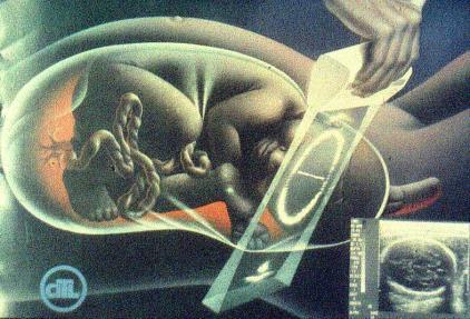
5 5 OBSTETRICAL IMAGE Display Modes B-SCAN B-SCAN (2-D) M-MODE PATIENT-ORIENTED B-SCAN PLANES OBSTETRICAL IMAGES 2D 3D/4D 3D/4D Freehand 3D, often called manual 3D uses a standard 2D transducer and produces a static volumetric image after the transducer is slowly moved along a scan plane. Ultrasound Transducers Automatic 3D requires a dedicated transducer and can produce a volumetric image from a fixed transducer position. 4D also requires a dedicated transducer, but the volumetric image is displayed in real-time.


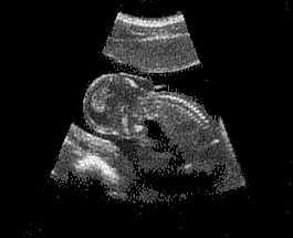
6 6 FLAT-LINEAR ARRAY (linear array) Labeled L FLAT-LINEAR ARRAY (linear array) FLAT-LINEAR ARRAY (linear array) FLAT-LINEAR ARRAY (linear array) Labeled C EMBRYO


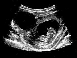
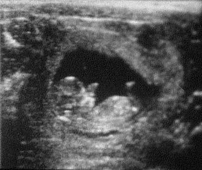
7 7 FIBROID UTERUS EMBRYO AND OVARIAN CYST FETUS


 PHASED")

8 8 FETUS ENDOVAGINAL TRANSDUCER SAGITTAL SCAN PLANES TRANSABDOMINAL ENDOVAGINAL ENDOVAGINAL UTERUS PHASED ARRAY (electronically steered) PHASED ARRAY (electronically steered) Labeled P or S FETAL HEAD
")
")

9 9 PHASED ARRAY (electronically steered) PHASED ARRAY (electronically steered) FETUS ENDOVAGINAL TRANSDUCER PHASED ARRAY (electronically steered) PHASED / VECTOR ARRAY (trapezoidal array) Labeled V ENDOVAGINAL UTERUS PHASED / VECTOR ARRAY (trapezoidal array) ULTRASOUND ARTIFACTS REVERBERATION ENHANCEMENT SHADOWING FETUS


10 10 Doppler Imaging DOPPLER DOPPLER TRANSDUCER SAME FREQUENCY TRANSDUCER LOWER FREQUENCY During Doppler operation, the reflected sound has the same frequency as the transmitted sound if the blood is stationary. During Doppler operation, the reflected sound has a lower frequency if the blood is moving away from the sound source.


11 11 DOPPLER SPECTRAL & COLOR-FLOW DOPPLER TRANSDUCER HIGHER FREQUENCY During Doppler operation, the reflected sound has a higher frequency if the blood is moving toward the sound source. KEY ITEMS FOR REVIEW #1 Frequencies used for medical diagnostic ultrasound are: #1 Frequencies used for medical diagnostic ultrasound are: above 1 MHz CATEGORIES OF SOUND INFRASOUND = below 20 Hz AUDIBLE SOUND = 20 Hz to 20 khz ULTRASOUND = above 20 khz o Medical Diagnostic Ultrasound = above 1 MHz
12 #2 #2 The average sound velocity in soft tissue is: The average sound velocity in soft tissue is: 1540 meters per second Material SOUND VELOCITIES Meters per second Air 330 Pure Water 1430 Fat 1450 Soft Tissue 1540 Muscle 1585 #3 High frequency transducers provide improved resolution and: Bone 4080 #3 High frequency transducers provide improved resolution and: poor penetration RESOLUTION & PENETRATION HIGH-FREQUENCY TRANSDUCERS BETTER RESOLUTION GREATER ATTENUATION POORER PENETRATION LOW-FREQUENCY TRANSDUCERS POORER RESOLUTION LESS ATTENUATION BETTER PENETRATION 12
13 #4 #4 Major categories of ultrasound bioeffects are heat and: Major categories of ultrasound bioeffects are heat and: cavitation ULTRASOUND BIOEFFECTS #5 HEAT (Thermal) Two basic scanning formats are: CAVITATION (Mechanical) Stable Transient #5 Two basic scanning formats are: linear and sector 13
14 14
Physical Principles of Ultrasound
 Physical Principles of Ultrasound Grateful appreciation to Richard A. Lopchinsky, MD, FACS and Nancy H. Van Name, RDMS, RTR, and MarleneKattaron, RDMS 2000 UIC All Rights Reserved. Course Objectives Identify
Lesson 07: Ultrasound Transducers. This lesson contains 73 slides plus 16 multiple-choice questions.
 Lesson 07: Ultrasound Transducers This lesson contains 73 slides plus 16 multiple-choice questions. This lesson was derived from pages 33 through 42 in the textbook: Ultrasound Transducers Ultrasound Transducers
What is Ultrasound? What is Ultrasound? B A. Basic Principles of Ultrasound. Basic Principles of Ultrasound. Basic Principles of Ultrasound
 Introduction to Ultrasound Principles Mani Montazemi, RDMS Baylor College of Medicine Division of Maternal-Fetal Medicine Department of Obstetrics and Gynecology Manager, Maternal Fetal Center Imaging
Diploma of Medical Ultrasonography (DMU) Physical Principles of Ultrasound and Instrumentation Syllabus
 Physical Principles of Ultrasound and Instrumentation Syllabus") Diploma of Medical Ultrasonography (DMU) Physical Principles of Ultrasound and Instrumentation Syllabus Page 1 of 7 11/18 Candidates are expected to cover all of the content of this syllabus when preparing
Diploma of Medical Ultrasonography (DMU) Physical Principles of Ultrasound and Instrumentation Syllabus Page 1 of 7 11/18 Candidates are expected to cover all of the content of this syllabus when preparing
Preamble (disclaimer)
") Preamble (disclaimer) PHYSICS AND PRINCIPLES OF HEAD/NECK ULTRASOUND Joseph C. Sniezek, MD FACS LTC, MC, USA Otolaryngology/H&N Surgery Tripler Army Medical Center 1. I am not a physicist 2. ACS has recommended
Preamble (disclaimer) PHYSICS AND PRINCIPLES OF HEAD/NECK ULTRASOUND Joseph C. Sniezek, MD FACS LTC, MC, USA Otolaryngology/H&N Surgery Tripler Army Medical Center 1. I am not a physicist 2. ACS has recommended
Diagnostic Ultrasound. Sutiporn Khampunnip, M.D.
 Diagnostic Ultrasound Sutiporn Khampunnip, M.D. Definition of Ultrasound Ultrasound is simply sound waves, like audible sound. High-frequency sound and refers to mechanical vibrations above 20 khz. Human
Lesson 03: Sound Wave Propagation and Reflection. This lesson contains 15 slides plus 14 multiple-choice questions.
 Lesson 03: Sound Wave Propagation and Reflection This lesson contains 15 slides plus 14 multiple-choice questions. Accompanying text for the slides in this lesson can be found on pages 8 through 14 in
Ultrasound Physics & Terminology
 Ultrasound Physics & Terminology This module includes the following: Basic physics terms Basic principles of ultrasound Ultrasound terminology and terms Common artifacts seen Doppler principles Terms for
Ultrasound Principles cycle Frequency Wavelength Period Velocity
 ! Teresa S. Wu, MD, FACEP Director, EM Ultrasound Program & Fellowship Co-Director, Simulation Based Training Program & Fellowship Associate Program Director, EM Residency Program Maricopa Medical Center
The Physics of Ultrasound. The Physics of Ultrasound. Claus G. Roehrborn. Professor and Chairman. Ultrasound Physics
 The Physics of Ultrasound Pipe Organ 10-8000 Emission Dog 452-1080 Man 85-1100 Spectrum Bat 10,000-120,000 Porpoise 7000-120,000 Claus G. Roehrborn Professor and Chairman 10 20 Cycles per second Reception
ULTRASOUND IMAGING EE 472 F2018. Prof. Yasser Mostafa Kadah
 ULTRASOUND IMAGING EE 472 F2018 Prof. Yasser Mostafa Kadah www.k-space.org Recommended Textbook Diagnostic Ultrasound: Physics and Equipment, 2nd ed., by Peter R. Hoskins (Editor), Kevin Martin (Editor),
Concepts of Imaging and Knobology
 Concepts of Imaging and Knobology Pravin Patil, MD FACC FASE Associate Professor of Medicine Director, Cardiovascular Disease Training Program Lewis Katz School of Medicine at Temple University Disclosures
Basic of Ultrasound Physics E FAST & Renal Examination. Dr Muhammad Umer Ihsan MBBS,MD, DCH CCPU,DDU1,FACEM
 Basic of Ultrasound Physics E FAST & Renal Examination Dr Muhammad Umer Ihsan MBBS,MD, DCH CCPU,DDU1,FACEM What is Sound? Sound is Mechanical pressure waves What is Ultrasound? Ultrasounds are sound waves
Basic Training Programme. 16 Februrary 2018, ROTTERDAM. Pre and Post-Course Test Answers
 Basic Training Programme 16 Februrary 2018, ROTTERDAM Pre and Post-Course Test Answers Your details: Name: Conference registration number/ BT delegate number: Email address: Are you already performing
Principles of Ultrasound. Cara C. Prideaux, M.D. University of Utah PM&R Sports Medicine Fellow March 14, 2012
 Principles of Ultrasound Cara C. Prideaux, M.D. University of Utah PM&R Sports Medicine Fellow March 14, 2012 None Disclosures Outline Introduction Benefits and Limitations of US Ultrasound (US) Physics
Dr Emma Chung. Safety first - Physical principles for excellent imaging
 Safety first - Physical principles for excellent imaging Dr Emma Chung Lecturer in Medical Physics, University of Leicester Clinical Scientist, University Hospitals of Leicester NHS Trust Thanks to Caroline
Ultrasound. Principles of Medical Imaging. Contents. Prof. Dr. Philippe Cattin. MIAC, University of Basel. Oct 17th, 2016
 Ultrasound Principles of Medical Imaging Prof. Dr. Philippe Cattin MIAC, University of Basel Contents Abstract 1 Image Generation Echography A-Mode B-Mode M-Mode 2.5D Ultrasound 3D Ultrasound 4D Ultrasound
Introduction to Biomedical Imaging
 Alejandro Frangi, PhD Computational Imaging Lab Department of Information & Communication Technology Pompeu Fabra University www.cilab.upf.edu Basic principles. Comparison to X-rays Ultrasound > 20kHz
Sound in medicine. CH.12. Dr.Rajaa أ.م.د. رجاء سهيل جنم جامعة تكريت كلية طب االسنان. General Properties of Sound
 CH.12. Dr.Rajaa Sound in medicine أ.م.د. رجاء سهيل جنم جامعة تكريت كلية Sound : It is the audible waves of frequency between 20 Hz and 20 khz. Infrasound : refers to the sound of frequency below the normal
2015 ARDMS Sonography Principles & Instrumentation Job Task Analysis Summary Report
 P a g e 1 2015 ARDMS Sonography Principles & Instrumentation Job Task Analysis Summary Report American Registry for Diagnostic Medical Sonography (ARDMS) P a g e 2 Table of Contents ABOUT THE REPORT...
CONTENTS. Test Number cpd Tanya Reynolds (Nat. Dip. Diag. Rad., B. Tech. Diag. Rad., B. Tech. Ultrasound)
") CONTENTS page 1-15 page 16 BASIC 2-DIMENSIONAL ULTRASOUND PRINCIPLES Multiple Choice Test Test Number cpd 41640 Tanya Reynolds (Nat. Dip. Diag. Rad., B. Tech. Diag. Rad., B. Tech. Ultrasound) Tanya is
CONTENTS page 1-15 page 16 BASIC 2-DIMENSIONAL ULTRASOUND PRINCIPLES Multiple Choice Test Test Number cpd 41640 Tanya Reynolds (Nat. Dip. Diag. Rad., B. Tech. Diag. Rad., B. Tech. Ultrasound) Tanya is
1 Fundamentals. Basic Definitions and Physics Principles. Fundamentals
 1 To become versed in the language of ultrasonography, it is necessary to review some of the basic principles of physics. The wave physics principles of ordinary (i.e., audible) sound apply to ultrasound
Basic Physics of Ultrasound and Knobology
 WELCOME TO UTMB Basic Physics of Ultrasound and Knobology By Daneshvari Solanki, FRCA Laura B. McDaniel Distinguished Professor Anesthesiology and Pain Medicine University of Texas Medical Branch Galveston,
Ultrasound Physics and Knobology Alan Macfarlane. Consultant Anaesthetist Glasgow Royal Infirmary
 Ultrasound Physics and Knobology Alan Macfarlane Consultant Anaesthetist Glasgow Royal Infirmary RAPM 2009; 34: 40-46 Ultrasound Proficiency Understanding US image generation and device operation Image
Ultrasound 10/1/2014. Basic Echocardiography for the Internist. Mechanical (sector) transducer Piezoelectric crystal moved through a sector sweep
 transducer Piezoelectric crystal moved through a sector sweep") Ultrasound Basic Echocardiography for the Internist Carol Gruver, MD, FACC UT Erlanger Cardiology Mechanical wave of compression and rarefaction Requires a medium for transmission Ultrasound frequency
Ultrasound Basic Echocardiography for the Internist Carol Gruver, MD, FACC UT Erlanger Cardiology Mechanical wave of compression and rarefaction Requires a medium for transmission Ultrasound frequency
Supplement (videos)
") Supplement (videos) Ruben s tube (sound): http://www.youtube.com/watch?v=gpcquuwqayw Doppler US (diagnostic use): http://www.youtube.com/watch?v=fgxzg-j_hfw http://www.youtube.com/watch?v=upsmenyoju8 High
Supplement (videos) Ruben s tube (sound): http://www.youtube.com/watch?v=gpcquuwqayw Doppler US (diagnostic use): http://www.youtube.com/watch?v=fgxzg-j_hfw http://www.youtube.com/watch?v=upsmenyoju8 High
Pulse-Echo Ultrasound Imaging. Resolution in Ultrasound Imaging. Doppler Ultrasound. Resolution vs Penetration. Medical Imaging (EL582/BE620/GA4426)
") Medical Imaging (EL582/BE620/GA4426) Pulse-Echo Ultrasound Imaging Ultrasound Imaging Lecture 2 Daniel (Dan) Turnbull, Ph.D. Skirball Institute and Dept of Radiology NYU School of Medicine (daniel.turnbull@med.nyu.edu)
Medical Imaging (EL582/BE620/GA4426) Pulse-Echo Ultrasound Imaging Ultrasound Imaging Lecture 2 Daniel (Dan) Turnbull, Ph.D. Skirball Institute and Dept of Radiology NYU School of Medicine (daniel.turnbull@med.nyu.edu)
The Value. Ultrasound System. Professional Concept for. Women s. Health
 The Value Innovative Ultrasound System Professional Concept for Women s Health 9 Beyond Women s Healthcare E-CUBE 9 - Powerful Imaging Performance for All Women s Health Diagnostic Applications Diagnostic
ULTRASOUND NOMENCLATURE
 Chapter 1: Ultrasound Nomenclature, Image Orientation, and Basic Instrumentation CYNTHIA SIKOWSKI Ultrasound waves are sound waves that have a frequency exceeding 20,000 Hz. When sound waves are transmitted
Basic Physics of Ultrasound in Transesophageal Echocardiography
 SPECIAL ARTICLE IJUTPC Basic Physics of Ultrasound in Transesophageal Echocardiography Basic Physics of Ultrasound in Transesophageal Echocardiography 1 Mary Korula, 2 Ravi Hebballi 1 Senior Consultant,
Ultrasound Physics & Doppler
 Ultrasound Physics & Doppler Endocrine University 2018 Mark Lupo, MD, FACE, ECNU Objectives Review the essential components of ultrasound physics in neck sonography Demonstrate the importance of ultrasound
Application of Phased Array Radar Theory to Ultrasonic Linear Array Medical Imaging System
 Application of Phased Array Radar Theory to Ultrasonic Linear Array Medical Imaging System R. K. Saha, S. Karmakar, S. Saha, M. Roy, S. Sarkar and S.K. Sen Microelectronics Division, Saha Institute of
The 2 nd Cambridge Advanced Emergency Ultrasound Course
 The 2 nd Cambridge Advanced Emergency Ultrasound Course Addenbrooke s Hospital Cambridge Sept 2008 1 2 Faculty! UK! USA! Australia! Toshiba! Emergency Medicine! Radiology 3 Programme! Day 1 Introduction
Ultrasonic Testing Level I:
 Ultrasonic Testing Level I: 1- Sound Wave - Introduction - ASNT Level I - Sound Wave Propagation - Velocity / Frequency / Wave Length - Acoustic Impedance - Energy / Intensity 2- Ultrasound Wave Modes
Terminology Tissue Appearance
 By Marc Nielsen, MD Advantages/Disadvantages Generation of Image Ultrasound Machine/Transducer selection Modes of Ultrasound Terminology Tissue Appearance Scanning Technique Real-time Portable No ionizing
Basic Ultrasound Physics Board Review Questions
 Basic Ultrasound Physics Board Review Questions Sidney K. Edelman, PhD ESP Ultrasound The Woodlands, TX Question 1 What is the wavelength of 2 MHz sound in soft tissue? 1. 1.54 mm 2. 0.75 mm 3. 0.75 cm
DIGITAL IMAGE PROCESSING IN ULTRASOUND IMAGES
 DIGITAL IMAGE PROCESSING IN ULTRASOUND IMAGES Kamaljeet Kaur Computer Science & Engineering Department Guru Nanak Dev Engg. College, Ludhiana. Punjab-India meetk.89@gmail.com ABSTRACT-- Image processing
Exam Practice Guide. Units 1 & 2 Physics: Detailed Study 5 - Investigations: Medical physics Examination Questions
 Exam Practice Guide Units 1 & 2 Physics: Detailed Study 5 - Investigations: Medical physics Examination Questions Key Features: 22 original examination style questions on all examinable topics. Full solutions
Introduction & Physics of ED Ultrasound. Objectives. What? - Limited Studies. Who? - ED Docs
 Introduction & Physics of ED Ultrasound Martine Sargent, MD Ultrasound Director, Assistant Professor UCSF Department of Emergency Medicine San Francisco General Hospital & Trauma Center Objectives Who?
Point-of-Care Ultrasound: An Introduction
 Point-of-Care Ultrasound: An Introduction Delegation Teaching Package for Registered Respiratory Therapists and Anesthesia Assistants Developed by: Rob Bryan RRT, AA Edited by: Kelly Hassall RRT, FCSRT,
Underwater Acoustic Measurements in Megahertz Frequency Range.
 Underwater Acoustic Measurements in Megahertz Frequency Range. Current State and Prospects of Development in Russia Alexander M. Enyakov,, Many medical applications of underwater acoustic measurements
DOW-RAD, DOW DIAGNOSTIC COMPLEX, DUHS TRAINING PROGRAM HANDBOOK 2013
 DOW-RAD, DOW DIAGNOSTIC COMPLEX, DUHS TRAINING PROGRAM HANDBOOK 2013 CERTIFICATE COURSE INVASCULAR/DOPPLER ULTRASOUND: Introduction: Ultrasound is an evolving technology with wide spectrum application
The table below shows the density and velocity of waves in two different substances. Density / kg m 3 Velocity / m s 1
 Q1.(a) When ultrasound is incident at an interface between two different media some energy is transmitted and some is reflected. The ratio of the reflected energy intensity I r to the incident energy intensity
Ultrasound: Past and Present. Lecturer: Dr. John M Hudson, PhD
 Ultrasound: Past and Present Lecturer: Dr. John M Hudson, PhD Disclosures 2 No conflicts of interest to declare Course Outline 3 1. Survey of ultrasound physics & applications 2. (Sep 21) 3. (Sep 28) 4.
Linear Ultrasonic Wave Propagation in Biological Tissues
 Indian Journal of Biomechanics: Special Issue (NCBM 7-8 March 29) Linear Ultrasonic Wave Propagation in Biological Tissues Narendra D Londhe R. S. Anand 2, 2 Electrical Engineering Department, IIT Roorkee,
What is Ultrasound? Resolution Image production Attenuation Imaging modes Ultrasound artifacts... 7
 What is Ultrasound?... 1 Resolution... 3 Image production... 3 Attenuation... 4 Imaging modes... 5 Ultrasound artifacts... 7 0 What is Ultrasound? High frequency sound of frequencies 2-50 MHz is used in
Development of Ultrasound Based Techniques for Measuring Skeletal Muscle Motion
 Development of Ultrasound Based Techniques for Measuring Skeletal Muscle Motion Jason Silver August 26, 2009 Presentation Outline Introduction Thesis Objectives Mathematical Model and Principles Methods
Ultrasound Measurements and Non-destructive Testing Educational Laboratory
 Session 3548 Ultrasound Measurements and Non-destructive Testing Educational Laboratory Vladimir Genis, Horacio Sosa Goodwin College of Professional Studies, Drexel University, Philadelphia, 19104 Emil
4.17. RESEARCHING MODELS WITH AN ULTRASONIC ECHOSCOPE
 4.17. RESEARCHING MODELS WITH AN ULTRASONIC ECHOSCOPE Purpose of experiment Determine the main characteristics of ultrasound waves, and the distances and positions of models using an ultrasonic echoscope.
An Overview of Ultrasound Testing For Lesion Detection in Human Kidney
 Journal of Tomography System & Sensors Application Vol.1, Issue 1, June 2018 An Overview of Ultrasound Testing For Lesion Detection in Human Kidney Aina Fadhilah Abd Rahim 1, Zawin Najah Abd Halim 1, Jaysuman
1. Fig. 1 shows data for the intensity of a parallel beam of X-rays after penetration through varying thicknesses of a material
 1. Fig. 1 shows data for the intensity of a parallel beam of X-rays after penetration through varying thicknesses of a material. intensity / MW m 2 thickness / mm 0.91 0.40 0.69 0.80 0.52 1.20 0.40 1.60
Medical Imaging. Ultrasound Imaging
 Medical Imaging Ultrasound Imaging Prof. Ed X. Wu Overview History Physics of Ultrasound wave propagation, attenuation, scattering, and reflection Generation and Detection of Ultrasound Piezoelectric Transducers
WELCOME! Introduction to Bedside Ultrasound
 WELCOME! Introduction to Bedside Ultrasound TEACHERS University of California-Irvine School of Medicine Nathan Molina nathan.d.molina@gmail.com Trevor Plescia taplescia90@gmail.com Jack Silva jpsilva42@gmail.com
4.17. RESEARCHING MODELS WITH AN ULTRASONIC ECHOSCOPE
 4.17. RESEARCHING MODELS WITH AN ULTRASONIC ECHOSCOPE Purpose of experiment Determine the main characteristics of ultrasound waves, and the distances and positions of models using an ultrasonic echoscope.
Descriptions of NDT Projects Fall 2004 October 31, 2004
 Descriptions of NDT Projects Fall 2004 October 31, 2004 Introduction There are two separate NDT labs in Magister: ULTRA for ultrasound and EDDY for eddy current. Both labs are equipped with mechanical
Ultrasonic Testing of Composite Structures
 I. Introduction Ultrasonic Testing of Composite Structures This section of this work defines ultrasound basic concepts and Ultrasonic Technique. It describes the details of how ultrasonic testing works,
RPVI Exam Review ecourse
 RPVI Exam Review ecourse The RPVI Exam Review ecourse consists of ten Vascular Physics Modules and fourteen Vascular Specialty Modules. Detailed descriptions of module content are listed below. During
Ultrasound in Medicine
 Ultrasound in Medicine Experimental Equipment for Medical Education Universities Colleges Medical Schools Medical and Med-Technical Training Education can befun! WELCOME TO GAMPT Devices and accessories
Medical Imaging. By: Engr. Joseph Ronald Canedo
 Medical Imaging By: Engr. Joseph Ronald Canedo Medical Sonography (Ultrasound) is an ultrasound-based diagnostic imaging technique used to visualize muscles and internal organs, their size, structures
Table of contents. Foreword. Preface. 1 Introduction Historical Perspective 00
 Table of contents Foreword Preface 1 Introduction 00 1.1 Historical Perspective 00 2 Fundamentals of musculoskeletal ultrasound 00 2.1 Frequency and wavelength 00 2.2 Generating ultrasound waves 00 2.3
Ultrasound in Anesthesia: Applying Scientific Principles to Clinical Practice
 AANA Journal Course Update for Nurse Anesthetists 3 6 CE Credits* Ultrasound in Anesthesia: Applying Scientific Principles to Clinical Practice Christian R. Falyar, CRNA, DNAP The use of ultrasound as
Ultrasound System DUS ULT. Ultrasound. September 2016 DUS 7000 Rev. 03
 ULT Ultrasound System DUS - 7000 Ultrasound September 2016 DUS 7000 Rev. 03 3D, 4D Volumetric, S-Live DUS - 7000 Ultrasound Touch screen color Display with articulated arm Second display Four active transducer
ULTRASOUND QA SOLUTIONS. Ensure Accurate Screening, Diagnosis & Monitoring DOPPLER FLOW PHANTOMS MULTI-PURPOSE PHANTOMS TRANSDUCER TEST PHANTOMS
 ULTRASOUND QA SOLUTIONS Ensure Accurate Screening, Diagnosis & Monitoring DOPPLER FLOW PHANTOMS MULTI-PURPOSE PHANTOMS TRANSDUCER TEST PHANTOMS INNOVATORS IN ADVANCED ULTRASOUND TECHNIQUES Gammex is the
APPLICATION AND DEPLOYMENT OF ADVANCED NDE TECHNIQUES IN HIGH PRESSURE VESSELS
 APPLICATION AND DEPLOYMENT OF ADVANCED NDE TECHNIQUES IN HIGH PRESSURE VESSELS Jeffrey P. Milligan, Daniel T. Peters, Structural Integrity Associates, Inc., USA Many advances in Non-Destructive Examination
ULTRASOUND QA SOLUTIONS. Ensure Accurate Screening, Diagnosis & Monitoring DOPPLER FLOW PHANTOMS MULTI-PURPOSE PHANTOMS TRANSDUCER TEST PHANTOMS
 ULTRASOUND QA SOLUTIONS Ensure Accurate Screening, Diagnosis & Monitoring DOPPLER FLOW PHANTOMS MULTI-PURPOSE PHANTOMS TRANSDUCER TEST PHANTOMS INNOVATORS IN ADVANCED ULTRASOUND TECHNIQUES Gammex is the
Feng Xiujuan National Institute of Metrology (NIM),China
,China") The acoustic calibration service in transportation at NIM Feng Xiujuan National Institute of Metrology (NIM),China 1. Calibration requirements 2. Calibration service at NIM 2.1 Microphone 2.2 Ultrasonic
The acoustic calibration service in transportation at NIM Feng Xiujuan National Institute of Metrology (NIM),China 1. Calibration requirements 2. Calibration service at NIM 2.1 Microphone 2.2 Ultrasonic
High resolution ultrasound scanner for skin imaging
 High resolution ultrasound scanner for skin imaging Christine Turlat Sales Director Atys medical 17 Parc d Arbora 69510 SOUCIEU EN JARREST Atys company Principle of ultrasound imaging DERMCUP Normal image
Introduction to Ultrasound Guided Region Anesthesia
 Introduction to Ultrasound Guided Region Anesthesia Brian D. Sites, MD Dept of Anesthesiology Dartmouth-Hitchcock Medical Center INTRODUCTION Welcome to Introduction to Ultrasound Guided Regional Anesthesia.
Emergency Medicine Interest Group (EMIG) 2016
 2016") Emergency Medicine Interest Group (EMIG) 2016 Welcome to the flipped classroom (learning objectives summary) for the 2016 Emergency Medicine Interest Group (EMIG) Procedures Workshop. Overview - Tuesday
Emergency Medicine Interest Group (EMIG) 2016 Welcome to the flipped classroom (learning objectives summary) for the 2016 Emergency Medicine Interest Group (EMIG) Procedures Workshop. Overview - Tuesday
ULTRASOUND QA SOLUTIONS. Ensure Accurate Screening, Diagnosis and Monitoring DOPPLER FLOW PHANTOMS MULTI-PURPOSE PHANTOMS TRAINING PHANTOMS
 ULTRASOUND QA SOLUTIONS Ensure Accurate Screening, Diagnosis and Monitoring DOPPLER FLOW PHANTOMS MULTI-PURPOSE PHANTOMS TRAINING PHANTOMS INNOVATORS IN ADVANCED ULTRASOUND TECHNIQUES Gammex is the only
TG-128: Quality Assurance for Prostate Brachytherapy Ultrasound
 TG-128: Quality Assurance for Prostate Brachytherapy Ultrasound STEVEN SUTLIEF DOUG PFEIFFER (HEATHER PIERCE, WENGZHENG FENG, JIM KOFLER) AAPM ANNUAL MEETING 2010 Educational Objectives To describe the
Aixplorer MultiWave Ultrasound System An Innovation in Breast Ultrasound Imaging
 b r e a s t i m a g i n g S O L U T I O N S Aixplorer MultiWave Ultrasound System An Innovation in Breast Ultrasound Imaging The new wave in breast ultrasound Aixplorer is a next-generation ultrasound
Optimising your Doppler settings for an accurate PI. Alison McGuinness Mid Yorks Hospitals
 Optimising your Doppler settings for an accurate PI Alison McGuinness Mid Yorks Hospitals Applications Both maternal uterine and fetal circulations can be studied with doppler sonography Uterine arteries
Chapter 3 Physical Principles of Ultrasound of the Male Genitalia
 Chapter 3 Physical Principles of Ultrasound of the Male Genitalia Bruce R. Gilbert and Pat Fox Fulgham Introduction The value of ultrasound evaluation of the male genitalia depends, in large part, on the
Chapter 14. Imaging Artifacts
 Chapter 14 Image Artifacts The complex physical interactions that occur between an ultrasound beam and human anatomy and the intricate and sophisticated technological components of a sonographic imaging
Women s Lifetime Partner
 Women s Lifetime Partner ALPINION MEDICAL SYSTEMS We are Ultrasound Professionals E-CUBE 7, the reliable healthcare partner. Obstetrics 3D/4D Imaging The E-CUBE 7 is a comprehensive solution for all your
Lectures on Medical Biophysics Dept. Biophysics, Medical faculty, Masaryk University in Brno
 Lectures on Medical Biophysics Dept. Biophysics, Medical faculty, Masaryk University in Brno Lectures on Medical Biophysics Department of Biophysics, Medical Faculty, Masaryk University, Brno Ultrasound
Insight, Intelligence, and more
 DC-80 Diagnostic Ultrasound System Insight, Intelligence, and more P/N:ENG-DC-80-210285X12P-20170725 Overview The best patient care is your ultimate goal. To achieve this requires confident diagnosis even
Tissue Strain Analytics Virtual Touch Tissue Imaging and Quantification
 Whitepaper Tissue Strain Analytics Virtual Touch Tissue Imaging and Quantification ACUSON S2000 Ultrasound System Answers for life. Page 1 Tissue Strain Analytics: Virtual Touch Tissue Imaging and Quantification
The Evolution and Benefits of Phased Array Technology for the Every Day Inspector
 ECNDT 2006 - Poster 198 The Evolution and Benefits of Phased Array Technology for the Every Day Inspector Dan KASS, Tom NELLIGAN, and Erich HENJES Olympus NDT, Waltham, USA Abstract. Phased arrays were
Physical Principles of Ultrasound
 Physical Principles of Ultrasound Pat F. Fulgham 2 Introduction The use of ultrasound is fundamental to the practice of urology. In order for urologists to best use this technology on behalf of their patients,
Ultrasound Knobology
 Ultrasound Knobology Raj Dasgupta MD, FACP, FCCP, FASSM Assistant Professor of Clinical Medicine Pulmonary / Critical Care / Sleep Medicine University of Southern California (USC) Objectives Physics of
GE Healthcare. Logiq P5 Premium
 GE Healthcare Logiq P5 Premium LOGIQ P5 PREMIUM The GE Logiq P5 premium is an economical shared service ultrasound machine that is unique in it s price range by offering deep support for all applications
ALPINION MEDICAL SYSTEMS We are Ultrasound Professionals
 ALPINION MEDICAL SYSTEMS We are Ultrasound Professionals S l i m & P o w e r f u l The E-CUBE 7 delivers compact and powerful image performance for a fast and confident clinical decision, a smart-software
Annular Array Transducer and Matched Amplifier for Therapeutic Ultrasound
 ARCHIVES OF ACOUSTICS 35, 4, 653 660 (2010) DOI: 10.2478/v10168-010-0049-6 Annular Array Transducer and Matched Amplifier for Therapeutic Ultrasound Wojciech SECOMSKI, Andrzej NOWICKI, Janusz WÓJCIK, Marcin
Ultrasonography of the Neck as an Adjunct to FNA. Nicole Massoll M.D.
 Ultrasonography of the Neck as an Adjunct to FNA Nicole Massoll M.D. Basic Features of Head and Neck Ultrasound and Anatomy Nicole Massoll M.D. University of Arkansas for Medical Sciences, Little Rock
Principles of echocardiography for the anesthesiologist.
 Principles of echocardiography for the anesthesiologist. Nikolaos Skubas, MD PhD Abstract Ultrasound-based diagnostic techniques are now part of the cardiological patients chart, while echocardiography
The complete, portable patient care solution
 The complete, portable patient care solution ACUSON P300 Ultrasound System www.siemens.com/ultrasound Answers for life. Mobile. Powerful. And extraordinarily versatile. Sleek design Portable Complete 2
DMS-2350: SONOGRAPHIC INSTRUMENT/PHYSICS
 DMS-2350: Sonographic Instrument/Physics 1 DMS-2350: SONOGRAPHIC INSTRUMENT/PHYSICS Cuyahoga Community College Viewing:DMS-2350 : Sonographic Instrument/Physics Board of Trustees: 2014-03-20 Academic Term:
Ultrasound guidance in regional anesthesia has
 Ultrasound and Regional Anesthesia Artifacts and Pitfall Errors Associated With Ultrasound-Guided Regional Anesthesia. Part I: Understanding the Basic Principles of Ultrasound Physics and Machine Operations
Improved ultrasound-based navigation for robotic drilling at the lateral skull base
 International Congress Series 1268 (2004) 662 666 Improved ultrasound-based navigation for robotic drilling at the lateral skull base U.W. Geisthoff a, *, S.H. Tretbar b, Ph.A. Federspil a, P.K. Plinkert
Chapter 1. Principles of medical ultrasound. Overview. Background history: first steps to the piezo-electric effect.
 Chapter 1 Principles of medical ultrasound GRAHAM ARTHURS, PATRICK HILL AND TREVOR FRANKEL Overview This chapter provides an introduction to the ultrasound process for trainees in anesthesia and other
ACUSON X150 Ultrasound System. Transducer Options Release 1.0
 ACUSON X150 Ultrasound System Transducer Options Release 1.0 P4-2 Transducer Part Number: 08648045 4 2 MHz 2.0*, 2.5, 2.9*, 3.1, 3.6 MHz 1.3*, 1.5, 1.8, 2.5* MHz 2.0, 2.7 MHz 2.0, 2.5*, 2.7 MHz Number
Image optimization for critical care US
 Image optimization for critical care US 1 Although we assume you are already familiar with focused US in the ED, it might not hurt to revise the basics: Machines & transducers US appearance of normal tissues
ACUSON X500 Ultrasound System. Transducers
 ACUSON X500 Ultrasound System Transducers 1 C8-5 Transducer 8 5 MHz SONOLINE G60 S ultrasound system SONOLINE G50 ultrasound system Neonatal head, pediatric, neonatal abdomen Wide bandwidth curved array
NMIJ measurement service on ultrasonic field parameters available to demonstrate performance and safety of ultrasonic medical equipment
 NMIJ measurement service on ultrasonic field parameters available to demonstrate performance and safety of ultrasonic medical equipment Masahiro Yoshioka National Metrology Institute of Japan (NMIJ) National
NCVH. Ultrasongraphy: State of the Art Vein Forum 2015 A Multidisciplinary Approach to Otptimizing Venous Circulation From Wounds to WOW
 Ultrasongraphy: State of the Art 2015 NCVH New Cardiovascular Horizons Vein Forum 2015 A Multidisciplinary Approach to Otptimizing Venous Circulation From Wounds to WOW Anil K. Chagarlamudi, M.D. Cardiovascular
Reliable versatility. Philips HD5 ultrasound system
 Reliable versatility Philips HD5 ultrasound system Affordability and features in one Designed to perform as you need it Every day, your patients come to you for high-quality care. Now, there s an ultrasound
Critical Care Ultrasound Study Notes
 Critical Care Ultrasound Study Notes Compiled by David Tripp October 2014 Ultrasound Physics 2 Ultrasound in Tissue 2 Ultrasound Interaction with Tissue 2 Pulsed Ultrasound and Imaging 3 Image Formation