LARYNX ANATOMY. Elena Rizzo Riera R1 ORL HUSE
|
|
|
- Lester Stanley
- 5 years ago
- Views:
Transcription


1 LARYNX ANATOMY Elena Rizzo Riera R1 ORL HUSE

2 INTRODUCTION v Odd and median organ v Infrahyoid region v Phonation, swallowing and breathing v Triangular pyramid v Postero- superior base à pharynx and hyoid bone v Bottom point à upper orifice of the trachea
3 INTRODUCTION C4-C6 Tongue trachea In women it is somewhat higher than in men. Male Female Length 44mm 36mm Transverse diameter 43mm 41mm Anteroposterior diameter 36mm 26mm

4 SKELETAL STRUCTURE Framework: 11 cartilages linked by joints and fibroelastic structures 3 odd-and median cartilages: the thyroid, cricoid and epiglottis cartilages. 4 pair cartilages: corniculate cartilages of Santorini, the cuneiform cartilages of Wrisberg, the posterior sesamoid cartilages and arytenoid cartilages. Intrinsic and extrinsic muscles
 M:90º F: 120º Children: intrathyroid")
5 THYROID CARTILAGE Shield shaped cartilage Right and left vertical laminaà laryngeal prominence (Adam s apple) M:90º F: 120º Children: intrathyroid cartilage

6 THYROID CARTILAGE Outer surface à oblique line Inner surface Superior border à superior thyroid notch Inferior border à inferior thyroid notch Superior horns à lateral thyrohyoid ligaments Inferior horns à cricothyroid articulation

7 THYROID CARTILAGE The oblique line gives attachement to the following muscles: Thyrohyoid muscle Sternothyroid muscle Inferior constrictor muscle Ligaments attached to the thyroid cartilage Thyroepiglottic lig Vestibular lig Vocal lig


8 CRICOID CARTILAGE Complete signet ring Anterior arch and posterior lamina Ridge and depressions Cricothyroid articulation Cricothyroid membrane Cricotracheal ligament Cricoarytenoid articulation Ridge à longitudinal muscle of the esophagous.


9 EPIGLOTTIC CARTILAGE Racket shaped cartilage Anterior wall of the laryngeal aditus Covered by mucous membrane. Hyoepligottic ligament Thyroepiglottic ligament Aryepiglottic folds Laryngeal surface Lingual surface Preepligottic spaceà carcinoma New borns: omega shaped
10 EPIGLOTTIC CARTILAGE
 Base à cricoarytenoid")
11 ARYTENOID CARTILAGES On the upper surface of the cricoid cartilage Vocal process à vocal folds Vestibular ligaments Muscular process à posterior and lateral cricoarytenoid muscles Posterior surfaceà transverse arytenoid muscle Apex à corniculate cartilages (Santorini) Base à cricoarytenoid joint
 articulating with the apices of")
 in each")
12 OTHER CARTILAGES Corniculate cartilages (of Santorini) articulating with the apices of arytenoid cartilages. Cuneiform cartilages (of Wrisberg) in each margin of the aryepiglottic fold.
 From the sides of the epiglottic cartilage to the corniculate and arytenoid")

13 ELASTIC TISSUE INTRINSIC LIGAMENTS (connect the laryngeal cartilages) Quadrangular membrane (upper part) From the sides of the epiglottic cartilage to the corniculate and arytenoid cartilages. ü Aryepiglottic folds ü Vestibular ligaments and vestibular folds Ventricular segment of fibroelastic tissue Conus elasticus (lower part) ü Cricothyroid ligament (anteriorly) ü Vocal ligaments
 Ø Mobility of the hyoid bone and larynx during swallowing and fonation.")
14 ELASTIC TISSUE EXTRINSIC LIGAMENTS (connect the laryngeal cartilages to the hyoid bone above and trachea below) Thyrohyoid membrane Ø Median thyrohyoid ligament Ø Lateral thyroyoid ligaments Ø Pierced by the superior laryngeal artery and nerve (i.b.) Ø Mobility of the hyoid bone and larynx during swallowing and fonation. Cricotracheal membrane Ø Cricoid cartilage and the first tracheal ring Cricothyroid membrane Ø Middle cricothyroid ligament Ø External branch of the superior laryngeal nerve and the middle laryngeal artery perfore it.

15 ELASTIC TISSUE LIGAMENTS OF THE EPIGLOTTIS Ø Middle and lateral glosso epiglottic ligaments Ø Pharyngoepiglottic ligaments Ø Thyroepiglottic ligament Ø Hyoepiglottic membrane, muscle and ligament Ø Aryepiglottic ligaments


16 JOINTS OF THE LARYNX Cricothyroid joint Allows to tilt forward or behind the thyroid cartilage changing the tension of the vocal chords. Cricoarytenoid joint A llows fronto-translational movements to get away or close the arytenoid cartilages. Separate or approximate the vocal chords.
 Corniculate cartilages a n d arytenoideus muscle")

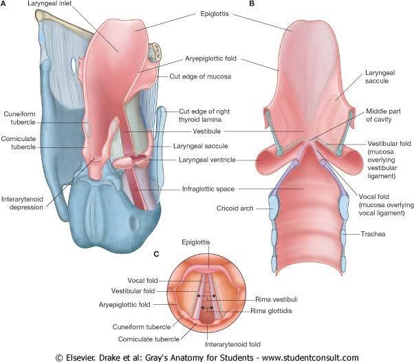
17 INTERNAL ANATOMY LARYNGEAL ADITUS Epiglottis (ant) Aryepiglottic folds (laterally) Corniculate cartilages a n d arytenoideus muscle (post) ü Glossoepiglottic folds and valleculae ü Piriform recesses LARYNX Vestibule Ventricle Subglottic cavity Vestibular folds Vocal folds
: contains mucous glands to lubricate the vocal folds.")
18 INTERNAL ANATOMY Vestibule -Aditus à vestibular folds Vestibular folds -Mucous membrane + connective tissue (vestibular ligament) - Thyroid cartilage à arytenoid cartilage Ventricle of the larynx - Recess between the vestibular and the vocal folds. - Laryngeal saccule (ant): contains mucous glands to lubricate the vocal folds. - Thyroarytenoid muscle
19


20 Glottis: vocal folds + space between them - The narrowest portion of the larynx - Anterior 3/5 à vocal chords à intermembranous portion - Posterior 2/5 à vocal process à intercartilaginous portion

21 STRUCTURE OF ADULT VOCAL FOLD The Cover - Epithelium (mucosa) - Basal lamina - Superficial layer of lamina propria The transition - Intermediate layer of lamina propria - Deep layer of lamina propria The body - Vocalis muscle (thyroarytenoid muscle)
22 STRUCTURE OF ADULT VOCAL FOLD COVER Epithelium -Anterior glottis à stratified squamous - Posterior glottis à pseudostratified ciliated Basal lamina à physical suport - Lamina lucida - Lamina densa Superficial layer of lamina propria - Reinke s space (potential space)à Reinke s edema - Fibrous components + extracellular matrix
23 STRUCTURE OF ADULT VOCAL FOLD TRANSITION Intermediate layer of the lamina propria - Elastic fibers Deep layer of the lamina propria - Collagenous fibers BODY - The vocalis muscle
24

")


25 MUSCULATURE EXTRINSIC MUSCLES GROUP 1: INFRAHYOID: omohyoid, sternohyoid, sternothyroid and thyrohyoid muscles Depressors (exception thyrohyoid) GROUP 2: (suprahyoid) Stylohyoid, digastric and mylohyoid muscles Elevate the larynx GROUP 3: Stilofaryngeal and palatofaryngeal muscles Elevate the larynx and the pharynx GROUP 4: Middle and inferior constrictor muscles of the pharynx


26 INTRINSIC MUSCLES MUSCULATURE Shape and size of the aditus and glottis Cricothyroid à lateral and outer aspect of the larynx Posterior and lateral cricoarytenoid Transverse and oblique arytenoid Lateral and medial thyroarytenoid (vocalis muscle)


27 Cricothyroid muscle External surface of the arch of the cricoid cartilage ad - Straight part - Oblique part - To lengthen, tense and adduction of the vocal chords - Superior laringeal nerve


28 Posterior cricoarytenoid muscle ü Origin: posterior surface of the cricoid lamina ü Insertion: muscular process of the arythenoid cartilage ü Midline crest - Cricoesophageal tendon ü Function: the only ABDUCTOR of the vocal chords


29 Lateral cricoarytenoid muscle ü Origin: upper border and outer surface of the cricoid arch ü Insertion: anterior surface of the muscular process of the arytenoid cartilage ü Antagonist of the posterior cricoarytenoidà adduction vocal chords


30 Transverse arytenoid muscle ü Between the posterior surfaces of the the two arytenoid cartilages ü It approximates the two arytenoid cartilages. Oblique arytenoid muscle ü ü ü Origin: posterior surface of the transverse arytenoid muscle Insertion: the tip of the opposite arytenoid cartilage Adduction of the vocal chords.


31 (Lateral) Thyroarytenoid muscle ü Origin: inner surface of thyroid cartilage ü Insertion: lateral surface of arytenoid cartilage ü Thyroepiglottic muscle ü Function: to shorten the vocal ligaments. ü Adduction of vestibular folds ü ü ü ü Origin: the same Insertion: vocal process of arytenoid cartilage Thicker, deeper and better developed. Function: adducts the vocal fold
 2.")
32 Extrinsic muscles Pull up (high tones) and pull down (low tones) the larynx Intrinsic muscles 1. Adduction of the chords PHONATION Lateral cricoarytenoid / transverse arytenoyd / vocalis muscles (medial thyroarytenoid) 2. Variation of the lenght and tenseness of the chords Cricothyroid and thyroarytenoid muscles à tone of the voice. 3. Abduction of the chords Posterior cricoarytenoid


33 LARYNGEAL SPACES Subglottic: - Glottis - Lower border of the cricoid cartilage Preepiglottic - Ant.:thyroid cartilage and thyrohyoid m. - Above: hyoepiglottic lig.and vallecula - Posteriorly: epiglottis The epiglottic tumors can extend in this region. Paraglottic - Anterolat: thyroid cartilage - Medially: laryngeal ventricle, quadrangular m. and conus elasticus

 - Inf border of inf.")
34 BLOOD SUPPLY Superior laryngeal artery - Arises from the sup. thyroid aa - Internal branch of the superior laryngeal nerve - Runs horizontally across the thyrohyoid membrane. Inferior laryngeal artery - Inf. thyroid artery - Killian-Jamieson area (cricothyroid art) - Inf border of inf. Constrictor m. Cricothyroid artery - Sup. thyroid artery - Cricothyroid membrane

35 Veins Superior laryngeal vein - Sup. thyroid vein - Internal jugular vein Inferior laryngeal vein - Inferior thyroid vein - Brachiocefalic vein


36 Lymphatics Superior group Inferior group Vocal folds - Prelaryngeal nodes - Upper portion of deep cervical chain - Pre and paratracheal nodes - Laryngeal nodes -Lower deep cervical nodes - Supraclavicular nodes


37 NERVES SUPERIOR LARYNGEAL NERVE ü Inferior ganglion of X nerve ü External branch - Ends in the cricothyroid muscle
38 ü Internal branch - Thyrohyoid membrane/ superior laryngeal artery - Sensitive - Root of tongue - Epiglottis and valleculae - Piriform recess - Vestible, vestibular folds, ventricl - Posterior wall of larynx - Anterior wall of pharynx - Mucosa of hipopharynx - Sympathetic/parasympathetic fibres
39 NERVES INFERIOR LARYNGEAL (RECURRENT) NERVE ü The most important MOTOR nerve ü Longer on the left side ü Related to the inferior thyroid aa and the thyroid gland ü The left nerve originates in the chest and surrounds the aortic arc. It penetrates into the larynx behind the cricothyroid articulation. ü The right nerve originates in the base of the neck, anteriorly to the subclavian aa. It penetrates into the larynx behind the cricoarytenoid articulation.

40
41 ü Anterior branch (adductor) - Lateral cricoarytenoid m. - Thyroaritenoid m. - Vocalis and aryepiglottic m. ü Posterior branch (abductor) 2 branches - Posterior cricoarytenoid m.(abductor) - Arytenoid m. (adductors) ü Anastomotic branch - With a branch from the superior laryngeal - Interarytenoid m. - Posterior cricoarytenoid m. - Pressure of subglottic airflow - Reflex of the cough
42 BIBLIOGRAPHY Surgical anatomy of the head and neck. P. Janfaza, J. B. Nadol, R. J. Galla, R. L. Fabian, W. W. Montgomery. Otorrinolaringología. W. Becker, H. H. Naumann, C. R. Pfaltz. Otorhinolaryngology, Head & Neck Surgery. M. Anniko, M. Bernal- Sprekelsen, V. Bonkowsky, P. Bradley, S. Iurato Cummings otolaryngology head and neck surgery. Paul W. Flint, K. Thomas Robbins, Bruce H. Haughey, J. Regan Thomas et al.
Larynx. Rudimentary. Behind the posterior surface : -stylopharyngeus - salpingopharyngeus -platopharyngeus
 Larynx The larynx is an organ that provides a protective sphincter at the inlet of the air passages and is responsible for voice production. It extends from C3-C6: *Posterior: the pharynx *Lateral: the
Larynx - cartilaginous structure holding the vocal folds which protrude into airstream
 1! Larynx - cartilaginous structure holding the vocal folds which protrude into airstream 2! Flow increase - like thumb over garden hose Pressure drop - narrower space forces pressure drop due to speed
Please refer back to the slides as these are extra notes only. Slide 2 -The Larynx is a Box of cartilage.
 [ANATOMY #3] 1 بسم رلاهللا Please refer back to the slides as these are extra notes only. Slide 2 -The Larynx is a Box of cartilage. -The lower border of c6 is the lower border of cricoid cartilage. -The
The Larynx. Prof. Dr.Mohammed Hisham Al-Muhtaseb
 The Larynx Prof. Dr.Mohammed Hisham Al-Muhtaseb The Larynx Extends from the middle of C3 vertebra till the level of the lower border of C6 Continue as Trachea Above it opens into the laryngo-pharynx Suspended
12 Larynx. I - Cartilages. Learning Objectives
 12 Larynx Learning Objectives By the end of this topic you should be able to: Identify the cartilages, membranes, muscles and nerves of the larynx. Describe the attachments of the larynx to other structures
Structure and Nerve Supply of The Larynx
 Kingdom of Bahrain Arabian Gulf University College of Medicine and Medical sciences Structure and Nerve Supply of The Larynx This presentation was originally prepared by: Dr. Kumar Notes were added by:
Upper Respiratory Tract
 Upper Respiratory Tract Lectures Objectives Describe the structure of nasal cavity including nasal septum. Describe the structure of lateral wall of nasal cavity including conchae and meatuses. Locate
Anatomy of the Airway
 Anatomy of the Airway Nagelhout, 5 th edition, Chapter 26 Morgan & Mikhail, 5 th edition, Chapter 23 Mary Karlet, CRNA, PhD Airway Anatomy The airway consists of the nose, pharynx, larynx, trachea, and
Respiratory System. Ling Shucai
 Respiratory System Ling Shucai General Description Ⅰ. Constituents: Respiratory tract Lungs Pleura and plural cavity Ⅱ. Function: exchange O 2 and CO 2 mainly Mediastinum Respiratory tract Upper respiratory
Laryngeal Anatomy. Dr.Hani Abdulsattar Shaker
 1/14 Laryngeal Anatomy The larynx ("organ of voice") is a valve separating the trachea from the upper aerodigestive tract. It is placed at the upper part of the air passage. It is situated between the
Larynx, Trachea & Bronchi
 Larynx, Trachea & Bronchi Respiratory block-anatomy-lecture 3 Editing file Objectives By the end of the lecture, you should be able to: Describe the Extent, structure and functions of the larynx. Describe
NURSE-UP RESPIRATORY SYSTEM
 NURSE-UP RESPIRATORY SYSTEM FUNCTIONS OF THE RESPIRATORY SYSTEM Pulmonary Ventilation - Breathing Gas exchanger External Respiration between lungs and bloodstream Internal Respiration between bloodstream
Hypopharynx and larynx anatomy
 Hypopharynx and larynx anatomy Poster No.: C-0786 Congress: ECR 2016 Type: Educational Exhibit Authors: A. I. Fernández Martín, N. Delgado Ronda, E. Dominguez 1 3 2 4 5 Franjo, M. Martínez Martínez-Losa,
SCHOOL OF ANATOMICAL SCIENCES Mock Run Questions. 4 May 2012
 SCHOOL OF ANATOMICAL SCIENCES Mock Run Questions 4 May 2012 1. With regard to the muscles of the neck: a. the platysma muscle is supplied by the accessory nerve. b. the stylohyoid muscle is supplied by
Compartmentalization of the larynx Sites and subsites Supraglottis Glottis subglottis Spaces Pre-epiglottic epiglottic space Para-glottic space
 Stroboscopy Rounds February 8, 2008 C. Matt Stewart, M.D.,Ph.D. Compartmentalization of the larynx Sites and subsites Supraglottis Glottis subglottis Spaces Pre-epiglottic epiglottic space Para-glottic
Vocal Systems * Marcos Gridi-Papp. 1 The Human Larynx
 OpenStax-CNX module: m66861 1 Vocal Systems * Marcos Gridi-Papp This work is produced by OpenStax-CNX and licensed under the Creative Commons Attribution License 4.0 Abstract The human larynx is protects
Anterior triangle of neck
 Anterior triangle of neck Dept. of Anatomy Zhou Hong Ying Outline boundary and subdivisions of ant. triangle contents of the triangle Muscles: suprahyoid muscles, infrahyoid muscles Nerves: CNⅩ, CNⅪ, CNⅫ,
CHAPTER 22 RESPIRATORY
 pulmonary ventilation move air external respiration exchange gases transportation of gases internal respiration exchange gases CHAPTER 22 RESPIRATORY in / out lungs air - blood blood - cells cell respiration
Respiratory System. Cambridge University Press Concise Anatomy for Anaesthesia Andreas G. Erdmann Excerpt More information
 Respiratory System 1 The mouth DESCRIPTION The mouth extends from the lips (anterior) to the isthmus of the fauces (posterior). There are two sections: Vestibule slit-like cavity between the cheeks/lips
Prevertebral Region, Pharynx and Soft Palate
 Unit 20: Prevertebral Region, Pharynx and Soft Palate Dissection Instructions: Step1 Step 2 Step 1: Insert your fingers posterior to the sternocleidomastoid muscle, vagus nerve, internal jugular vein,
Lecture 01. The Thyroid & Parathyroid Glands. By: Dr Farooq Khan PMC Date: 12 th March. 2018
 Lecture 01 The Thyroid & Parathyroid Glands By: Dr Farooq Khan PMC Date: 12 th March. 2018 INTRODUCTION LAYERS OF THE NECK The neck has four major compartments or layer which are enclosed by an outer musculofascial
Alexander C Vlantis. Total Laryngectomy 57
 07 Total Laryngectomy Alexander C Vlantis Total Laryngectomy 57 Total Laryngectomy STEP 1 INCISION AND POSITION OF STOMA A superiorly based apron flap incision is marked with the horizontal limb placed
The Pharynx. Dr. Nabil Khouri MD. MSc, Ph.D
 The Pharynx Dr. Nabil Khouri MD. MSc, Ph.D Introduction The pharynx is the Musculo-fascial halfcylinder that links the oral and nasal cavities in the head to the larynx and esophagus in the neck Common
THE INTERIOR OF THE PHARYNX. By Dr. Muhammad Imran Qureshi
 THE INTERIOR OF THE PHARYNX By Dr. Muhammad Imran Qureshi The Cavity The cavity of the pharynx is divided into: 1. The Nasal part (called Nasopharynx) 2. The Oral part (called the Oropharynx), 3. And the
Oral cavity : consist of two parts: the oral vestibule and the oral cavity proper. Oral vestibule : is slit like space between.
 Oral cavity Oral cavity : consist of two parts: the oral vestibule and the oral cavity proper Oral vestibule : is slit like space between the teeth, buccal gingiva, lips, and cheeks 1 Oral cavity Oral
The Respiratory System
 The Respiratory System Respiration Includes Pulmonary ventilation Air moves in and out of lungs Continuous replacement of gases in alveoli (air sacs) External respiration Gas exchange between blood and
The Neck the lower margin of the mandible above the suprasternal notch and the upper border of the clavicle
 The Neck is the region of the body that lies between the lower margin of the mandible above and the suprasternal notch and the upper border of the clavicle below Nerves of the neck Cervical Plexus Is formed
Neck-2. Dr. Heba Kalbouneh Associate Professor of Anatomy and Histology
 Neck-2 ` Dr. Heba Kalbouneh Associate Professor of Anatomy and Histology Triangles of the neck Side of the neck Midline Lower border of mandible Line between angle of mandible and mastoid Superior nuchal
Lies in front and sides of the neck. Consists of two lobe connected anterior to the trachea by an isthmus.
 THYROID GLAND 1 Lies in front and sides of the neck. Consists of two lobe connected anterior to the trachea by an isthmus. A small pyramidal lobe projects upwards from the left lobe in 40% of cases. The
Bio 322 Human Anatomy Objectives for the laboratory exercise Respiratory System
 Bio 322 Human Anatomy Objectives for the laboratory exercise Respiratory System Required reading before beginning this lab: Saladin, KS: Human Anatomy 5 th ed (2017) Chapter 23 For this lab you will use
THYROID & PARATHYROID. By Prof. Saeed Abuel Makarem & Dr. Sanaa Al-Sharawy
 THYROID & PARATHYROID By Prof. Saeed Abuel Makarem & Dr. Sanaa Al-Sharawy 1 OBJECTIVES By the end of the lecture, the student should be able to: Describe the shape, position, relations and structure of
THE RESPIRATORY SYSTEM
 THE RESPIRATORY SYSTEM Functions of the Respiratory System Provides extensive gas exchange surface area between air and circulating blood Moves air to and from exchange surfaces of lungs Protects respiratory
The Neck. BY: Lina Abdullah & Rahaf Jreisat
 The Neck BY: Lina Abdullah & Rahaf Jreisat Boundaries of the Neck: generally from base of the skull to root of the neck Superior margin :From superior nuchal line of occipital bone up to mastoid process
Tympanic Bulla Temporal Bone. Digastric Muscle. Masseter Muscle
 Superior view Hyoid Bone The hyoid bone does not articulate with any other bones. It is held in place by ligaments to the styloid process of the temporal bone and the thyroid cartilage of the larynx. It
Lower respiratory tract
 Lower respiratory tract 1. Larynx 2. Windpipe (trachea) 3. Bronchi The larynx, larynx The larynx, larynx a wind and stringed musical instrument: an air passage an organ of phonation Topography skeletotopy
Hyoid Bone. Lower Airway. Aspiration. Larynx. Cartilages of the Larynx. Larynx Tracheobronchial Tree (TB Tree) Trachea Bronchi Bronchioles
 Trachea Bronchi Bronchioles") Lower Airway Larynx Tracheobronchial Tree (TB Tree) Trachea Bronchi Bronchioles Respiratory Terminal Hyoid Bone Not part of the larynx. The Hyoid bone is an anchor for the anterior muscles of the neck
Lower Airway Larynx Tracheobronchial Tree (TB Tree) Trachea Bronchi Bronchioles Respiratory Terminal Hyoid Bone Not part of the larynx. The Hyoid bone is an anchor for the anterior muscles of the neck
Thyroid and Parathyroid Glands
 Thyroid and Parathyroid Glands Please view our Editing File before studying this lecture to check for any changes. Color Code Important Doctors Notes Notes/ explanation Objectives: By the end of the lecture,
THE ANATOMY AND PHYSIOLOGY OF THE RESPIRATORY SYSTEM
 42790_01_ch01_001-052.qxd 7/6/07 12:23 PM Page 1 CHAPTER ONE THE ANATOMY AND PHYSIOLOGY OF THE RESPIRATORY SYSTEM O B J E C T I V E S By the end of this chapter, the student should be able to: 1. List
VOCAL CORD PALSY. Department of ENT, Head and Neck Surgery DR OSEGHALE DR AKPALABA
 VOCAL CORD PALSY Department of ENT, Head and Neck Surgery DR OSEGHALE DR AKPALABA Case Presentation M /70 years Pensioner Christain Bini Resides in Benin Had total thyroidectomy. Follicular Ca of thyroid
safety margin, To leave a functioning i larynx i.e. respiration, phonation & swallowing.
 The aim of the horizontal supra-glottic laryngectomy is: To remove the tumour with good safety margin, To leave a functioning i larynx i.e. respiration, phonation & swallowing. Disadvantages of classical
I. Anatomy of the Respiratory System A. Upper Respiratory System Structures 1. Nose a. External Nares (Nostrils) 1) Vestibule Stratified Squamous
 1) Vestibule Stratified Squamous") I. Anatomy of the Respiratory System A. Upper Respiratory System Structures 1. Nose a. External Nares (Nostrils) 1) Vestibule Stratified Squamous Epithelium b. Nasal Cartilages 1) Nasal Cavity Pseudostratified
I. Anatomy of the Respiratory System A. Upper Respiratory System Structures 1. Nose a. External Nares (Nostrils) 1) Vestibule Stratified Squamous Epithelium b. Nasal Cartilages 1) Nasal Cavity Pseudostratified
The PHARYNX. Dr. Nabil Khouri MD Ph.D
 The PHARYNX Dr. Nabil Khouri MD Ph.D PHARYNX Fibromuscular tube lined with mucous membrane extends from base of skull to lower border of cricoid cartilage (C-6). 12-14 cm long At the lower border of cricoid
CHAPTER 24. Respiratory System
 CHAPTER 24 Respiratory System RESPIRATION INCLUDES Air moves in and out of lungs Continuous replacement of gases in alveoli (air sacs) Gas exchange between blood and air at alveoli Transport of respiratory
B. Correct! As air travels through the nasal cavities, it is warmed and humidified.
 Human Anatomy - Problem Drill 20: The Respiratory System Question No. 1 of 10 1. Which of the following statements about the portion of the respiratory system labeled in the image below is correct? Question
Dr. Sami Zaqout Faculty of Medicine IUG
 The Nose External Nose Nasal Cavity External Nose Blood and Nerve Supplies of the External Nose Blood Supply of the External Nose The skin of the external nose Branches of the ophthalmic and the maxillary
Contents. Part A Clinical Evaluation of Laryngeal Disorders. 3 Videostroboscopy and Dynamic Voice Evaluation with Flexible Laryngoscopy...
 Contents Part A Clinical Evaluation of Laryngeal Disorders 1 Anatomy and Physiology of the Larynx....... 3 1.1 Anatomy.................................. 3 1.1.1 Laryngeal Cartilages........................
Anatomy of the Thyroid Gland
 Anatomy of the Thyroid Gland Introduction Nomenclature G, thyreos= shield, eidos= like Location Root of the neck ventrally (C5-T1) Function endocrine gland that secretes: Thyroxine (T4) T3 Calcitonin LWW,
NASAL ANATOMY. Elena Rizzo Riera R1 ORL HUSE
 NASAL ANATOMY Elena Rizzo Riera R1 ORL HUSE NASAL ANATOMY The nose is a highly contoured pyramidal structure situated centrally in the face and it is composed by: ü Skin ü Mucosa ü Bone ü Cartilage ü Supporting
The Respiratory System
 PowerPoint Lecture Slide Presentation by Vince Austin Human Anatomy & Physiology FIFTH EDITION Elaine N. Marieb The Respiratory System Dr Nabil Khouri. MD, Ph.D Respiratory System Consists of a conducting
Evaluation of the Hoarse Patient
 Evaluation of the Hoarse Patient Herve J. LeBoeuf, MD The University of Texas Medical Branch Department of Otolaryngology Grand Rounds Presentation May 17, 2000 Anatomy- Vagus N. ambiguus: Motor - skeletal
Karachi King s College of Nursing
 Karachi King s College of Nursing Badil Dass Lecturer Respiratory system Respiratory System Respiratory system consist of: Nose Pharynx (Throat) Larynx (Voice Box) Trachea (Wind Pipe) Bronchi Bronchioles
Thyroid gland. importance. relations and connections. external laryngeal nerves. malformations.
 Thyroid gland 1. Recognize and understand the coverings of the thyroid gland and their clinical importance. 2. Recognize and understand the main parts of the thyroid gland and their locations, relations
Esophagus Stomach 4/2/15
 Collecting Cancer Data: Larynx & Thyroid 2014-2015 NAACCR Webinar Series May 7, 2015 Q&A Please submit all questions concerning webinar content through the Q&A panel. Reminder: If you have participants
CERVICAL LYMPH NODES
 CERVICAL LYMPH NODES (ANATOMY & EXAMINATION) Hemant (DTCD 1 st YEAR) 1. Lymphatic Tissues: A Type of connective tissue that contains large numbers of lymphocytes. 2. Lymphatic Vessels: Are Tubes that assist
A220: Larynx cancer tissues. (formalin fixed)
") A220: Larynx cancer tissues (formalin fixed) For research use only Specifications: No. of cases: 45 Tissue type: Larynx cancer tissues No. of spots: 2 spots from each cancer case (90 spots) 4 non-neoplastic
A220: Larynx cancer tissues (formalin fixed) For research use only Specifications: No. of cases: 45 Tissue type: Larynx cancer tissues No. of spots: 2 spots from each cancer case (90 spots) 4 non-neoplastic
AIRWAY MANAGEMENT SUZANNE BROWN, CRNA
 AIRWAY MANAGEMENT SUZANNE BROWN, CRNA OBJECTIVE OF LECTURE Non Anesthesia Sedation Providers Review for CRNA s Informal Questions encouraged 2 AIRWAY MANAGEMENT AWARENESS BASICS OF ANATOMY EQUIPMENT 3
Organ preservation in laryngeal cancer
 Organ preservation in laryngeal cancer Wojciech Golusiński Department of Head and Neck Surgery The Great Poland Cancer Centre, Poznan, Poland Poznan University of Medical Sciences, Poznan, Poland Silver
The Respiratory System
 The Respiratory System Cells continually use O2 & release CO2 Respiratory system designed for gas exchange Cardiovascular system transports gases in blood Failure of either system rapid cell death from
Laryngeal Manifestations of Systemic Diseases
 Laryngeal Manifestations of Systemic Diseases Plural_Hamdan_FM.indd 1 1/10/2019 1:41:29 AM Plural_Hamdan_FM.indd 2 1/10/2019 1:41:29 AM Laryngeal Manifestations of Systemic Diseases Abdul-Latif Hamdan,
The Ear The ear consists of : 1-THE EXTERNAL EAR 2-THE MIDDLE EAR, OR TYMPANIC CAVITY 3-THE INTERNAL EAR, OR LABYRINTH 1-THE EXTERNAL EAR.
 The Ear The ear consists of : 1-THE EXTERNAL EAR 2-THE MIDDLE EAR, OR TYMPANIC CAVITY 3-THE INTERNAL EAR, OR LABYRINTH 1-THE EXTERNAL EAR Made of A-AURICLE B-EXTERNAL AUDITORY MEATUS A-AURICLE It consists
Respiratory System Structures and Gas Exchange
 A. Respiratory medium the oxygen source 1. Air 2. Water Respiratory Medium Organism Cellular Respiration O 2 CO 2 B. Respiratory surface the structure where exchange of gases with the surrounding environment
Anatomy of the Thorax
 Anatomy of the Thorax A) THE THORACIC WALL Boundaries Posteriorly by the thoracic part of the vertebral column Anteriorly by the sternum and costal cartilages Laterally by the ribs and intercostal spaces
AJCC Cancer Staging 8 th edition. Lip and Oral Cavity Oropharynx (p16 -) and Hypopharynx Larynx
 and Hypopharynx Larynx") AJCC Cancer Staging 8 th edition Lip and Oral Cavity Oropharynx (p16 -) and Hypopharynx Larynx AJCC 7 th edition Lip and Oral cavity Pharynx Larynx KEY CHANGES Skin of head and neck (Vermilion of the lip)
AJCC Cancer Staging 8 th edition Lip and Oral Cavity Oropharynx (p16 -) and Hypopharynx Larynx AJCC 7 th edition Lip and Oral cavity Pharynx Larynx KEY CHANGES Skin of head and neck (Vermilion of the lip)
Biomechanics of Voice
 Biomechanics of Voice Stephen F. Austin, M.M., Ph.D. Chair, Division of Vocal Studies College of Music University of North Texas Goals: To offer medical professionals a review of the function of the vocal
REVIEW/PREVIEW OF HEAD AND NECK ANATOMY FOR ENT EXAM
 REVIEW/PREVIEW OF HEAD AND NECK ANATOMY FOR ENT EXAM - 2017 PALPATE CAROTID ARTERY: AT LEVEL OF CAROTID BIFURCATION VERTEBRAL LEVEL C4 Sternocleidomastoid Muscle INTERNAL CAROTID EXTERNAL CAROTID COMMON
Anatomy: head and Neck (6 questions) 1. Prevertebral Flexor Musculature (lying in front of the vertebrae) include all, EXCEPT: Longus Colli.
 1. Prevertebral Flexor Musculature (lying in front of the vertebrae) include all, EXCEPT: Longus Colli.") Anatomy: head and Neck (6 questions) 1. Prevertebral Flexor Musculature (lying in front of the vertebrae) include all, EXCEPT: Longus Colli. Rectus Capitis Anterior. Rectus Capitis Lateralis. Rectus Capitis
Anatomy: head and Neck (6 questions) 1. Prevertebral Flexor Musculature (lying in front of the vertebrae) include all, EXCEPT: Longus Colli. Rectus Capitis Anterior. Rectus Capitis Lateralis. Rectus Capitis
Organs Histology D. Sahar AL-Sharqi. Respiratory system
 Respiratory system The respiratory system provides for exchange of O2 and CO2 to and from the blood. Respiratory organs include the lungs and a branching system of bronchial tubes that link the sites of
NAACCR Hospital Registry Webinar Series
 NAACCR Hospital Registry Webinar Series Shannon Vann, CTR Jim Hofferkamp, CTR Webinar Series 1 Abstracting Larynx Cancer Incidence & Treatment Data Estimated new cases and deaths from laryngeal cancer
Chapter 26: The temporomandibular joint, pharynx and larynx. The Temporomandibular Joint. Ligaments. (a) Capsular
 Capsular") Chapter 26: The temporomandibular joint, pharynx and larynx The Temporomandibular Joint This is a synovial joint of a condyloid (modified hinge) variety between the condyle of the mandible and the mandibular
Chapter 26: The temporomandibular joint, pharynx and larynx The Temporomandibular Joint This is a synovial joint of a condyloid (modified hinge) variety between the condyle of the mandible and the mandibular
NAACCR Webinar Series 11/2/2017
 COLLECTING CANCER DATA: LARYNX 2017 2018 NAACCR WEBINAR SERIES Q&A Please submit all questions concerning webinar content through the Q&A panel. Reminder: If you have participants watching this webinar
THE THORACIC WALL. Boundaries Posteriorly by the thoracic part of the vertebral column. Anteriorly by the sternum and costal cartilages
 THE THORACIC WALL Boundaries Posteriorly by the thoracic part of the vertebral column Anteriorly by the sternum and costal cartilages Laterally by the ribs and intercostal spaces Superiorly by the suprapleural
Neck of Condylar. Process. Anterior Border of Ramus. Mandibular. Foramen. Posterior Border of Ramus Incisive Fossa.
 Learning Outcomes The Mandible Surface Anatomy Muscle Attachments The (FOM) Muscles of the FOM The Tongue Muscles of the Tongue The Submandibular Region Submandibular Gland Sublingual Gland Lingual The
Anatomy 2. Parotid bed (V.imp): meaning that gland is sleeping on structures and they are:
: meaning that gland is sleeping on structures and they are:") Anatomy 2 Parotid Gland: "refer to previous sheet for extra details." Its pyramidal in shape, apex is toward pharynx. Its Medial surface is divided into Anterio-medial and posterio-medial and its posterio-medial
Anatomy 2 Parotid Gland: "refer to previous sheet for extra details." Its pyramidal in shape, apex is toward pharynx. Its Medial surface is divided into Anterio-medial and posterio-medial and its posterio-medial
COMD #6305 The Phonatory system Chapters 4, 5
 COMD #6305 The Phonatory system Chapters 4, 5 Laryngeal function Theory of phonation Jitter, shimmer Vocal registers Normal vs. abnormal voice qualities Measurements of voice quality Clinical applications
Respiratory System. Functional Anatomy of the Respiratory System
 Respiratory System Overview of the Respiratory System s Job Major Duty Respiration Other important aspects ph control Vocalization Processing incoming air Protection Metabolism (ACE) What structures allow
LECTURE 2 THE RESPIRATORY SYSTEM
 LECTURE 2 THE RESPIRATORY SYSTEM Respiratory system - a complex of organs and anatomical structures exercising function of external respiration. Functions of the respiratory system: - Provides the organism
Right lung. -fissures:
 -Right lung is shorter and wider because it is compressed by the right copula of the diaphragm by the live.. 2 fissure, 3 lobes.. hilum : 2 bronchi ( ep-arterial, hyp-arterial ), one artery mediastinal
Anatomy of the Lungs. Dr. Gondo Gozali Department of anatomy
 Anatomy of the Lungs Dr. Gondo Gozali Department of anatomy 1 Pulmonary Function Ventilation and Respiration Ventilation is the movement of air in and out of the lungs Respiration is the process of gas
Lab Activity 27. Anatomy of the Respiratory System. Portland Community College BI 233
 Lab Activity 27 Anatomy of the Respiratory System Portland Community College BI 233 1 Terminology Pulmonary Ventilation: aka breathing, is the movement of air into and out of the lungs External Respiration:
Laser Cordectomy. Glottic Carcinoma
 Laser Cordectomy in Glottic Carcinoma Department of Otolaryngology gy Head & Neck Surgery Alexandria University Historical Review Endolaryngeal extirpation of vocal cord cancers is a controversial o issue
ANATOMY OF THE VOICE. "The physical working and structure ofthe vocal tract"
 Sweet Adelines International Britt-Helene Bonnedahl, 2007 International Education Symposium Handout Page 1 "The physical working and structure ofthe vocal tract" I. WHY DO THE VOCAL FOLDS VIBRATE? It is
Surgical anatomy of the tracheobronchial tree
 Review Article Surgical anatomy of the tracheobronchial tree Gabrielle Drevet, Massimo Conti, Jean Deslauriers Division of Thoracic Surgery, Institut Universitaire de Cardiologie et de Pneumologie de Québec
Subdivided into Vestibule & Oral cavity proper
 Extends from the lips to the oropharyngeal isthmus The oropharyngeal isthmus: Is the junction of mouth and pharynx. Is bounded: Above by the soft palate and the palatoglossal folds Below by the dorsum
This is not a required assignment but it is recommended.
 SU 12 Name: This is not a required assignment but it is recommended. BIO 116 - Anatomy & Physiology II Practice Assignment 2 - The Respiratory and Cardiovascular Systems 1. The exchange of oxygen and carbon
HEAD & NECK ANATOMY - MCQ HEAD & NECK ANATOMY
 . ' HEAD & NECK ANATOMY I. Deep investing layer of cervical fascia splits to enclose: A. Sternocleidomastoid B. Trapezius C. Parotid gland D. Omohyoid 2. Regarding the prevertebral fascia, the following
The Art of Choral Techniques
 Source: Phillips, TKTS, p. 145/157. Main Sources for Images: Phillips, Kenneth. Teaching Kids to Sing (TKTS), 2nd ed. U.S.: Schirmer CENGAGE Learning, 2014. Doscher, Barbara. The Functional Unity of the
OBJECTIVE: To obtain a fundamental knowledge of the root of the neck with respect to structure and function
 The root of the neck Jeff Dupree, Ph.D. e mail: jldupree@vcu.edu OBJECTIVE: To obtain a fundamental knowledge of the root of the neck with respect to structure and function READING ASSIGNMENT: Moore and
The RESPIRATORY System
 The RESPIRATORY System Respira5on The exchange of gases between the atmosphere, blood, and cells Pulmonary Ven5la5on - the exchange of air between the atmosphere and lungs External (Pulmonary) Respira5on
Diaphragm and intercostal muscles. Dr. Heba Kalbouneh Associate Professor of Anatomy and Histology
 Diaphragm and intercostal muscles Dr. Heba Kalbouneh Associate Professor of Anatomy and Histology Skeletal System Adult Human contains 206 Bones 2 parts: Axial skeleton (axis): Skull, Vertebral column,
Organs of the Respiratory System Laboratory Exercise 52
 Organs of the Respiratory System Laboratory Exercise 52 Background The organs of the respiratory system include the nose, nasal cavity, sinuses, pharynx, larynx, trachea, bronchial tree, and lungs. They
The Thoracic wall including the diaphragm. Prof Oluwadiya KS
 The Thoracic wall including the diaphragm Prof Oluwadiya KS www.oluwadiya.com Components of the thoracic wall Skin Superficial fascia Chest wall muscles (see upper limb slides) Skeletal framework Intercostal
1. Thyroxine (inactive form) also called T4 (90% of the secretion). 2. Triiodothyronine (active form) also called T3 (10% of the secretion).
 also called T4 (90% of the secretion). 2. Triiodothyronine (active form) also called T3 (10% of the secretion).") A Introduction The nomenclature of the thyroid gland comes from its close relation to the thyroid cartilage (the thyroid cartilage was named like this because thyroid means shield and it is shielding the
A Introduction The nomenclature of the thyroid gland comes from its close relation to the thyroid cartilage (the thyroid cartilage was named like this because thyroid means shield and it is shielding the
Anatomy Sheet #5. In the previous lecture, we finished discussion about the larynx; now we continue with trachea, lungs and pleura.
 Anatomy Sheet #5 In the previous lecture, we finished discussion about the larynx; now we continue with trachea, lungs and pleura. Trachea and lungs The knowledge about the pleura and lungs is very important
General Human Histology. The Respiratory System
 General Human Histology Lecture 5 Assist. Prof. Ahmed Anwar Albir The Respiratory System The respiratory system includes the lungs and a system of tubes that link the sites of gas exchange with the external
Thyroidectomy. Siu Kwan Ng. Modified Radical Neck Dissection Type II 47
 06 Thyroidectomy Siu Kwan Ng Modified Radical Neck Dissection Type II 47 Thyroidectomy STEP 1. EXPOSING THE THYROID GLAND The collar incision Figure 1 (curvilinear skin crease incision) is made at 1.5-2
ANTERIOR CERVICAL TRIANGLE (Fig. 2.1 )
") 2 Neck Anatomy ANTERIOR CERVICAL TRIANGLE (Fig. 2.1 ) The boundaries are: Lateral: sternocleidomastoid muscle Superior: inferior border of the mandible Medial: anterior midline of the neck This large triangle
2 Neck Anatomy ANTERIOR CERVICAL TRIANGLE (Fig. 2.1 ) The boundaries are: Lateral: sternocleidomastoid muscle Superior: inferior border of the mandible Medial: anterior midline of the neck This large triangle
Clinical Anatomy of the Thyroid and Adrenal Glands
 Clinical Anatomy of the Thyroid and Adrenal Glands Handout download: http://www.oucom.ohiou.edu/dbms-witmer/gs-rpac.htm 28 October 2003 Lawrence M. Witmer, PhD Department of Biomedical Sciences College
Lung & Pleura. The Topics :
 Lung & Pleura The Topics : The Trachea. The Bronchi. The Brochopulmonary Segments. The Lungs. The Hilum. The Pleura. The Surface Anatomy Of The Lung & Pleura. The Root & Hilum. - first of all, the lung
The Respiratory System:
 The Respiratory System: Respiration Involves both the respiratory and the circulatory systems Four processes that supply the body with O 2 and dispose of CO 2 Respiration Pulmonary ventilation (breathing):