CERVICAL LYMPH NODES
|
|
|
- Rosemary Shannon McCarthy
- 6 years ago
- Views:
Transcription
1 CERVICAL LYMPH NODES (ANATOMY & EXAMINATION) Hemant (DTCD 1 st YEAR)
2 1. Lymphatic Tissues: A Type of connective tissue that contains large numbers of lymphocytes. 2. Lymphatic Vessels: Are Tubes that assist the cardiovascular system in the removal of tissue fluid from the tissue spaces of the body; The vessels then return the fluid to the blood. 3. Lymph : name given to tissue fluid once it has entered a Lymphatic Vessel Snell S. Richard; Clinical anatomy for medical students, 1999;6:19
3 Before Lymph is returned to the blood stream, it passes through at least one lymph node and often through several The Lymph vessels that carry lymph to a lymph node are referred to as afferent those that transport it away from a node are called efferent vessels. Snell S. Richard; Clinical anatomy for medical students, 1999;6:22
4 Lymph reaches the blood stream at the root of neck by large lymph vessels called the right lymphatic duct and the thoracic duct. White - thoracic duct Black - right lymphatic duct Snell S. Richard; Clinical anatomy for medical students, 1999;6:22

5 Structure of Lymph Nodes : Lymph nodes are oval-shaped of bean-shaped structures. Some are as small as a pinhead and others as large as a lima bean. Each lymph node is enclosed by a fibrous capsule. Once lymph enters the node, it "percolates" slowly through the spaces known as sinuses before draining into a single efferent draining vessel. One-way valves in both the afferent and efferent vessels keep lymph flowing in one direction.
6 STRUCTURE OF LYMPH NODE Fibrous septa or trabeculae extend from the covering capsule toward the center of the node. Cortical nodules found within the sinuses along the outer region of the node are separated from each other by these trabeculae. Each cortical nodule is composed of packed lymphocytes that surround a less dense area called a germinal center. When an infection is present, germinal centers form and the node begins to release lymphocytes. Lymphocytes begin their final stages of maturation within the germinal center of the nodule and then are pushed to the more densely packed outer layers as they mature to become antibody-producing plasma cells. The center or medulla of a lymph node is composed of sinuses and cords. Both the cortical and medullary sinuses are lined with specialized reticuloendothelial cells (fixed macrophages) capable of phagocytosis.
7 STRUCTURE OF LYMPH NODE
8 ANATOMY OF CERVICAL LYMPH NODES Classification 1. Upper horizontal chain of nodes. a) Submental b) Submandibular c) Parotid d) Postauricular e) Occipital Dhingra P.L.; Diseases of EAR, NOSE and THROAT, 2004;3: 447,448
9 2. Lateral cervical nodes. They include nodes, superficial and deep to sternocleidomastoid muscle and in the posterior triangle. a) Superficial external jugular group b) Deep group i. Internal jugular chain (upper, middle and lower groups) ii. iii. Spinal accessory chain Transverse cervical chain Dhingra P.L.; Diseases of EAR, NOSE and THROAT, 2004;3: 447,448,
10 3. Anterior cervical nodes a) Anterior jugular chain b) Justavisceral chain i. Prelaryngeal ii. iii. Pretracheal Paratracheal Dhingra P.L.; Diseases of EAR, NOSE and THROAT, 2004;3: 447,448,
11 Classification of Neck Nodes According to Levels Level I Level II Level III Level IV Level V Level VI Level VII Submental (IA) Submandibular (IB) Upper jugular middle jugular Lower jugular Posterior triangle group (Spinal accessory and transverse cervical chains) Prelaryngeal Pretracheal Paratrachal Nodes of upper mediastinum Dhingra P.L.; Diseases of EAR, NOSE and THROAT, 2004;3: 450
12 Level I includes : IA Submental nodes, which lie in the submental triangle i.e. between right and left anterior bellies of diagastric muscles and the hyoid bone. IB Submandibular ones, lying between anterior and posterior bellies of diagastric muscle and the body of mandible. Dhingra P.L.; Diseases of EAR, NOSE and THROAT, 2004;3: 450


13 Snell S. Richard; Clinical anatomy for medical students, 1999;6:808
14 Dhingra P.L.; Diseases of EAR, NOSE and THROAT, 2004;3: 448
15 Level II Upper Jugular Nodes They are located along the upper third of jugular vein I.e. between the skull base above, and the level of hyoid bone (or bifurcation of carotid artery) below Dhingra P.L.; Diseases of EAR, NOSE and THROAT, 2004;3: 451
16 Level III Middle Jugular Nodes They are located along the middle third of jugular vein, from the level of hyoid bone above, to the level of upper border of cricoid cartilage. Dhingra P.L.; Diseases of EAR, NOSE and THROAT, 2004;3: 451
17 Level IV Lower Jugular Nodes They are located along the lower third of jugular vein; from upper border of cricoid cartilage to the clavicle. Dhingra P.L.; Diseases of EAR, NOSE and THROAT, 2004;3: 451
18 Level V Posterior Cervical Group They are located in the posterior triangle i.e. between posterior border of sternocleidomastoid (anteriorly), anterior border of trapezius (posteriorly), and the clavicle below. They include lymph nodes of spinal accessary chain, transverse cervical nodes and supraclavicular nodes. Dhingra P.L.; Diseases of EAR, NOSE and THROAT, 2004;3: 451
19 Level VI Anterior Compartment Nodes They are located between the medial borders of sternocleidomastoid muscles (or carotid sheaths) on each side, hyoid bone above and superasternal notch below. They include prelaryngeal, pretracheal, paratracheal nodes. Dhingra P.L.; Diseases of EAR, NOSE and THROAT, 2004;3: 451
20 Level VII They are located below the suprasternal notch and include nodes of the upper mediastinum. Dhingra P.L.; Diseases of EAR, NOSE and THROAT, 2004;3: 451
21 Submental Nodes Lie on mylohyoid muscle in the submental triangle 2 to 8 in number Drainage afferents come from the chin, middle part of lower lip, anterior gums, anterior floor of mouth and tip of tongue. Efferents - they go to submandibular and internal jugular chain
22 Submandibular they lie in submandibular triangle in relation to submandibular gland. Afferents come from lateral part of the lower lip, upper lip, cheek, nasal vestibule and anterior part of nasal cavity, gums teeth medial canthus, soft palate, anterior pillar aneerior part of tongue, submandibular and sublingual salivary glands and floor of mouth. Efferents go to internal jugular chain.
23 Parotid nodes they lie in relation to the parotid salivary gland. Afferents come from the scalp, pinna, external auditory canal, face buccal mucosa. Efferents go to internal jugular or external jugular chain.
24 Post auricular nodes (mastoid nodes) they lie behind the the pinna over the mastoid. Afferents come from the scalp, posterior surface of pinna and skin of mastoid. Efferents drain into internal jugular chain
25 Occipital Nodes They lie at the apex of the posterior triangle Afferents come from scalp, skin of upper neck. Efferents drain into upper accessory chain of nodes.
26 Lateral Cervical Nodes a) Superficial group it lies along external jugular vein and drains into internal jugular and transverse cervical nodes.
27 Deep Group It consists of three chains, the internal jugular, spinal accessory and transverse cervical. Internal jugular chain Lymph nodes of internal jugular chain lie anterior, lateral and posterior to internal jugular vein. Upper group (jugulodigastric node) drains oral cavity, orpharynx, nasopharynx, hypopharynx, larynx and parotid. Middle group drains hypopharynx, larynx, throid, oral cavity, oropharynx. Lower jugular group drains larynx, thyroid and cervical oesophagus. Dhingra P.L.; Diseases of EAR, NOSE and THROAT, 2004;3: 449

28 Drainage of Deep group of nodes
29 Spinal accessory chain Lies along the spinal accessory nerve. Spinal accessory chain drains the scalp, skin of the neck, the nasopharynx, occipital and postauricular nodes. Efferents from this chain drain into transverse cervical chain.

30 Transverse cervical chain (supraclavicular nodes) It lies horizontally, along the trasverse cervical vessels, in the lower part of the posterior triangle. The medial nodes of the group called scalene nodes. Afferents to those nodes come from the accessory chain and also infraclavicular structures, e.d. breast, lung, stomach, colon, ovary and testis.
31 Anterior Cervical Nodes They lie between the two carotids and below the level of hyoid bone and consist of two chains: (a) Anterior jugular chian - It lies along anterior jugular vein and drains the skin of anterior neck. (b) Juxtavisceral chain It consists of prelaryngeal, pretracheal, and paratracheal n (i) Prelaryngeal node (Delphian node) - lies on cricothyroid membrane and drains subgottic region of larynx and pyriform sinuses. (ii) Pretracheal nodes lie in front of the trachea, and drain thyroid gland and the trachea. Efferents from these nodes go to paratracheal, lower internal jugular and anterior mediastinal nodes. (iii) Paratracheal Nodes drain the thyroid lobes, subglottic larynx, tracha and cervical oesophagus. Dhingra P.L.; Diseases of EAR, NOSE and THROAT, 2004;3: 449
32 Drainage of anterior cervical group

33 EXAMINATION OF NECK NODES Examination of neck nodes is important, particularly in head and neck malignancies and a systematic approach should be followed. Neck nodes are better palpated while standing at the back of the patient. Neck is slightly flexed to achieve relaxation of muscles.
34 (i) (ii) When a node or nodes are palpable, look for the following points: Location of nodes Number of nodes (iii) Size Abnormal Nodes Greater than 1.5 c.m. in jugulo digastric area (level 1,2,3) Greater than 1 c.m. elsewhere. (iv) Consistency. Metastatic nodes are hard; lymphoma nodes are firm and rubbery; hyperplastic nodes are soft. Nodes of metastatic melanoma are also soft. (v) Discrete or matted nodes. (vi) Tenderness. Inflammatory nodes are tender. (vii) Fixity to overlying skin or deeper structures. Mobility should be checked both in the vertical and horizontal planes. Dhingra P.L.; Diseases of EAR, NOSE and THROAT, 2004;3: 450
35 The nodes are examined in the following manner so that none is missed. a) Upper horizontal chain. b) External jugular chain c) Internal jugular chain d) Spinal accessory chain e) Transverse cervical chain f) Anterior jugular chain g) Juxtavisceral chain. Dhingra P.L.; Diseases of EAR, NOSE and THROAT, 2004;3: 450

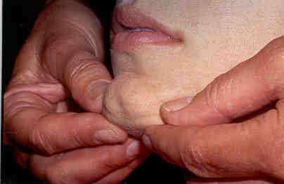
36 Submental Nodes


37 Submandibular Nodes Roll your fingers against inner surface of Mandible with patient's head gently tilted towards one side.

38 Parotid (Preauricular) Nodes Roll your finger in front of the ear, against the maxilla.

39 Post auricular (Mastoid Nodes)

40 Occipital Nodes


41 External jugular chain It lies superficial to sternomastoid. Internal jugular chain Examine the upper, middle and lower groups. Many of them lie deep to sternomastoid muscle which may need to be displaced posteriorly.

42 Transverse Cervical Nodes Supraclavicular (Scalene Nodes) roll your fingers gently behind the clavicles. Instruct the patient to cough or to bear down like they are having a bowel movement. Occasionally an enlarged lymph node may pop up.
43 Thank you!
Lecture 07. Lymphatic's of Head & Neck. By: Dr Farooq Amanullah Khan PMC
 Lecture 07 Lymphatic's of Head & Neck By: Dr Farooq Amanullah Khan PMC Dated: 28.11.2017 Lymphatic Vessels Of the 800 lymph nodes in the human body, 300 are in the Head & neck region. The lymphatic vessels
Neck-2. Dr. Heba Kalbouneh Associate Professor of Anatomy and Histology
 Neck-2 ` Dr. Heba Kalbouneh Associate Professor of Anatomy and Histology Triangles of the neck Side of the neck Midline Lower border of mandible Line between angle of mandible and mastoid Superior nuchal
Veins of the Face and the Neck
 Veins of the Face and the Neck Facial Vein The facial vein is formed at the medial angle of the eye by the union of the supraorbital and supratrochlear veins. connected through the ophthalmic veins with
The Neck the lower margin of the mandible above the suprasternal notch and the upper border of the clavicle
 The Neck is the region of the body that lies between the lower margin of the mandible above and the suprasternal notch and the upper border of the clavicle below Nerves of the neck Cervical Plexus Is formed
PCM1 Physical Exam Skills Session: Head and Neck FACILITATOR & STUDENT COPY
 PATIENT CENTERED MEDICINE - 1 GOALS & OUTCOMES: PCM1 Physical Exam Skills Session: Head and Neck FACILITATOR & STUDENT COPY 1. To introduce the applied anatomy relevant for the examination of the head
Head & Neck Contouring
 Head & Neck Contouring Presented by James Wheeler, MD Center for Cancer Care Goshen, IN 46526 September 12, 2014 Special Thanks to: Spencer Boulter, Director of Operations (AAMD) Adam Moore, RT(T), CMD
The Neck. BY: Lina Abdullah & Rahaf Jreisat
 The Neck BY: Lina Abdullah & Rahaf Jreisat Boundaries of the Neck: generally from base of the skull to root of the neck Superior margin :From superior nuchal line of occipital bone up to mastoid process
Anatomy: head and Neck (6 questions) 1. Prevertebral Flexor Musculature (lying in front of the vertebrae) include all, EXCEPT: Longus Colli.
 1. Prevertebral Flexor Musculature (lying in front of the vertebrae) include all, EXCEPT: Longus Colli.") Anatomy: head and Neck (6 questions) 1. Prevertebral Flexor Musculature (lying in front of the vertebrae) include all, EXCEPT: Longus Colli. Rectus Capitis Anterior. Rectus Capitis Lateralis. Rectus Capitis
Anatomy: head and Neck (6 questions) 1. Prevertebral Flexor Musculature (lying in front of the vertebrae) include all, EXCEPT: Longus Colli. Rectus Capitis Anterior. Rectus Capitis Lateralis. Rectus Capitis
REVIEW/PREVIEW OF HEAD AND NECK ANATOMY FOR ENT EXAM
 REVIEW/PREVIEW OF HEAD AND NECK ANATOMY FOR ENT EXAM - 2017 PALPATE CAROTID ARTERY: AT LEVEL OF CAROTID BIFURCATION VERTEBRAL LEVEL C4 Sternocleidomastoid Muscle INTERNAL CAROTID EXTERNAL CAROTID COMMON
Chapter 13: Mass in the Neck. Raymond P. Wood II:
 Chapter 13: Mass in the Neck Raymond P. Wood II: In approaching the problem of a mass in the neck, one immediately encounters the fact that there are normally palpable masses in the neck (eg, almost all
AJCC Staging of Head & Neck Cancer (7 th edition, 2010) -LIP & ORAL CAVITY-
 -LIP & ORAL CAVITY-") TX: primary tumor cannot be assessed T0: no evidence of primary tumor Tis: carcinoma in situ. T1: tumor is 2 cm or smaller AJCC Staging of Head & Neck Cancer (7 th edition, 2010) -LIP & ORAL CAVITY- T2:
TX: primary tumor cannot be assessed T0: no evidence of primary tumor Tis: carcinoma in situ. T1: tumor is 2 cm or smaller AJCC Staging of Head & Neck Cancer (7 th edition, 2010) -LIP & ORAL CAVITY- T2:
Dr.Ban I.S. head & neck anatomy 2 nd y. جامعة تكريت كلية طب االسنان املرحلة الثانية أ.م.د. بان امساعيل صديق 6102/6102
 جامعة تكريت كلية طب االسنان التشريح مادة املرحلة الثانية أ.م.د. بان امساعيل صديق 6102/6102 Parotid region The part of the face in front of the ear and below the zygomatic arch is the parotid region. The
The importance of knowing the lymphatic spread patterns of head and neck cancer for accurate nodal staging on CT: A practical schematic guide
 The importance of knowing the lymphatic spread patterns of head and neck cancer for accurate nodal staging on CT: A practical schematic guide Alba L. Reyes Ortiz, MD Elena Capilla, MD. Lina Cruz Hernández,
Alexander C Vlantis. Selective Neck Dissection 33
 05 Modified Radical Neck Dissection Type II Alexander C Vlantis Selective Neck Dissection 33 Modified Radical Neck Dissection Type II INCISION Various incisions can be used for a neck dissection. The incision
Posterior Triangle of the Neck By Prof. Dr. Muhammad Imran Qureshi
 Posterior Triangle of the Neck By Prof. Dr. Muhammad Imran Qureshi For the purpose of anatomical description the neck is sub divided into two major triangles, the Anterior and the Posterior by muscle bellies
Tikrit University collage of dentistry Dr.Ban I.S. head & neck anatomy 2 nd y. Lec [5] / Temporal fossa :
![Tikrit University collage of dentistry Dr.Ban I.S. head & neck anatomy 2 nd y. Lec [5] / Temporal fossa :](/thumbs/88/115294566.jpg "Tikrit University collage of dentistry Dr.Ban I.S. head & neck anatomy 2 nd y. Lec [5] / Temporal fossa :") Lec [5] / Temporal fossa : Borders of the Temporal Fossa: Superior: Superior temporal line. Inferior: gap between zygomatic arch and infratemporal crest of sphenoid bone. Anterior: Frontal process of the
Lec [5] / Temporal fossa : Borders of the Temporal Fossa: Superior: Superior temporal line. Inferior: gap between zygomatic arch and infratemporal crest of sphenoid bone. Anterior: Frontal process of the
Thyroid INTRODUCTION ANATOMY SUMMARY OF CHANGES
 AJC 7/14/06 1:19 PM Page 67 Thyroid C73.9 Thyroid gland SUMMARY OF CHANGES Tumor staging (T) has been revised and the categories redefined. T4 is now divided into T4a and T4b. Nodal staging (N) has been
Surgical Anatomy of the Neck. M. J. Jurkiewicz, John Bostwick. Surgical Clinics of North America, Vol 54, No 6, December 1974.
 Surgical Anatomy of the Neck M. J. Jurkiewicz, John Bostwick Surgical Clinics of North America, Vol 54, No 6, December 1974. The radical neck dissection is a safe, effective therapeutic procedure for eradication
Prevertebral Region, Pharynx and Soft Palate
 Unit 20: Prevertebral Region, Pharynx and Soft Palate Dissection Instructions: Step1 Step 2 Step 1: Insert your fingers posterior to the sternocleidomastoid muscle, vagus nerve, internal jugular vein,
ANTERIOR CERVICAL TRIANGLE (Fig. 2.1 )
") 2 Neck Anatomy ANTERIOR CERVICAL TRIANGLE (Fig. 2.1 ) The boundaries are: Lateral: sternocleidomastoid muscle Superior: inferior border of the mandible Medial: anterior midline of the neck This large triangle
2 Neck Anatomy ANTERIOR CERVICAL TRIANGLE (Fig. 2.1 ) The boundaries are: Lateral: sternocleidomastoid muscle Superior: inferior border of the mandible Medial: anterior midline of the neck This large triangle
Dr. Muhammad Shamim. Assistant Professor, Dept. of Surgery College of Medicine, Prince Sattam bin Abdulaziz University
 Dr. Muhammad Shamim FCPS (Pak), FACS (USA), FICS (USA). JMHPE (Nl & Eg) Assistant Professor, Dept. of Surgery College of Medicine, Prince Sattam bin Abdulaziz University Email: surgeon.shamim@gmail.com
Anterior triangle of neck
 Anterior triangle of neck Dept. of Anatomy Zhou Hong Ying Outline boundary and subdivisions of ant. triangle contents of the triangle Muscles: suprahyoid muscles, infrahyoid muscles Nerves: CNⅩ, CNⅪ, CNⅫ,
Lymphatic System and Immunity. Lymphatic System
 Lymphatic System and Immunity Lymphatic System Lymphatic System High hydrostatic pressure in the arterioles and capillaries at the arterial part of the circulation leads to move plasma fluid from the capillaries
University of Palestine. Midterm Exam 2013/2014 Total Grade:
 Course No: DNTS2208 Course Title: Head and Neck Anatomy Date: 09/11/2013 No. of Questions: (50) Time: 1hour Using Calculator (No) University of Palestine Midterm Exam 2013/2014 Total Grade: Instructor
Subdivided into Vestibule & Oral cavity proper
 Extends from the lips to the oropharyngeal isthmus The oropharyngeal isthmus: Is the junction of mouth and pharynx. Is bounded: Above by the soft palate and the palatoglossal folds Below by the dorsum
THYROID & PARATHYROID. By Prof. Saeed Abuel Makarem & Dr. Sanaa Al-Sharawy
 THYROID & PARATHYROID By Prof. Saeed Abuel Makarem & Dr. Sanaa Al-Sharawy 1 OBJECTIVES By the end of the lecture, the student should be able to: Describe the shape, position, relations and structure of
Face. Definition: The area between the two ears and from the chin to the eye brows. The muscles of the face
 Face Definition: The area between the two ears and from the chin to the eye brows. The muscles of the face The muscle of facial expression (include the muscle of the face and the scalp). All are derived
Since the first description of the radical neck dissection by George Crile almost a century
 ORIGINAL ARTICLE Neck Dissection Classification Update Revisions Proposed by the American Head and Neck Society and the American Academy of Otolaryngology Head and Neck Surgery K. Thomas Robbins, MD; Garry
For the following questions, indicate the letter that corresponds to the SINGLE MOST APPROPRIATE ANSWER
 GROSS ANATOMY EXAMINATION May 15, 2000 For the following questions, indicate the letter that corresponds to the SINGLE MOST APPROPRIATE ANSWER 1. Pain associated with an infection limited to the middle
Head and Neck Examination
 Head and Neck Examination Statement of Goals Understand and perform an examination of the head and neck. Learning Objectives Head Ears Nose Sinus A. Describe the anatomy of the head, including regions
Lymphatic and Immune Systems
 Lymphatic and Immune www.vastaccess.com 2 Specialized component of circulatory system Lymphatic system functions: Maintenance of internal fluid balance Immunity Lymph derived from blood and tissue fluid
A Pathologist s Guide to Neck Dissection. Neck Dissections. Lymphatics of head and neck. Neck Dissections
 A Pathologist s Guide to Neck Dissection North American Society for Head and Neck Pathology Companion Meeting 2006 Sigrid Wayne, M.D. Department of Pathology University of Iowa The presence of cervical
Tikrit University College of Dentistry Dr.Ban I.S. head & neck anatomy 2 nd y.
 Lec [3]/The scalp The scalp extends from the supraorbital margins anteriorly to the nuchal lines at the back of the skull and down to the temporal lines at the sides. The forehead, from eyebrows to hairline,
Head & Neck Clinical Sub Group. Network Agreed Imaging Guidelines for UAT and Thyroid Cancer. Measure Nos: 11-1C-105i & 11-1C-106i
 Greater Manchester, Lancashire & South Cumbria Strategic Clinical Network & Senate Head & Neck Clinical Sub Group Network Agreed Imaging Guidelines for UAT and Thyroid Cancer Measure Nos: 11-1C-105i &
Lecture 01. The Thyroid & Parathyroid Glands. By: Dr Farooq Khan PMC Date: 12 th March. 2018
 Lecture 01 The Thyroid & Parathyroid Glands By: Dr Farooq Khan PMC Date: 12 th March. 2018 INTRODUCTION LAYERS OF THE NECK The neck has four major compartments or layer which are enclosed by an outer musculofascial
BIOE221. Session 03. Head and Neck Assessment. Bioscience Department. Endeavour College of Natural Health endeavour.edu.au
 BIOE221 Session 03 Head and Neck Assessment Bioscience Department Session Objectives Understand the importance and rationale for the examinations of the mouth and throat Understand the importance and rationale
Thyroid and Parathyroid Glands
 Thyroid and Parathyroid Glands Please view our Editing File before studying this lecture to check for any changes. Color Code Important Doctors Notes Notes/ explanation Objectives: By the end of the lecture,
Tympanic Bulla Temporal Bone. Digastric Muscle. Masseter Muscle
 Superior view Hyoid Bone The hyoid bone does not articulate with any other bones. It is held in place by ligaments to the styloid process of the temporal bone and the thyroid cartilage of the larynx. It
Basic Anatomy and Physiology of the Lips and Oral Cavity. Dr. Faghih
 Basic Anatomy and Physiology of the Lips and Oral Cavity Dr. Faghih It is divided into seven specific subsites : 1. Lips 2. dentoalveolar ridges 3. oral tongue 4. retromolar trigone 5. floor of mouth 6.
Dr. Sami Zaqout Faculty of Medicine IUG
 Auricle External Ear External auditory meatus The Ear Middle Ear (Tympanic Cavity) Auditory ossicles Internal Ear (Labyrinth) Bony labyrinth Membranous labyrinth External Ear Auricle External auditory
Thyroid gland. importance. relations and connections. external laryngeal nerves. malformations.
 Thyroid gland 1. Recognize and understand the coverings of the thyroid gland and their clinical importance. 2. Recognize and understand the main parts of the thyroid gland and their locations, relations
OBJECTIVE: To obtain a fundamental knowledge of the root of the neck with respect to structure and function
 The root of the neck Jeff Dupree, Ph.D. e mail: jldupree@vcu.edu OBJECTIVE: To obtain a fundamental knowledge of the root of the neck with respect to structure and function READING ASSIGNMENT: Moore and
The Pharynx. Dr. Nabil Khouri MD. MSc, Ph.D
 The Pharynx Dr. Nabil Khouri MD. MSc, Ph.D Introduction The pharynx is the Musculo-fascial halfcylinder that links the oral and nasal cavities in the head to the larynx and esophagus in the neck Common
AIRWAY MANAGEMENT SUZANNE BROWN, CRNA
 AIRWAY MANAGEMENT SUZANNE BROWN, CRNA OBJECTIVE OF LECTURE Non Anesthesia Sedation Providers Review for CRNA s Informal Questions encouraged 2 AIRWAY MANAGEMENT AWARENESS BASICS OF ANATOMY EQUIPMENT 3
Carcinoma of Unknown Primary site (CUP) in HEAD & NECK SURGERY
 in HEAD & NECK SURGERY") Carcinoma of Unknown Primary site (CUP) in HEAD & NECK SURGERY SEARCHING FOR THE PRIMARY? P r o f J P P r e t o r i u s H e a d : C l i n i c a l U n i t C r i t i c a l C a r e U n i v e r s i t y O f
Carcinoma of Unknown Primary site (CUP) in HEAD & NECK SURGERY SEARCHING FOR THE PRIMARY? P r o f J P P r e t o r i u s H e a d : C l i n i c a l U n i t C r i t i c a l C a r e U n i v e r s i t y O f
The Lymphatic System
 94 The Lymphatic System Lymph Lymph is the name for tissue fluid that enters lymph capillaries. Filtration in capillaries creates tissue fluid, most of which returns almost immediately to the blood in
CLINICAL MEDICATION POLICY
 CLINICAL MEDICATION POLICY Policy Name: Opdivo (nivolumab) injection Policy Number: Approved By: Medical Management, Clinical Pharmacy Products: Highmark Health Options Application: All participating hospitals
General Anatomic Layout
 General Anatomic Layout 2 Core Messages At the start of the dissection exercise, we must take a panoramic look for orientation. We then establish the limits of the area of operation and the main landmarks.
Ultrasonographic identification of the anatomical landmarks that define cervical lymph nodes spaces
 Review Med Ultrason 2013, Vol. 15, no. 1, 29-34 DOI: 10.11152/mu.2013.2066.151.lml1uia2 Ultrasonographic identification of the anatomical landmarks that define cervical lymph nodes spaces Lavinia Manuela
APRIL
 APRIL - 2003 OCTOBER - 2003 February 2009 [KU 652] Sub. Code : 4131 FIRST B.D.S DEGREE EXAMINATION (Modified Regulations III) Paper I HUMAN ANATOMY, HISTOLOGY AND EMBRYOLOGY Time : Three hours
SCHOOL OF ANATOMICAL SCIENCES Mock Run Questions. 4 May 2012
 SCHOOL OF ANATOMICAL SCIENCES Mock Run Questions 4 May 2012 1. With regard to the muscles of the neck: a. the platysma muscle is supplied by the accessory nerve. b. the stylohyoid muscle is supplied by
Oral cavity : consist of two parts: the oral vestibule and the oral cavity proper. Oral vestibule : is slit like space between.
 Oral cavity Oral cavity : consist of two parts: the oral vestibule and the oral cavity proper Oral vestibule : is slit like space between the teeth, buccal gingiva, lips, and cheeks 1 Oral cavity Oral
PTERYGOPALATINE FOSSA
 PTERYGOPALATINE FOSSA Outline Anatomical Structure and Boundaries Foramina and Communications with other spaces and cavities Contents Pterygopalatine Ganglion Especial emphasis on certain arteries and
AJCC Cancer Staging 8 th edition. Lip and Oral Cavity Oropharynx (p16 -) and Hypopharynx Larynx
 and Hypopharynx Larynx") AJCC Cancer Staging 8 th edition Lip and Oral Cavity Oropharynx (p16 -) and Hypopharynx Larynx AJCC 7 th edition Lip and Oral cavity Pharynx Larynx KEY CHANGES Skin of head and neck (Vermilion of the lip)
AJCC Cancer Staging 8 th edition Lip and Oral Cavity Oropharynx (p16 -) and Hypopharynx Larynx AJCC 7 th edition Lip and Oral cavity Pharynx Larynx KEY CHANGES Skin of head and neck (Vermilion of the lip)
Anatomy of Oral Cavity DR. MAAN AL-ABBASI
 Anatomy of Oral Cavity DR. MAAN AL-ABBASI By the end of this lecture you should be able to: 1. Differentiate different parts of the oral cavity 2. Describe the blood and nerve supply of mucosa and muscles
Head and Neck Tumours
 Head and Neck Tumours Introductory Notes The following sites are included: Lip, oral cavity Pharynx: oropharynx, nasopharynx, hypopharynx Larynx: supraglottis, glottis, subglottis Nasal cavity and paranasal
Lies in front and sides of the neck. Consists of two lobe connected anterior to the trachea by an isthmus.
 THYROID GLAND 1 Lies in front and sides of the neck. Consists of two lobe connected anterior to the trachea by an isthmus. A small pyramidal lobe projects upwards from the left lobe in 40% of cases. The
SYLLABUS BDS I PROFESSIONAL GENERAL HUMAN ANATOMY INCLUDING EMBRYOLOGY AND HISTOLOGY
 GENERAL HUMAN ANATOMY INCLUDING EMBRYOLOGY AND HISTOLOGY I. General Anatomy 1. Anatomical terms 2. Skin, superficial fascia & deep fascia 3. Cardiovascular system, portal system, collateral circulation
Candidate s instructions Look at this cross-section taken at the level of C5. Answer the following questions.
 Section 1 Anatomy Chapter 1. Trachea 1 Candidate s instructions Look at this cross-section taken at the level of C5. Answer the following questions. Pretracheal fascia 1 2 5 3 4 Questions 1. Label the
Ultrasound Interpretation of Non-Thyroid Neck Pathology
 Ultrasound Interpretation of Non-Thyroid Neck Pathology Kevin T. Brumund, M.D., F.A.C.S. Associate Professor of Surgery Head and Neck Surgery University of California, San Diego Health Sciences VA Medical
The importance of knowing the lymphatic spread patterns of head and neck cancer for accurate nodal staging on CT: A practical schematic guide
 The importance of knowing the lymphatic spread patterns of head and neck cancer for accurate nodal staging on CT: A practical schematic guide Poster No.: C-0483 Congress: ECR 2014 Type: Educational Exhibit
Anatomy and Physiology. Bones, Sutures, Teeth, Processes and Foramina of the Human Skull
 Anatomy and Physiology Chapter 6 DRO Bones, Sutures, Teeth, Processes and Foramina of the Human Skull Name: Period: Bones of the Human Skull Bones of the Cranium: Frontal bone: forms the forehead and the
Infratemporal fossa: Tikrit University college of Dentistry Dr.Ban I.S. head & neck Anatomy 2 nd y.
 Infratemporal fossa: This is a space lying beneath the base of the skull between the lateral wall of the pharynx and the ramus of the mandible. It is also referred to as the parapharyngeal or lateral pharyngeal
HEAD & NECK ANATOMY - MCQ HEAD & NECK ANATOMY
 . ' HEAD & NECK ANATOMY I. Deep investing layer of cervical fascia splits to enclose: A. Sternocleidomastoid B. Trapezius C. Parotid gland D. Omohyoid 2. Regarding the prevertebral fascia, the following
Introduction to Head and Neck Anatomy
 Introduction to Head and Neck Anatomy Nervous Tissue Controls and integrates all body activities within limits that maintain life Three basic functions 1. sensing changes with sensory receptors 2. interpreting
The peripheral (secondary) lymphoid tissues
 lymphoid tissues") The peripheral (secondary) lymphoid tissues The peripheral (secondary) lymphoid tissues : are the lymph nodes, spleen, Mucosal associated lymphoid tissue (MALT). All secondary lymphoid organs have one
The peripheral (secondary) lymphoid tissues The peripheral (secondary) lymphoid tissues : are the lymph nodes, spleen, Mucosal associated lymphoid tissue (MALT). All secondary lymphoid organs have one
ANATOMY & PHYSIOLOGY ONLINE COURSE - SESSION 11 THE LYMPHATIC SYSTEM AND IMMUNITY
 ANATOMY & PHYSIOLOGY ONLINE COURSE - SESSION 11 THE LYMPHATIC SYSTEM AND IMMUNITY Functions of the Lymphatic System The lymphatic system has three primary functions. First of all, it returns excess interstitial
FACULTY OF MEDICINE SIRIRAJ HOSPITAL
 Neck Dissection Pornchai O-charoenrat MD, PhD Division of Head, Neck and Breast Surgery Department of Surgery FACULTY OF MEDICINE SIRIRAJ HOSPITAL Introduction Status of the cervical lymph nodes is the
Mediastinum and pericardium
 Mediastinum and pericardium Prof. Abdulameer Al-Nuaimi E-mail: a.al-nuaimi@sheffield.ac.uk E. mail: abdulameerh@yahoo.com The mediastinum: is the central compartment of the thoracic cavity surrounded by
Over the past 18 years, numerous classifications have been proposed to distinguish
 ORGNAL ARTCLE An maging-based Classification for the Cervical Nodes Designed as an Adjunct to Recent Clinically Based Nodal Classifications Peter M. Som, MD; Hugh D. Curtin, MD; Anthony A. Mancuso, MD
WOMEN'S INTERAGENCY HIV STUDY ORAL PROTOCOL FORM OP 4: ORAL MUCOSAL TISSUE EXAM
 WOMEN'S INTERAGENCY HIV STUDY ORAL PROTOCOL FORM OP 4: ORAL MUCOSAL TISSUE EXAM COMPLETING THE FORM GENERAL INFORMATION Affix the Participant ID label in the space indicated. Record the visit number. Be
Chapter 84: Surgical Anatomy. Raleigh E. Lingeman
 Chapter 84: Surgical Anatomy Raleigh E. Lingeman Surgeons doing head and neck surgery must first master the surgical anatomy and technique of doing neck dissection. Neck dissection is either the classic
ANTERIOR CERVICAL TRIANGLE (FIG. 2.1 )
") 2 Neck Anatomy ANTERIOR CERVICAL TRIANGLE (FIG. 2.1 ) The boundaries are: Lateral: sternocleidomastoid muscle Superior: inferior border of the mandible Medial: anterior midline of the neck This large triangle
2 Neck Anatomy ANTERIOR CERVICAL TRIANGLE (FIG. 2.1 ) The boundaries are: Lateral: sternocleidomastoid muscle Superior: inferior border of the mandible Medial: anterior midline of the neck This large triangle
Cervical Lymph Nodes
 Cervical Lymph Nodes Diana Gaitini, MD Unit of Ultrasound, Department of Medical Imaging Rambam Medical Center and Faculty of Medicine Technion, Israel Institute of Technology Haifa, Israel Learning Targets
Human Anatomy and Physiology - Problem Drill 20: Immunity and the Lymphatic System
 Human Anatomy and Physiology - Problem Drill 20: Immunity and the Lymphatic System Question No. 1 of 10 The lymphatic system is formed early during human development. Which of the following statements
Sinusoids and venous sinuses
 LYMPHOID SYSTEM General aspects Consists of organs that are made of lymphoid tissue; Immune defense Breakdown of red blood cells. 1 Sinusoids In place of capillaries Endothelium; often fenestrated More
Chapter 5: Other mediastinal structures. The Large Arteries. The Aorta. Ascending aorta
 Chapter 5: Other mediastinal structures The Large Arteries The Aorta The aorta is the main arterial trunk of the systemic circulation and in the healthy state its wall contain a large amount of yellow
Nasal Cavity CS Tumor Size (Revised: 02/03/2010)
") Nasal Cavity C30.0 C30.0 Nasal cavity (excludes nose, NOS C76.0) Note: Laterality must be coded for this site, except subsites Nasal cartilage and Nasal septum, for which laterality is coded 0. CS Tumor
Nasal Cavity C30.0 C30.0 Nasal cavity (excludes nose, NOS C76.0) Note: Laterality must be coded for this site, except subsites Nasal cartilage and Nasal septum, for which laterality is coded 0. CS Tumor
Tracing the Cranial Nerves Osteologically
 CN I II III IV V 1 Supra-orbital ethmoidal nn. Ext. nasal V 2 Tracing the Cranial Nerves Osteologically Nucleus of Origin Olfactory tracts of frontal lobe of cerebrum Optic tracts from optic chiasma and
General Anatomy p. 1 Organization of the Human Body p. 1 Skeleton of the Human Body p. 4 Ossification of the Bones p. 6 Bone Structure p. 8 Joints p.
 General Anatomy p. 1 Organization of the Human Body p. 1 Skeleton of the Human Body p. 4 Ossification of the Bones p. 6 Bone Structure p. 8 Joints p. 10 Principal Joints (Immovable) p. 12 Synovial Joints
Anatomical Considerations for Lab Practical II
 Anatomical Considerations for Lab Practical II For each of the following please be prepared to provide: Identification System Organ(s) or ducts to Function(s) location which it is attached Use your lecture
DESCRIPTION: This is the part of the trunk, which is located between the root of the neck and the superior border of the abdominal region.
 1 THE THORACIC REGION DESCRIPTION: This is the part of the trunk, which is located between the root of the neck and the superior border of the abdominal region. SHAPE : T It has the shape of a truncated
ANATOMY & PHYSIOLOGY II
 ANATOMY & PHYSIOLOGY II THE BODY SYSTEMS Anatomy & Physiology II The Body Systems Michelle Cochrane 2014 All rights reserved. This material is subject to copyright and may not be reprinted or reproduced
INTRA/EXTRA ORAL EXAMINATION
 INTRA/EXTRA ORAL EXAMINATION www.dentalelle.com Dentalelle Tutoring Intraoral/Extraoral Examination 1. If you notice a typical finding after reviewing the head and neck on a new patient, what does this
Temporal region. temporal & infratemporal fossae. Zhou Hong Ying Dept. of Anatomy
 Temporal region temporal & infratemporal fossae Zhou Hong Ying Dept. of Anatomy Temporal region is divided by zygomatic arch into temporal & infratemporal fossae. Temporal Fossa Infratemporal fossa Temporal
Cranial nerves.
 Cranial nerves eaglezhyxzy@163.com Key Points of Learning Name Components Passing through Peripheral distribution Central connection Function Cranial nerves Ⅰ olfactory Ⅱ optic Ⅲ occulomotor Ⅳ trochlear
The following images were all acquired using a CTI Biograph
 Positron Emission Tomography/ Computed Tomography Imaging of Head and Neck Tumors: An Atlas Michael M. Graham, MD, PhD, and Yusuf Menda, MD Department of Radiology, University of Iowa, Iowa City, IA. Address
ORAL CAVITY, ESOPHAGUS AND STOMACH
 ORAL CAVITY, ESOPHAGUS AND STOMACH 1 OBJECTIVES By the end of the lecture you should be able to: Describe the anatomy the oral cavity, (boundaries, parts, nerve supply). Describe the anatomy of the palate,
Case Scenario #1 Larynx
 Case Scenario #1 Larynx 56 year old white female who presented with a 2 month history of hoarseness treated with antibiotics, but with no improvement. In the last 3 weeks, she has had a 15 lb weight loss,
Dr.Ban I.S. head & neck anatomy 2 nd y. جامعة تكريت كلية طب االسنان املرحلة الثانية
 جامعة تكريت كلية طب االسنان التشريح مادة املرحلة الثانية أ.م.د. بان امساعيل صديق 6102-6102 1 The Palate The palate forms the roof of the mouth and the floor of the nasal cavity. It is divided into two
Anatomy of the Thyroid Gland
 Anatomy of the Thyroid Gland Introduction Nomenclature G, thyreos= shield, eidos= like Location Root of the neck ventrally (C5-T1) Function endocrine gland that secretes: Thyroxine (T4) T3 Calcitonin LWW,
3-Deep fascia: is absent (except over the parotid gland & buccopharngeal fascia covering the buccinator muscle)
") The Face 1-Skin of the Face The skin of the face is: Elastic Vascular (bleed profusely however heal rapidly) Rich in sweat and sebaceous glands (can cause acne in adults) It is connected to the underlying
The Face 1-Skin of the Face The skin of the face is: Elastic Vascular (bleed profusely however heal rapidly) Rich in sweat and sebaceous glands (can cause acne in adults) It is connected to the underlying
Nose & Mouth OUTLINE. Nose. - Nasal Cavity & Its Walls. - Paranasal Sinuses. - Neurovascular Structures. Mouth. - Oral Cavity & Its Contents
 Dept. of Human Anatomy, Si Chuan University Zhou hongying eaglezhyxzy@163.com Nose & Mouth OUTLINE Nose - Nasal Cavity & Its Walls - Paranasal Sinuses - Neurovascular Structures Mouth - Oral Cavity & Its
Ch16: Respiratory System
 Ch16: Respiratory System Function: - O2 in and CO2 out of the blood vessels in the lungs - O2 out and CO2 into the blood vessels around the cells - Gas exchange happens in - Other organs purify, humidify,
Temporal fossa Infratemporal fossa Pterygopalatine fossa Terminal branches of external carotid artery Pterygoid venous plexus
 Outline of content Temporal fossa Infratemporal fossa Pterygopalatine fossa Terminal branches of external carotid artery Pterygoid venous plexus Boundary Content Communication Mandibular division of trigeminal
Structure and Nerve Supply of The Larynx
 Kingdom of Bahrain Arabian Gulf University College of Medicine and Medical sciences Structure and Nerve Supply of The Larynx This presentation was originally prepared by: Dr. Kumar Notes were added by:
Compliance Department ELEMENTS OF EAR, NOSE AND THROAT EXAMINATION 11/2010
 Compliance Department ELEMENTS OF EAR, NOSE AND THROAT EXAMINATION 11/2010 System/ Body Area Constitutional Measurement of any three of the following seven vital signs: 1) sitting or standing blood pressure,