Approach to Liver Lesions. Anjana A. Pillai, MD Associate Professor of Medicine Director, Liver Tumor Clinic The University of Chicago Medical Center
|
|
|
- Barnard Butler
- 5 years ago
- Views:
Transcription


1 Approach to Liver Lesions Anjana A. Pillai, MD Associate Professor of Medicine Director, Liver Tumor Clinic The University of Chicago Medical Center
2 Objectives Identify common clinical features and imaging characteristics of benign and malignant liver masses Identify which benign lesions require further follow-up and management Review key imaging characteristics of hepatocellular carcinoma and cholangiocarcinoma
3 Three Categories of Liver Masses Benign lesions typically requiring no further imaging Benign lesions requiring further follow-up and management Malignant lesions requiring appropriate therapy Lewis, R. Clinical Approach to Liver Mass Lesions. In: Mayo Clin Gastro and Hep Board Review, Hauser SC, ed. 2015, 5 th ed, Ch 24:
4 How do I approach liver lesions? First thing is first - take a good history! Is there underlying liver disease? Is the patient symptomatic vs incidentally found? Is the lesion new? Is the lesion growing? Is the imaging study adequate? Then examine the patient! What did their physical exam tell me? Finally, ALWAYS, ALWAYS personally review the imaging
5 BENIGN LESIONS NOT REQUIRING FOLLOW-UP Simple liver cysts Cavernous hemangioma Focal Nodular Hyperplasia Focal fatty change (fat sparing) in liver Angiolipoma BENIGN LESIONS REQUIRING FOLLOW-UP Hepatic adenoma Pyogenic Liver Abscess Nodular Regenerative Hyperplasia Biliary Cystadenoma Inflammatory pseudotumor Granulomatous abscesses Amebic Liver abscess Echinococcal Cysts
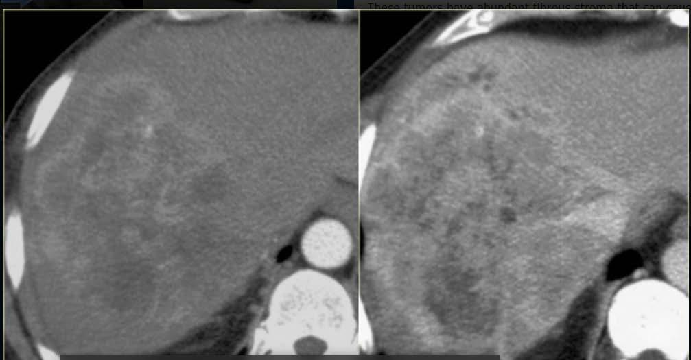
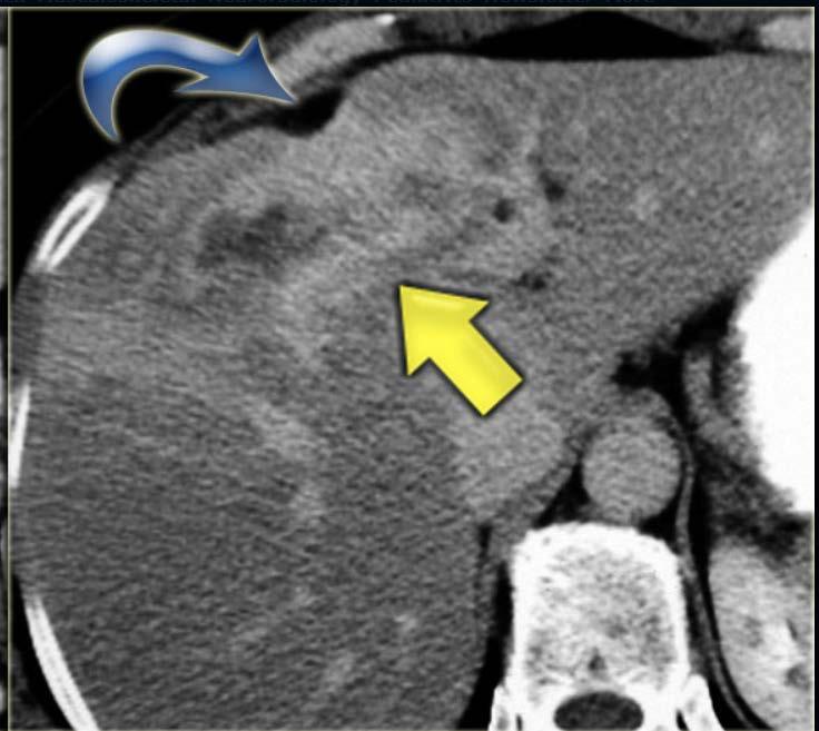
6 Question #1 A 25 yo AA woman with long standing asthma was noted to have an incidentally found liver lesion on cuts from her CT chest. A dedicated MRI and CT with contrast of the abdomen described the lesion as a large 7 cm central arterially enhancing mass (extending into segment 7,8,4A and1) with fading/isoenhancement on venous phase with no wash out on delayed images. No definitive central scar noted. The liver appeared morphologically normal.
 with fading/isoenhancement on venous")
7 Question #1 A 25 yo AA woman with long standing asthma was noted to have an incidentally found liver lesion on cuts from her CT chest. A dedicated MRI and CT with contrast of the abdomen described the lesion as a large 7 cm central arterially enhancing mass (extending into segment 7,8,4A and1) with fading/isoenhancement on venous phase with no wash out on delayed images. No definitive central scar noted. The liver appeared morphologically normal.
8 What is your next step in management? A. No further work-up needed B. Repeat imaging in 6-12 months C. Repeat imaging now with a different modality D. Biopsy the lesion E. Surgical resection
9
10 What is your next step in management? A. No further work-up needed B. Repeat imaging in 6-12 months C. Repeat imaging now with a different modality D. Biopsy the lesion E. Surgical resection
11 Liver Cysts Cysts are thin-walled structures lined by cuboidal bile duct epithelium and filled with isotonic fluid. Female predominance Can be solitary or multiple Often asymptomatic unless very large Often coexist with other mass lesions Prevalence increases with age Ultrasound best imaging modality Biopsy is not needed
 Smooth with round margins No internal echoes No flow on")
12 Liver Cyst - US Anechoic = black Increased through transmission (white around it) Smooth with round margins No internal echoes No flow on doppler

13 Liver Cyst - CT <10 Hounsfield units Same density as water Don t change with contrast


14 Liver Cyst MRI Bright on T2 Dark on T1 No enhancement T2 MR T1 Portal venous phase
15 Case Presentation 55 yo Indian woman presents with dyspnea on exertion. She also notes new right sided abdominal pain/ache for the last 2-3 weeks which is persistent. Pain is worse when she lies on the right side. She has also noted early satiety and poor appetite for last few years but never sought treatment for this because she was able to maintain her weight for the most part. She has noticed progressive mild lower extremity edema for the last few years. No jaundice or dark colored urine. No fevers/chills. No recent travel. Labs: Viral hepatitis serologies negative. LFTs nl, Alb 4.2, Cr 0.7, WBC 2.9 H/H 11.8/35 platelets 138, MCV nl
16
17 Cavernous Hemangioma Extensive network of vascular spaces lined by endothelial cells and separated by thin, fibrous stroma Most common benign liver lesion Most common in women Often asymptomatic Do not undergo malignant transformation and rupture is very rare Can be multicentric in 30% of cases Only require intervention if symptomatic Biopsy not needed Kasabach-Merritt syndrome (DIC, thrombocytopenia, usu in infants)

18 Hemangioma - US Hyperechoic = bright Smooth, wellcircumscribed Homogenous

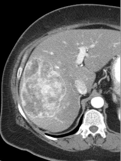
19 Hemangioma CT Peripheral discontinuous enhancement with gradual fill-in.


20 Bright T2 signal (as bright as CSF) Hemangioma MRI Peripheral discontinuous enhancement T2 MR Arterial phase MR with gradual fill-in. Portal venous phase MR Delayed phase MR
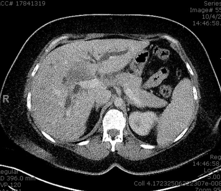
21 Case Presentation 65 yo Somalian man with no known liver disease presents with recent onset of abdominal discomfort, early satiety, 30 lb weight loss and lower extremity edema Labs unremarkable except for lowish albumin and mildly elevated INR Viral serologies negative Imaging showed Went to OR for resection with unremarkable post op course and resolution of symptoms

22 Case Presentation 65 yo Somalian man with no known liver disease presents with recent onset of abdominal discomfort, early satiety, 30 lb weight loss and lower extremity edema Labs unremarkable except for lowish albumin and mildly elevated INR Viral serologies negative Imaging showed Went to OR for resection with unremarkable post op course and resolution of symptoms
23 Focal Fat or Fat Sparing Usually occurs in obese patients or those with diabetes, heavy ETOH use or malnourished (chemotherapy) Focal fat can appear as a mass or alternatively an area of fat sparing can appear as a mass in the background of a fatty liver Asymptomatic Does not displace vessels or distort appearance of the liver Usually found near falciform ligament and around the gallbladder

24 Focal Fat - US Ultrasound Hyperechoic = bright


25 Focal Fat - CT Normal coursing of hepatic vessels through mass

26 Fat Sparing -CT


27 Focal Fat Sparing - MRI In-phase Out-of-phase
28 Focal Nodular Hyperplasia Non-malignant response to a congenital arterial malformation Female predominance Central scar (arterial malformation) is classic but not always seen Relationship to OCP controversial and no data that discontinuing OCPs helps Most are asymptomatic unless large and space-occupying (rare)
29 Focal Nodular Hyperplasia No potential for malignant transformation The term telangiectatic FNH is no longer used and are instead classified as adenoma inflammatory subtype which can have malignant potential Biopsy can be useful in differentiating from adenoma but both lesions may show benign appearing hepatocytes on FNA

30 FNH - US Varies: can be hypo/hyper or isoechoic Most hypoechoic except for central scar Color Doppler increased blood flow in central stellate scar


31 FNH - CT Hyperenhances in arterial phase Isoattenuating in venous phase Delayed phase fades out

32 FNH - MRI Hyperenhances in arterial phase Isoattenuating in venous phase
33 BENIGN LESIONS NOT REQUIRING FOLLOW-UP Simple liver cysts Cavernous hemangioma Focal Nodular Hyperplasia Focal fatty change (fat sparing) in liver Angiolipoma BENIGN LESIONS REQUIRING FOLLOW-UP Hepatic adenoma Pyogenic Liver Abscess Nodular Regenerative Hyperplasia Biliary Cystadenoma Inflammatory pseudotumor Granulomatous abscesses Amebic Liver abscess Echinococcal Cysts
34 Hepatic Adenoma Characterized by sheets of hepatocytes separated by dilated sinusoids and supplied by naked arteries (no venous supply or bile ducts) 4 molecular subgroups: 1) HNF1A-inactivated adenoma 2) telangiectatic or inflammatory subtype 3) B-catenin activated adenomas 4) unclassified hepatocellular adenoma Present in women in the 3 rd and 4 th decade of life Associated with OCPs (can decrease in size after withdrawal, usu no change) relative risk of adenoma formation 2.5x greater if OCP use 3-5 yrs vs 1 yr; if 9 yrs, risk is 25x greater
35 Hepatic Adenoma Obesity and metabolic syndrome also increases risk (esp inflammatory subtype) Associated with MODY-3, glycogen storage disease 1A or 3 and androgenic hormone use Multiple adenomas more common in obese patients and in MODY -3 or glycogen storage disease Hepatic adenomatosis >10 adenomas Rupture also rare but occur in large adenomas (>5 cm)
36 Hepatic Adenoma Malignant transformation rare but seen in: Size >5 cm Male sex Positive B-catenin staining Surgical resection recommended for large adenomas (>5 cm), any size in men, B-catenin +, or unable to distinguish from well-diff HCC Adenomas in pregnant patients need to be monitored closely


37 Hepatic Adenoma - MRI Pre T2 Arterial Delayed
 or gadoxetate disodium (Eovist): not taken up by adenomas but light up with FNH Why?")
38 Is there imaging that can distinguish FNH from hepatic adenoma? Gadobenate dimenglumaine (Multihance) or gadoxetate disodium (Eovist): not taken up by adenomas but light up with FNH Why? Because adenomas have no bile ducts and do not light up in the hepatobiliary scan
39 Biliary Cystadenoma Composed of multiple cysts lined by cuboidal or columnar epithelium that resemble normal biliary epithelium Cystic neoplasms can be unilocular or multilocular More common in middle aged women Asymptomatic unless large Usually benign Can recur after excision Cannot differentiate from cystadenocarcinoma without surgical resection


40 Biliary Cystadenoma CT/MRI CT septae enhances with contrast MRI- Bright on T2
41 Liver Abscesses

42 Hepatic Abscess - US Heterogenous on US


43 Hepatic Abscess - MRI Bright on T2 Dark on T1 T1 portal venous phase - enhances


44 Echinococcal Cysts CT Portal venous phase Large solitary mass or multiple well-defined lesions with internal daughter cysts

45 Echinococcal Cysts MRI Bright on T2
46 Question #2: What is the most common malignant lesion seen in the liver? A. Hepatocellular carcinoma B. Cholangiocarinoma C. Metastases D. Lymphoma E. Cystadenoma
47
48 Question #2: What is the most common malignant lesion seen in the liver? A. Hepatocellular carcinoma B. Cholangiocarinoma C. Metastases D. Lymphoma E. Cystadenoma
49 MALIGNANT LESIONS REQUIRING TREATMENT Liver Metastases Primary HCC Cholangiocarcinoma Mixed HCC/CCA Hemangioendotheliomas Epitheliod angiomyolipoma Mixed epithelial and stromal tumors Sarcomas
50 Case Presentation 29 yo woman becomes jaundiced after delivering her 3 rd child via C-section. Noted to have ALT 319 AST 171 TB 1.2 AP 353 One week later ALT 159 AST 137 TB 14.9 AP 356 Abdominal US shows multiple lesions throughout the liver with IH ductal dilatation CT abdomen shows central hepatic lesion measuring 6x6.3, similar density smaller lesions throughout the liver

51 Liver Metastases Hypoattenuating/hypovascular lesions on CT
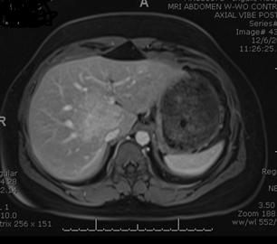
52 Question #3 60 yo Egyptian man with genotype 4 HCV cirrhosis presents with new onset ascites and a new mass in the liver. Physical exam reveals mild temporal wasting, anicteric sclera, moderate abdominal distension with small reducible umbilical hernia, palpable spleen tip, mild lower extremity edema and palmar erythema Labs reveal TB 2, ALT 33 AST 30 alb 2.8 INR 1.6 platelets 55 AFP 249 MRI liver protocol shows a 4.5 cm mass in segment 6 with a pseudocapsule and a hyperenhancing right and main portal vein thrombus.
53 What is the next step in this patient s management? A. Liver transplant evaluation B. Biopsy of lesion C. Microwave ablation/radiofrequency ablation D. Sorafenib E. Surgical resection
54
55 What is the next step in this patient s management? A. Liver transplant evaluation B. Biopsy of lesion C. Microwave ablation/radiofrequency ablation D. Sorafenib E. Surgical resection
56 HCC Always consider HCC if lesion present in cirrhotic or chronic HBV patient Male predominance Biopsy not required for diagnosis (risk of seeding) HCC is supplied by the hepatic artery Classic HCC: enhances in the arterial phase and washes out in portal venous and delayed phase, often has pseudocapsule Treatment options: liver transplant (Milan criteria), resection (small percentage), liver directed therapy (RFA/MWA/TACE/TARE/SBRT), systemic chemo Patients with cirrhosis or chronic HBV 6 month surveillance AFP not sensitive or specific (~40% of HCC does not secrete AFP)

57 HCC - US Heterogenous on US


58 HCC - MRI Enhances in arterial phase Washes out in portal venous/delayed phase


59 HCC - MRI Enhances in arterial phase Washes out in portal venous/delayed phase 20 sec 180 sec


60 Multifocal HCC - MRI Enhances in arterial phase Washes out in portal venous/delayed phase 20 sec 180 sec
61 Imaging Characteristics of HCC FINDING CT MRI Arterial hyperenhancement X X Delayed washout X X T2 bright signal Intralesional fat X X
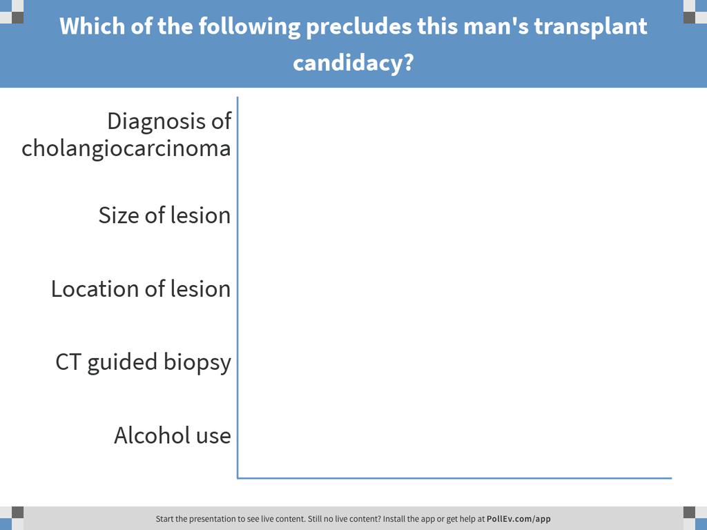
62 Question #4 A 42 yo Korean physician is seen in the local ER with painless jaundice. Upon pressing him further he has noted a loss of appetite, fatigue and 10 lb weight loss in the last 3 months. He otherwise feels well. Does not smoke. Has a glass of wine with dinner most nights. CT scan of the abdomen reveals a 2.5 cm lesion at the bifurcation of the R/L hepatic ducts with upstream L sided ductal dilatation Physical exam reveals healthy appearing man with scleral icterus but otherwise unremarkable exam Labs reveal TB 4.2, ALT 45 AST 30 alb 3.2 INR 1.1 platelets 255 AFP 24 CA CT guided biopsy of the lesion reveals a well-differentiated cholangiocarcinoma ERCP reveals a mass at the bifurcation with upstream ductal dilatation with stent placement. Brushings + adenocarcinoma
63 Question #4: Which of the following precludes this man s transplant candidacy? A. Diagnosis of cholangiocarcinoma B. Size of lesion C. Location of lesion D. CT guided biopsy E. Alcohol use
64
65 Question #4: Which of the following precludes this man s transplant candidacy? A. Diagnosis of cholangiocarcinoma B. Size of lesion C. Location of lesion D. CT guided biopsy E. Alcohol use
66 Cholangiocarcinoma Arise from bile duct epithelium; pathology reveals adenoca Risk factors include PSC (most common in W. countries), liver fluke (Asia), choledochal and cystic disorders Most have no known risk factors and present with advanced disease (painless jaundice and biliary obstruction) Classified as intra- or extra-hepatic CA 19-9 usually elevated (>100 ~65-75% sensitive and 85-95% specific) 60-70% tumors occur at bifurcation of the CHD, 20-30% extrahepatic and 5-15% intrahepatic
67 Cholangiocarcinoma Pain is usually a constant, dull ache in the right upper quadrant Resection and liver transplantation are options for early stage disease; otherwise systemic chemotherapy Most present in late stages with dismal prognosis

68 Mayo protocol for cholangiocarcinoma for Liver Transplantation
69 Cholangiocarcinoma Imaging Characteristics HYPOechoic on US HYPOenhancing (or minimal enhancement) on arterial phase with delayed or progressive HYPERenhancement in venous phase on CT HYPOintense on T1, HYPERintense on T2, peripheral contrast enhancement that progresses into the venous phase on MRI, similar to CT


70 Cholangiocarcinoma HYPOenhancement Delayed HYPERenhancement Arterial phase Delayed phase
71 Cholangiocarcinoma Arterial phase Delayed phase Delayed phase
72 Case Presentation 41 yo woman with no history of chronic liver disease began to experience right sided abdominal pain after the birth of her 3 rd child An abdominal US showed 10 cm mass in liver A dedicated MRI liver showed multiple masses throughout the liver, largest being 10 cm in right inferior lobe TB 0.3 ALT 9 AST 57 AP 152 CA Biopsy of lesion shows adenocarcinoma

73 Case Presentation Arterial phase Venous phase
74 Take Home Points Be cognizant of the quality of the imaging study and phases available Most lesions can be diagnosed with proper imaging and biopsy is infrequently needed Many benign lesions are found incidentally and are not the cause of symptomatology Always error on serial monitoring if lesion is indeterminate and not clearly benign Distinction between FNH and adenoma is not always clear and biopsy may be beneficial Always suspect HCC in a patient with cirrhosis or chronic hepatitis B Early stage cholangiocarcinoma can be treated successfully with liver transplant and knowing criteria can be life saving!
75
Evaluation of Liver Mass Lesions. American College of Gastroenterology 2013 Regional Postgraduate Course
 Evaluation of Liver Mass Lesions American College of Gastroenterology 2013 Regional Postgraduate Course Lewis R. Roberts, MB ChB, PhD Division of Gastroenterology and Hepatology Mayo Clinic College of
Financial Disclosure
 Benign Liver Masses Adil Abdalla, MBBS Creighton University-CHI Health August 25, 2018 Financial Disclosure Nothing to disclose Financial Disclosure 1 Objectives To assess patients with benign liver tumors
Jesse Civan, M.D. Medical Director, Jefferson Liver Tumor Center
 Liver Tumors Jesse Civan, M.D. Medical Director, Jefferson Liver Tumor Center Differential Diagnosis Malignant Metastatic from non-hepatic primary Hepatocellular carcinoma Cholangiocarcinoma Biliary cystcarcinoma
Malignant Focal Liver Lesions
 Malignant Focal Liver Lesions Other Than HCC Pablo R. Ros, MD, MPH, PhD Departments of Radiology and Pathology University Hospitals Cleveland Medical Center Case Western Reserve University Pablo.Ros@UHhospitals.org
HEPATO-BILIARY IMAGING
 HEPATO-BILIARY IMAGING BY MAMDOUH MAHFOUZ MD PROF.OF RADIOLOGY CAIRO UNIVERSITY mamdouh.m5@gmail.com www.ssregypt.com CT ABDOMEN Indications Patient preparation Patient position Scanogram Fasting 4-6 hours
Interesting Cases from Liver Tumor Board. Jeffrey C. Weinreb, M.D.,FACR Yale University School of Medicine
 Interesting Cases from Liver Tumor Board Jeffrey C. Weinreb, M.D.,FACR Yale University School of Medicine jeffrey.weinreb@yale.edu Common Liver Diseases Hemangioma Cyst FNH Focal Fat/Sparing THID Non-Cirrhotic
HEPATOCYTE SPECIFIC CONTRAST MEDIA: WHERE DO WE STAND?
 HEPATOCYTE SPECIFIC CONTRAST MEDIA: WHERE DO WE STAND? Andrew T. Trout, MD @AndrewTroutMD Disclosures No relevant disclosures Outline Review of hepatocyte specific contrast media Review of hepatocellular
Liver Tumors. Prof. Dr. Ahmed El - Samongy
 Liver Tumors Prof. Dr. Ahmed El - Samongy Objective 1. Identify the most important features of common benign liver tumors 2. Know the risk factors, diagnosis, and management of hepatocellular carcinoma
Essentials of Clinical MR, 2 nd edition. 65. Benign Hepatic Masses
 65. Benign Hepatic Masses Pulse sequences acquired for abdominal MRI typically consist of fast acquisition schemes such as single-shot turbo spin echo (i.e. HASTE) and gradient echo schemes such as FLASH
CTA/MRA of Pediatric Hepatic Masses Radiology-Pathology Correlation
 Acta Radiológica Portuguesa, Vol.XVIII, nº70, pág. 41-50, Abr.-Jun., 2006 CTA/MRA of Pediatric Hepatic Masses Radiology-Pathology Correlation Marilyn J. Siegel Mallinckrodt Institute of Radiology, Washington
Radiology of hepatobiliary diseases
 GI cycle - Lecture 14 436 Teams Radiology of hepatobiliary diseases Objectives 1. To Interpret plan x-ray radiograph of abdomen with common pathologies. 2. To know the common pathologies presentation.
Liver Cancer (Hepatocellular Carcinoma or HCC) Overview
 Overview") Liver Cancer (Hepatocellular Carcinoma or HCC) Overview Recent advances in liver cancer care seek to address the rising incidence of liver cancer, which has steadily increased over the past three decades.
Liver Cancer (Hepatocellular Carcinoma or HCC) Overview Recent advances in liver cancer care seek to address the rising incidence of liver cancer, which has steadily increased over the past three decades.
MRI OF FOCAL LESIONS IN
 Introduction MRI OF FOCAL LESIONS IN THE NON-CIRRHOTIC LIVER Ivan Pedrosa M.D. Ph.D. Associate Professor of Radiology and Advanced Imaging Research Center University of Texas Southwestern. Dallas, TX Incidental
Workup of a Solid Liver Lesion
 Workup of a Solid Liver Lesion Joseph B. Cofer MD FACS Chief Quality Officer Erlanger Health System Affiliate Professor of Surgery UTHSC-Chattanooga I have no financial or other relationships with any
ACG Clinical Guideline: Diagnosis and Management of Focal Liver Lesions
 ACG Clinical Guideline: Diagnosis and Management of Focal Liver Lesions Jorge A. Marrero, MD, 1 Joseph Ahn, MD, FACG, 2 K. Rajender Reddy, MD, FACG 3 1 University of Texas at Southwestern, Dallas, Texas,
Hepatobiliary and Pancreatic Malignancies
 Hepatobiliary and Pancreatic Malignancies Gareth Eeson MD MSc FRCSC Surgical Oncologist and General Surgeon Kelowna General Hospital Interior Health Consultant, Surgical Oncology BC Cancer Agency Centre
LIVER IMAGING TIPS IN VARIOUS MODALITIES. M.Vlychou, MD, PhD Assoc. Professor of Radiology University of Thessaly
 LIVER IMAGING TIPS IN VARIOUS MODALITIES M.Vlychou, MD, PhD Assoc. Professor of Radiology University of Thessaly Hepatocellular carcinoma is a common malignancy for which prevention, screening, diagnosis,
Lewis R. Roberts, MB, ChB, PhD, FACG
 2B: Hot Topics in Liver Disease Evaluation of Liver Mass Lesions Lewis R. Roberts, MB, ChB, PhD, FACG Clinical Classification of Liver Mass Lesions It is helpful to subclassify lesions into three clinical
Normal Sonographic Anatomy
 hapter 2:The Liver DUNSTAN ABRAHAM Normal Sonographic Anatomy Homogeneous, echogenic texture (Figure 2-1) Measures approximately 15 cm in length and 10 12.5 cm anterior to posterior; measurement taken
Approach to the Patient with Liver Disease
 Approach to the Patient with Liver Disease Diagnosis of liver disease Careful history taking Physical examination Laboratory tests Radiologic examination and imaging studies Liver biopsy Liver diseases
Resident Teaching Conference:
 Resident Teaching Conference: Evaluation of the Dreaded Liver Mass May 6, 2011 Sunil Geevarghese, MD, HS 00 Matt Landman, MD Anatomy Overview Couinaud anatomy Resection nomenclature Functional Assessment
What is Liver Cancer? About the Liver
 Your liver is important and it has many functions. The top three are that it cleans your blood of toxins, gives you energy and produces bile for digestion. What is Liver Cancer? Cancer starts when cells
Pitfalls in the diagnosis of well-differentiated hepatocellular lesions
 2013 Colorado Society of Pathology Pitfalls in the diagnosis of well-differentiated hepatocellular lesions Sanjay Kakar, MD University of California, San Francisco Outline Hepatocellular adenoma: new WHO
Chief Complain. Liver lesion found in routine health check 41 days ago
 Chief Complain Liver lesion found in routine health check 41 days ago Present Illness On 2005-7-26 at 台北署立醫院 he underwent a health check for the first time. Abdominal US showed suspicious of a 6*5 cm hepatoma,
CT & MRI of Benign Liver Neoplasms Srinivasa R Prasad
 CT & MRI of Benign Liver Neoplasms Srinivasa R Prasad No financial disclosures Acknowledgements Many thanks to Drs. Heiken, Narra & Menias (MIR) Dr. Sahani (MGH) for sharing images Benign Liver Tumors:
Interesting case. Vikas Kundra, M.D., Ph.D. October Vikas Kundra, M.D., Ph.D.
 Interesting case October 2012 Disclosure Information Vikas Kundra, M.D, Ph.D. I have no financial relationships to disclose. I WILL NOT include discussion of investigational or off-label use of a product
Enhancements in Hepatobiliary Imaging:
 Enhancements in Hepatobiliary Imaging: S. Channual 1, MD; A. Pahwa 2, MD; S. Raman 1, MD. 1 UCLA Medical Center, Department of Radiologic Sciences 2 Olive-View UCLA Medical Center, Department of Radiology
Cystic Disease of the Liver Work Up and Management. Louis Ferrari MD, PGY 3 6/9/16 SUNY Downstate Medical Center
 Cystic Disease of the Liver Work Up and Management Louis Ferrari MD, PGY 3 6/9/16 SUNY Downstate Medical Center The Case 73F presents to clinic after diagnostic laparoscopy at OSH. Known liver mass for
Newcastle HPB MDM updated radiology imaging protocol recommendations. Author Dr John Scott. Consultant Radiologist Freeman Hospital
 Newcastle HPB MDM updated radiology imaging protocol recommendations Author Dr John Scott. Consultant Radiologist Freeman Hospital This document is intended as a guide to aid radiologists and clinicians
Liver Cancer And Tumours
 Liver Cancer And Tumours What causes liver cancer? Many factors may play a role in the development of cancer. Because the liver filters blood from all parts of the body, cancer cells from elsewhere can
Liver Tumors. Patient Education. Treatment options 8 4A. About the Liver. Surgical Specialties
 Patient Education Treatment options This handout describes different kinds of tumors that form in the liver and how they are treated. About the Liver Your liver is the largest organ in your abdomen. It
A Review of Liver Function Tests. James Gray Gastroenterology Vancouver
 A Review of Liver Function Tests James Gray Gastroenterology Vancouver Copyright 2017 by Sea Courses Inc. All rights reserved. No part of this document may be reproduced, copied, stored, or transmitted
Acknowledgements. Update of Focal Liver Lesions Goals. Focal Liver Lesions. Imaging Choices For Liver Lesions. Focal Liver Lesions
 Acknowledgements Update of Focal Liver Lesions 2012 Giles Boland Massachusetts General Hospital Harvard Medical School No disclosures Dushyant Sahani Mukesh Harisinghani Goals Focal liver lesions Imaging
The Focal Hepatic Lesion: Radiologic Assessment
 The Focal Hepatic Lesion: Radiologic Assessment Kevin Kuo, Harvard Medical School Year III Our Patient: PS 67 y/o female w/ long history of alcohol use Drinking since age 18, up to one bottle of wine/day
Diagnostic Studies Then. It s important to be able to distinguish. Diagnostic Studies Now
 Jonathan S. Fisher, MD, FACS It s important to be able to distinguish Diagnostic Studies Then Diagnostic Studies Then History Biopsy Diagnostic Studies Now History Biopsy Serum markers (AFP, CA19 9, CEA)
LIVER. Question 1 ~ Anatomy. Answer 1 ~ Anatomy 1/5/2018. SEMCME Board Review January 11-12, 2017
 SEMCME Board Review January 11-12, 2017 Surgical Treatment of Hepatobiliary and Splenic Disorders Michael J. Jacobs, MD, FACS, FICS Clinical Professor of Surgery- MSU CHM Associate Chair of Surgery Director
Surgical Treatment of Hepatobiliary and Splenic Disorders
 SEMCME Board Review January 10-11, 2019 Surgical Treatment of Hepatobiliary and Splenic Disorders Michael J. Jacobs, MD, FACS, FICS Clinical Professor of Surgery- MSU CHM Associate Chair of Surgery Director
Imaging of common diseases of hepatobiliary and GI system
 Imaging of common diseases of hepatobiliary and GI system Natthaporn Tanpowpong, M.D. Diagnostic radiology Faculty of Medicine, Chulalongkorn University Normal plain radiograph A = Common bile duct
State of the Art Imaging for Hepatic Malignancy: My Assignment
 State of the Art Imaging for Hepatic Malignancy: My Assignment CT vs MR vs MRCP Which one to choose for HCC vs Cholangiocarcinoma What special protocols to use for liver tumors Role of PET and Duplex US
Hepatocellular Carcinoma: Diagnosis and Management
 Hepatocellular Carcinoma: Diagnosis and Management Nizar A. Mukhtar, MD Co-director, SMC Liver Tumor Board April 30, 2016 1 Objectives Review screening/surveillance guidelines Discuss diagnostic algorithm
Alice Fung, MD Oregon Health and Science University
 Alice Fung, MD Oregon Health and Science University Disclosure Comments The speaker Alice Fung, MD Has relevant financial relationships to disclose. Received honorarium from (Guerbet). This individual
Imaging of liver and pancreas
 Imaging of liver and pancreas.. Disease of the liver Focal liver disease Diffusion liver disease Focal liver disease Benign Cyst Abscess Hemangioma FNH Hepatic adenoma HCC Malignant Fibrolamellar carcinoma
Hepato-Pancreatico-Biliary Surgery. Dr. Ankur J. Shah. MS, DNB, MNAMS, MRCSEd (UK), FRCS (UK)
, FRCS (UK)") Hepato-Pancreatico-Biliary Surgery Dr. Ankur J. Shah MS, DNB, MNAMS, MRCSEd (UK), FRCS (UK) Consultant Hepato-Pancreatico-Biliary and Liver Transplant Surgeon Ansh Liver Clinic Prevention to Cure Address
Hepato-Pancreatico-Biliary Surgery Dr. Ankur J. Shah MS, DNB, MNAMS, MRCSEd (UK), FRCS (UK) Consultant Hepato-Pancreatico-Biliary and Liver Transplant Surgeon Ansh Liver Clinic Prevention to Cure Address
Liver and Pancreatic Case discussion
 The Royal Marsden Liver and Pancreatic Case discussion Dr Ian Chau Consultant Medical Oncologist The Royal Marsden 77 year old gentleman with 2 months history of vague abdominal ache and clinically finding
Innovations in HCC Imaging: MDCT/MRI
 Innovations in HCC Imaging: MDCT/MRI Anthony E. Cheng, M.D. Cardinal MRI Center Cardinal Santos Medical Center, Wilson Street, San Juan Innovations in HCC Imaging: Goals/Objectives MDCT/MRI Learn the diagnostic
Primary Hepatic Neoplasms. estimated 560,000 new cases per year. There is tremendous regional variation in incidence of
 Primary Hepatic Neoplasms Hepatocellular Carcinoma Incidence and Epidemiology Worldwide, hepatocellular carcinoma is the 3 rd most common causes of cancer death with an estimated 560,000 new cases per
Chief Complaint. Retroperitoneal cystic mass incidentally found at health examination center.
 Personal Information Age: 34 y/o Sex: female Past history: major systemic medical history(-) surgical history(-), family history(-) Denied food or drug allergy Chief Complaint Retroperitoneal cystic mass
Hepatocellular carcinoma Cholangiocarcinoma. Jewels of hepatobiliary cancer imaging : what to look for? Imaging characteristics of HCC.
 Outline : Imaging Jewels Jewels of hepatobiliary cancer imaging : what to look for? Hepatocellular carcinoma Cholangiocarcinoma Surachate Siripongsakun, M.D. Chulabhorn Cancer Center Imaging characteristics
Cholangiocarcinoma (Bile Duct Cancer)
") Cholangiocarcinoma (Bile Duct Cancer) The Bile Duct System (Biliary Tract) A network of bile ducts (tubes) connects the liver and the gallbladder to the small intestine. This network begins in the liver
Cholangiocarcinoma (Bile Duct Cancer) The Bile Duct System (Biliary Tract) A network of bile ducts (tubes) connects the liver and the gallbladder to the small intestine. This network begins in the liver
Liver imaging takes a step forward with Ingenia
 Publication for the Philips MRI Community ISSUE 49 2013 / 2 Liver imaging takes a step forward with Ingenia Lyon South Hospital strives to move from several studies first CT, then MR or PET to using just
Recently role of non-invasive diagnostics methods
 CERTAIN ASPECTS OF NLS-DIAGNOSTICS OF LIVER FOCAL PATHOLOGY A.Y. Shvack, V.I. Nesterov, N.L. Ogluzdina This article contains information about NLS-graphy application in diagnostics of liver focal affections:
US LI-RADS v2017 CORE
 US LI-RADS v2017 CORE Screening or surveillance US in patient at high risk for HCC US category US-1 US-2 US-3 Negative Subthreshold Positive Category Concept Definition US-1 Negative US-2 Subthreshold
Liver Ultrasound - Beyond the Basics. Pamela Parker Lead Sonographer
 Liver Ultrasound - Beyond the Basics Pamela Parker Lead Sonographer Aims Review what we know about the liver Reasons for imaging Focal lesions Diffuse disease Can we do more? The Liver The Liver The Liver
Simplifying liver assessment in internal medicine
 Ultrasound Customer story Simplifying liver assessment in internal medicine Philips Affiniti ultrasound for elastography and contrast-enhanced ultrasound (CEUS) Where Sonography Institute, Uster, Switzerland
CHOLANGIOCARCINOMA (CCA)
") CHOLANGIOCARCINOMA (CCA) Deepak Hariharan MD (Research), FRCS, Locum Consultant HPB Surgeon AIM Outline essential facts & principles Present 4 cases Discuss Challenges /Controversies INTRODUCTION Most
CHOLANGIOCARCINOMA (CCA) Deepak Hariharan MD (Research), FRCS, Locum Consultant HPB Surgeon AIM Outline essential facts & principles Present 4 cases Discuss Challenges /Controversies INTRODUCTION Most
GASTROINTESTINAL IMAGING STUDY GUIDE
 GASTROINTESTINAL IMAGING STUDY GUIDE Pharynx Diverticula Foreign bodies Trauma o Motility Disorders Esophagus Diverticula Trauma Esophagitis Barrett esophagus Rings, webs, and strictures Varices Benign
Jaundice. Agnieszka Dobrowolska- Zachwieja, MD, PhD
 Jaundice Agnieszka Dobrowolska- Zachwieja, MD, PhD Jaundice definition Jaundice, as in the French jaune, refers to the yellow discoloration of the skin. It arises from the abnormal accumulation of bilirubin
R.Sotoudehmanesh, MD Professor of Gastroenterology Digestive Disease Research Institute Tehran University of Medical Sciences Pancreatobiliary
 R.Sotoudehmanesh, MD Professor of Gastroenterology Digestive Disease Research Institute Tehran University of Medical Sciences Pancreatobiliary /Advanced Endoscopy group Most common biliary malignancy and
IT 의료융합 1 차임상세미나 복부질환초음파 이재영
 IT 의료융합 1 차임상세미나 2013-4-3 복부질환초음파 이재영 나는오늘누구를위하여 종을울리나? 전통적의료 의사 공학설계자 의사 최첨단진단장비들 USG, CT, MRI 환자 환자 현대의료 사용자중심의사고 US in the Abdomen Detection DDx Look Behavior Response by external stimuli Guiding Tool
NEOPLASMS AND TUMOR-LIKE CONDITIONS OF LIVER
 NEOPLASMS AND TUMOR-LIKE CONDITIONS OF LIVER Epithelial Tumors Focal nodular hyperplasia Focal nodular hyperplasia is a localized hyperplasic overgrowth of hepatocytes around a vascular anomaly, particularly
Cirrhosis and Portal Hypertension Gastroenterology Teaching Project American Gastroenterological Association
 CIRRHOSIS AND PORTAL HYPERTENSION Cirrhosis and Portal Hypertension Gastroenterology Teaching Project American Gastroenterological Association WHAT IS CIRRHOSIS? What is Cirrhosis? DEFINITION OF CIRRHOSIS
Liver Specialty Evening Conference. Matthew M. Yeh, MD, PhD Professor of Pathology Adjunct Professor of Medicine University of Washington, Seattle
 Liver Specialty Evening Conference Matthew M. Yeh, MD, PhD Professor of Pathology Adjunct Professor of Medicine University of Washington, Seattle Case History A 65 year-old man presents with abdominal
Multiple Primary Quiz
 Multiple Primary Quiz Case 1 A 72 year old man was found to have a 12 mm solid lesion in the pancreatic tail by computed tomography carried out during a routine follow up study of this patient with adult
Diseases of liver. Dr. Mohamed. A. Mahdi 4/2/2019. Mob:
 Diseases of liver Dr. Mohamed. A. Mahdi Mob: 0123002800 4/2/2019 Cirrhosis Cirrhosis is a complication of many liver disease. Permanent scarring of the liver. A late-stage liver disease. The inflammation
X-Ray Corner. Imaging Approach to Cystic Liver Lesions. Pantongrag-Brown L. Solitary cystic liver lesions. Hepatic simple cyst (Figure 1)
") THAI J 136 Imaging Approach to Cystic Liver Lesions GASTROENTEROL 2013 X-Ray Corner Imaging Approach to Cystic Liver Lesions Pantongrag-Brown L Cystic liver lesions are common findings in daily practice
THAI J 136 Imaging Approach to Cystic Liver Lesions GASTROENTEROL 2013 X-Ray Corner Imaging Approach to Cystic Liver Lesions Pantongrag-Brown L Cystic liver lesions are common findings in daily practice
MALIGNANT HEPATIC NEOPLASMS: USING ULTRASONOGRAPHY AS A MEANS OF DEFINING HEPATIC LESIONS. 1.5 Contact Hours. Presented by: CEU Professor 7
 MALIGNANT HEPATIC NEOPLASMS: USING ULTRASONOGRAPHY AS A MEANS OF DEFINING HEPATIC LESIONS 1.5 Contact Hours Presented by: CEU Professor 7 www.ceuprofessoronline.com Copyright 8 2007 The Magellan Group,
CASES FOR DISCUSSION. Yohannes B
 CASES FOR DISCUSSION Yohannes B HCV CASE-1 A 34 years old apparently healthy lady who came to us after being told to have HCV infection on medical checkup done as part of a visa approval process to travel
Pediatric Hepatobiliary, Pancreatic & Splenic US
 Pediatric Hepatobiliary, Pancreatic & Splenic US Susan J. Back, MD Department of Radiology, The Children s Hospital of Philadelphia No Disclosures Objectives Normal Abnormal: cases and US advances Objectives
Primary Hepatic Undifferentiated Pleomorphic Sarcoma: CT and angiographic findings in two cases
 J Radiol Sci 2013; 38: 15-19 Primary Hepatic Undifferentiated Pleomorphic Sarcoma: CT and angiographic findings in two cases Jan-Wen Ku Ying-Chi Tseng Kuo-Luon Kung Hsien-Chang Shen Yen-Lin Huang Chi-Jen
Benign liver tumors : Diagnosis and management
 4th International Hepatology Conference 2016 HEPATOLOGY SOCIETY, DHAKA, BANGLADESH Benign liver tumors : Diagnosis and management Pr Laurence Chiche Hepato biliary surgery and transplantation Bordeaux,
Unusual Pancreatic Neoplasms RTC 2/11/2011
 Unusual Pancreatic Neoplasms RTC 2/11/2011 Objectives Intraductal Papillary Mucinous Neoplasm (IPMN) Mucinous Cystic Neoplasm (MCN) Islet Cell Tumors Insulinoma Glucagonoma VIPoma Somatostatinoma Gastrinoma
Pancreas Case Scenario #1
 Pancreas Case Scenario #1 An 85 year old white female presented to her primary care physician with increasing abdominal pain. On 8/19 she had a CT scan of the abdomen and pelvis. This showed a 4.6 cm mass
Management of Cholangiocarcinoma. Roseanna Lee, MD PGY-5 Kings County Hospital
 Management of Cholangiocarcinoma Roseanna Lee, MD PGY-5 Kings County Hospital Case Presentation 37 year old male from Yemen presented with 2 week history of epigastric pain, anorexia, jaundice and puritis.
Biliary tract tumors
 Short Course 2010 Annual Fall Meeting of the Korean Society for Pathologists Biliary tract tumors Joon Hyuk Choi, M.D., Ph.D. Professor, Department of Pathology, Yeungnam Univ. College of Medicine, Daegu,
Contrast Enhanced Ultrasound of Parenchymal Masses in Children
 Contrast Enhanced Ultrasound of Parenchymal Masses in Children Sue C Kaste, DO On behalf of Beth McCarville, MD St. Jude Children s Research Hospital Memphis, TN Overview Share St. Jude experience with
We are IntechOpen, the world s leading publisher of Open Access books Built by scientists, for scientists. International authors and editors
 We are IntechOpen, the world s leading publisher of Open Access books Built by scientists, for scientists 3,350 108,000 1.7 M Open access books available International authors and editors Downloads Our
Gemstone Spectral Imaging quantifies lesion characteristics for a confident diagnosis
 GE Healthcare Gemstone Spectral Imaging quantifies lesion characteristics for a confident diagnosis CT clinical case study lesion characterization Desiree Morgan, MD Vice Chair of Clinical Research Professor
Noncalculous Biliary Disease Dean Abramson, M.D. Gastroenterologists, P.C. Cedar Rapids. Cholestasis
 Noncalculous Biliary Disease Dean Abramson, M.D. Gastroenterologists, P.C. Cedar Rapids Cholestasis Biochemical hallmark Impaired bile flow from liver to small intestine Alkaline phosphatase is primary
Elastography in the. technically difficult patient. EPIQ ultrasound system. Ultrasound
 Ultrasound Elastography in the technically difficult patient EPIQ ultrasound system Chairman Department of Diagnostic Radiology Allegheny General Hospital Pittsburgh, PA, USA You can offer more information
End Stage Liver Disease & Disease Specific Indications for Liver Transplant. Susan Kang, RN, MSN, ANP-BC
 End Stage Liver Disease & Disease Specific Indications for Liver Transplant Susan Kang, RN, MSN, ANP-BC Introduction (https://www.srtr.org) What does the liver do? STORAGE METABOLIC DETOXIFICATION SYNTHETIC
End Stage Liver Disease & Disease Specific Indications for Liver Transplant Susan Kang, RN, MSN, ANP BC
 End Stage Liver Disease & Disease Specific Indications for Liver Transplant Susan Kang, RN, MSN, ANP BC Introduction (https://www.srtr.org) 1 What does the liver do? STORAGE METABOLIC DETOXIFICATION SYNTHETIC
간암의조직검사 : 언제, 어떻게? 계명대학교의과대학내과학교실 정우진
 간암의조직검사 : 언제, 어떻게? 계명대학교의과대학내과학교실 정우진 간생검한다 vs 안한다? M/81 Alcoholic LC, albumin 4.0, bil 0.6, Cr 1.06, glucose 141, afp 2.2, CA19-9 12.41 CT: R/O HCC in S8, R/O CC M/69 HBV(-), HCV(-), social alcoholics
Use of Ultrasound in NAFLD
 Institute for Liver and Digestive Health Use of Ultrasound in NAFLD Dr. Davide Roccarina Specialist in General Medicine Specialist Doctor in Clinical Ultrasound and non-invasive liver assessment Hepatology
Histology. The pathology of the. bile ducts. pancreas. liver. The lecture in summary. Vt-2006
 Vt-2006 The pathology of the liver, bile ducts and pancreas Richard Palmqvist Docent, ST-läkare, Klin Pat Lab, Labcentrum The lecture in summary Introduction, histology & physiology in brief General phenomenon
Intrahepatic Sarcomatoid Cholangiocarcinoma with Portal Vein Thrombosis: A Case Report 1
 Intrahepatic Sarcomatoid Cholangiocarcinoma with Portal Vein Thrombosis: A Case Report 1 Jae-Hoon Lim, M.D., Jin Woong Kim, M.D., Suk Hee Heo, M.D., Yong Yeon Jeong, M.D., Heoung Keun Kang, M.D. A 53-year-old
Anatomy Jessica Ferguson Ashley Dobos May 31, 2006 LIVER
 Anatomy Jessica Ferguson Ashley Dobos May 31, 2006 LIVER 1) Other Names: Reidel s Lobe normal anatomic variant; projection of the right lobe that can extend as far as the iliac crest (Tempkin, p.54, Anatomy).
Personal Profile. Name: 劉 XX Gender: Female Age: 53-y/o Past history. Hepatitis B carrier
 Personal Profile Name: 劉 XX Gender: Female Age: 53-y/o Past history Hepatitis B carrier Chief complaint Fever on and off for 2 days Present illness 94.10.14 Sudden onset of epigastric pain 94.10.15 Fever
Diagnostic Challenges and Pitfalls in MR Imaging with Hepatocyte-specific
 Note: This copy is for your personal non-commercial use only. To order presentation-ready copies for distribution to your colleagues or clients, contact us at www.rsna.org/rsnarights. ABDOMINAL AND GASTROINTESTINAL
O Farrell Legacy UPDATE ON WHO NOMENCLATURE. World Health Organization, 2010 DISCLOSURES WITH EMPHASIS ON PROBLEM HEPATOCELLULAR TUMORS
 O Farrell Legacy UPDATE ON WHO NOMENCLATURE WITH EMPHASIS ON PROBLEM HEPATOCELLULAR TUMORS Linda Ferrell, MD University of California San Francisco Vice Chair, Director of Surgical Pathology World Health
Hepatobiliary Malignancies Retrospective Study at Truman Medical Center
 Hepatobiliary Malignancies 206-207 Retrospective Study at Truman Medical Center Brandon Weckbaugh MD, Prarthana Patel & Sheshadri Madhusudhana MD Introduction: Hepatobiliary malignancies are cancers which
LIVER CIRRHOSIS. The liver extracts nutrients from the blood and processes them for later use.
 LIVER CIRRHOSIS William Sanchez, M.D. & Jayant A. Talwalkar, M.D., M.P.H. Advanced Liver Disease Study Group Miles and Shirley Fiterman Center for Digestive Diseases Mayo College of Medicine Rochester,
Appendix 5. EFSUMB Newsletter. Gastroenterological Ultrasound
 EFSUMB Newsletter 87 Examinations should encompass the full range of pathological conditions listed below A log book listing the types of examinations undertaken should be kept Training should usually
Liver nodules mimicking metastatic disease
 Liver nodules mimicking metastatic disease Poster No.: C-1703 Congress: ECR 2011 Type: Educational Exhibit Authors: F. Vandenbroucke, B. Ilsen, B. Op de Beeck, J. de Mey ; 1 1 2 2 3 2 3 Brussels/BE, Brussel/BE,
Management of Rare Liver Tumours
 Gian Luca Grazi Hepato-Biliary-Pancreatic Surgery National Cancer Institute Regina Elena Rome Fibrolamellar Carcinoma Mixed Hepato Cholangiocellular Carcinoma Hepatoblastoma Carcinosarcoma Primary Hepatic
Objectives. HCC Incidence and Mortality. Disclosure Statement HCC. Imaging of Hepatocellular Carcinoma. Treatment of Hepatocellular Carcinoma
 Imaging of Hepatocellular Carcinoma and the use of LI RADS Treatment of Hepatocellular Carcinoma Aaron D. Anderson, D.O. AOCR April 2015 Objectives Show how the use of LI RADS can simplify the diagnosis
Surgical conditions of liver Somkit Mingphruedhi, M.D.
 Surgical conditions of liver Somkit Mingphruedhi, M.D. Division of HPB Surgery, Department of Surgery Ramathibodi Hospital Anatomy IVC Portal Vein Hepatic Artery Splenic Vein Gallbladder CBD SMV Anatomy
Hematologic Malignancies of the Liver : Spectrum of Disease. Zhou Jian
 Hematologic Malignancies of the Liver : Spectrum of Disease Zhou Jian 2015-7-8 Hematologic malignancies include a wide spectrum of lymphoproliferative and myeloproliferative disorders with nodal and extranodal