Dr. S. K. Vijayasarathi Head, Pathology Advinus, Bangalore
|
|
|
- Tyrone Reed
- 6 years ago
- Views:
Transcription

1 PITUITARY IN PRECLINICAL TOXICOLOGY Dr. S. K. Vijayasarathi Head, Pathology Advinus, Bangalore
2 Disclaimer The ideas and thoughts expressed in this presentation are purely that of the presenter and do not necessarily reflect the thinking of the organization or represented by the presenter.


3

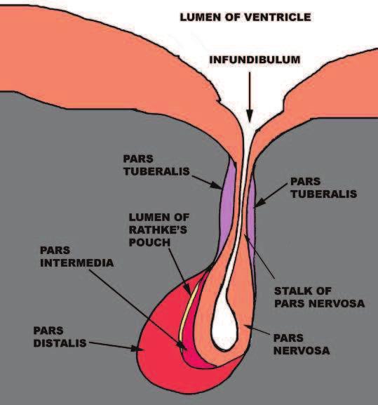
4 Anatomy and Histology


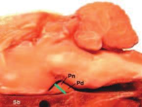
5 Normal Pituitary Gland Pd Pi Pn Pn: pars nervosa, Pi: pars intermedia, Pd: pars distalis
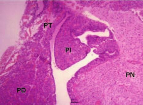
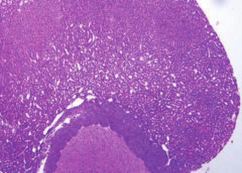
6 Histology Comparison Pd Rat Mouse Pd Pi Pi Pn Pn Rabbit Dog Pd PT PI Pi PN Pn PD

7 Histology Comparison Dog 20x


8 Pituitary Gross Lesions ü Discoloration ü Focus/foci ü Enlarged ü Mass

9 Gross Lesions

10 Common Histopathological Findings NON-PROLIFERATIVE LESIONS Congenital changes ü Dilatation/Persistence of Rathke s Pouch / Cleft ü Stomatodeal remnants in neurohypophysis Vascular changes ü Hemangiectasis ü Hemorrhage ü Thrombosis Inflammatory changes ü Inflammation Miscellaneous changes ü Cysts/Hemocyst

11 PROLIFERATIVE LESIONS qpars Distalis üfocal hypertrophy üfocal hyperplasia üadenoma ücarcinoma qpars Intermedia ühyperplasia ühypertrophy üadenoma qpars Nervosa üpituicytoma Tumor of Embryonic Remnant ücraniopharyngioma

12 Induced Lesions qpars Distalis/Pars Intermedia ühypertrophy/hyperplasia üatrophy üincreased no. of vacuolated cells
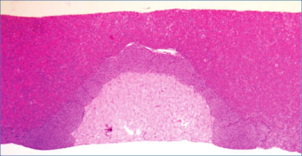
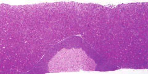
13 Congenital changes Dilatation/Persistence of Rathke s Pouch / Cleft ümay be located in pars distalis, pars intermedia, or pars nervosa üoften associated withrathke s cleft üepithelial lining is usually ciliated but can be squamous, cuboidal, or columnar occasional lesion


14 Congenital changes Persistence of Rathke s Pouch
15 Congenital changes Stomatodeal remnants in neurohypophysis üconsisting of duct like structure of varying sizes ülined by cuboidal/columnar type of epithelium üoccasionally lumen may contains secretions


16 Congenital changes 10x 20x Stomatodeal remnants in neurohypophysis
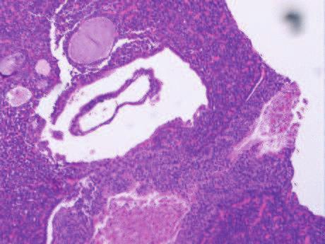
17 Vascular changes Hemangiectasis üdilated, blood-filled spaces lined by endothelium üusually present in pars distalis ümay be accompanied by proteinaceous fluid or hemorrhage

18 Vascular changes 40X Hemangiectasis
19 Vascular changes Hemorrhage üfree erythrocytes outside of vascular channels ümay be accompanied by hemosiderin-laden macrophages, cholesterol clefts, and fibrosis if long-standing

20 Vascular changes 20X Hemorrhage - pars distalis


21 Vascular changes Thrombosis üsolid mass within lumen of blood vessels ücomposed of fibrin and platelets, and may contain trapped erythrocytes
22 Inflammatory changes Inflammation - Rare üinflammatory celluar infiltrate üin acute cases, inflammatory cells are predominantly neutrophils üin chronic cases, inflammatory cells are plasma cells and lymphocytes ücell degeneration or loss
23 Miscellaneous changes Cysts/hemocyst üsimple or multilocular ücommon in pars distalis ü Usually contain eosinophilic to amphophilic mucoproteinaceous material ü Single layer of cuboidal to columnar epithelium ü Epithelial lining is generally ciliated and contain mucous cells
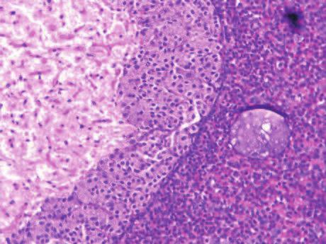
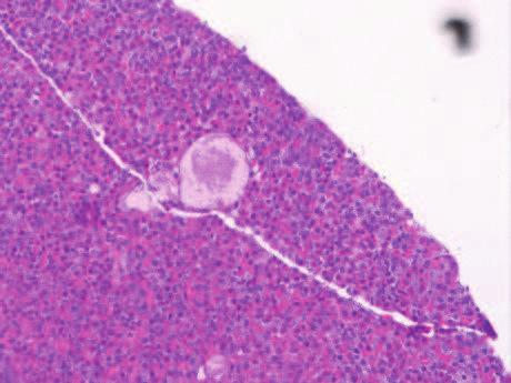
24 Miscellaneous changes 20X 20X Cyst
25 Miscellaneous changes 20X 10X Multiple Cyst Hemocyst


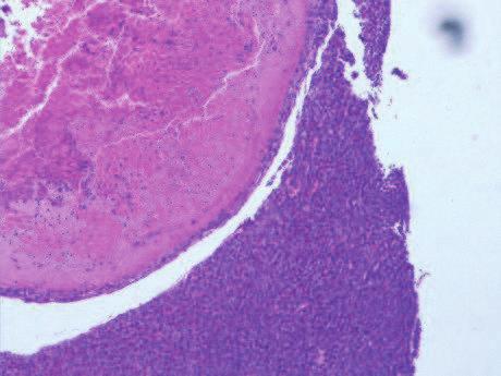
26
27 PROLIFERATIVE LESIONS In routine long-term rat studies neoplasms of the pituitary gland are among the most commonly encountered in most strains The vast majority of these neoplasms occurs in the pars distalis
28 PARS DISTALIS Focal hypertrophy üslightly delineated focus or area üno apparent compression üindistinct borders ücells enlarged due to increase in eosinophilic cytoplasm-droplets or vacuoles ünuclei normal or enlarged


29 PARS DISTALIS 20X 40X Focal hypertrophy- Pars distalis
30 PARS DISTALIS Focal hyperplasia üfocal proliferation ülittle or no compression ürelatively indistinct borders ücells slightly larger and paler than normal ünuclei often similar to normal cells üoccasional "normal" cell types interspersed in focus


31 PARS DISTALIS 40X 40X Focal hyperplasia- Pars distalis


32 PARS DISTALIS 40X 40X Focal hyperplasia- Pars distalis
33 PARS DISTALIS Adenoma ügrowth by expansion with prominent compression üsharp delineation from normal areas ücells usually larger with abundant, pale cytoplasm ü Nuclei more vesicular üvariety of architectural patterns diffuse, angiomatous. glandular, pseudofollicular. Pleomorphic


34 PARS DISTALIS 4X 20X Adenoma Pars distalis


35 PARS DISTALIS 20X 20X Adenoma Pars distalis


36 PARS DISTALIS 4X 10X Adenoma Pars distalis


37 PARS DISTALIS 10X 40X Adenoma Pars distalis


38 PARS DISTALIS 40X 4X Adenoma Pars distalis


39 PARS DISTALIS Carcinoma ümorphologically similar to adenoma ürarely metastasize ülocal invasion into brain
40 PARS INTERMEDIA Atrophy üdecreased width/small sized cells Hypertrophy üdiffuse broadening of intermediate zone ülarge sized cells üpale in color Hyperplasia üdiffuse broadening of intermediate zone ünormal cell morphology üpresence of mitotic figure
41 PARS INTERMEDIA Adenoma Non-encapsulated and poorly circumscribed Cells extend into other /ones of gland Cells larger and more basophilic than normal Nuclear pleomorphism common Lobular or whorled pattern Single/multiple


42 PARS INTERMEDIA 20X 20X Normal Atrophy Atrophy Pars intermedia


43 PARS INTERMEDIA Male Mice 4X 4X Normal Hypertrophy Hypertrophy Pars intermedia


44 PARS INTERMEDIA Female Mice 4X 4X Normal Hypertrophy Hypertrophy Pars intermedia


45 PARS INTERMEDIA 20X 20X Adenoma Pars intermedia
46 PARS NERVOSA Pituicytoma ücircumscribed, nonencapsulated üdense packets of spindle cells üindistinct cords and interlacing bundles ü Dark ovoid to round nuclei and minimal indistinct cytoplasm ünuclear pleomorphism seen in some regions üfoci of mineralization sometimes present

47 Tumor of Embryonic Remnant Craniopharyngioma ükeratinizing squamous epithelium ücells arranged in cords, columns, nests ücystic spaces filled with keratin and debris üinvasion of brain or pituitary tissue


48 LYMPHORETICULAR/ HEMOPOIETIC SYSTEM TUMOR 20X 20X Lymphoma; malignant; multicentric

49 LYMPHORETICULAR/ HEMOPOIETIC SYSTEM TUMOR 40X Lymphoma; malignant; multicentric


50 LYMPHORETICULAR/ HEMOPOIETIC SYSTEM TUMOR 40X 100X Myeloid leukemia; malignant; multicentric
51 NTP Historical Controls Data - June 2013 All Routes & Vehicles - Overall Incidence Location Pars Distalis or Unspecified Site Lesion Adenoma Carcinoma Carcinoma or Adenoma Rats/ Fischer 344N Rats/ Fischer 344N TAC Species/Strain Rats/ Wistar Han Rats/ Harlan Sprague Dawley Mice/ B6C3F1 M F M F M F M F M F 408/698 (58.45%) 4/698 (0.57%) 412/698 (59.03%) 411/699 (58.8%) 11/699 (1.57%) 422/699 (60.37%) 53/100 (53%) 52/100 (52%) 54/149 (36.24%) 88/150 (58.67%) /100 (53%) 52/100 (52%) 54/149 (36.24%) 88/150 (58.67%) 5/50 (10%) 0/50 (0%) 5/50 (10%) 35/102 (34.31%) 2/102 (1.96%) 37/102 (36.27%) 3/933 (0.32%) 0/933 (0%) 3/933 (0.32%) 72/933 (7.72%) 1/933 (0.11%) 73/933 (7.82%) Ganglioneuroma 1/698 (0.14%) 1/699 (0.14%) Pars Intermedia Adenoma Carcinoma or Adenoma 2/698 (0.29%) 2/698 (0.29%) 3/699 (0.43%) 3/699 (0.43%) /149 (3.36%) 5/149 (3.36%) 7/150 (4.67%) 7/150 (4.67%) /933 (0.21%) 2/933 (0.21%) 7/933 (0.75%) 7/933 (0.75%) Pars Nervosa Craniopharyngioma 0/698 (0%) 1/699 (0.14%) *: Denominator is number of animals with tissues examined microscopically


52
Pancreas. Atrophy, acinar cell. Pathogenesis: Diagnostic key features:
 Pancreas Atrophy, acinar cell Pathogenesis: Decrease in number and/or size of acinar cells may be due to spontaneous or experimentally induced degenerative changes, apoptosis, or a sequel of chronic inflammation.
WSC , Conference 9, Case 1. Tissue from a nyala.
 WSC 2009-2010, Conference 9, Case 1. Tissue from a nyala. MICROSCOPIC DESCRIPTION: Heart, atrium (1 pt.): Approximately 40% of the atrial myocardium is replaced by areas of fibrous connective tissue (1
Salivary Glands 3/7/2017
 Salivary Glands 3/7/2017 Goals and objectives Focus on the entities unique to H&N Common board type facts Information for your future practice Salivary Glands Salivary Glands Major gland. Paratid. Submandibular.
The Endocrine System Pituitary
 The Endocrine System Pituitary Look at your slide of the human pituitary with your naked eye. You should see a cellular region and a more fibrous region. Then view each region with your microscope under
Diseases of the breast (1 of 2)
") Diseases of the breast (1 of 2) Introduction A histology introduction Normal ducts and lobules of the breast are lined by two layers of cells a layer of luminal cells overlying a second layer of myoepithelial
Diseases of the breast (1 of 2) Introduction A histology introduction Normal ducts and lobules of the breast are lined by two layers of cells a layer of luminal cells overlying a second layer of myoepithelial
1 NORMAL HISTOLOGY AND METAPLASIAS
 1 NORMAL HISTOLOGY AND METAPLASIAS, MD Anatomy and Histology 1 Metaplasias 2 ANATOMY AND HISTOLOGY The female breast is composed of a branching duct system, which begins at the nipple with the major lactiferous
WSC , Conference 9. Case 1. Tissue from a rhesus macaque.
 Case 1. Tissue from a rhesus macaque. MICROSCOPIC DESCRIPTION: Esophagus: There is multifocal loss of the mucosal lining (1 pt). In these areas, the denuded subepithelial fibrous connective tissue is infiltrated
Epithelia will be discussed according to the following scheme: Type Number of layers Shape Line drawing. Squamous Cuboidal Columnar
 Epithelia Epithelia will be discussed according to the following scheme: Type Number of layers Shape Line drawing Simple Squamous Cuboidal Columnar Covering and Lining epithelium Pseudostratified Stratified
Benign and malignant epithelial lesions: Seborrheic keratosis: A common benign pigmented epidermal tumor occur in middle-aged or older persons more
 Benign and malignant epithelial lesions: Seborrheic keratosis: A common benign pigmented epidermal tumor occur in middle-aged or older persons more common on the trunk; but extremities, head and neck are
XIII. Tumours of the liver and biliary system
 XIII. Tumours of the liver and biliary system V. PONOMARKOV 1 & L. J. MACKEY 2 In this histological classification of liver and gall bladder tumours the tumour types largely correspond to those found in
SESSION 1: GENERAL (BASIC) PATHOLOGY CONCEPTS Thursday, October 16, :30am - 11:30am FACULTY COPY
 PATHOLOGY CONCEPTS Thursday, October 16, :30am - 11:30am FACULTY COPY") SESSION 1: GENERAL (BASIC) PATHOLOGY CONCEPTS Thursday, October 16, 2008 9:30am - 11:30am FACULTY COPY GOAL: Describe the basic morphologic (structural) changes which occur in various pathologic conditions.
SESSION 1: GENERAL (BASIC) PATHOLOGY CONCEPTS Thursday, October 16, 2008 9:30am - 11:30am FACULTY COPY GOAL: Describe the basic morphologic (structural) changes which occur in various pathologic conditions.
Note: The cause of testicular neoplasms remains unknown
 - In the 15- to 34-year-old age group, they are the most common tumors of men. - Tumors of the testis are a heterogeneous group of neoplasms that include: I. Germ cell tumors : 95%; all are malignant.
Brain Tumors. Medulloblastoma. Pilocytic astrocytoma: Ahmed Koriesh, MD. Pathological finding
 NeuroPathology Page 8 Brain Tumors Pathological finding Pseudorosette Rosenthal fibers Rosettes Wet Keratin Psammoma bodies Fried egg Tumor Ependymoma, SEGA Pilocytic astrocytoma Medulloblastoma Craniopharyngioma
Diagnostically Challenging Cases in Gynecologic Pathology
 Diagnostically Challenging Cases in Gynecologic Pathology Eric C. Huang, M.D., Ph.D. Department of Pathology and Laboratory Medicine University of California, Davis Medical Center Case 1 Presentation 38
Gross appearance of nodular hyperplasia in material obtained from suprapubic prostatectomy. Note the multinodular appearance and the admixture of
 Tiền liệt tuyến Tiền liệt tuyến Gross appearance of nodular hyperplasia in material obtained from suprapubic prostatectomy. Note the multinodular appearance and the admixture of solid and microcystic areas.
Pathological Classification of Hepatocellular Carcinoma
 3 rd APASL Single Topic Conference: HCC in 3D Pathological Classification of Hepatocellular Carcinoma Glenda Lyn Y. Pua, M.D. HCC Primary liver cancer is the 2 nd most common cancer in Asia HCC is the
CINtec p16 INK4a Staining Atlas
 CINtec p16 INK4a Staining Atlas Rating Rating Positive The rating positive will be assigned if the p16 INK4a -stained slide shows a continuous staining of cells of the basal and parabasal cell layers of
Synonyms. Nephrogenic metaplasia Mesonephric adenoma
 Nephrogenic Adenoma Synonyms Nephrogenic metaplasia Mesonephric adenoma Definition Benign epithelial lesion of urinary tract with tubular, glandular, papillary growth pattern Most frequently in the urinary
(CYLINDROMA) ATLAS OF HEAD AND NECK PATHOLOGY ADENOID CYSTIC CARCINOMA
 ATLAS OF HEAD AND NECK PATHOLOGY ADENOID CYSTIC CARCINOMA") (CYLINDROMA) This malignant tumor is poorly encapsulated and while seemingly well defined within the affected gland, there is usually infiltration of surrounding tissue on closer examination. The cut surface
(CYLINDROMA) This malignant tumor is poorly encapsulated and while seemingly well defined within the affected gland, there is usually infiltration of surrounding tissue on closer examination. The cut surface
Normal thyroid tissue
 Thyroid Pathology Overview Normal thyroid tissue Normal thyroid tissue with follicles filled with colloid. Thyroid cells form follicles, spheres of epithelial cells (always single layered in health, usually
2015 Descriptive Vet Path Course. Histo Exam #3 KEY
 2015 Descriptive Vet Path Course Histo Exam #3 KEY Test 3, Slide 1 Tissue from a guinea pig. MORPHOLOGIC DIAGNOSIS: Heart: Multifocally and randomly (1 pt), within the left and right ventricular myocardium
CYTOMORPHOLOGY MODULE 28.1 INTRODUCTION OBJECTIVES 28.2 GENERAL GUIDELINES. Notes
 28 CYTOMORPHOLOGY 28.1 INTRODUCTION Light microscopic examination of stained cells in smears is the method of choice of diagnostic cytology. It allows classification of most normal cells as to type and
G3.02 The malignant potential of the neoplasm should be recorded. CG3.02a
 G3.02 The malignant potential of the neoplasm should be recorded. CG3.02a Conventional adrenocortical neoplasm. Each of the below parameters is scored 0 when absent and 1 when present. 3 or more of these
Demystifying Endometrial Hyperplasia
 Demystifying Endometrial Hyperplasia A review from Diagnostic Histopathology 19:7 Dr R Hadden ST5 Histopathology Derriford Hospital Plymouth Endometrium Target for sex-steroid hormones Glands Stroma Proliferate
XX. Tumours of the nasal cavity *
 XX. Tumours of the nasal cavity * H. STONZI 1 & B. HAUSER2 Tumours of the nasal cavity are rare in domestic animals, most cases occurring in the dog. Epithelial tumours are the most common type in carnivores
Disorders of Cell Growth & Neoplasia. Histopathology Lab
 Disorders of Cell Growth & Neoplasia Histopathology Lab Paul Hanna April 2010 Case #84 Clinical History: 5 yr-old, West Highland White terrier. skin mass from axillary region. has been present for the
Diagnostic Cytology of Cancer Cases
 Diagnostic Cytology of Cancer Cases Somporn Techangamsuwan Companion Animal Cancer Research Unit (CAC-RU) Department of Pathology, Faculty of Veterinary Science, Chulalongkorn University 1 Tumor or Non-tumor
Endocrine System. Organs and Tissues: Pituitary Adrenals Pancreas Thyroid Parathyroids
 Endocrine System Organs and Tissues: Pituitary Adrenals Pancreas Thyroid Parathyroids Bruce A. Fenderson, Ph.D. Pathology, Anatomy & Cell Biology Sidney Kimmel Medical College Bruce.Fenderson@Jefferson.edu
Biliary tract tumors
 Short Course 2010 Annual Fall Meeting of the Korean Society for Pathologists Biliary tract tumors Joon Hyuk Choi, M.D., Ph.D. Professor, Department of Pathology, Yeungnam Univ. College of Medicine, Daegu,
PRACTICAL ROADMAP. GLANDS AFFECTING LIFESTYLE WJ van der Spuy & T Tshabalala
 PRACTICAL ROADMAP GLANDS AFFECTING LIFESTYLE WJ van der Spuy & T Tshabalala GLANDS AFFECTING LIFESTYLE Submandibular gland (salivary gland) Liver Pancreas Hypophysis (pituitary gland) Thyroid Suprarenal
21/07/2017. Hobnail endothelial cells are not the same as epithelioid endothelial cells
 UPDATE IN CUTANEOUS VASCULAR S DERMATOPATHOLOGY SESSION BELFAST PATHOLOGY JUNE 21/2017 Dr E Calonje St John s Institute of Dermatology, London, United Kingdom THE FAMILY OF VASCULAR S WITH EPITHELIOID
Neoplasia 2018 Lecture 2. Dr Heyam Awad MD, FRCPath
 Neoplasia 2018 Lecture 2 Dr Heyam Awad MD, FRCPath ILOS 1. List the differences between benign and malignant tumors. 2. Recognize the histological features of malignancy. 3. Define dysplasia and understand
Proceeding of the SEVC Southern European Veterinary Conference
 www.ivis.org Proceeding of the SEVC Southern European Veterinary Conference Oct. 17-19, 2008 Barcelona, Spain http://www.sevc.info Reprinted in the IVIS website with the permission of the SEVC www.ivis.org
Papillary Lesions of the Breast A Practical Approach to Diagnosis. (Arch Pathol Lab Med. 2016;140: ; doi: /arpa.
 Papillary Lesions of the Breast A Practical Approach to Diagnosis (Arch Pathol Lab Med. 2016;140:1052 1059; doi: 10.5858/arpa.2016-0219-RA) Papillary lesions of the breast Span the spectrum of benign,
Neoplasms of the Canine, Feline and Lemur Liver:
 Neoplasms of the Canine, Feline and Lemur Liver: Classification and Prognosis Annual Seminar of the French Society of Veterinary Pathology John M. Cullen VMD PhD DACVP North Carolina State University Primary
Salivary Gland Cytology
 Salivary Gland Cytology Diagnostic challenges and potential pitfalls Tarik M. Elsheikh, MD Professor and Medical Director Anatomic Pathology Cleveland Clinic FNA Salivary Gland Lesions Indications Distinguish
ARTHUR PURDY STOUT SOCIETY COMPANION MEETING: DIFFICULT NEW DIFFERENTIAL DIAGNOSES IN PROSTATE PATHOLOGY. Jonathan I. Epstein.
 1 ARTHUR PURDY STOUT SOCIETY COMPANION MEETING: DIFFICULT NEW DIFFERENTIAL DIAGNOSES IN PROSTATE PATHOLOGY Jonathan I. Epstein Professor Pathology, Urology, Oncology The Reinhard Professor of Urological
1. To describe the gross structure of the pituitary gland and be able to identify the pars nervosa, pars intermedia and pars distalis.
 ENDOCRINE Objectives 1. To describe the gross structure of the pituitary gland and be able to identify the pars nervosa, pars intermedia and pars distalis. 2. Identify and describe the histological features
Histopathology: Cervical HPV and neoplasia
 Histopathology: Cervical HPV and neoplasia These presentations are to help you identify basic histopathological features. They do not contain the additional factual information that you need to learn about
CONTINUING EDUCATION IN TOXICOLOGIC PATHOLOGY REPRODUCTIVE SYSTEM
 CONTINUING EDUCATION IN TOXICOLOGIC PATHOLOGY REPRODUCTIVE SYSTEM ORGANIZED BY SOCIETY FOR TOXICOLOGIC PATHOLOGY IN INDIA (STPI) OCTOBER 29-31, 2010 The Atria Hotel, # 1, Palace Road, Bangalore - 560 001
Histology Review Can you identify the Cell Structures? Can you identify the Stain? Can you identify the Cell type?
 Histology Review Can you identify the Cell Structures? Can you identify the Stain? Can you identify the Cell type? 2.01 Border of Epithelia (fluorescence) M A: lamina basalis B: epithelium C: other tissues
AN OBSERVATION OF PRIMARY TUMORS OF THE PITUITARY, OVARIES, AND MAMMARY GLANDS IN A MOUSE
 AN OBSERVATION OF PRIMARY TUMORS OF THE PITUITARY, OVARIES, AND MAMMARY GLANDS IN A MOUSE W. U. GARDNER,' L. C. STRONG,' AND G. M. SMITH (Front the Department of Anatomy, Yale Univerdy School of Medicine)
number Done by Corrected by Doctor Maha Shomaf
 number 16 Done by Waseem Abo-Obeida Corrected by Zeina Assaf Doctor Maha Shomaf MALIGNANT NEOPLASMS The four fundamental features by which benign and malignant tumors can be distinguished are: 1- differentiation
Proliferative Epithelial lesions of the Breast. Sami Shousha, MD, FRCPath Charing Cross Hospital & Imperial College, London
 Proliferative Epithelial lesions of the Breast Sami Shousha, MD, FRCPath Charing Cross Hospital & Imperial College, London Amman, November2013 Proliferative Epithelial Lesions of the Breast Usual type
Pitfalls in thyroid tumor pathology. Prof.Valdi Pešutić-Pisac MD, PhD
 Pitfalls in thyroid tumor pathology Prof.Valdi Pešutić-Pisac MD, PhD Too many or... Tumour herniation through a torn capsule simulating capsular invasion fibrous capsule with a sharp discontinuity, suggestive
Cytyc Corporation - Case Presentation Archive - March 2002
 FirstCyte Ductal Lavage History: 68 Year Old Female Gail Index: Unknown Clinical History: Negative Mammogram in 1995 6 yrs. later presents with bloody nipple discharge Subsequent suspicious mammogram Suspicious
Prepared By Jocelyn Palao and Layla Faqih
 Prepared By Jocelyn Palao and Layla Faqih The structure of the suspected atypical cell should always be compared to the structure of other similar, benign, cells which are present in the smears. The diagnosis
PATHOLOGY OF LIVER TUMORS
 PATHOLOGY OF LIVER TUMORS Pathobasic, 31.05.2016 WHO Classification Approach to a Liver Mass Lesion in a patient with chronic liver disease? Lesion in a patient without chronic liver disease? Malignant
Pathology Slides. [Pathology]
![Pathology Slides. [Pathology]](/thumbs/94/120604575.jpg "Pathology Slides. [Pathology]") Pathology Slides MedicoNotes provides real laboratory pathological slides to aid you to differentiate between different pathological structures under microscope. www.mediconotes.com Histology slides example
Pathology Slides MedicoNotes provides real laboratory pathological slides to aid you to differentiate between different pathological structures under microscope. www.mediconotes.com Histology slides example
CYTOLOGY OF THE LIVER
 CYTOLOGY OF THE LIVER Maxey L. Wellman, DVM, PhD, DACVP (Clinical Pathology) Professor, Department of Veterinary Biosciences, College of Veterinary Medicine, The Ohio State University, Columbus, OH, USA
Breast pathology. 2nd Department of Pathology Semmelweis University
 Breast pathology 2nd Department of Pathology Semmelweis University Breast pathology - Summary - Benign lesions - Acute mastitis - Plasma cell mastitis / duct ectasia - Fat necrosis - Fibrocystic change/
Avian Pathology. Bacterial diseases: histo slides. ECVP-ESVP Summer School 2012 Frédérique NGUYEN
 Avian Pathology Bacterial diseases: histo slides ECVP-ESVP Summer School 2012 Frédérique NGUYEN Bacterial diseases: histo slides B1. Turkey. Organs? Morphologic diagnosis? Special procedure? B2. Hen. Organ?
Macro- and microacinar proliferations of the prostate
 Macro- and microacinar proliferations of the prostate (with emphasis on cancer mimics) Rodolfo Montironi, MD (IT), FRCPath (UK), IFCAP (USA) Polytechnic University of Marche Region (Ancona) School of Medicine,
the urinary system pathology Dr. Fairoz A Eltorgman
 the urinary system pathology Dr. Fairoz A Eltorgman Tumors of the renal pelvis & kidney Benign tumors of the renal pelvis: Hemangioma Leiomyoma Malignant tumors: Transitional cell carcinoma Squamous cell
Bio & 241 A&P Unit 1 / Lecture 3
 Bio & 241 A&P Unit 1 / Lecture 3 Tissues All body tissues arise from three fundamental embryonic tissues. Endoderm: forms epithelial tissues lining internal organs such as the GI tract Mesoderm: connective
Pancreatitis: A Potential Pitfall in Endoscopic Ultrasound Guided Pancreatic FNA
 Pancreatitis: A Potential Pitfall in Endoscopic Ultrasound Guided Pancreatic FNA Jack Yang, MD Department of Pathology, Medical University of South Carolina Objectives Understand the indication of EUS
EMBRYONAL NEPHROMA IN THE CHICKEN: REPORT OF TWO CASES
 EMBRYONAL NEPHROMA IN THE CHICKEN: REPORT OF TWO CASES FRANK D. McKENNEY, V.M.D. (Di1!ision of Experimental Surgery and Pathology, The Mayo Foundation, Rochester, Minnesota) Few data have been collected
Papillary Lesions of the breast
 Papillary Lesions of the breast Emad Rakha Professor of Breast Pathology The University of Nottingham Papillary lesions of the breast are a heterogeneous group of disease, which are characterised by neoplastic
Outline 11/2/2017. Pancreatic EUS-FNA general aspects. Cytomorphologic features of solid neoplasms/lesions of the pancreas
 ENDOSCOPIC ULTRASOUND GUIDED-FINE NEEDLE ASPIRATION CYTOLOGY OF PANCREAS Khalid Amin M.D. Assistant Professor Department of Laboratory Medicine and Pathology University of Minnesota Outline Pancreatic
MORPHOLOGIC DIAGNOSIS: Liver: Hepatitis, necrotizing, multifocal to coalescing, severe, with numerous trichomonads. (3 pt)
") Case 1. Tissue from a pelican. MICROSCOPIC DESCRIPTION: Liver: Approximately 80% (1 pt) of the liver is replaced by multifocal to coalescing areas of coagulative and lytic necrosis. Centrally, within these
Case 1. Tissue from a pelican. MICROSCOPIC DESCRIPTION: Liver: Approximately 80% (1 pt) of the liver is replaced by multifocal to coalescing areas of coagulative and lytic necrosis. Centrally, within these
-The cause of testicular neoplasms remains unknown
 - In the 15- to 34-year-old age group, they are the most common tumors of men. - include: I. Germ cell tumors : (95%); all are malignant. II. Sex cord-stromal tumors: from Sertoli or Leydig cells; usually
2 to 3% of All New Visceral Cancers Peak Incidence is 6th Decade M:F = 2:1 Grossly is a Bright Yellow, Necrotic Mass with a Pseudocapsule
 GENITOURINARY PATHOLOGY Kathleen M. O Toole, M.D. Renal Cell Carcinoma 2 to 3% of All New Visceral Cancers Peak Incidence is 6th Decade M:F = 2:1 Grossly is a Bright Yellow Necrotic Mass Grossly is a Bright
BREAST PATHOLOGY. Fibrocystic Changes
 BREAST PATHOLOGY Lesions of the breast are very common, and they present as palpable, sometimes painful, nodules or masses. Most of these lesions are benign. Breast cancer is the 2 nd most common cause
Histopathology: skin pathology
 Histopathology: skin pathology These presentations are to help you identify, and to test yourself on identifying, basic histopathological features. They do not contain the additional factual information
LYMPHATIC DRAINAGE AXILLARY (MOSTLY) INTERNAL MAMMARY SUPRACLAVICULAR
 INTERNAL MAMMARY SUPRACLAVICULAR") BREAST LYMPHATIC DRAINAGE AXILLARY (MOSTLY) INTERNAL MAMMARY SUPRACLAVICULAR HISTOLOGY LOBE: (10 in whole breast) LOBULE: (many per lobe) ACINUS/I, aka ALVEOLUS/I: (many per lobule) DUCT(S): INTRA- or
BREAST LYMPHATIC DRAINAGE AXILLARY (MOSTLY) INTERNAL MAMMARY SUPRACLAVICULAR HISTOLOGY LOBE: (10 in whole breast) LOBULE: (many per lobe) ACINUS/I, aka ALVEOLUS/I: (many per lobule) DUCT(S): INTRA- or
Epithelium tissue system
 Epithelium tissue system Histology : is the study of the microscopic anatomy (microanatomy) of cells and tissues of plants and animals. It is commonly performed by examining cells and tissues under a light
They Do Look Alike : Mimics of Prostate Cancer in Biopsy Samples
 They Do Look Alike : in Biopsy Samples Gladell P. Paner, MD Departments of Pathology and Surgery (Urology) University of Chicago, IL USA Gladell.paner@uchospitals.edu Benign in Needle Biopsy 1. Benign
RENAL CELL CARCINOMA 2 to 3% of All New Visceral Cancers Peak Incidence is 6th Decade M:F = 2:1 Grossly is a Bright Yellow, Necrotic Mass with a Pseud
 GENITOURINARY PATHOLOGY Kathleen M. O Toole Toole, M.D. RENAL CELL CARCINOMA 2 to 3% of All New Visceral Cancers Peak Incidence is 6th Decade M:F = 2:1 Grossly is a Bright Yellow, Necrotic Mass with a
Unit I Problem 9 Histology: Basic Tissues of The Body
 Unit I Problem 9 Histology: Basic Tissues of The Body - What is the difference between cytology and histology? Cytology: it is the study of the structure and functions of cells and their contents. Histology:
BOSNIAN-TURKISH CYTOPATHOLOGY SCHOOL June 18-19, 2016 Sarajevo. Case Discussions. 60 year old woman Routine gynecologic control LBC
 BOSNIAN-TURKISH CYTOPATHOLOGY SCHOOL June 18-19, 2016 Sarajevo Case Discussions Prof Dr Sıtkı Tuzlalı Tuzlalı Pathology Laboratory 60 year old woman Routine gynecologic control LBC 1 2 Endometrial thickening
Almost any suspected tumor can be aspirated easily and safely. Some masses are more risky to aspirate including:
 DOES THIS PATIENT HAVE CANCER? USING IN-HOUSE CYTOLOGY TO HELP YOU MAKE THIS DIAGNOSIS. Joyce Obradovich, DVM, Diplomate, ACVIM (Oncology) Animal Cancer & Imaging Center, Canton, Michigan Almost every
My Journey into the World of Salivary Gland Sebaceous Neoplasms
 My Journey into the World of Salivary Gland Sebaceous Neoplasms Douglas R. Gnepp Warren Alpert Medical School at Brown University Rhode Island Hospital Pathology Department Providence RI Asked to present
Disclosures. Parathyroid Pathology. Objectives. The normal parathyroid 11/10/2012
 Disclosures Parathyroid Pathology I have nothing to disclose Annemieke van Zante MD/PhD Assistant Professor of Clinical Pathology Associate Chief of Cytopathology Objectives 1. Review the pathologic features
This is the second learning component (Learning Component 2) in our first learning module (Learning Module 1). In this component we review a very
 in our first learning module (Learning Module 1). In this component we review a very") This is the second learning component (Learning Component 2) in our first learning module (Learning Module 1). In this component we review a very basic response to injury inflammation. We ll look at examples
This is the second learning component (Learning Component 2) in our first learning module (Learning Module 1). In this component we review a very basic response to injury inflammation. We ll look at examples
Pituitary Gland (Hypophysis)
") Endocrine Organs Pituitary Gland (Hypophysis) Function o Production of hormones Location o Connected to the hypothalamus via an infundibulum situated within the sella turcica of the sphenoid bone Structure
Endocrine Organs Pituitary Gland (Hypophysis) Function o Production of hormones Location o Connected to the hypothalamus via an infundibulum situated within the sella turcica of the sphenoid bone Structure
Cells and Tissues. Lesson 2.1: Molecules of Life Lesson 2.2: Cells Lesson 2.3: Tissues
 2 Cells and Tissues Lesson 2.1: Molecules of Life Lesson 2.2: Cells Lesson 2.3: Tissues Chapter 2: Cells and Tissues Lesson 2.1 Molecules of Life Molecules of Life carbohydrates proteins lipids nucleic
Update in Salivary Gland Pathology. Benjamin L. Witt University of Utah/ARUP Laboratories February 9, 2016
 Update in Salivary Gland Pathology Benjamin L. Witt University of Utah/ARUP Laboratories February 9, 2016 Objectives Review the different appearances of a selection of salivary gland tumor types Establish
Lách
 Lách Lách Lách Lách Splenogonadal fusion. Splenic tissue is attached to testicular tissue. Pseudocyst (false or secondary cyst). A, Outer aspect. Pseudocyst (false or secondary cyst). B, Inner surface.
Gastrooesophageal reflux disease. Jera Jeruc Institute of pathology, Faculty of Medicine, Ljubljana, Slovenia
 Gastrooesophageal reflux disease Jera Jeruc Institute of pathology, Faculty of Medicine, Ljubljana, Slovenia Reflux esophagitis (RE) GERD: a spectrum of clinical conditions and histologic alterations resulting
21 Endocrine organs and cells
 21 Endocrine organs and cells The endocrine system consists of discrete organs, portions of organs and distributed cells that secrete hormones into surrounding tissues or structures. Objectives You should
Cerebral Parenchymal Lesions: I. Metastatic Neoplasms
 Chapter 4 Cerebral Parenchymal Lesions: I. Metastatic Neoplasms After one has reasonably ruled out the possibility of a nonneoplastic diagnosis (see Chap. 3), one is left with considering a diagnosis of
Cellular Pathology. Histopathology Lab #2 (web) Paul Hanna Jan 2018
 Paul Hanna Jan 2018") Cellular Pathology Histopathology Lab #2 (web) Paul Hanna Jan 2018 Slide #91 Clinical History: a necropsy was performed on an aged cat the gross pathological changes included: widespread subcutaneous edema
Cellular Pathology Histopathology Lab #2 (web) Paul Hanna Jan 2018 Slide #91 Clinical History: a necropsy was performed on an aged cat the gross pathological changes included: widespread subcutaneous edema
 THE EFFECTS OF AMMONIUM PERCHLORATE ON THYROIDS PATHOLOGY WORKING GROUP REPORT Ammonium perchlorate is an oxidant utilized in solid fuel for rockets. Due to its potential environmental impact, a number
Histology. There are four basic tissue types in the body are :-
 Histology Lab.I There are four basic tissue types in the body are :- 1- Epithelial tissues (Epithelium) 2- Connective tissues 3- Muscular tissues 4- Nervous tissues 1-Epithelial tissues epithelial tissues
Neoplasia part I. Dr. Mohsen Dashti. Clinical Medicine & Pathology nd Lecture
 Neoplasia part I By Dr. Mohsen Dashti Clinical Medicine & Pathology 316 2 nd Lecture Lecture outline Review of structure & function. Basic definitions. Classification of neoplasms. Morphologic features.
ACCME/Disclosures. Cribriform Lesions of the Prostate. Case
 Cribriform Lesions of the Prostate Ming Zhou, MD, PhD Departments of Pathology and Urology New York University Langone Medical Center New York, NY Ming.Zhou@NYUMC.ORG ACCME/Disclosures The USCAP requires
Mody. AIS vs. Invasive Adenocarcinoma of the Cervix
 Common Problems in Gynecologic Pathology Michael T. Deavers, M.D. Houston Methodist Hospital, Houston, Texas Common Problems in Gynecologic Pathology Adenocarcinoma in-situ (AIS) of the Cervix vs. Invasive
Oncocytic carcinoma: A rare malignancy of the parotid gland
 ISPUB.COM The Internet Journal of Pathology Volume 8 Number 2 Oncocytic carcinoma: A rare malignancy of the parotid gland K Mardi, J Sharma Citation K Mardi, J Sharma.. The Internet Journal of Pathology.
Endometrial Metaplasia, Hyperplasia & Other Cancer Mimics: a Consultant s Experience
 Endometrial Metaplasia, Hyperplasia & Other Cancer Mimics: a Consultant s Experience Pacific Northwest Society of Pathologists Vancouver, B.C. September 26, 2015 Teri A. Longacre, M.D. longacre@stanford.edu
A classification of epithelial tissues
 A classification of epithelial tissues Ramray Bhat Molecular Reproduction Development and Genetics ramray@iisc.ac.in Textbooks for my portion Molecular Biology of the Cell (Bruce Alberts) 6 th Edition
Exocrine vs. Endocrine Glands. Dr. Sami Zaqout IUG
 Exocrine vs. Endocrine Glands Hypophysis (Pituitary Gland) It lies in a cavity of the sphenoid bone the sella turcica Weighs about 0.5 g, and its normal dimensions in humans are about 10 x 13 x 6 mm.
Trichofolliculoma of the Guinea Pig 1,2
 Trichofolliculoma of the Guinea Pig 1,2 Raymond D. Ediger, Garrett S. Dill, Jr., and Robert M. Kovatch, Aerobiology and Evaluation Laboratories and Medical Sciences Laboratories, Fort Detrick, Frederick,
ENDOCRINE SYSTEM ENDOCRINE SYSTEM
 Endocrine system consists of organs that produce and secrete hormones "endocrine" = internal secretion into capillaries Hormones carried by the blood to another organ; exert effects Hormones manipulate
Normal Morphology. Anatomic Considerations. Normal Urothelial Histology and Cytology
 1 Normal Morphology Anatomic Considerations The urinary tract can be divided into three regions: the kidney; the calyces, pelves and ureters (upper collecting system or upper tract); and the bladder and
Lesions Mimicking Adenoid Cystic Carcinoma. Diagnostic Problems in Salivary Gland Pathology An Update 5/29/2009
 Diagnostic Problems in Salivary Gland Pathology An Update Lesions Mimicking Adenoid Cystic Carcinoma Stacey E. Mills, M.D. W.S. Royster Professor of Pathology Director of Surgical and Cytopathology University
Fourth Practical Pathology. Circulatory disturbances
 Fourth Practical Pathology Circulatory disturbances 12.12.2018 1 Organ: Lung (40X, low power) 1) The blood capillaries within the alveolar septa are engorged with blood 2) Pinkish proteinaceous fluid,
Case: The patient is a 62 year old woman with a history of renal cell carcinoma that was removed years ago. A 2.4 cm liver mass was found on CT
 Case: The patient is a 62 year old woman with a history of renal cell carcinoma that was removed years ago. A 2.4 cm liver mass was found on CT during follow- up. ALT, AST, Alk Phos and bilirubin were
Tinh hoàn
 Tinh hoàn Tinh hoàn Tinh hoàn Tiền liệt tuyến Tiền liệt tuyến Mào tinh hoàn Mào tinh hoàn Túi tinh Túi tinh Túi tinh Túi tinh So-called cystadenoma of seminal vesicle. Gross appearance of granulomatous
Laurie A. Loevner, MD
 Laurie A. Loevner, MD Chief, Division of Neuroradiology UPHS Professor of Radiology, Otorhinolaryngology: Head & Neck Surgery, Neurosurgery, and Ophthalmology University of Pennsylvania Health System Disclosures
Normal endometrium: A, proliferative. B, secretory.
 Normal endometrium: A, proliferative. B, secretory. Nội mạc tử cung Nội mạc tử cung Cyclic changes in endometrium.. Approximate relationship of useful microscopic changes. Arias-Stella reaction in endometrial