Salivary Gland Pathology
|
|
|
- Edmund McBride
- 6 years ago
- Views:
Transcription

1 IN THE NAME OF GOD


2 Salivary Gland Pathology CHAPTER 11 Dr.kheirandish Oral and maxillofacial pathology
3 Sialadenosis Adenomatoid Hyperplasia of the Minor Salivary Glands Necrotizing Sialometaplasia Pleomorphic Adenoma Oncocytoma

4 Sialadenosis (Sialosis) Unusual Noninflammatory disorder Salivary gland enlargement Parotid glands Underlying systemic problem Endocrine, nutritional, or neurogenic Diabetes mellitus, general malnutrition, alcoholism, and bulimia
5 Dysregulation of the autonomic innervation of the salivary acini Accumulation of secretory granules : enlargement of the acinar cells. CLINICAL FEATURES Slowly evolving swelling May or may not be painful Usually bilateral Submandibular glands Minor salivary glands : rare
6
7 HISTOPATHOLOGIC FEATURES Hypertrophy of the acinar cells Two to three times greater than normal size. Nuclei are displaced to the cell base Cytoplasm is engorged with zymogen granules Treatment Control of the underlying cause. Cosmetic concern : partial parotidectomy
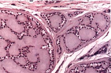
8 ADENOMATOID HYPERPLASIA OF THE MINOR SALIVARY GLANDS Rare lesion Minor salivary glands Mimics a neoplasm Pseudotumor Hard or soft palate Local trauma

9
10 HISTOPATHOLOGIC FEATURES Normal-appearing mucous acini Greater in number than normally Increased in size Chronic inflammation TREATMENT Biopsy is necessary
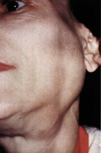
11 NECROTIZING SIALOMETAPLASIA Uncommon Locally destructive Inflammatory condition Ischemia leads to local infarction Mimics a malignant process, both clinically and microscopically.
12 Predisposing factors : Traumatic injuries Dental injections Ill-fitting dentures Upper respiratory infections Adjacent tumors Previous surgery Compromising the blood supply to the involved glands, resulting in ischemic necrosis. However, many cases occur without any known predisposing factors.
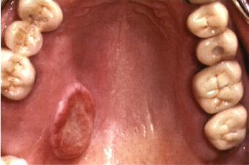
13 CLINICAL FEATURES Palatal salivary glands More than 75% : posterior palate Hard palate Two thirds : unilateral Any age Adults
14 CLINICAL FEATURES Males : twice time Initially : nonulcerated swelling, often associated with pain or paresthesia Within 2 to 3 weeks, necrotic tissue sloughs out, leaving a craterlike ulcer ( 1 cm to more than 5 cm in diameter) A part of my palate fell out." At this point, the pain often subsides

15
16 HISTOPATHOLOGIC FEATURES Acinar necrosis in early lesions, followed by associated squamous metaplasia (ducts) lobular architecture : helpful histopathologic Squamous cell carcinoma(scc) or mucoepidermoid carcinoma(mec) Pseudoepitheliomatous hyperplasia Squamous proliferation has a bland cytologic appearance

17
18 TREATMENT Biopsy usually is indicated to rule out the possibility of malignant disease. The lesion typically resolves on its own accord, with an average healing time of 5 to 6 weeks.
19 SALIVARY GLAND TUMORS Parotid gland most common site Pleomorphic adenom Mucoepidermoid carcinoma Submandibular gland Malignancy is double of the parotid gland Pleomorphic adenoma Adenoid cystic carcinoma Sublingual gland Rare 70% to 90% are malignant
20 SALIVARY GLAND TUMORS Minor salivary glands Palate : most frequent site Pleomorphic adenoma Mucoepidermoid carcinoma and adenoid cystic carcinoma Lips : second most common Upper lip Up to 91% of retromolar tumors and most tumors in the floor of the mouth and tongue are malignant.
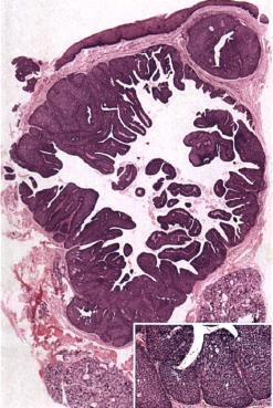
21 PLEOMORPHIC ADENOMA (BENIGN MIXED TUMOR) Most common salivary neoplasm. The terms pleomorphic adenoma and mixed tumor both represent attempts to describe this tumor's unusual Histopathologic. Pleomorphic Mixed neoplasm : derived from more than one germ layer.
22 CLINICAL FEATURES Painless Slowly growing Firm mass Many months or years Any age (most common in young and middle-aged adults) Most common childhood Female Superficial lobe / 10% deep lobe
23 CLINICAL FEATURES Facial nerve palsy and pain are rare. Initially : movable / Grows larger: less mobile Bilateral (Parotid glands) : synchronous or metachronous Palate: posterior lateral aspect /smooth-surfaced/domeshaped masses/ not movable Upper lip : mobile Traumatized : secondary ulceration

24
25
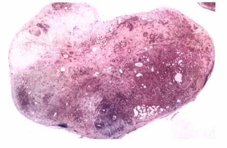
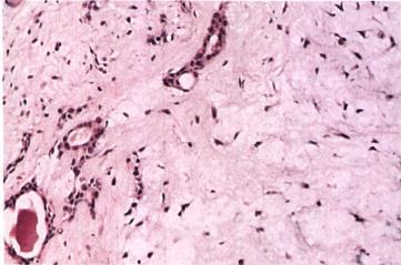
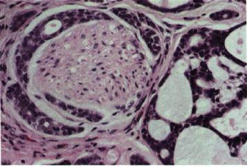
26 HISTOPATHOLOGIC FEATURES Well circumscribed, encapsulated tumor (may be incomplete or show infiltration by tumor cells). Encapsulation : minor gland tumors(palatal) Mixture of glandular epithelium and myoepithelial cells within a mesenchyme-like background. Variable ratio of the epithelial elements and the mesenchyme-like component
27 Epithelium : ducts and cystic structures / islands or sheets Myoepithelial cells : large percentage /variable morphology (angular or spindle) Plasmacytoid myoepithelial cells (minor glands) Stromal" changes (myoepithelial cells) Mucoid Myxomatous Chondroid Hyalinized Osteoid Fat Myoepithelioma : No ductal elements.
28


29
30 TREATMENT Surgical excision Superficial lobe : superficial parotidectomy ( preservation of the facial nerve) Deep lobe : total parotidectomy (preservation of the facial nerve) Local enucleation:should be avoided Submandibular tumors : total removal Adequate surgery: excellent prognosis Predominantly myxoid : more recurrence Carcinoma ex pleomorphic adenoma
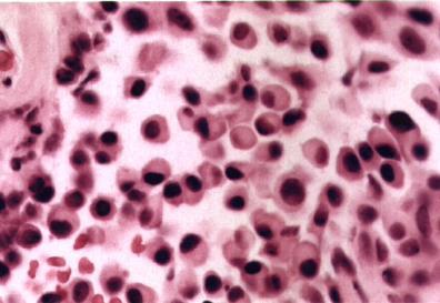
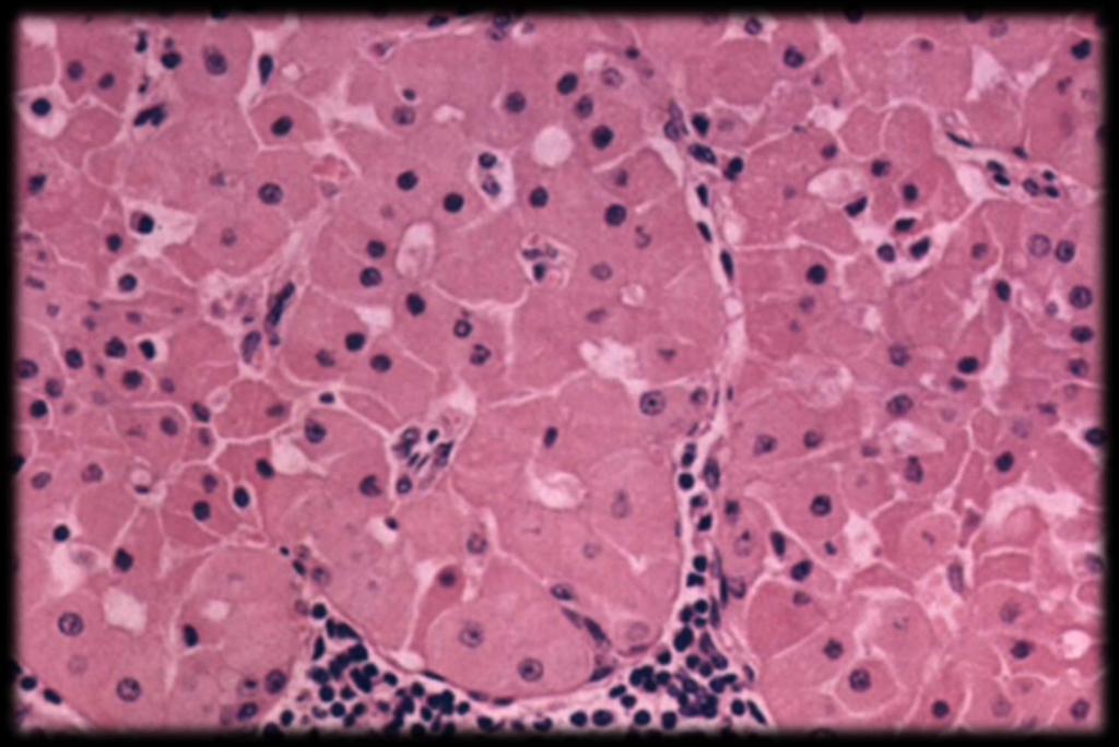
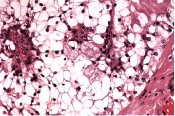
31 ONCOCYTE o Large epithelial cells o Greek word onkoustai (swell) o Swollen granular cytoplasm o Mitochondria o Focal oncocytic metaplasia :age o Uncommon :younger than 50 o Other organs: thyroid, parathyroid, and kidney.
32 ONCOCYTOMA (OXYPHILIC ADENOMA) Older adults(eighth decade) Female Major salivary glands(parotid) Firm Slowly growing Painless Superficial lobe Bilateral Benign Oncocytes Rare
33 HISTOPATHOLOGIC FEATURES Well circumscribed Sheets of large polyhedral cells (oncocytes) Abundant granular. Eosinophilic cytoplasm Centrally nuclei (small and hyperchromatic to large and vesicular). Little stroma Mitochondria(electron microscopy) light microscopic Phosphotungstic acid hematoxylin (PTAH) Glycogen : periodic acid-schiff (PAS) Clear cells
34
35 TREATMENT Surgical excision Partial parotidectomy Submandibular gland (total removal) Low rate of recurrence Malignant oncocytomas

36 IN THE NAME OF GOD
37 ONCOCYTOSIS (NODULAR ONCOCYTIC HYPERPLASIA) Oncocytic metaplasia Transformation of ductal and acinar cells to oncocytes Older than 50 y/o Both the proliferation and the accumulation of oncocytes Mimic a tumor (clinically and microscopically)
38 CLINICAL FEATURES Parotid gland Proliferation is multifocal and nodular Entire gland can be replaced by oncocytes HISTOPATHOLOGIC FEATURES Focal nodular collections of oncocytes Oncocytoma
39 WARTHIN TUMOR (PAPILLARY CYSTADENOMA LYMPHOMATOSUM) Benign Parotid gland Second most common benign parotid Tumor Adenolymphoma : should be avoided Smoking Polyclonal : tumorlike
40 Pathogenesis : uncertain Hypothesis 1)Heterotopic salivary gland tissue found within parotid lymph nodes 2)Proliferation of salivary gland ductal epithelium that is associated with secondary formation of lymphoid tissue
41 Older adults (sixth and seventh decades) Whites Male (10: 1) Smoking and bilaterality.
42 Slowly growing Painless Nodular mass Firm or fluctuant Parotid tail Bilaterally (metachronous) Submandibular or minor salivary glands
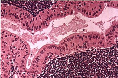
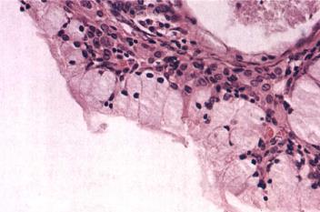
43 HISTOPATHOLOGIC FEATURES Papillary cystadenoma Iymphomatosum Ductal epithelium and a lymphoid stroma Epithelium (oncocytic nature)
44 Two layers : A) inner luminal layer tall columnar cells with centrally placed, palisaded, and slightly hyperchromatic nuclei. B) second layer of cuboidal or polygonal multiple papillary cells with more vesicular nuclei Cystic spaces Multiple papillary infoldings lymphoid stroma


45
46 TREATMENT AND PROGNOSIS Surgical removal Superficial parotidectomy Malignant Warthin tumors (carcinoma ex papillary cystadenoma Iymphomatosum)
47 MONOMORPHIC ADENOMA Benign salivary gland tumors demonstrating a more uniform histopathologic pattern than the common pleomorphic adenoma. Basal cell adenoma Canalicular adenoma
48 CANALICULAR ADENOMA Female Slowly growing Painless mass Firm or fluctuant Mucocele
49 Uncommon Minor salivary glands Upper lip Parotid gland Older adults

50
51 HISTOPATHOLOGIC FEATURES Fibrous capsule Single-layered cords of columnar or cuboidal epithelial cells with deeply basophilic nuclei Ductal structures Cystic spaces Papillary projections
52
53 BASAL CELL ADENOMA Benign Uncommon Basaloid appearance Parotid gland Minor glands : second most common site Middle-aged and older adults Women
54 Slowly growing Movable mass Superficial lobe Membranous basal cell adenoma Hereditary Combination with skin appendage tumors (dermal cylindromas and trichoepitheliomas) Multiple bilateral Dermal analogue tumors
55 HISTOPATHOLOGIC FEATURES Encapsulated or well circumscribed Ductal epithelium and myoepithelial cells Subtypes : 1. Solid (Most common subtype) 2. Trabecular 3. Tubular 4. Membranous
56
57 Solid subtype : Multiple islands and cords of epithelial cells Fibrous stroma Peripheral cells (palisaded and cuboidal to columnar) Basal cell carcinoma Central cells
58 Tubular subtype: Is characterized by the formation of small, round, duct-like structures. Trabecular subtype: Demonstrates narrow cordlike epithelial strands
59 Membranous basal cell adenoma : Multiple Large lobular islands Jigsaw puzzle fashion Similar to dermal cylindroma,
60 TREATMENT AND PROGNOSIS Complete surgical removal Recurrence is rare, BUT membranous subtype has a 25% to 37% recurrence rate Malignant counterpart (basal cell adenocarcinoma)
61 DUCTAL PAPILLOMAS 1) SIALADENOMA PAPILLIFERUM 2) INTRADUCTAL PAPILLOMA 3) INVERTED DUCTAL PAPILLOMA

62 Microscopically : papillomatous pattern Rare Squamous papilloma
63 SIALADENOMA PAPILLIFERUM Minor salivary glands (palate) Older adults Male Exophytic Papillary surface Clinically similar to the squamous papilloma
64
65 HISTOPATHOLOGIC FEATURES Similar to the squamous papilloma Exophytic papillary projections Covered by stratified squamous epithelium Cutaneous syringocystadenoma papilliferum
66
67 INTRADUCTAL PAPILLOMA Ill-defined lesion Adults Minor salivary glands Submucosal swelling HISTOPATHOLOGIC FEATURES Unicystic structure Cuboidal or columnar epithelium Papillary projections into the cystic lumen
68 INVERTED DUCTAL PAPILLOMA Rare Minor salivary glands Adults Lower lip and mandibular vestibule Asymptomatic nodule HISTOPATHOLOGIC FEATURES Proliferation of squamoid epithelium Multiple thick, bulbous papillary projections that fill the ductal lumen

69
70
71 TREATMENT AND PROGNOSIS Conservative surgical excision Recurrence is rare
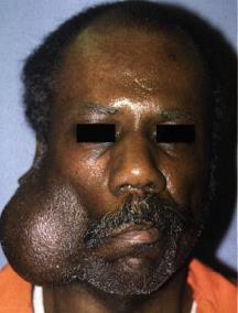
72 MUCOEPIDERMOID CARCINOMA One of the most common salivary gland malignancies Second to seventh decades Most common malignant salivary gland tumor in children Previous history of radiation therapy to the head and neck Parotid gland Asymptomatic swelling
73 Pain or facial nerve palsy (high-grade tumors) Minor glands : second most common site (palate) Mucocele Intraosseous Lower lip. floor of mouth. tongue. and retromolar pad areas are uncommon locations for salivary gland neoplasia. the mucoepidermoid carcinoma is the most common salivary tumor in each of these sites.


74
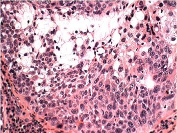
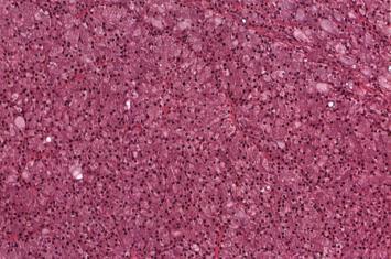
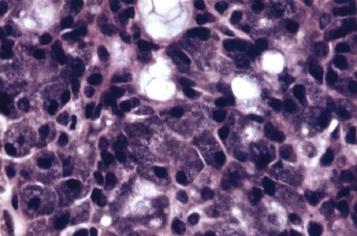
75 HISTOPATHOLOGIC FEATURES A. Mucus-producing cells : Abundant foamy cytoplasm Mucin stains + B. Squamous (epidermoid) cells : Squamoid features Polygonal Shape C. Intermediate cells : Progenitor of both the mucous and the epidermoid cells Vary in appearance ( small. Basaloid) Clear cells
76
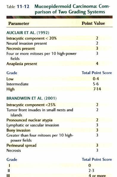
77 Traditionally categorized into three histopathologic grades : 1. Amount of cyst formation 2. Degree of cytologic atypia 3. Relative numbers of Mucous. Epidermoid and Intermediate cells
78 Low-grade tumors Prominent cyst formation Minimal cellular atypia High proportion of mucous cells High-grade tumors Solid islands of squamous and intermediate cells Pleomorphism Mitotic activity Mucus-producing cells (infrequent)

79
80 Intermediate-grade tumors Between those of the low-grade and high-grade neoplasms Cyst formation occurs (less prominent than lowgrade) Cellular atypia +/_ All three major cell types are present, but intermediate cells usually predominate.
81
82 TREATMENT AND PROGNOSIS Treatment : Location, Histopathologic grade, and Clinical stage Early-stage : parotidectomy (preservation of the facial nerve) Advanced : total removal of the parotid gland (sacrifice of the facial nerve) low-grade: modest margin of surrounding normal tissue removed High-grade or large : wider resection (involved bone must be excised)
83 Submandibular gland : total removal Radical neck dissection : Metastasis Large High-grade Postoperative radiation therapy (more aggressive tumors) Prognosis : depends on the grade and stage of the Submandibular gland tumors : poorer outlook than parotid gland

84 IN THE NAME OF GOD
85 INTRAOSSEOUS MUCOEPIDERMOID CARCINOMA (CENTRAL MUCOEPIDERMOID CARCINOMA) Rare Most common Adenoid cystic carcinoma, benign and malignant mixed tumors, adenocarcinoma, acinic cell adenocarcinoma, epithelial-myoepithelial carcinoma, and monomorphic adenoma.
86 Hypotheses : 1) Ectopic salivary 2) Sinus lining 3) Odontogenic epithelium (most likely source) o Association with impacted teeth or odontogenic cysts
87 Female (middle-aged adults) Mandible (molar-ramus area) Cortical swelling Pain, trismus, paresthesia Unilocular or multilocular radiolucency Well-defined borders Irregular and ill-defined area of bone destruction Unerupted tooth (odontogenic cyst or tumor)

88
89 HISTOPATHOLOGIC FEATURES Similar to soft tissue counterpart Low-grade TREATMENT AND PROGNOSIS Radical surgical resection Adjunctive radiation therapy Metastasis Prognosis : good
90 ACINIC CELL ADENOCARCINOMA Serous acinar differentiation Acinic cell tumor Metastasis Death Low-grade malignancy
91 Parotid gland Submandibular gland, buccal mucosa, lips, palate Second to the seventh decades (40s) women Slowly growing Many months or years Asymptomatic Facial nerve paralysis : ominous sign for parotid tumors
92
93 HISTOPATHOLOGIC FEATURES Highly variable Well circumscribed Most characteristic cell : serous acinar cell Abundant granular basophilic cytoplasm and a round. darkly Stained eccentric nucleus Uniform Mitotic activity : uncommon Clear cells
94 Growth patterns : 1. Solid : resembles normal parotid gland tissue 2. Micro cystic : small cystic spaces 3. Papillary-cystic : papillary projections into the cystic spaces 4. Follicular : similar to thyroid tissue
95
96 TREATMENT AND PROGNOSIS Superficial lobectomy Total parotidectomy One of the better prognoses of any of the malignant salivary gland tumors The prognosis for minor gland tumors is better
97 MALIGNANT MIXED TUMORS : I. CARCINOMA EX PLEOMORPHIC ADENOMA (CARCINOMA EX MIXED TUMOR) I. CARCINOSARCOMA II. METASTASIZING MIXED TUMOR


98
99 CARCINOMA EX PLEOMORPHIC ADENOMA Most common Malignant transformation of the epithelial component 15 years older than benign pleomorphic adenoma Sixth to eighth decades Mass (many years) Recent rapid growth
100 Pain Painless Ulceration Some tumors :short duration Risk for malignant change : duration Major glands : parotid gland Palate Facial nerve palsy
101 Variable microscopic appearance Areas of typical benign (most or only a small portion) Cellular pleomorphism Abnormal mitotic activity Poorly differentiated adenocarcinoma o Polymorphous lowgrade adenocarcinoma. salivary duct carcinoma. mucoepidermoid carcinoma, and adenoid cystic carcinoma.
102 Aggressive growth pattern Capsular invasion Noninvasive or carcinoma in situ ex mixed tumor
103
104 Wide excision Local lymph node dissection Adjunctive radiation therapy Prognosis : guarded Histopathologic subtype of the malignant component In situ (noninvasive) carcinoma ex mixed tumor
105 CARCINOSARCOMA Rare Parotid gland Submandibular gland and minor salivary glands Clinical signs and symptoms(similar to those of the carcinoma ex pleomorphic Adenoma) Previous history of a benign pleomorphic adenoma De novo
106 Biphasic tumor Both carcinomatous and sarcomatous areas Epithelial component : poorly differentiated adenocarcinoma or an undifferentiated Carcinoma Sarcomatous portion : Chondrosarcoma (osteosarcoma. fibrosarcoma. liposarcoma. rhabdomyosarcoma. malignant fibrous histiocytoma) Evidence of an origin (benign mixed tumor)
107 Radical surgical excision Radiation therapy Chemotherapy Prognosis : poor Recurrent local tumor Metastases
108 METASTASIZING MIXED TUMOR Rare Parotid gland Submandibular gland or minor salivary glands Metastases (bones or lung) History of a benign mixed tumor Multiple recurrences
109 Benign microscopic appearance Both the primary and the metastatic sites Malignant histopathologic changes Surgical excision of both the primary tumor and the metastatic sites
110 ADENOID CYSTIC CARCINOMA ADCC Common Cylindroma Any salivary gland Minor salivary glands (palate) Parotid and submandibular glands

111
112 Middle-aged adults Slowly growing Pain : common / important finding / early (before swelling) Facial nerve paralysis
113 HISTOPATHOLOGIC FEATURES Myoepithelial and ductal cells Varied arrangement Three major pattern : 1) Cribriform 2) Tubular 3) Solid Combination (predominant pattern)
114 Cribriform pattern: Most classic Islands Basaloid epithelial cells Multiple cylindrical, cystlike spaces (resembling Swiss cheese) Tumor cells : small / cuboidal / basophilic nuclei / little cytoplasm /uniform /mitotic activity is rarely Tubular pattern : Multiple small ducts or tubules

115
116 Solid variant : Larger islands or sheets of tumor cells Cellular pleomorphism Mitotic activity Focal necrosis Perineural invasion (not pathognomonic)
117
118 TREATMENT AND PROGNOSIS Local recurrence Distant metastasis Surgical excision Neck dissection - Prognosis : Poor
119 Late recurrence and metastasis (5-year survival rate has little significance) Solid pattern (worse outlook) Maxillary sinus and submandibular gland : poor prognosis Death : local recurrence or distant metastases Metastases : lungs and bones
120 POLYMORPHOUS LOW-GRADE ADENOCARCINOMA (LOBULAR CARCINOMA; TERMINAL DUCT CARCINOMA) Common Minor salivary Major glands : rare De novo Malignant component of a carcinoma ex pleomorphic adenoma
121 Palate Upper lip and buccal mucosa Females (Older adults) Painless mass Slow growth Erode or infiltrate the underlying bone
122
123 HISTOPATHOLOGIC FEATURES Tumor cells : Uniform Round to polygonal Indistinct cell borders Nuclei : round, ovoid (pale staining) Different growth patterns : (polymorphous term) Solid / cords / ducts / cystic spaces / cribriform

124
125 Mitotic : uncommon Well circumscribed Peripheral : infiltrative / single-file Perineural invasion : common Adenoid cystic carcinoma (different prognoses) Immunohistochemical staining : helpful
126
127 TREATMENT AND PROGNOSIS Wide surgical excision (including underlying bone) Metastasis (regional lymph nodes): uncommon Distant metastasis : rare Overall prognosis : good
128 SALIVARY ADENOCARCINOMA, NOT OTHERWISE SPECIFIED (NOS) Diverse group of neoplasms Parotid gland Minor glands and submandibular gland
129 Asymptomatic Pain Facial nerve paralysis Microscopic : highly variable Low-grade neoplasms to high-grade malignancies

130
Salivary Glands 3/7/2017
 Salivary Glands 3/7/2017 Goals and objectives Focus on the entities unique to H&N Common board type facts Information for your future practice Salivary Glands Salivary Glands Major gland. Paratid. Submandibular.
DISORDERS OF THE SALIVARY GLANDS Neoplasms Dr.M.Baskaran Selvapathy S IV
 DISORDERS OF THE SALIVARY GLANDS Neoplasms Dr.M.Baskaran Selvapathy S IV NEOPLASMS A) Epithelial I. Benign Pleomorphic adenoma( Mixed tumour) Adenolymphoma (Warthin s tumour) Oxyphil adenoma (Oncocytoma)
Lesions Mimicking Adenoid Cystic Carcinoma. Diagnostic Problems in Salivary Gland Pathology An Update 5/29/2009
 Diagnostic Problems in Salivary Gland Pathology An Update Lesions Mimicking Adenoid Cystic Carcinoma Stacey E. Mills, M.D. W.S. Royster Professor of Pathology Director of Surgical and Cytopathology University
My Journey into the World of Salivary Gland Sebaceous Neoplasms
 My Journey into the World of Salivary Gland Sebaceous Neoplasms Douglas R. Gnepp Warren Alpert Medical School at Brown University Rhode Island Hospital Pathology Department Providence RI Asked to present
FNA OF SALIVARY GLANDS: A PRACTICAL APPROACH
 FNA OF SALIVARY GLANDS: A PRACTICAL APPROACH FNA of Salivary Glands: Challenges Wide range of neoplastic and non-neoplastic lesions Cytological overlap between the different benign and malignant tumors
(CYLINDROMA) ATLAS OF HEAD AND NECK PATHOLOGY ADENOID CYSTIC CARCINOMA
 ATLAS OF HEAD AND NECK PATHOLOGY ADENOID CYSTIC CARCINOMA") (CYLINDROMA) This malignant tumor is poorly encapsulated and while seemingly well defined within the affected gland, there is usually infiltration of surrounding tissue on closer examination. The cut surface
(CYLINDROMA) This malignant tumor is poorly encapsulated and while seemingly well defined within the affected gland, there is usually infiltration of surrounding tissue on closer examination. The cut surface
Objectives. Salivary Gland FNA: The Milan System. Role of Salivary Gland FNA 04/26/2018
 Salivary Gland FNA: The Milan System Dr. Jennifer Brainard Section Head Cytopathology Cleveland Clinic Objectives Introduce the Milan System for reporting salivary gland cytopathology Define cytologic
Differential Diagnosis of Oral Masses. Palatal Lesions
 Differential Diagnosis of Oral Masses Palatal Lesions Palatal Masses Periapical Abscess Torus Palatinus Mucocele Lymphoid Hyperplasia Adenomatous Hyperplasia Benign Salivary Neoplasms Malignant Salivary
PLEOMORPHIC ADENOMA ( BENIGN MIXED TUMOR )
") ( BENIGN MIXED TUMOR ) Grossly, the tumor is freely movable, solid, sometimes lobulated and occasionally cystic. If recurrent, multinodular masses are common. Histologically, within a fibrous capsule,
( BENIGN MIXED TUMOR ) Grossly, the tumor is freely movable, solid, sometimes lobulated and occasionally cystic. If recurrent, multinodular masses are common. Histologically, within a fibrous capsule,
Salivary gland Workshop Trondheim 31th may 2012
 Salivary gland Workshop Trondheim 31th may 2012 Peter Jebsen cytopathologist Oslo University Hospital Rikshospitalet Anna Bofin ass. Professor St. Olavs Hospital, Trondheim Drying artifacts Lymfocytes
Salivary Gland Cytology
 Salivary Gland Cytology Diagnostic challenges and potential pitfalls Tarik M. Elsheikh, MD Professor and Medical Director Anatomic Pathology Cleveland Clinic FNA Salivary Gland Lesions Indications Distinguish
Los Angeles Society Of Pathologists Dr. Shobha Castelino Prabhu
 Los Angeles Society Of Pathologists Dr. Shobha Castelino Prabhu Loma Linda University Medical Center June 12, 2007 CASE 1 76 year-old gentleman Status post right parotidectomy 1 year ago for a rare tumor
ARIZONA SOCIETY OF PATHOLOGISTS 13 TH APRIL 2013 HEAD AND NECK CYTOPATHOLOGY. F ZAHRA ALY, MD, PhD
 ARIZONA SOCIETY OF PATHOLOGISTS 13 TH APRIL 2013 HEAD AND NECK CYTOPATHOLOGY F ZAHRA ALY, MD, PhD The main areas sites amenable for cytopathology include lymph nodes, thyroid, major salivary glands especially
Educational Cases EQA November T.J. Palmer Raigmore Hospital Inverness
 Educational Cases EQA November 2013 T.J. Palmer Raigmore Hospital Inverness Case 2 Clinical Details Dob 11 February 1951 PMH: 1964 Extraction of 45 aet 13 yr 1966 Cyst between 44 and 46 enucleated 1973
TYPES and FREQUENCY of SALIVARY GLAND TUMORS in MAJOR and MINOR. Karl Donath Department of Oral Pathology (Director:Prof. Dṛ Dr.
 TYPES and FREQUENCY of SALIVARY GLAND TUMORS in MAJOR and MINOR SALIVARY GLANDS Karl Donath Department of Oral Pathology (Director:Prof. Dṛ Dr. Karl Donath) University of Hamburg, Salivary gland tumors
Oncocytic-Appearing Salivary Gland Tumors. Oncocytic, Cystic, Mucinous, and High Grade Salivary Gland Tumors SALIVARY GLAND FNA: PART II
 William C. Faquin, MD, PhD Professor of Pathology Harvard Medical School Director of Head and Neck Pathology Massachusetts Eye and Ear Massachusetts General Hospital SALIVARY GLAND FNA: PART II Oncocytic,
SALIVARY GLAND DISEASES. Omar alnoubani MD,MRCS
 SALIVARY GLAND DISEASES Omar alnoubani MD,MRCS Salivary Glands Overview Parotid gland Sublingual gland Submandibular gland Salivary glands - Types 3 Major Salivary Glands Parotid Submandibular Sublingual
Polymorphous Low-Grade. December 5 th, 2008
 Polymorphous Low-Grade Adenocarcinoma December 5 th, 2008 Epidemiology Represents 2 nd or 3 rd most common minor salivary gland malignancy (17-26%) 1 st mucoepidermoid carcinoma Rare in reported Asian
PRELIMINARY CYTOLOGIC DIAGNOSIS: Suspicious for Acinic Cell Carcinoma. Cell Block: Immunohistochemical Studies CYTOLOGIC DIAGNOSIS:
 1 PRELIMINARY CYTOLOGIC DIAGNOSIS: Suspicious for Acinic Cell Carcinoma. Cell Block: Immunohistochemical Studies GCDFP-15 S-100 CYTOLOGIC DIAGNOSIS: Consistent with mammary analogue secretory carcinoma.
Salivary gland cytology. Salivary gland cytology. Triage helps the clinician. Salivary gland tumors. Diagnostic difficulties
 Salivary gland cytology Salivary Gland Cytology Pınar Fırat, MD Professor of Pathology İ.U. İstanbul Faculty of Medicine Çapa, İstanbul It is a reliable diagnostic test However, definitive subclassification
Update in Salivary Gland Pathology. Benjamin L. Witt University of Utah/ARUP Laboratories February 9, 2016
 Update in Salivary Gland Pathology Benjamin L. Witt University of Utah/ARUP Laboratories February 9, 2016 Objectives Review the different appearances of a selection of salivary gland tumor types Establish
Problem diagnoses. Current issues in Anatomic pathology. Problem Diagnoses in Tumors of the Oral Cavity 5/29/2009
 Current issues in Anatomic pathology Problem Diagnoses in Tumors of the Oral Cavity Richard Jordan DDS PhD FRCPath Professor of Oral Pathology & Pathology Director, UCSF Oral Pathology Diagnostic Laboratory
04/09/2018. Salivary Gland Pathology in the Molecular Era Old Friends, Old Foes, & New Acquaintances
 Salivary Gland Pathology in the Molecular Era Old Friends, Old Foes, & New Acquaintances Jennifer L. Hunt, MD, MEd Aubrey J. Hough Jr, MD, Endowed Professor of Pathology Chair of Pathology and Laboratory
Oncocytic carcinoma: A rare malignancy of the parotid gland
 ISPUB.COM The Internet Journal of Pathology Volume 8 Number 2 Oncocytic carcinoma: A rare malignancy of the parotid gland K Mardi, J Sharma Citation K Mardi, J Sharma.. The Internet Journal of Pathology.
DISCUSSION: PLGA accounts for about 2% of all salivary gland tumours and occurs almost exclusively in the minor salivary glands.
 SWELLING ON THE HARD PALATE PRESENTING AS POLYMORPHOUS LOW GRADE ADENOCARCINOMA: A AND REVIEW OF LITERATURE Swapnil D. Chandekar 1, Sunita S. Dantkale 2, Rahul R. Narkhede 3, Snehal V. Chavhan 4, Khushboo
IN THE NAME OF GOD Dr. Kheirandish Oral and maxillofacial pathology
 IN THE NAME OF GOD Dr. Kheirandish Oral and maxillofacial pathology ORAL FOCAL MUCINOSIS Uncommon Tumorlike Cutaneous myxoid cyst Overproduction of hyaluronic acid by firoblasts Young adults Female Gingiva
Epithelial tumors. Dr. F.F. Khuzin, PhD Dr. M.O. Mavlikeev
 Epithelial tumors Dr. F.F. Khuzin, PhD Dr. M.O. Mavlikeev Epithelial tumors Tumors from the epithelium are the most frequent among tumors. There are 2 group features of these tumors: The presence in most
Papillary Lesions of the breast
 Papillary Lesions of the breast Emad Rakha Professor of Breast Pathology The University of Nottingham Papillary lesions of the breast are a heterogeneous group of disease, which are characterised by neoplastic
Diseases of the breast (1 of 2)
") Diseases of the breast (1 of 2) Introduction A histology introduction Normal ducts and lobules of the breast are lined by two layers of cells a layer of luminal cells overlying a second layer of myoepithelial
Diseases of the breast (1 of 2) Introduction A histology introduction Normal ducts and lobules of the breast are lined by two layers of cells a layer of luminal cells overlying a second layer of myoepithelial
Salivary gland tumor cytologic and histologic correlation: Algorithmic and risk stratification based approaches
 Salivary gland tumor cytologic and histologic correlation: Algorithmic and risk stratification based approaches Christopher C. Griffith, MD, PhD Raja R. Seethala, MD 1. Salivary gland tumor cytology: A
2007 June Cheng-Chung Lin Prof. in Oral Pathology Kaohsiung Med University
 Salivary Gland Pathology 2007 June Cheng-Chung Lin Prof. in Oral Pathology Kaohsiung Med University E-mail: cclin99@hotmail.com Histological Classification of Salivary Gland Tumors (WHO) 1. Adenomas 2.13.
SESSION 1: GENERAL (BASIC) PATHOLOGY CONCEPTS Thursday, October 16, :30am - 11:30am FACULTY COPY
 PATHOLOGY CONCEPTS Thursday, October 16, :30am - 11:30am FACULTY COPY") SESSION 1: GENERAL (BASIC) PATHOLOGY CONCEPTS Thursday, October 16, 2008 9:30am - 11:30am FACULTY COPY GOAL: Describe the basic morphologic (structural) changes which occur in various pathologic conditions.
SESSION 1: GENERAL (BASIC) PATHOLOGY CONCEPTS Thursday, October 16, 2008 9:30am - 11:30am FACULTY COPY GOAL: Describe the basic morphologic (structural) changes which occur in various pathologic conditions.
Salivary gland neoplasms: an update 29th Annual Meeting of Arab Division of the International Academy of Pathology MUSCAT, OMAN 2017
 Salivary gland neoplasms: an update 29th Annual Meeting of Arab Division of the International Academy of Pathology MUSCAT, OMAN 2017 Dr Mary Toner Consultant Pathologist St James Hospital Trinity College
Proliferative Epithelial lesions of the Breast. Sami Shousha, MD, FRCPath Charing Cross Hospital & Imperial College, London
 Proliferative Epithelial lesions of the Breast Sami Shousha, MD, FRCPath Charing Cross Hospital & Imperial College, London Amman, November2013 Proliferative Epithelial Lesions of the Breast Usual type
Breast pathology. 2nd Department of Pathology Semmelweis University
 Breast pathology 2nd Department of Pathology Semmelweis University Breast pathology - Summary - Benign lesions - Acute mastitis - Plasma cell mastitis / duct ectasia - Fat necrosis - Fibrocystic change/
See the latest estimates for new cases of salivary gland cancers in the US and what research is currently being done.
 About Salivary Gland Cancer Overview and Types If you have been diagnosed with salivary gland cancer or are worried about it, you likely have a lot of questions. Learning some basics is a good place to
THE NATURAL HISTORY OF TUMORS PECULIAR TO THE SALIVARY GLANDS
 THE NATURAL HISTORY OF TUMORS PECULIAR TO THE SALIVARY GLANDS FRANK VELLIOS, M.D., AND DALE DAVIDSON, M.D. Departments of Clinical Pathology and Surgery, Indiana University School of Medicine Tumors of
Acute bacterial sialadenitis:
 Developmental anomalies: Aplasia Atresia Ectopic tissue Sialadenitis: Acute bacterial sialadenitis: Uncommon Parotid Xerostomia Seen in pts with: Sjogren s syndrome Tricyclic antidepressants Immunosuppression
The Pathology and Surgery of the Salivary Glands. R. A. Cawson, M. J. Gleeson, J. W. Eveson. Chapter 7: Carcinoma of salivary glands.
 The Pathology and Surgery of the Salivary Glands R. A. Cawson, M. J. Gleeson, J. W. Eveson Chapter 7: Carcinoma of salivary glands Introduction In terms of registration in England and Wales, cancers of
04/10/2018. Intraductal Papillary Neoplasms Of Breast INTRADUCTAL PAPILLOMA
 Intraductal Papillary Neoplasms Of Breast Savitri Krishnamurthy MD Professor of Pathology Deputy Division Head The University of Texas MD Anderson Cancer Center 25 th Annual Seminar in Pathology Pittsburgh,
Note: The cause of testicular neoplasms remains unknown
 - In the 15- to 34-year-old age group, they are the most common tumors of men. - Tumors of the testis are a heterogeneous group of neoplasms that include: I. Germ cell tumors : 95%; all are malignant.
Basement membrane in lobule.
 Bahram Memar, MD Basement membrane in lobule. Normal lobule-luteal phase Normal lobule-follicular phase Lactating breast Greater than 95% are adenocarcinomas in situ carcinomas and invasive carcinomas.
Synonyms. Nephrogenic metaplasia Mesonephric adenoma
 Nephrogenic Adenoma Synonyms Nephrogenic metaplasia Mesonephric adenoma Definition Benign epithelial lesion of urinary tract with tubular, glandular, papillary growth pattern Most frequently in the urinary
Histopathologic spectrum of salivary gland neoplasms
 Original article Histopathologic spectrum of salivary gland neoplasms Dr. Dipkana Das 1, Dr.Subhasish Saha 1, Dr V Satyanarayana 2 Name & Address of Institution: Department of Pathology, Kamineni Institute
Salivary Gland Imaging. Mary Scanlon MD FACR October 2016
 Salivary Gland Imaging Mary Scanlon MD FACR October 2016 Objectives Recognize normal and abnormal anatomy Discuss work up, management and differential diagnosis of commonly referred clinical scenarios
Normal thyroid tissue
 Thyroid Pathology Overview Normal thyroid tissue Normal thyroid tissue with follicles filled with colloid. Thyroid cells form follicles, spheres of epithelial cells (always single layered in health, usually
Diagnostically Challenging Cases in Gynecologic Pathology
 Diagnostically Challenging Cases in Gynecologic Pathology Eric C. Huang, M.D., Ph.D. Department of Pathology and Laboratory Medicine University of California, Davis Medical Center Case 1 Presentation 38
Papillary Lesions of the Breast A Practical Approach to Diagnosis. (Arch Pathol Lab Med. 2016;140: ; doi: /arpa.
 Papillary Lesions of the Breast A Practical Approach to Diagnosis (Arch Pathol Lab Med. 2016;140:1052 1059; doi: 10.5858/arpa.2016-0219-RA) Papillary lesions of the breast Span the spectrum of benign,
POLYMORPHOUS LOW GRADE ADENOCARCINOMA - CASE REPORT AND REVIEW OF LITERATURE
 POLYMORPHOUS LOW GRADE ADENOCARCINOMA - CASE REPORT AND REVIEW OF LITERATURE S.Sunil 1 B.S. Sreenivasan 2 Jisha Titus 3 Soma Susan 4 Jubin Thomas 4 Antony George 4 Devi Gopakumar 5 1 Reader, 2 Professor,
Gross appearance of nodular hyperplasia in material obtained from suprapubic prostatectomy. Note the multinodular appearance and the admixture of
 Tiền liệt tuyến Tiền liệt tuyến Gross appearance of nodular hyperplasia in material obtained from suprapubic prostatectomy. Note the multinodular appearance and the admixture of solid and microcystic areas.
Carcinoma ex Pleomorphic Adenoma (CXPA)-A rare parotid malignancy
-A rare parotid malignancy") Indian Journal of Mednodent and Allied Sciences, pp- 54-58 Indian journals.com Case Report Carcinoma ex Pleomorphic Adenoma (CXPA)-A rare parotid malignancy Vani Padmaja GJ 1 *, Sireesha A 2, Sunderi Devi
Indian Journal of Mednodent and Allied Sciences, pp- 54-58 Indian journals.com Case Report Carcinoma ex Pleomorphic Adenoma (CXPA)-A rare parotid malignancy Vani Padmaja GJ 1 *, Sireesha A 2, Sunderi Devi
doi: /j.anl
 doi: 10.1016/j.anl.2006.07.001 Synchronous unilateral parotid gland neoplasms of three different histological types Shuho Tanaka 1, Keiji Tabuchi 1, Keiko Oikawa 1, Rika Kohanawa 1, Hideki Okubo 1, Dai
Pathology of Selected Head and Neck Lesions. Adel Assaad MD Department of Pathology
 Pathology of Selected Head and Neck Lesions Adel Assaad MD Department of Pathology 1 NOSE Infections 2 Zygomycosis (Mucormycosis) Opportunistic infection caused by "bread mold fungi," including Rhizopus,
BREAST PATHOLOGY. Fibrocystic Changes
 BREAST PATHOLOGY Lesions of the breast are very common, and they present as palpable, sometimes painful, nodules or masses. Most of these lesions are benign. Breast cancer is the 2 nd most common cause
Malignant Peripheral Nerve Sheath Tumor
 C H A P T E R 120 Malignant Peripheral Nerve Sheath Tumor Currently, malignant peripheral nerve sheath tumor (MPNST) is the most commonly used generic name for the neoplasms known in the past as neurosarcoma,
4Ps LUMPS AND BUMPS B.L.&T. BUMPS, LUMPS, AND TATTOOS. Most Common BUMP in the oral cavity Fibroma INTERDENTAL PAPILLAE LESIONS
 B.L.&T. BUMPS, LUMPS, AND TATTOOS LUMPS AND BUMPS DIFFERENTIAL DIAGNOSIS FOR LUMPS AND BUMPS Traumatic Fibroma Papilloma Epulis Fissuratum Inflammatory Papillary Hyperplasia Lesions of Attached Gingiva
XX. Tumours of the nasal cavity *
 XX. Tumours of the nasal cavity * H. STONZI 1 & B. HAUSER2 Tumours of the nasal cavity are rare in domestic animals, most cases occurring in the dog. Epithelial tumours are the most common type in carnivores
University Journal of Pre and Para Clinical Sciences
 ISSN 2455 2879 Volume 2 Issue 1 2016 Metaplastic carcinoma breast a rare case report Abstract : Metaplastic carcinoma of the breast is a rare malignancy with two distinct cell lines described as a breast
Pancreas. Atrophy, acinar cell. Pathogenesis: Diagnostic key features:
 Pancreas Atrophy, acinar cell Pathogenesis: Decrease in number and/or size of acinar cells may be due to spontaneous or experimentally induced degenerative changes, apoptosis, or a sequel of chronic inflammation.
FNA Biopsy of Salivary Gland
 FNA Biopsy of Salivary Gland Richard M. DeMay, MD Professor of Pathology Director of Cytopathology The University of Chicago Objective: To describe the use of FNA Bx to diagnose salivary gland lesions
Central Poorly Differentiated Adenocarcinoma of the Maxilla: Report of a Case
 Kobe J. Med. Sci., Vol. 49, No. 2, pp. 45-49, 2003 Central Poorly Differentiated Adenocarcinoma of the Maxilla: Report of a Case MASAHIRO UMEDA 1), SATOSHI YOKOO 1), YASUYUKI SHIBUYA 1), TAKAHIDE KOMORI
Diseases of oral cavity
 Diseases of oral cavity Diseases of Teeth and Supporting Structures Inflammatory/Reactive Lesions Infections Oral Manifestations of Systemic Disease Precancerous and Cancerous Lesions Odontogenic Cysts
Kidney Case 1 SURGICAL PATHOLOGY REPORT
 Kidney Case 1 Surgical Pathology Report February 9, 2007 Clinical History: This 45 year old woman was found to have a left renal mass. CT urography with reconstruction revealed a 2 cm medial mass which
2 to 3% of All New Visceral Cancers Peak Incidence is 6th Decade M:F = 2:1 Grossly is a Bright Yellow, Necrotic Mass with a Pseudocapsule
 GENITOURINARY PATHOLOGY Kathleen M. O Toole, M.D. Renal Cell Carcinoma 2 to 3% of All New Visceral Cancers Peak Incidence is 6th Decade M:F = 2:1 Grossly is a Bright Yellow Necrotic Mass Grossly is a Bright
Glycogen-rich adenocarcinoma in the lower lip: report of a case with particular emphasis on differential diagnosis
 Open Journal of Stomatology, 2011, 1, 109-113 doi:10.4236/ojst.2011.13017 Published Online September 2011 (http://www.scirp.org/journal//). Glycogen-rich adenocarcinoma in the lower lip: report of a case
NEOPLASIA-I CANCER. Nam Deuk Kim, Ph.D.
 NEOPLASIA-I CANCER Nam Deuk Kim, Ph.D. 1 2 Tumor in the hieroglyphics of the Edwin Smith papyrus (1,600 B.C., Breasted s translation 1930) 3 War on Cancer (National Cancer Act, 1971) 4 Cancer Acts in Korea
Slide Seminar of the Head and Neck Session of the European Congress of Pathology Bilbao, Spain, 2018.
 Slide Seminar of the Head and Neck Session of the European Congress of Pathology Bilbao, Spain, 2018. Prof Sulen Sarioglu, MD Dokuz Eylul University Faculty of Medicine Department of Pathology Graduate
An Integrated Cytologic and Histologic Approach to the Diagnosis of Salivary Gland Tumors
 An Integrated Cytologic and Histologic Approach to the Diagnosis of Salivary Gland Tumors W.C. Faquin, M.D., Ph.D. Massachusetts General Hospital Massachusetts Eye and Ear Infirmary Boston, MA An Integrated
Slide seminar. Asist. Prof. Jože Pižem, MD, PhD Institute of Pathology Medical Faculty, University of Ljubljana
 Slide seminar Asist. Prof. Jože Pižem, MD, PhD Institute of Pathology Medical Faculty, University of Ljubljana Case 5 A 57-year-old man with a dermal/subcutaneous lesion on the scalp, which was interpreted
Abid Irshad, MD Director Breast Imaging. Medical University of South Carolina Charleston
 Abid Irshad, MD Director Breast Imaging Medical University of South Carolina Charleston Cases Financial disclosure: I or my family have no financial interest related to the material discussed in this presentation
XIII. Tumours of the liver and biliary system
 XIII. Tumours of the liver and biliary system V. PONOMARKOV 1 & L. J. MACKEY 2 In this histological classification of liver and gall bladder tumours the tumour types largely correspond to those found in
Review of the AP Part II Practical Examination. Dr David Clift Co Chief Examiner
 Review of the AP Part II Practical Examination Dr David Clift Co Chief Examiner General Remarks The part II practical examination involved 15 cases which were presented with sufficient clinical data to
Disorders of Cell Growth & Neoplasia. Histopathology Lab
 Disorders of Cell Growth & Neoplasia Histopathology Lab Paul Hanna April 2010 Case #84 Clinical History: 5 yr-old, West Highland White terrier. skin mass from axillary region. has been present for the
ARTHUR PURDY STOUT SOCIETY COMPANION MEETING: DIFFICULT NEW DIFFERENTIAL DIAGNOSES IN PROSTATE PATHOLOGY. Jonathan I. Epstein.
 1 ARTHUR PURDY STOUT SOCIETY COMPANION MEETING: DIFFICULT NEW DIFFERENTIAL DIAGNOSES IN PROSTATE PATHOLOGY Jonathan I. Epstein Professor Pathology, Urology, Oncology The Reinhard Professor of Urological
Pitfalls in thyroid tumor pathology. Prof.Valdi Pešutić-Pisac MD, PhD
 Pitfalls in thyroid tumor pathology Prof.Valdi Pešutić-Pisac MD, PhD Too many or... Tumour herniation through a torn capsule simulating capsular invasion fibrous capsule with a sharp discontinuity, suggestive
Pathology Slides. [Pathology]
![Pathology Slides. [Pathology]](/thumbs/94/120604575.jpg "Pathology Slides. [Pathology]") Pathology Slides MedicoNotes provides real laboratory pathological slides to aid you to differentiate between different pathological structures under microscope. www.mediconotes.com Histology slides example
Pathology Slides MedicoNotes provides real laboratory pathological slides to aid you to differentiate between different pathological structures under microscope. www.mediconotes.com Histology slides example
Disclosures. Parathyroid Pathology. Objectives. The normal parathyroid 11/10/2012
 Disclosures Parathyroid Pathology I have nothing to disclose Annemieke van Zante MD/PhD Assistant Professor of Clinical Pathology Associate Chief of Cytopathology Objectives 1. Review the pathologic features
Mucoepidermoid carcinoma
 CASE REPORT Mucoepidermoid carcinoma Ramaraju Devaraju, Ramlal Gantala, Harisha Aitha, Srikanth Goud Gotoor Department of Oral Medicine and Radiology, SVS Institute of Dental Sciences, Mahabubnagar, Andhra
2018 Oregon Dental Conference Course Handout Denis Lynch, DDS, PhD
 2018 Oregon Dental Conference Course Handout Denis Lynch, DDS, PhD Course 9120: Spit Happens (and Sometimes it Doesn t): The Diagnosis and Treatment of Salivary Gland Disease Thursday, April 5 9 am - 12
Fine-Needle Aspiration Cytology of Low-Grade Cribriform Cystadenocarcinoma with Many Psammoma Bodies of the Salivary Gland
 The Korean Journal of Pathology 2013; 47: 481-485 CASE STUDY Fine-Needle Aspiration Cytology of Low-Grade Cribriform Cystadenocarcinoma with Many Psammoma Bodies of the Salivary Gland Ji Yun Jeong Dongbin
Case study 1. Rie Horii, M.D., Ph.D. Division of Pathology Cancer Institute Hospital, Japanese Foundation for Cancer Research
 NCCN/JCCNB Seminar in Japan April 15, 2012 Case study 1 Rie Horii, M.D., Ph.D. Division of Pathology Cancer Institute Hospital, Japanese Foundation for Cancer Research Present illness: A 50y.o.premenopausal
Basal cell carcinoma 5/28/2011
 Goal of this Presentation A practical approach to the diagnosis of cutaneous carcinomas and their mimics Thaddeus Mully, MD University of California San Francisco To review common non-melanoma skin cancers
Molecular Diagnostics of Head and Neck Tumors Justin A. Bishop, M.D. Associate Professor of Pathology The Johns Hopkins University Baltimore, Maryland
 Molecular Diagnostics of Head and Neck Tumors Justin A. Bishop, M.D. Associate Professor of Pathology The Johns Hopkins University Baltimore, Maryland Two Main Topics Molecular insights in salivary gland
Salivary Gland FNA ATYPICAL : Criteria and Controversies
 Salivary Gland FNA ATYPICAL : Criteria and Controversies W.C. Faquin, M.D., Ph.D. Director, Head and Neck Pathology Massachusetts General Hospital Massachusetts Eye and Ear Infirmary Harvard Medical School
Pleomorphic adenoma of breast - a case report and distinction with metaplastic carcinoma D Gupta, S Agrawal, N Trivedi, A Tewari
 of breast - a case report and distinction with metaplastic carcinoma D Gupta, S Agrawal, N Trivedi, A Tewari Introduction, also known as mixed tumour, is a benign tumour which typically presents as a painless,
The Pathology and Surgery of the Salivary Glands. R. A. Cawson, M. J. Gleeson, J. W. Eveson. Chapter 6: Adenomas of salivary glands
 The Pathology and Surgery of the Salivary Glands R. A. Cawson, M. J. Gleeson, J. W. Eveson Chapter 6: Adenomas of salivary glands One of the many difficulties of salivary gland surgery is the great variety
Diseases of the salivary glands:
 Diseases of the salivary glands: Sialoadenitis Sialolithiasis Mikulicz syndrome/disease Sialolithiasis Most frequently occures in the submandibular duct, rarely in the parotid duct and in the ducts of
encapsulated thyroid nodule with a follicular architecture and some form of atypia. The problem is when to diagnose
 Histological Spectrum of Papillary Carcinoma of Thyroid A Two Years Study Gomathi Srinivasan 1, M. Vennila 2 1 Associate Professor Pathology, Government Medical College, Omandurar Estate, Chennai 600 002
S1.04 Principal clinician. G1.01 Comments. G2.01 *Specimen dimensions (prostate) S2.02 *Seminal vesicles
 S2.02 *Seminal vesicles") Prostate Cancer Histopathology Reporting Proforma (Radical Prostatectomy) Includes the International Collaboration on Cancer reporting dataset denoted by * Family name Given name(s) Date of birth Sex Male
Prostate Cancer Histopathology Reporting Proforma (Radical Prostatectomy) Includes the International Collaboration on Cancer reporting dataset denoted by * Family name Given name(s) Date of birth Sex Male
Parotid Disease Case Discussions. Valerie Jefford November 28, 2002
 Parotid Disease Case Discussions Valerie Jefford November 28, 2002 Case 1 44 y.o. man referred with lump anterior to R ear. Q1 What do you want to know? no pain 2 years but bigger now Smoker Q2 What to
CPC 4 Breast Cancer. Rochelle Harwood, a 35 year old sales assistant, presents to her GP because she has noticed a painless lump in her left breast.
 CPC 4 Breast Cancer Rochelle Harwood, a 35 year old sales assistant, presents to her GP because she has noticed a painless lump in her left breast. 1. What are the most likely diagnoses of this lump? Fibroadenoma
Enterprise Interest None
 Enterprise Interest None What are triple negative breast cancers? A synopsis of their histological patterns Ian Ellis Molecular Medical Sciences, University of Nottingham Department of Histopathology,
Intraductal carcinoma of the prostate on needle biopsy: histologic features and clinical significance
 & 2006 USCAP, Inc All rights reserved 0893-3952/06 $30.00 www.modernpathology.org Intraductal carcinoma of the prostate on needle biopsy: histologic features and clinical significance Charles C Guo 1 and
Case Scenario 1: Thyroid
 Case Scenario 1: Thyroid History and Physical Patient is an otherwise healthy 80 year old female with the complaint of a neck mass first noticed two weeks ago. The mass has increased in size and is palpable.
Outline 11/2/2017. Pancreatic EUS-FNA general aspects. Cytomorphologic features of solid neoplasms/lesions of the pancreas
 ENDOSCOPIC ULTRASOUND GUIDED-FINE NEEDLE ASPIRATION CYTOLOGY OF PANCREAS Khalid Amin M.D. Assistant Professor Department of Laboratory Medicine and Pathology University of Minnesota Outline Pancreatic
Ameloblastic Carcinoma of the Mandible: a Rare Case Report
 IBIMA Publishing Journal of Research and Practice in Dentistry http://www.ibimapublishing.com/journals/dent/dent.html Vol. 2015 (2015), Article ID 672596, 6 pages DOI: 10.5171/2015.672596 Case Report Ameloblastic
Maram Abdaljaleel, MD Dermatopathologist and Neuropathologist University of Jordan, School of Medicine
 Maram Abdaljaleel, MD Dermatopathologist and Neuropathologist University of Jordan, School of Medicine The most common non-skin malignancy of women 2 nd most common cause of cancer deaths in women, following
A CASE OF A Huge Submandibular Pleomorphic Adenoma
 ISPUB.COM The Internet Journal of Head and Neck Surgery Volume 4 Number 2 S VERMA Citation S VERMA.. The Internet Journal of Head and Neck Surgery. 2009 Volume 4 Number 2. Abstract Pleomorphic adenoma
Desmoplastic Melanoma R/O BCC. Clinical Information. 74 y.o. man with lesion on left side of neck r/o BCC
 R/O BCC Sabine Kohler, M.D. Professor of Pathology and Dermatology Dermatopathology Service Stanford University School of Medicine Clinical Information 74 y.o. man with lesion on left side of neck r/o