RADIOLOGIC EVALUATION OF PULMONARY NTM INFECTION. Tilman Koelsch, MD National Jewish Health - Department of Radiology
|
|
|
- Dominic O’Neal’
- 6 years ago
- Views:
Transcription


1 Pr N op ot e fo rty rr o f ep Pr ro es du en ct te io r n RADIOLOGIC EVALUATION OF PULMONARY NTM INFECTION Tilman Koelsch, MD National Jewish Health - Department of Radiology
2 Disclosures None
3 Goals Identify the imaging features of pulmonary NTM infection on CT and X-ray Understand radiological phenotypes of pulmonary NTM infection Understand the role of other imaging modalities

4 Overview I. CT technique II. NTM imaging signs III. Radiological/Clinical Phenotypes & Secondary Causes and Underlying Disease

5 CT Technique Spiral & Volumetric CT
6 CT Technique Spiral CT Most CTs in US. Quick - One breath hold (10-30 sec) Reconstruct in: Any plane, Any thickness, 3D

7 CT Technique HRCT (1 mm) Why? Fine Details! Subtle bronchiectasis Air trapping/ Semi-Functional Evaluation Subtle interstitial disease (i.e. mild ILD or mild change)


8 CT Technique Our Protocol HRCT for initial eval Low-Dose CT if any follow-up CASE - 83 y/o M with indoor hot tub. + NTM sputum 1.5 mm HRCT 5 mm
9 CT Technique HRCT (1 mm) Often reconstructed from Spiral CT Often also: 1) End expiration (air trapping) 2) Possible Prone

10 Slice Thickness NTM from same CT, with 3 different thickness reconstructions 1.5 mm 3 mm 5 mm


11 CT Technique Contrast? Usually not needed Use for Soft Tissue Mediastinum/Hila? Pleural evaluation?
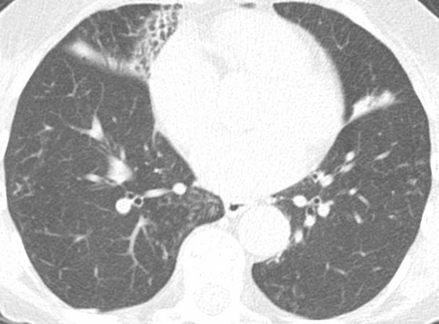
12 NTM Imaging Signs Tree-In-Bud and Centilobular Nodules Bronchiectasis Cavities Ground-Glass and Consolidation Atelectasis


13 NTM Imaging Signs Example Case - RML and Lingula - Where we are going


14 NTM Imaging Signs Centrilobular Nodules and Tree-In-Bud Typically from Airways Infection, Infection, Mucus Plugs/Aspiration


15


16 NTM Imaging Signs Bronchiectasis


17 Bronchiectasis NTM Imaging Signs


18 Bronchiectasis NTM Imaging Signs
19 figures from Bronchiectasis NTM Imaging Signs

20 Slice Thickens Bronchiectasis? 5 mm 1 mm Maybe? Yes! Bronchi bigger than arteries


21 Slice Thickens Bronchiectasis? 5 mm 1 mm Maybe? Yes! Bronchi bigger than arteries

22 Kim et al AJR 2005; 184: NTM Imaging Signs Cavities - and feeding bronchus sign


23 NTM Imaging Signs Cavities


24 NTM Imaging Signs Cavities

25 NTM Imaging Signs Cavities - and feeding bronchus sign
26 NTM Imaging Signs Cavities - and feeding bronchus sign Kim et al AJR 2005; 184:

27 NTM Imaging Signs Consolidation and Ground-Glass

28 NTM Imaging Signs Consolidation and Ground-Glass


29 Atelectasis NTM Imaging Signs RML

30 Atelectasis NTM Imaging Signs

31 Atelectasis NTM Imaging Signs RML


32 Aside: NTM with Normal CXR

33 Overview I. CT technique II. NTM imaging signs III. Radiological/Clinical Phenotypes & Secondary Causes and Underlying Disease
34 Radiological/Clinical Phenotypes of NTM I. Bronchiectasis/Tree-in-bud - Right middle lobe/lingular bronchiectasis II. Upper Lobe Cavities III. Solitary Pulmonary Nodule IV. NTM superimposed on underlying disease - Chronic Obstructive Pulmonary Disease (COPD) - Idiopathic Pulmonary Fibrosis (IPF) - Cystic Fibrosis (CF) V. Hot Tub Lung
35 Radiological/Clinical Phenotypes of NTM I. Bronchiectasis & Tree-in-Bud - CASE 1 CASE 1 Mild Look for active disease Tree-in-bud, consolidation possible look for stability (& clinical) GGO typically active inflammation

36 Radiological/Clinical Phenotypes of NTM I. Bronchiectasis & Tree-in-Bud - CASE 1 CASE 1 Mild Look for active disease Tree-in-bud, consolidation possible look for stability, (& clinical) GGO typically active inflammation


37 Radiological/Clinical Phenotypes of NTM I. Bronchiectasis & Tree-in-Bud - CASE 1 Inspiratory Expiratory Mild - no significant/minimal air trapping
38 Radiological/Clinical Phenotypes of NTM I. Bronchiectasis & Tree-in-Bud - CASE 2 CASE 2 Note RUL cystic bronchiectasis More severe: Nodules/nodular consolidations Bronchiectasis


39 CASE 2 Progression from

40 Radiological/Clinical Phenotypes of NTM I. Bronchiectasis & Tree-in-Bud - CASE 2 CASE 2 More severe, progressive Cavity formation


41 Radiological/Clinical Phenotypes of NTM II. Upper Lobe Cavities- CASE 3 CASE 3 RUL Cavity. Also bronchiectasis elsewhere Note soft tissue in cavity: Debris? Fungus? Rare cancer? Progressive enlarging solid


42 Radiological/Clinical Phenotypes of NTM II. Upper Lobe Cavities- CASE 3


43 Radiological/Clinical Phenotypes of NTM II. Upper Lobe Cavities- CASE 4 CASE 4 - More severe/ Classic upper lobe cavitary dz.


44 Radiological/Clinical Phenotypes of NTM II. Upper Lobe Cavities- CASE 4


45 Radiological/Clinical Phenotypes of NTM II. Upper Lobe Cavities- CASE Surgical Biopsy 2014 CASE 5 - Upper lobe cavitary dz. Mixed Change. Overall worsening


46 Radiological/Clinical Phenotypes of NTM II. Upper Lobe Cavities- CASE 5 CASE 5 Mixed change with progression Developed bronchocutaneous fistula. Rare. Here after surgery. Broncho-pleural fistula still

47 Radiological/Clinical Phenotypes of NTM III. Solitary Pulmonary Nodule - CASE 6 CASE 6 Solitary nodule. less common. Must still rule out other causes of nodule (i.e neoplasm)


48 Radiological/Clinical Phenotypes of NTM III. Solitary Pulmonary Nodule - CASE 7 CASE 7 Solitary Cavity Not squamous
49 Honda et al Clin Chest Med 2015; 36:1-11 Radiological/Clinical Phenotypes of NTM IV. NTM & underlying lung disease Often underlying lung disease Structural Non-structural Radiology also has role also in Risk factors for pulmonary NTM underlying disease


50 Radiological/Clinical Phenotypes of NTM IV. NTM & underlying lung disease- CASE 8 NTM in COPD/Emphysema Cavities can form: with bronchiectasis OR in preexisting disease Can spill contents


51 Radiological/Clinical Phenotypes of NTM IV. NTM & underlying lung disease- CASE 8


52 Radiological/Clinical Phenotypes of NTM IV. NTM & underlying lung disease- CASE 9 NTM in Chronic Aspiration Nothing Specific with known NTM Few months later Migratory Ground-Glass/Consolidation most suggestive Location? Anywhere, but: lower-posterior - most common. unilateral - sided sleeper? upper - gardening, yoga, cough?


53 Radiological/Clinical Phenotypes of NTM IV. NTM & underlying lung disease- CASE 9 Inspiratory Expiratory

54 Radiological/Clinical Phenotypes of NTM IV. NTM & underlying lung disease- CASE 9 Aspiration Work-up 1. Esophogram Also evaluates dysmotility Only 2 min intermittent for GERD 2. Tailored Barium Swallow with Speech Pathology Oral motility issues 3. Esophageal ph testing


55 Radiological/Clinical Phenotypes of NTM IV. NTM & underlying lung disease- CASE 10 NTM in Adult CF - often NOT specific findings


56 Radiological/Clinical Phenotypes of NTM IV. NTM & underlying lung disease- CASE 10 NTM in Adult CF Fatty Pancreas Replacement Inspiratory Expiratory Dynamic Expiratory - over 70% from Insp. ALSO TBM


57 Radiological/Clinical Phenotypes of NTM IV. NTM & underlying lung disease- CASE 11 NTM in Adult CF - example 2 more classic upper lung adult CF but normal pancreas

58 Radiological/Clinical Phenotypes of NTM IV. NTM & underlying lung disease- CASE 12 NTM in Silicosis

59 Radiological/Clinical Phenotypes of NTM IV. NTM & underlying lung disease- CASE 13 NTM in IPF

60 Radiological/Clinical Phenotypes of NTM IV. NTM & underlying lung disease- CASE 14 NTM in RA with TNF antagonist


61 Radiological/Clinical Phenotypes of NTM IV. NTM & underlying lung disease- CASE 14 NTM in RA/TNF antagonist


 Congenital")
62 Radiological/Clinical Phenotypes of NTM IV. NTM & underlying lung disease- CASE 15 NTM in Tracheobronchomegally (aka.mounier-kuhn syndrome) Congenital large central airways

63 Radiological/Clinical Phenotypes of NTM V. Hot Tub Lung- CASE 16 Hot Tub Lung Ground-Glass Centrilobular nodules


64 Radiological/Clinical Phenotypes of NTM V. Hot Tub Lung- CASE 16 Inspiratory Expiratory Hot Tub Lung Air-trapping essentially always present Could be only finding by CT Normal CXR in 20+% Hartman et al. AJR Apr;188(4):1050-3
65 PET/CT and NTM NTM will cause increased uptake (like most infections) SUV typically about 8.5 ( ) So caution in evaluating for cancer with NTM May be useful for disease activity/response (but higher radiation) Hahm et al. Lung Jan-Feb;188(1):25-31 Treglia et al. J Comput Assist Tomogr. 2011;35(3):


66 PET/CT and NTM Lung Cancer with NTM 6/13 1/14 6/15


67 MRI and NTM Cavities - Excellent Good but not perfect for other findings. (may miss small/mild findings and change) NO Radiation Chung et al. Ann Am Thorac Soc Jan;13(1):49-
68 References Martinez S, McAdams HP, Batchu CS. The many faces of pulmonary nontuberculous mycobacterial infection. AJR Am J Roentgenol. 2007;189(1): Ellis SM. The spectrum of tuberculosis and non-tuberculous mycobacterial infection. Eur Radiol. 2004;14 Suppl 3(3):E34-E42. Ellis SM, Hansell DM. Imaging of Non-tuberculous (Atypical) Mycobacterial Pulmonary Infection. Clin Radiol. 2002;57(8): Jeong YJ, Lee KS, Koh W-J, Han J, Kim TS, Kwon OJ. Nontuberculous mycobacterial pulmonary infection in immunocompetent patients: comparison of thin-section CT and histopathologic findings. Radiology. 2004;231(3): Wittram C, Weisbrod GL. Mycobacterium avium complex lung disease in immunocompetent patients: radiography-ct correlation. BJR. 2002;75(892): Erasmus JJ, McAdams HP, Farrell MA, Patz EF. Pulmonary nontuberculous mycobacterial infection: radiologic manifestations. RadioGraphics. 1999;19(6):
RADIOLOGIC EVALUATION OF PULMONARY NTM INFECTION. Tilman Koelsch, MD National Jewish Health - Department of Radiology
 Pr N op ot er fo ty r R of ep Pr ro es du en ct te io r n RADIOLOGIC EVALUATION OF PULMONARY NTM INFECTION Tilman Koelsch, MD National Jewish Health - Department of Radiology Disclosures No relevant financial
Hypothesis on the Evolution of Cavitary Lesions in Nontuberculous Mycobacterial Pulmonary Infection: Thin-Section CT and Histopathologic Correlation
 CT of Nontuberculous Mycobacterial Pulmonary Infection Tae Sung Kim 1 Won-Jung Koh 2 Joungho Han 3 Myung Jin Chung 1 Ju Hyun Lee 1 Kyung Soo Lee 1 O Jung Kwon 2 Kim TS, Koh W-J, Han J, et al. Received
Bronchiectasis: An Imaging Approach
 Bronchiectasis: An Imaging Approach Travis S Henry, MD Associate Professor of Clinical Radiology Cardiac and Pulmonary Imaging Section University of California, San Francisco Large Middle Small 1 Bronchiectasis
Diagnostic Evaluation of NTM and Bronchiectasis
 Division of Pulmonary, Critical Care and Sleep Medicine Diagnostic Evaluation of NTM and Bronchiectasis Ashwin Basavaraj, MD, FCCP NTM patient education program November 9, 2016 Involves a combination
Financial disclosure COMMON DIAGNOSES IN HRCT. High Res Chest HRCT. HRCT Pre test. I have no financial relationships to disclose. Anatomy Nomenclature
 Financial disclosure I have no financial relationships to disclose. Douglas Johnson D.O. Cardiothoracic Imaging Gaston Radiology COMMON DIAGNOSES IN HRCT High Res Chest Anatomy Nomenclature HRCT Sampling
Pediatric High-Resolution Chest CT
 Pediatric High-Resolution Chest CT Alan S. Brody, MD Professor of Radiology and Pediatrics Chief, Thoracic Imaging Cincinnati Children s s Hospital Cincinnati, Ohio, USA Pediatric High-Resolution CT Short
Overview of Idiopathic Pulmonary Fibrosis: Diagnosis and Therapy
 Overview of Idiopathic Pulmonary Fibrosis: Diagnosis and Therapy Jeff Swigris, DO, MS Director, ILD Program National Jewish Health Disclosures Speaker - Boehringer Ingelheim and Genentech Objectives Describe
Nontuberculous Mycobacterial Lung Disease
 Non-TB Mycobacterial Disease Jeffrey P. Kanne, MD Nontuberculous Mycobacterial Lung Disease Jeffrey P. Kanne, M.D. Consultant Disclosures Perceptive Informatics Royalties (book author) Amirsys, Inc. Wolters
Consolidations in Nodular Bronchiectatic Mycobacterium Avium Complex Lung Disease: Mycobacterium Avium Complex or Other Infection?
 Original Article DOI 10.3349/ymj.2010.51.4.546 pissn: 0513-5796, eissn: 1976-2437 Yonsei Med J 51(4):546-551, 2010 Consolidations in Nodular Bronchiectatic Mycobacterium Avium Complex Lung Disease: Mycobacterium
Case 1 : Question. 1.1 What is the intralobular distribution? 1. Centrilobular 2. Perilymphatic 3. Random
 Interesting case Case 1 Case 1 : Question 1.1 What is the intralobular distribution? 1. Centrilobular 2. Perilymphatic 3. Random Case 1: Answer 1.1 What is the intralobular distribution? 1. Centrilobular
Mycobacterium avium complex disease: prognostic implication of high-resolution computed tomography findings
 Eur Respir J 2008; 32: 147 152 DOI: 10.1183/09031936.00074207 CopyrightßERS Journals Ltd 2008 Mycobacterium avium complex disease: prognostic implication of high-resolution computed tomography findings
TB Radiology for Nurses Garold O. Minns, MD
 TB Nurse Case Management Salina, Kansas March 31-April 1, 2010 TB Radiology for Nurses Garold O. Minns, MD April 1, 2010 TB Radiology for Nurses Highway Patrol Training Center Salina, KS April 1, 2010
HRCT V/S MDCT: IN DETECTION OF BRONCHIECTASIS Sowmya M 1, Shilpa Patel 2, Pravan Kumar Reddy 3
 HRCT V/S MDCT: IN DETECTION OF BRONCHIECTASIS Sowmya M 1, Shilpa Patel 2, Pravan Kumar Reddy 3 HOW TO CITE THIS ARTICLE: Sowmya M, Shilpa Patel, Pravan Kumar Reddy. HRCT v/s MDCT: In Detection of Bronchiectasis.
10/17/2016. Nuts and Bolts of Thoracic Radiology. Objectives. Techniques
 Nuts and Bolts of Thoracic Radiology October 20, 2016 Carleen Risaliti Objectives Understand the basics of chest radiograph Develop a system for interpreting chest radiographs Correctly identify thoracic
2018 Vindico Medical Education. Non-tuberculous Mycobacteria: Circumventing Difficulties in Diagnosis and Treatment
 Activity presentations are considered intellectual property. These slides may not be published or posted online without permission from Vindico Medical Education (cme@vindicocme.com). Please be respectful
Radiological Aspects of Pulmonary Tuberculosis in Immunocompetent Hosts
 Nov 2003 Radiological Aspects of Pulmonary Tuberculosis in Immunocompetent Hosts Josh Rempell, Harvard Medical School Year III Tuberculosis: the captain of all (wo)men of death Overall, one third of the
An Image Repository for Chest CT
 An Image Repository for Chest CT Francesco Frajoli for the Chest CT in Antibody Deficiency Group An Image Repository for Chest CT he Chest CT in Antibody Deficiency Group is an international and interdisciplinary
NTM AND BRONCHIECTASIS (BXSIS): THE CHICKEN AND THE EGG
: THE CHICKEN AND THE EGG") REMINGTON WINTER COURSE IN ID VAIL, CO FEBRUARY, 2010 NONTUBERCULOSIS MYCOBACTERIA AND BRONCHIECTASIS: Revised 1/28/10 10:31 AM- CQ MICHAEL D. ISEMAN, MD OR In some cases NTMs invade pre-existing BXSIS
REMINGTON WINTER COURSE IN ID VAIL, CO FEBRUARY, 2010 NONTUBERCULOSIS MYCOBACTERIA AND BRONCHIECTASIS: Revised 1/28/10 10:31 AM- CQ MICHAEL D. ISEMAN, MD OR In some cases NTMs invade pre-existing BXSIS
Swyer-James Syndrome: An Infrequent Cause Of Bronchiectasis?
 ISPUB.COM The Internet Journal of Pulmonary Medicine Volume 12 Number 1 Swyer-James Syndrome: An Infrequent Cause Of Bronchiectasis? A Huaringa, S Malek, M Haro, L Tapia Citation A Huaringa, S Malek, M
PULMONARY TUBERCULOSIS RADIOLOGY
 PULMONARY TUBERCULOSIS RADIOLOGY RADIOLOGICAL MODALITIES Medical radiophotography Radiography Fluoroscopy Linear (conventional) tomography Computed tomography Pulmonary angiography, bronchography Ultrasonography,
September 2014 Imaging Case of the Month. Michael B. Gotway, MD. Department of Radiology Mayo Clinic Arizona Scottsdale, AZ
 September 2014 Imaging Case of the Month Michael B. Gotway, MD Department of Radiology Mayo Clinic Arizona Scottsdale, AZ Clinical History: A 57-year-old non-smoking woman presented to her physician as
Diagnosis of TB: Radiology David Finlay, MD
 TB Intensive Tyler, Texas June 2-4, 2010 Diagnosis of TB: Radiology David Finlay, MD June 3, 2010 2stages stages- Tuberculosis 1. primary infection 2. reactivation, or post primary disease 2 1 Primary
Case of the Day Chest
 Case of the Day Chest Darin White MDCM FRCPC Department of Radiology, Mayo Clinic 76 th Annual Scientific Meeting Canadian Association of Radiologists Montreal, QC April 26, 2013 2013 MFMER slide-1 Disclosures
An Introduction to Radiology for TB Nurses
 An Introduction to Radiology for TB Nurses Garold O. Minns, MD September 14, 2017 TB Nurse Case Management September 12 14, 2017 EXCELLENCE EXPERTISE INNOVATION Garold O. Minns, MD has the following disclosures
11/10/2014. Multi-disciplinary Approach to Diffuse Lung Disease: The Imager s Perspective. Radiology
 Multi-disciplinary Approach to Diffuse Lung Disease: The Imager s Perspective Radiology Pathology Clinical 1 Role of HRCT Diagnosis Fibrosis vs. inflammation Next step in management Response to treatment
The radiological differential diagnosis of the UIP pattern
 5th International Conference on Idiopathic Pulmonary Fibrosis, Modena, 2015, June 12th The radiological differential diagnosis of the UIP pattern Simon Walsh King s College Hospital Foundation Trust London,
Micronodular Lung Disease an algorithm
 Micronodular Lung Disease an algorithm H. Page McAdams, MD Department of Radiology Duke University Medical Center Durham, NC USA page.mcadams@duke.edu Question Which of the following lung diseases is MOST
How to Analyse Difficult Chest CT
 How to Analyse Difficult Chest CT Complex diseases are:- - Large lesion - Unusual or atypical pattern - Multiple discordant findings Diffuse diseases are:- - Numerous findings in both sides 3 basic steps
Comparison of CT Findings
 Newly Detected Pulmonary Nontuberculous Mycobacterial Infection and Peripheral Lung Cancers in Patients During Follow-Up of Idiopathic Interstitial Pneumonia Comparison of CT Findings Sang Young Oh, MD,
TB Intensive Houston, Texas
 TB Intensive Houston, Texas October 15-17, 17 2013 Diagnosis of TB: Radiology Rosa M Estrada-Y-Martin, MD MSc FCCP October 16, 2013 Rosa M Estrada-Y-Martin, MD MSc FCCP, has the following disclosures to
The Egyptian Journal of Hospital Medicine (July 2017) Vol.68 (2), Page
 Vol.68 (2), Page") The Egyptian Journal of Hospital Medicine (July 2017) Vol.68 (2), Page 1135-1140 Role of High Resolution Computed Tomography in Diagnosis of Interstitial Lung Diseases in Patients with Collagen Diseases
The Egyptian Journal of Hospital Medicine (July 2017) Vol.68 (2), Page 1135-1140 Role of High Resolution Computed Tomography in Diagnosis of Interstitial Lung Diseases in Patients with Collagen Diseases
Comparison of CT findings between MDR-TB and XDR-TB
 Comparison of CT findings between MDR-TB and XDR-TB Poster No.: C-0757 Congress: ECR 2017 Type: Authors: Keywords: DOI: Scientific Exhibit K. Yoon, H. Soohee; Changwon-si/KR Thorax, Lung, Respiratory system,
Original Article Tree-in-bud pattern of chest CT images for diagnosis of Mycobacterium abscesses
 Int J Clin Exp Med 2015;8(10):18705-18712 www.ijcem.com /ISSN:1940-5901/IJCEM0013538 Original Article Tree-in-bud pattern of chest CT images for diagnosis of Mycobacterium Haiqing Chu 1, Bing Li 1, Lan
4/17/2010 C ini n ca c l a Ev E a v l a ua u t a ion o n of o ILD U dat a e t e i n I LDs
 Update in ILDs Diagnosis 101: Clinical Evaluation April 17, 2010 Jay H. Ryu, MD Mayo Clinic, Rochester MN Clinical Evaluation of ILD Outline General aspects of ILDs Classification of ILDs Clinical evaluation
Acute and Chronic Lung Disease
 KATHOLIEKE UNIVERSITEIT LEUVEN Faculty of Medicine Acute and Chronic Lung Disease W De Wever, JA Verschakelen Department of Radiology, University Hospitals Leuven, Belgium Clinical utility of HRCT To detect
Pulmonary manifestations of Rheumatoid Arthritis: what is there waiting to be found?
 Pulmonary manifestations of Rheumatoid Arthritis: what is there waiting to be found? Poster No.: C-1795 Congress: ECR 2015 Type: Educational Exhibit Authors: M. S. C. Rodrigues, R. Correia, A. Carvalho,
Liebow and Carrington's original classification of IIP
 Liebow and Carrington's original classification of IIP-- 1969 Eric J. Stern MD University of Washington UIP Usual interstitial pneumonia DIP Desquamative interstitial pneumonia BIP Bronchiolitis obliterans
Difficulties Diagnosing Idiopathic Pulmonary Fibrosis
 1. er Encuentro Entre Neumólogos y Radiólogos, Madrid, Spain, 2016, October 14th Difficulties Diagnosing Idiopathic Pulmonary Fibrosis Simon Walsh King s College Hospital Foundation Trust London, United
Case 1: Question. 1.1 What is the main pattern of this HRCT? 1. Intralobular line 2. Groundglass opacity 3. Perilymphatic nodule
 HRCT WORK SHOP Case 1 Case 1: Question 1.1 What is the main pattern of this HRCT? 1. Intralobular line 2. Groundglass opacity 3. Perilymphatic nodule Case 1: Question 1.2 What is the diagnosis? 1. Hypersensitivity
Outline Definition of Terms: Lexicon. Traction Bronchiectasis
 HRCT OF IDIOPATHIC INTERSTITIAL PNEUMONIAS Disclosures Genentech, Inc. Speakers Bureau Tadashi Allen, MD University of Minnesota Assistant Professor Diagnostic Radiology 10/29/2016 Outline Definition of
Primary multidrug-resistant tuberculosis versus drug-sensitive tuberculosis in non-hivinfected patients: Comparisons of CT findings
 RESEARCH ARTICLE Primary multidrug-resistant tuberculosis versus drug-sensitive tuberculosis in non-hivinfected patients: Comparisons of CT findings Duo Li, Wei He, Budong Chen, Pingxin Lv* Department
Daria Manos RSNA 2016 RC 401. https://medicine.dal.ca/departments/depar tment-sites/radiology/contact/faculty/dariamanos.html
 Daria Manos RSNA 2016 RC 401 https://medicine.dal.ca/departments/depar tment-sites/radiology/contact/faculty/dariamanos.html STEP1: Is this fibrotic lung disease? STEP 2: Is this a UIP pattern? If yes:
Tuberculosis: The Essentials
 Tuberculosis: The Essentials Kendra L. Fisher, MD, PhD THORACIC TUBERCULOSIS: THE BARE ESSENTIALS Kendra Fisher MD, FRCP (C) Department of Radiology Loma Linda University Medical Center TUBERCULOSIS ()
Extraordinary Patterns of Tuberculosis
 Extraordinary Patterns of Tuberculosis E. Kadakovska Infectology Center of Latvia, Clinic of Tuberculosis and Lung Diseases, Diagnostics and Radiology Department 1 Target Importance of recognizing of tuberculosis
Differential diagnosis
 Differential diagnosis Idiopathic pulmonary fibrosis (IPF) is part of a large family of idiopathic interstitial pneumonias (IIP), one of four subgroups of interstitial lung disease (ILD). Differential
Lung Allograft Dysfunction
 Lung Allograft Dysfunction Carlos S. Restrepo M.D. Ameya Baxi M.D. Department of Radiology University of Texas Health San Antonio Disclaimer: We do not have any conflict of interest or financial gain to
Mycobacterium intracellulare Pleurisy Identified on Liquid Cultures of the Pleural Fluid and Pleural Biopsy
 http://dx.doi.org/10.4046/trd.2013.74.3.124 ISSN: 1738-3536(Print)/2005-6184(Online) Tuberc Respir Dis 2013;74:124-128 CopyrightC2013. The Korean Academy of Tuberculosis and Respiratory Diseases. All rights
Nontuberculous Mycobacteria
 Nontuberculous Mycobacteria Epidemiology and Clinical Management Kevin L. Winthrop, MD, MPH Associate Professor, Divisions of Infectious Diseases, Public Health and Preventive Medicine Oregon Health &
TB Intensive San Antonio, Texas November 29-December 2, 2011
 TB Intensive San Antonio, Texas November 29-December 2, 2011 Diagnosis of TB: Radiology Michael McCarthy, MD, FACR November 30, 2011 Michael McCarthy, MD, FACR has the following disclosures to make: No
Neuroendocrine Cell Hyperplasia of Infancy: Diagnosis With High- Resolution CT
 Pediatric Imaging Original Research Brody et al. CT of Neuroendocrine Cell Hyperplasia of Infancy Pediatric Imaging Original Research Alan S. Brody 1 R. Paul Guillerman 2 Thomas C. Hay 3 Brandie D. Wagner
Nontuberculous Mycobacteria (NTM) in Patients with Cystic Fibrosis
 in Patients with Cystic Fibrosis") Transcript Details This is a transcript of an educational program accessible on the ReachMD network. Details about the program and additional media formats for the program are accessible by visiting: https://reachmd.com/programs/cystic-fibrosis-in-focus/nontuberculous-mycobacteria-ntm-in-patientswith-cystic-fibrosis/8337/
Transcript Details This is a transcript of an educational program accessible on the ReachMD network. Details about the program and additional media formats for the program are accessible by visiting: https://reachmd.com/programs/cystic-fibrosis-in-focus/nontuberculous-mycobacteria-ntm-in-patientswith-cystic-fibrosis/8337/
Eun-Young Kang, M.D., Jae Wook Lee, M.D., Ji Yung Choo, M.D., Hwan Seok Yong, M.D., Ki Yeol Lee, M.D., Yu-Whan Oh, M.D.
 Eun-Young Kang, M.D., Jae Wook Lee, M.D., Ji Yung Choo, M.D., Hwan Seok Yong, M.D., Ki Yeol Lee, M.D., Yu-Whan Oh, M.D. Department of Radiology, Korea University Guro Hospital, College of Medicine, Korea
Invasive Mucinous Adenocarcinoma Mimicking Organizing Pneumonia Associated with Mycobacterium fortuitum Infection
 CASE REPORT Invasive Mucinous Adenocarcinoma Mimicking Organizing Pneumonia Associated with Mycobacterium fortuitum Infection Daisuke Morichika 1, Nobuaki Miyahara 1, Katsuyuki Hotta 1, Yoshiko Okamoto
Chest Radiology Interpretation: Findings of Tuberculosis
 Chest Radiology Interpretation: Findings of Tuberculosis Get out your laptops, smart phones or other devices pollev.com/chestradiology Case #1 1 Plombage Pneumonia Cancer 2 Reading the TB CXR Be systematic!
Pulmonary Manifestations of Systemic Lupus Erythematosus 1
 Pulmonary Manifestations of Systemic Lupus Erythematosus 1 Kee Hyuk Yang, M.D., Yo Won Choi, M.D., Seok Chol Jeon, M.D., Choong Ki Park, M.D., Kyung in Joo, M.D., Chang Kok Hahm, M.D., Seung Ro Lee, M.D.
Mai ElMallah,MD Updates in Pediatric Pulmonary Care XII: An Interdisciplinary Program April 13, 2012
 Mai ElMallah,MD Updates in Pediatric Pulmonary Care XII: An Interdisciplinary Program April 13, 2012 Recognize the importance of Pulmonary Function Testing in Cystic Fibrosis Be aware of different types
Tests Your Pulmonologist Might Order. Center For Cardiac Fitness Pulmonary Rehab Program The Miriam Hospital
 Tests Your Pulmonologist Might Order Center For Cardiac Fitness Pulmonary Rehab Program The Miriam Hospital BASIC ANATOMY OF THE LUNGS Lobes of Lung 3 lobes on the Right lung 2 lobes on the Left Blood
Residents Section Pattern of the Month
 Residents Section Pattern of the Month Gosset et al. Tree-In-Bud Pattern Residents Section Pattern of the Month Residents inradiology Natacha Gosset 1 Alexander A. Bankier Ronald L. Eisenberg Gosset N,
Unit II Problem 2 Pathology: Pneumonia
 Unit II Problem 2 Pathology: Pneumonia - Definition: pneumonia is the infection of lung parenchyma which occurs especially when normal defenses are impaired such as: Cough reflex. Damage of cilia in respiratory
Imaging Small Airways Diseases: Not Just Air trapping. Eric J. Stern MD University of Washington
 Imaging Small Airways Diseases: Not Just Air trapping Eric J. Stern MD University of Washington What we are discussing SAD classification SAD imaging with MDCT emphasis What is a small airway? Airway with
August 2018 Imaging Case of the Month: Dyspnea in a 55-Year-Old Smoker. Michael B. Gotway, MD
 August 2018 Imaging Case of the Month: Dyspnea in a 55-Year-Old Smoker Michael B. Gotway, MD Department of Radiology Mayo Clinic Arizona Scottsdale, AZ USA Clinical History: A 55 year old woman presented
Back to Basics: What Imaging Test should I order? Jeanne G. Hill, M.D. Pediatric Radiology Medical University of South Carolina
 Back to Basics: What Imaging Test should I order? Jeanne G. Hill, M.D. Pediatric Radiology Medical University of South Carolina Disclosure Neither I nor any member of my immediate family has a relevant
Atypical radiologic appearances of pulmonary tuberculosis in non-hiv adult patients
 Atypical radiologic appearances of pulmonary tuberculosis in non-hiv adult patients Poster No.: R-0200 Congress: RANZCR-AOCR 2012 Type: Educational Exhibit Authors: K. N. Jeon, K. Bae, M. J. Park Keywords:
Manish Powari Regional Training Day 10/12/2014
 Manish Powari Regional Training Day 10/12/2014 Large number of different types of Interstitial Lung Disease (ILD). Most are very rare Most patients present with one of a smaller number of commoner diseases
Uses, limitations and interpretation of CT in pulmonary infections: A practical approach
 Uses, limitations and interpretation of CT in pulmonary infections: A practical approach Canadian Association of Radiologists 2013 DISCLOSURES Speakers honorarium, Siemens Canada Objectives 1. Recognize
Prevalence of Gastroesophageal Reflux Disease in Patients With Nontuberculous Mycobacterial Lung Disease*
 Original Research MYCOBACTERIAL DISEASE Prevalence of Gastroesophageal Reflux Disease in Patients With Nontuberculous Mycobacterial Lung Disease* Won-Jung Koh, MD; Jun Haeng Lee, MD; Yong Soo Kwon, MD;
Interesting Cases. Pulmonary
 Interesting Cases Pulmonary 54M with prior history of COPD, hep B/C, and possible history of TB presented with acute on chronic dyspnea, and productive cough Hazy opacity overlying the left hemithorax
Thoracic CT pattern in lung cancer: correlation of CT and pathologic diagnosis
 19 th Congress of APSR PG of Lung Cancer (ESAP): Update of Lung Cancer Thoracic CT pattern in lung cancer: correlation of CT and pathologic diagnosis Kazuma Kishi, M.D. Department of Respiratory Medicine,
Radiologic-pathologic correlation of pulmonary diseases
 The 1578 th Chest Conference/ 3 rd Biennial Clinical- Radiologic-Pathologic Correlation Radiologic-pathologic correlation of pulmonary diseases Harumi Itoh, M.D. University of Fukui, Japan Centriacinar
Parametric response mapping
 Parametric response mapping Utility of a novel imaging biomarker in pulmonary disease Dharshan Vummidi MD, Lama VN MD, Yanik G MD, Kazerooni EA MD, Meilan Han MD, Galban C PhD Radiology, Pulmonary & Critical
HRCT in CHILDREN. strengths and weaknesses in practice: Dr Catherine Owens BSc MBBS MRCP FRCR. Great Ormond Street Hospital for Children NHS Trust
 HRCT in CHILDREN strengths and weaknesses in practice: Dr Catherine Owens BSc MBBS MRCP FRCR Great Ormond Street Hospital for Children NHS Trust London WC1N 3JH Telephone 02074059200 Ext. 0493 or 5072
Systemic lupus erythematosus (SLE): Pleuropulmonary Manifestations
: Pleuropulmonary Manifestations") 08/30/10 09/26/10 Systemic lupus erythematosus (SLE): Pleuropulmonary Manifestations Camila Downey S. Universidad de Chile, School of Medicine, Year VII Harvard University, School of Medicine Sept 17,
08/30/10 09/26/10 Systemic lupus erythematosus (SLE): Pleuropulmonary Manifestations Camila Downey S. Universidad de Chile, School of Medicine, Year VII Harvard University, School of Medicine Sept 17,
Hyun Su Kim, MD Kyung Soo Lee, MD Won-Jung Koh, MD Kyeongman Jeon, MD Eun Ju Lee, MD Hee Kang, MD Joonghyun Ahn, PhD. Purpose: Materials and Methods:
 Note: This copy is for your personal non-commercial use only. To order presentation-ready copies for distribution to your colleagues or clients, contact us at www.rsna.org/rsnarights. Original Research
Lung Cancer Associated with Cystic Airspaces: Don t Let This Lesion Fool You!
 Lung Cancer Associated with Cystic Airspaces: Don t Let This Lesion Fool You! Annemie Snoeckx Antwerp University Hospital, University of Antwerp, Belgium Head of Department: Prof. dr. Paul M. Parizel June
5/9/2015. Multi-disciplinary Approach to Diffuse Lung Disease: The Imager s Perspective. No, I am not a pulmonologist! Radiology
 Multi-disciplinary Approach to Diffuse Lung Disease: The Imager s Perspective No, I am not a pulmonologist! Radiology Pathology Clinical 1 Everyone needs a CT Confidence in diagnosis Definitive HRCT +
HRCT Versus Volume Rendering (Three Colors, Three Densities Lung Images) in Diagnosis of Small Airway Disease: A Comparative Study
 in Diagnosis of Small Airway Disease: A Comparative Study") Med. J. Cairo Univ., Vol. 84, No. 1, March: 359-364, 2016 www.medicaljournalofcairouniversity.net HRCT Versus Volume Rendering (Three Colors, Three Densities Lung Images) in Diagnosis of Small Airway Disease:
Med. J. Cairo Univ., Vol. 84, No. 1, March: 359-364, 2016 www.medicaljournalofcairouniversity.net HRCT Versus Volume Rendering (Three Colors, Three Densities Lung Images) in Diagnosis of Small Airway Disease:
Nontuberculous Mycobacteria (NTM)
") Nontuberculous Mycobacteria (NTM) Bacteria, like plants and animals, have been classified into similar groups. The groups are called "families." One such family of bacteria is known as the Mycobacteriaceae.
Nontuberculous Mycobacteria (NTM) Bacteria, like plants and animals, have been classified into similar groups. The groups are called "families." One such family of bacteria is known as the Mycobacteriaceae.
Immunocompromised patients. Immunocompromised patients. Immunocompromised patients
 Value of CT in Early Pneumonia in Immunocompromised Patients Nantaka Kiranantawat, PSU Preventative Factors Phagocyts Cellular immunity Humoral immunity Predisposing Factors Infection, Stress, Poor nutrition,
IPF: Epidemiologia e stato dell arte
 IPF: Epidemiologia e stato dell arte Clinical Classification Diffuse parenchimal lung diseases Exposure-related: - occupational - environmental - medication Desquamative interstitial pneumonia Idiopathic
Cavitation in primary lung cancer:
 Cavitation in primary lung cancer: characteristic features and mechanisms Yoshie Kunihiro 1,2), Taiga Kobayashi 2), Nobuyuki Tanaka 3), Tsuneo Matsumoto 1), Naofumi Matsunaga 2) 1) National Hospital Organization,
INTERSTITIAL LUNG DISEASE. Radhika Reddy MD Pulmonary/Critical Care Long Beach VA Medical Center January 5, 2018
 INTERSTITIAL LUNG DISEASE Radhika Reddy MD Pulmonary/Critical Care Long Beach VA Medical Center January 5, 2018 Interstitial Lung Disease Interstitial Lung Disease Prevalence by Diagnosis: Idiopathic Interstitial
Cryptogenic Organizing Pneumonia Diagnosis Approach Based on a Clinical-Radiologic-Pathologic Consensus
 Cryptogenic Organizing Pneumonia Diagnosis Approach Based on a Clinical-Radiologic-Pathologic Consensus Poster No.: C-1622 Congress: ECR 2012 Type: Scientific Exhibit Authors: C. Cordero Lares, E. Zorita
Imaging: how to recognise idiopathic pulmonary fibrosis
 REVIEW IDIOPATHIC PULMONARY FIBROSIS Imaging: how to recognise idiopathic pulmonary fibrosis Anand Devaraj Affiliations: Dept of Radiology, St George s Hospital, London, UK. Correspondence: Anand Devaraj,
Radiology Pathology Conference
 Radiology Pathology Conference Sharlin Johnykutty,, MD, Cytopathology Fellow Sara Majewski, MD, Radiology Resident Friday, August 28, 2009 Presentation material is for education purposes only. All rights
Chest Imaging Original Research
 Chest Imaging Original Research Song et al. CT of Mycobacterium avium-intracellulare Complex Pulmonary Disease Chest Imaging Original Research Jong Woon Song 1 Won-Jung Koh 2 Kyung Soo Lee 1 Ji Young Lee
Chapter 16. Lung Abscess. Mosby items and derived items 2011, 2006 by Mosby, Inc., an affiliate of Elsevier Inc.
 Chapter 16 Lung Abscess 1 EDA PM C AFC RB A B Figure 16-1. Lung abscess. A, Cross-sectional view of lung abscess. B, Consolidation and (C) excessive bronchial secretions are common secondary anatomic alterations
CT findings in multifocal or diffuse non-mucinous bronchioloalveolar carcinoma (BAC)
") CT findings in multifocal or diffuse non-mucinous bronchioloalveolar carcinoma (BAC) Poster No.: C-2192 Congress: ECR 2014 Type: Educational Exhibit Authors: I. Sandu, A. R. Popita, I.-A. Brumboiu; Cluj-Napoca/RO
CT findings in multifocal or diffuse non-mucinous bronchioloalveolar carcinoma (BAC) Poster No.: C-2192 Congress: ECR 2014 Type: Educational Exhibit Authors: I. Sandu, A. R. Popita, I.-A. Brumboiu; Cluj-Napoca/RO
NONE OVERVIEW FINANCIAL DISCLOSURES UPDATE ON IDIOPATHIC PULMONARY FIBROSIS/IPF (UIP) FOR PATHOLOGISTS. IPF = Idiopathic UIP Radiologic UIP Path UIP
 FOR PATHOLOGISTS. IPF = Idiopathic UIP Radiologic UIP Path UIP") UPDATE ON IDIOPATHIC PULMONARY FIBROSIS/IPF () FOR PATHOLOGISTS Thomas V. Colby, M.D. Professor of Pathology (Emeritus) Mayo Clinic Arizona FINANCIAL DISCLOSURES NONE OVERVIEW IPF Radiologic Dx Pathologic
UPDATE ON IDIOPATHIC PULMONARY FIBROSIS/IPF () FOR PATHOLOGISTS Thomas V. Colby, M.D. Professor of Pathology (Emeritus) Mayo Clinic Arizona FINANCIAL DISCLOSURES NONE OVERVIEW IPF Radiologic Dx Pathologic
Bronchial syndrome. Atelectasis Draining bronchus Bronchiectasis
 Bronchial syndrome Atelectasis Draining bronchus Bronchiectasis Etienne Leroy Terquem Pierre L Her SPI / ISP Soutien Pneumologique International / International Support for Pulmonology Atelectasis Consequence
Pulmonary Aspergillosis
 May 2005 Pulmonary Aspergillosis Nancy Wei, Harvard Medical School, Year III Overview Pulmonary aspergillosis background information Patient presentations Common radiographic findings for each type of
Thoracic lung involvement in rheumatoid arthritis: Findings on HRCT
 Thoracic lung involvement in rheumatoid arthritis: Findings on HRCT Poster No.: C-2488 Congress: ECR 2015 Type: Educational Exhibit Authors: R. E. Correa Soto, M. J. Martín Sánchez, J. M. Fernandez 1 1
Pathologic Findings of Surgically Resected Nontuberculous Mycobacterial Pulmonary Infection
 The Korean Journal of Pathology 2010; 44: 56-62 DOI: 10.4132/KoreanJPathol.2010.44.1.56 Pathologic Findings of Surgically Resected Nontuberculous Mycobacterial Pulmonary Infection Hye-Jong Song Jung Suk
Diffuse Interstitial Lung Diseases: Is There Really Anything New?
 : Is There Really Anything New? Sujal R. Desai, MBBS, MD ESTI SPEAKER SUNDAY Society of Thoracic Radiology San Antonio, Texas March 2014 Diffuse Interstitial Lung Disease The State of Play DILDs Is There
Excavated pulmonary nodule: steps to diagnosis?
 Excavated pulmonary nodule: steps to diagnosis? Poster No.: C-1044 Congress: ECR 2014 Type: Authors: Keywords: DOI: Educational Exhibit W. Mnari, M. MAATOUK, A. Zrig, B. Hmida, M. GOLLI; Monastir/ TN Metastases,
Community-Acquired Acinetobacter baumannii Pneumonia: Initial Chest Radiographic Findings and Follow-up CT Findings in Helping Predict Patient Outcome
 Community-Acquired Acinetobacter baumannii Pneumonia: Initial Chest Radiographic Findings and Follow-up CT Findings in Helping Predict Patient Outcome Jeong Joo Woo, Dong Hyun Lee, Jin Kyung An Department
OBJECTIVES. Solitary Solid Spiculated Nodule. What would you do next? Case Based Discussion: State of the Art Management of Lung Nodules.
 Organ Imaging : September 25 2015 OBJECTIVES Case Based Discussion: State of the Art Management of Lung Nodules Dr. Elsie T. Nguyen Dr. Kazuhiro Yasufuku 1. To review guidelines for follow up and management
The Dr. Jae Yang Lecture: An Overview of the Radiographic Picture of TB
 The Dr. Jae Yang Lecture: An Overview of the Radiographic Picture of TB Harvey H. Wong, MD FRCPC MScCH Assistant Professor Department of Medicine Division of Respirology University of Toronto Financial