HEPATO-BILIARY IMAGING
|
|
|
- Alan Alexander
- 5 years ago
- Views:
Transcription


1 HEPATO-BILIARY IMAGING BY MAMDOUH MAHFOUZ MD PROF.OF RADIOLOGY CAIRO UNIVERSITY
2 CT ABDOMEN Indications Patient preparation Patient position Scanogram Fasting 4-6 hours Frontal Supine To assess equivocal imaging findings Staging of hepatic neoplasms Metastatic workup of primary malignancies Diagnosis of diffuse hepatic diseases Assessment of biliary problems Assessment of suspected post-traumatic injury

3 CT ABDOMEN Indications Patient preparation Patient position Scanogram Fasting 4-6 hours Frontal Supine No required preparation unless the patient is going to be sedated or injected with contrast material FASTING FOR 4-6 HOURS


4 Scanning techniques Standard Examination Spiral, Helical, volumetric CT Multi-Detector, Multi-Slice CT

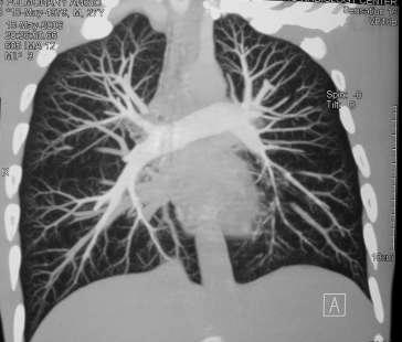
5 Normal pulmonary vascularity

6

7 Detailed examination of the Superior Mesenteric Artery and Celiac Artery. Scan time = 9.4 seconds. 1mm slice thickness

8 Non- contrast Study The inherent difference between attenuation value of normal liver parenchyma and most of the lesions is low Old indications for NCCT Hypervascular metastases Quantification of liver iron in cases of hemochromatosis Detection of confluent fibrosis in cirrhotic liver
9 Value of precontrast study
10 Arterial phase Hypervascular deposits

11 Single phase CECT 80% of hepatic parenchymal blood flow is supplied by portal vein Lesions are detected in the portal phase of enhancement [60 seconds after bolus contrast injection] Hepatocellular carcinoma


![injection] 20%of liver parenchymal](/docs-images/81/83647117/images/12-2.jpg "blood flow comes from the hepatic")

12 Dual- phase CECT Scanning the liver twice with a single contrast bolus Arterial phase [the 1 st seconds of contrast injection] 20%of liver parenchymal blood flow comes from the hepatic artery Most of hepatic tumours receive their main blood supply from the hepatic artery


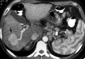
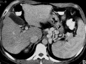
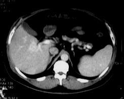
13 CT Portal venography in a 56Y Male with portal vein thrombosis

14 MS LS AS PS


15 Hepatic pathology Focal lesions Cystic Solid Mixed Diffuse lesions

16 Judgment is based only on either Contrast enhanced CT Contrast enhanced MRI

17 Cystic lesions of the liver Hepatic cysts Congenital lesions but detected late Isolated or associated with congenital cystic disease Usually asymptomatic Complications [ rupture or hage ] lead to symptoms Few mms to several cms in size Hepatic cysts


18 Hepatic cysts Typical cyst criteria Sharply defined margin Paper-thin wall Clear water contents 0-15 HU Absent Septations Calcification Enhancement Mural nodules
![Cystic lesions of the liver Liver abscess [Pyogenic] Frequently indolent with no signs of infection May present with profound septicemia Microabscesses](/docs-images/81/83647117/images/19-0.jpg "(>2cm) cluster or widely scattered Macroabscesses :Hypo dense lesion, unilocular or multilocular Marginal enhancement 6%?")
19 Cystic lesions of the liver Liver abscess [Pyogenic] Frequently indolent with no signs of infection May present with profound septicemia Microabscesses (>2cm) cluster or widely scattered Macroabscesses :Hypo dense lesion, unilocular or multilocular Marginal enhancement 6%?! Gas containing abscesses uncommon
20

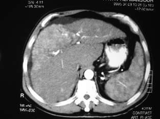
21 Gas containing hepatic abscess

22 Multiple Pyogenic hepatic abscesses
![Cystic lesions of the liver Liver abscess [Amebic ] Entameoba](/docs-images/81/83647117/images/23-0.jpg "Histolytica 10% world wide Patients are more often acutely ill")

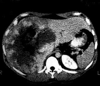
23 Cystic lesions of the liver Liver abscess [Amebic ] Entameoba Histolytica 10% world wide Patients are more often acutely ill Single or multiple near the liver capsule Enhancing wall is evident with peripheral zone of edema [ Common findings in amebic abscess]

24 Amebic abscess
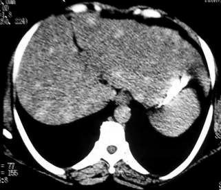
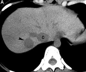
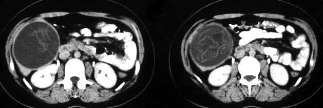
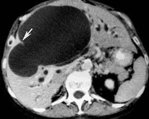
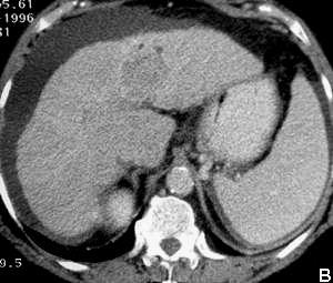
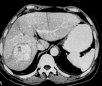
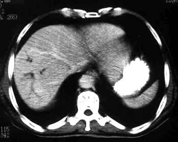
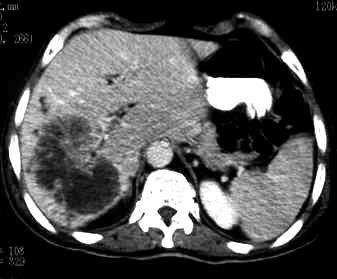
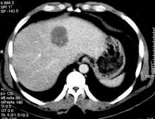
25 Benign lesions of the liver Hemangioma 20% of hepatic tumours The most common benign liver tumour 85% are asymptomatic Female: male = 5:1 50% are multiple Giant hemangioma 6-10 cm in diameter
26 Hemangioma with central area of fibrosis
27 Hemangioma
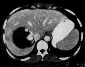
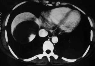
28 Hemangioma
29 Giant hemangioma
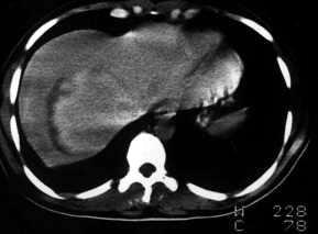
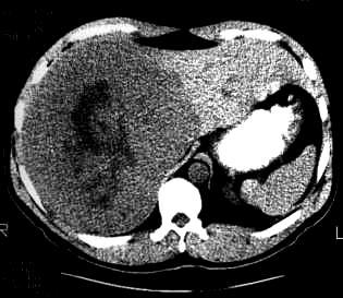
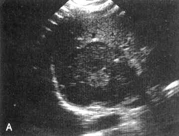
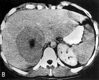
30 Cystic lesions of the liver Echinococcal disease [Hydatid cyst] Larval stage of E. granulosus Well defined unilocular or multilocular cyst Central and peripheral calcification Daughter cysts can be inside the large cyst


31 Hydatid cyst

32 Hydatid cyst

33 Hydatid cyst

34 Cystic lesions of the liver CT FEATURES TO DIGNOSE HYDATED CYST Other cysts specially in the lung Unilocular or multilocular cyst with marginal calcification Internal floating shadows Daughter cysts within the large cyst


35 35Y Male patient with lung cancer suspected to have liver deposits by US
36 Rare cystic lesions Biliary cystadenoma / carcinoma Cystic deposits
37 Biliary cystadenoma, cystadeno carcinoma 90% occur intrahypatic With ovarian stroma [seen in females+ good prognosis] Without ovarian stroma [males and females + bad prognosis] Large [3 40 cm] cystic multilocular tumour with mural nodularities [seen better by US] Distinction between cystadenoma and cystadenocarcinoma may not be possible by imaging and is not clinically critical, both will be excised
38 Biliary cystadenoma

39 Biliary cystadenoma

40 Malignant Hepatic Lesions Hepatocellular carcinoma The most common primary malignant hepatic neoplasms 3 rd 4 th decades male: female 8:1 80% of HCC occur in cirrhotic liver Serum AFP and ultrasound [screening]

![injection [ better triphasic study] Arterial phase heterogeneously](/docs-images/81/83647117/images/41-1.jpg "enhancing lesion with hypo dense capsule Portal phase hypo dense")
41 Hepatocellular carcinoma CT Single or multiple masses that are hypo dense to normal liver Calcification may be seen After contrast injection [ better triphasic study] Arterial phase heterogeneously enhancing lesion with hypo dense capsule Portal phase hypo dense lesion
42 Hepatocellular carcinoma CT [arterial phase] Detects a greater number of HCC than usual scanning Detects intravascular thrombosis [ portal vein] Better delineation of tumour capsule in capsulated lesions Detects early arteriovenous shunting [ sign of malignancy]
43 Hepatocellular carcinoma

44 Hepatocellular carcinoma

45 Poorly differentiated Hepatocellular carcinoma


46 HCC
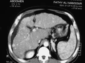
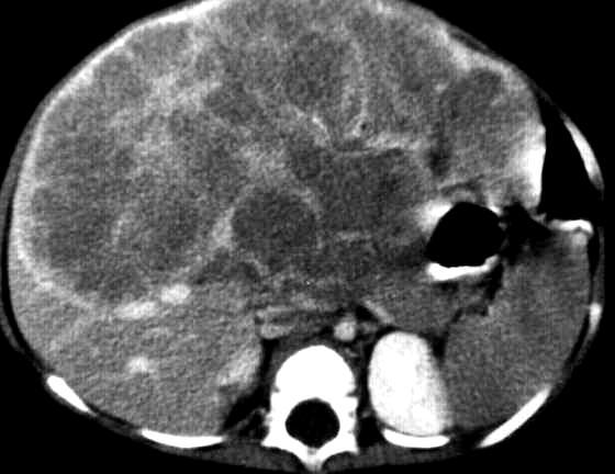
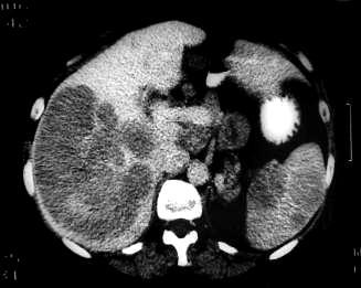
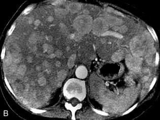
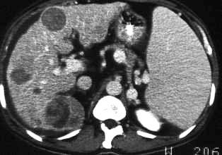
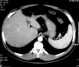
47 Hepatocellular carcinoma


48 M 49Y with elevated AFP


49 Portal Arterial Delayed M 59 Y with liver cirhhosis, splenomegaly and suspected focal lesion on US
50 Hepatoblastoma
51 Malignant Hepatic Lesions Cholangiocarcinoma The 2 nd most common primary malignant tumour Arise from bile duct epithelium [ 3 TYPES ] Intrahepatic tumour arised from small peripheral ducts Or the major ducts near the helium Orat the bifurcation of the common hepatic ducts [ Klatskin tumour] HCC: intrahepatic cholangiocarcinoma = 10:1 No strong association between the lesion and cirrhosis
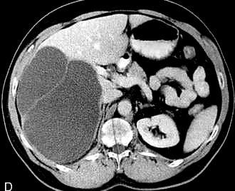
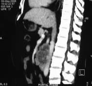
52 Cholangiocarcinoma CT Hypo dense lesion that shows heterogenous enhancement Portal vein invasion is rarely seen Small dilated ducts around the lesion may be seen
53

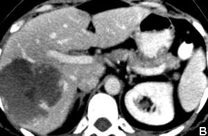
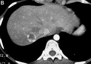
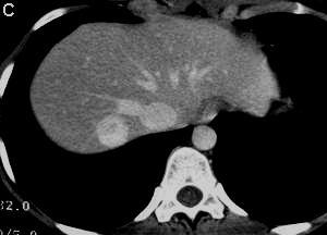
54 Cholangio carcinoma
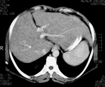
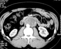
55 Lymphoma Primary hepatic lymphoma is rare compared to the 2ry type AIDS and organ transplant patients have an increased risk Non specific CT and MR appearance Diffuse hepatic lymphoma hypo dense liver similar to fatty infiltration
56 Lymphoma

57 Lymphoma

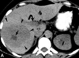
58 Hepatic deposits Liver is the 2 nd most common site for deposits after nodes 30% - 70% of patients who die of cancer have liver deposits CT NCCT hypodense lesions,calcification in mucin producing metastases CECT Dynamic bolus contrast injection with helical scanning in a single breath hold of seconds Single phase Dual phase Triphasic study CTHA & CTAP
59 Contrast enhanced CT Single phase [portal phase] Maximal enhancement of liver tissue 150ml. Pump injector 3ml/sec delay time 70 sec Dual phase [arterial & portal phase] 150ml. Pump injector 4ml/sec delay time 25/60 sec CTHA CTAP Selective enhancement of tumour Selective enhancement of liver tissue

60 Hepatic deposits Most of hepatic deposits are hypovascular Hepatic neoplasms receive most of their blood supply via hepatic artery Hypervascular deposits should be assessed by dual phase CT or dynamic MRI At present CTAP and intra operative US are the most sensitive methods for detection of hepatic deposits


61 Hyper vascular deposits

62 Hepatic deposits


63 portal M 54Y with liver deposits and cyst

64 Calcified hepatic metastases in a patient with mucinous adenocarcinoma of the colon


65 Multiple discrete hepatic metastases From segmoid cancer


66 M 83Y with abdominal pain, US showed 2 focal hepatic lesions


67 36Y Male with testicular tumor evaluated postoperatively
68 Benign solid lesions of the liver Rare lesions Hepatic adenoma Focal nodular hyperplasia [ FNH ] Angiomyolipoma Fat containing lesion occurs in patients with tuberous sclerosis where other lesions are present in the kidney


69 Focal nodular hyperplasia FNH 8% of all hepatic tumours 2 nd most common benign hepatic tumour after hemangioma 80%- 90% in women 3 rd 5 th decades 80%- 90% are solitary lesions <5cm in diameter Central fibrous scar is a characteristic feature Focal nodular hyperplasia


70 Focal nodular hyperplasia FNH Precontrast CT iso or hypodense lesion Arterial phase CT Marked enhancement Focal nodular hyperplasia dual phase helical CT

71 Hepatocellular adenoma

72 Thank you سبحانك اللهم و بحمدك نشهد ان ال اله اال انت نستغفرك و نتوب اليك
Liver Tumors. Prof. Dr. Ahmed El - Samongy
 Liver Tumors Prof. Dr. Ahmed El - Samongy Objective 1. Identify the most important features of common benign liver tumors 2. Know the risk factors, diagnosis, and management of hepatocellular carcinoma
CT & MRI of Benign Liver Neoplasms Srinivasa R Prasad
 CT & MRI of Benign Liver Neoplasms Srinivasa R Prasad No financial disclosures Acknowledgements Many thanks to Drs. Heiken, Narra & Menias (MIR) Dr. Sahani (MGH) for sharing images Benign Liver Tumors:
Malignant Focal Liver Lesions
 Malignant Focal Liver Lesions Other Than HCC Pablo R. Ros, MD, MPH, PhD Departments of Radiology and Pathology University Hospitals Cleveland Medical Center Case Western Reserve University Pablo.Ros@UHhospitals.org
Evaluation of Liver Mass Lesions. American College of Gastroenterology 2013 Regional Postgraduate Course
 Evaluation of Liver Mass Lesions American College of Gastroenterology 2013 Regional Postgraduate Course Lewis R. Roberts, MB ChB, PhD Division of Gastroenterology and Hepatology Mayo Clinic College of
CTA/MRA of Pediatric Hepatic Masses Radiology-Pathology Correlation
 Acta Radiológica Portuguesa, Vol.XVIII, nº70, pág. 41-50, Abr.-Jun., 2006 CTA/MRA of Pediatric Hepatic Masses Radiology-Pathology Correlation Marilyn J. Siegel Mallinckrodt Institute of Radiology, Washington
CT 101 :Pancreas and Spleen
 CT 101 :Pancreas and Spleen Shikha Khullar,, MD, MPH Division of Radiology University of South Alabama The Pancreas Normal Pancreas 3 Phase Pancreatic CT Non contrast Arterial phase : 30-35 35 second
The Focal Hepatic Lesion: Radiologic Assessment
 The Focal Hepatic Lesion: Radiologic Assessment Kevin Kuo, Harvard Medical School Year III Our Patient: PS 67 y/o female w/ long history of alcohol use Drinking since age 18, up to one bottle of wine/day
Imaging of liver and pancreas
 Imaging of liver and pancreas.. Disease of the liver Focal liver disease Diffusion liver disease Focal liver disease Benign Cyst Abscess Hemangioma FNH Hepatic adenoma HCC Malignant Fibrolamellar carcinoma
Brain Imaging. IC calcifications. Mamdouh mahfouz MD
 Brain Imaging IC calcifications www.ssregypt.com Mamdouh mahfouz MD mamdouh.m5@gmail.com CT Hyper dense [ more than100 HU ] MRI Low signal in T1 and T2 WIs [non mobile protons] Exceptions Minute calcifications
Interesting Cases from Liver Tumor Board. Jeffrey C. Weinreb, M.D.,FACR Yale University School of Medicine
 Interesting Cases from Liver Tumor Board Jeffrey C. Weinreb, M.D.,FACR Yale University School of Medicine jeffrey.weinreb@yale.edu Common Liver Diseases Hemangioma Cyst FNH Focal Fat/Sparing THID Non-Cirrhotic
Essentials of Clinical MR, 2 nd edition. 65. Benign Hepatic Masses
 65. Benign Hepatic Masses Pulse sequences acquired for abdominal MRI typically consist of fast acquisition schemes such as single-shot turbo spin echo (i.e. HASTE) and gradient echo schemes such as FLASH
Acknowledgements. Update of Focal Liver Lesions Goals. Focal Liver Lesions. Imaging Choices For Liver Lesions. Focal Liver Lesions
 Acknowledgements Update of Focal Liver Lesions 2012 Giles Boland Massachusetts General Hospital Harvard Medical School No disclosures Dushyant Sahani Mukesh Harisinghani Goals Focal liver lesions Imaging
Liver Cancer (Hepatocellular Carcinoma or HCC) Overview
 Overview") Liver Cancer (Hepatocellular Carcinoma or HCC) Overview Recent advances in liver cancer care seek to address the rising incidence of liver cancer, which has steadily increased over the past three decades.
Liver Cancer (Hepatocellular Carcinoma or HCC) Overview Recent advances in liver cancer care seek to address the rising incidence of liver cancer, which has steadily increased over the past three decades.
Normal Sonographic Anatomy
 hapter 2:The Liver DUNSTAN ABRAHAM Normal Sonographic Anatomy Homogeneous, echogenic texture (Figure 2-1) Measures approximately 15 cm in length and 10 12.5 cm anterior to posterior; measurement taken
Radiology of hepatobiliary diseases
 GI cycle - Lecture 14 436 Teams Radiology of hepatobiliary diseases Objectives 1. To Interpret plan x-ray radiograph of abdomen with common pathologies. 2. To know the common pathologies presentation.
Renal masses - the role of diagnostic imaging
 Renal masses - the role of diagnostic imaging Poster No.: C-2471 Congress: ECR 2015 Type: Educational Exhibit Authors: V. Rai#; Bjelovar/HR Keywords: Cysts, Cancer, Structured reporting, Ultrasound, MR,
MRI OF FOCAL LESIONS IN
 Introduction MRI OF FOCAL LESIONS IN THE NON-CIRRHOTIC LIVER Ivan Pedrosa M.D. Ph.D. Associate Professor of Radiology and Advanced Imaging Research Center University of Texas Southwestern. Dallas, TX Incidental
We are IntechOpen, the world s leading publisher of Open Access books Built by scientists, for scientists. International authors and editors
 We are IntechOpen, the world s leading publisher of Open Access books Built by scientists, for scientists 3,350 108,000 1.7 M Open access books available International authors and editors Downloads Our
X-Ray Corner. Imaging Approach to Cystic Liver Lesions. Pantongrag-Brown L. Solitary cystic liver lesions. Hepatic simple cyst (Figure 1)
") THAI J 136 Imaging Approach to Cystic Liver Lesions GASTROENTEROL 2013 X-Ray Corner Imaging Approach to Cystic Liver Lesions Pantongrag-Brown L Cystic liver lesions are common findings in daily practice
THAI J 136 Imaging Approach to Cystic Liver Lesions GASTROENTEROL 2013 X-Ray Corner Imaging Approach to Cystic Liver Lesions Pantongrag-Brown L Cystic liver lesions are common findings in daily practice
Newcastle HPB MDM updated radiology imaging protocol recommendations. Author Dr John Scott. Consultant Radiologist Freeman Hospital
 Newcastle HPB MDM updated radiology imaging protocol recommendations Author Dr John Scott. Consultant Radiologist Freeman Hospital This document is intended as a guide to aid radiologists and clinicians
Spinal Imaging. ssregypt.com. Mamdouh Mahfouz MD
 Spinal Imaging Degenerative diseases ssregypt.com Mamdouh Mahfouz MD mamdouh.m5@gmail.com MRI Open MRI Closed Extremity MRI Dynamic MRI Dynamic MRI The bed rotates from Upright to Recumbent, stopping at
ACG Clinical Guideline: Diagnosis and Management of Focal Liver Lesions
 ACG Clinical Guideline: Diagnosis and Management of Focal Liver Lesions Jorge A. Marrero, MD, 1 Joseph Ahn, MD, FACG, 2 K. Rajender Reddy, MD, FACG 3 1 University of Texas at Southwestern, Dallas, Texas,
Alice Fung, MD Oregon Health and Science University
 Alice Fung, MD Oregon Health and Science University Disclosure Comments The speaker Alice Fung, MD Has relevant financial relationships to disclose. Received honorarium from (Guerbet). This individual
Hepatocellular carcinoma Cholangiocarcinoma. Jewels of hepatobiliary cancer imaging : what to look for? Imaging characteristics of HCC.
 Outline : Imaging Jewels Jewels of hepatobiliary cancer imaging : what to look for? Hepatocellular carcinoma Cholangiocarcinoma Surachate Siripongsakun, M.D. Chulabhorn Cancer Center Imaging characteristics
State of the Art Imaging for Hepatic Malignancy: My Assignment
 State of the Art Imaging for Hepatic Malignancy: My Assignment CT vs MR vs MRCP Which one to choose for HCC vs Cholangiocarcinoma What special protocols to use for liver tumors Role of PET and Duplex US
Workup of a Solid Liver Lesion
 Workup of a Solid Liver Lesion Joseph B. Cofer MD FACS Chief Quality Officer Erlanger Health System Affiliate Professor of Surgery UTHSC-Chattanooga I have no financial or other relationships with any
Imaging of common diseases of hepatobiliary and GI system
 Imaging of common diseases of hepatobiliary and GI system Natthaporn Tanpowpong, M.D. Diagnostic radiology Faculty of Medicine, Chulalongkorn University Normal plain radiograph A = Common bile duct
Approach to Liver Lesions. Anjana A. Pillai, MD Associate Professor of Medicine Director, Liver Tumor Clinic The University of Chicago Medical Center
 Approach to Liver Lesions Anjana A. Pillai, MD Associate Professor of Medicine Director, Liver Tumor Clinic The University of Chicago Medical Center Objectives Identify common clinical features and imaging
GASTROINTESTINAL IMAGING STUDY GUIDE
 GASTROINTESTINAL IMAGING STUDY GUIDE Pharynx Diverticula Foreign bodies Trauma o Motility Disorders Esophagus Diverticula Trauma Esophagitis Barrett esophagus Rings, webs, and strictures Varices Benign
Financial Disclosure
 Benign Liver Masses Adil Abdalla, MBBS Creighton University-CHI Health August 25, 2018 Financial Disclosure Nothing to disclose Financial Disclosure 1 Objectives To assess patients with benign liver tumors
Neoplasms of the Canine, Feline and Lemur Liver:
 Neoplasms of the Canine, Feline and Lemur Liver: Classification and Prognosis Annual Seminar of the French Society of Veterinary Pathology John M. Cullen VMD PhD DACVP North Carolina State University Primary
International Journal of Current Medical Sciences- Vol. 6, Issue,, pp , June, 2016 A B S T R A C T
 ISSN: 2320-8147 International Journal of Current Medical Sciences- Vol. 6, Issue,, pp. 122-126, June, 2016 COMPUTED TOMOGRAPHY IN HEPATIC METASTASES Ananthakumar P and Adaikkappan M., Available online
Hepatobiliary and Pancreatic Malignancies
 Hepatobiliary and Pancreatic Malignancies Gareth Eeson MD MSc FRCSC Surgical Oncologist and General Surgeon Kelowna General Hospital Interior Health Consultant, Surgical Oncology BC Cancer Agency Centre
Innovations in HCC Imaging: MDCT/MRI
 Innovations in HCC Imaging: MDCT/MRI Anthony E. Cheng, M.D. Cardinal MRI Center Cardinal Santos Medical Center, Wilson Street, San Juan Innovations in HCC Imaging: Goals/Objectives MDCT/MRI Learn the diagnostic
Recently role of non-invasive diagnostics methods
 CERTAIN ASPECTS OF NLS-DIAGNOSTICS OF LIVER FOCAL PATHOLOGY A.Y. Shvack, V.I. Nesterov, N.L. Ogluzdina This article contains information about NLS-graphy application in diagnostics of liver focal affections:
Cystic lesions of the liver
 Cystic lesions of the liver Poster No.: C-0408 Congress: ECR 2014 Type: Educational Exhibit Authors: E. Rosado, J. Pereira, S. El Bouchaibi, M. A. A. Bali ; 1 1 2 2 3 3 4 4 Amadora/PT, Lisboa/PT, Bruxelles/BE,
Liver Cancer And Tumours
 Liver Cancer And Tumours What causes liver cancer? Many factors may play a role in the development of cancer. Because the liver filters blood from all parts of the body, cancer cells from elsewhere can
Radiological Reasoning: Incidentally Discovered Liver Mass
 AJR Integrative Imaging LIFELONG LEARNING FOR RADIOLOGY This Radiological Reasoning article is available for SAM credit and CME credits when completed with the additional educational material provided
Hepato-Pancreatico-Biliary Surgery. Dr. Ankur J. Shah. MS, DNB, MNAMS, MRCSEd (UK), FRCS (UK)
, FRCS (UK)") Hepato-Pancreatico-Biliary Surgery Dr. Ankur J. Shah MS, DNB, MNAMS, MRCSEd (UK), FRCS (UK) Consultant Hepato-Pancreatico-Biliary and Liver Transplant Surgeon Ansh Liver Clinic Prevention to Cure Address
Hepato-Pancreatico-Biliary Surgery Dr. Ankur J. Shah MS, DNB, MNAMS, MRCSEd (UK), FRCS (UK) Consultant Hepato-Pancreatico-Biliary and Liver Transplant Surgeon Ansh Liver Clinic Prevention to Cure Address
Pediatric Retroperitoneal Masses Radiologic-Pathologic Correlation
 Acta Radiológica Portuguesa, Vol.XVIII, nº 70, pág. 61-70, Abr.-Jun., 2006 Pediatric Retroperitoneal Masses Radiologic-Pathologic Correlation Marilyn J. Siegel Mallinckrodt Institute of Radiology, Washington
Role of imaging in RCC. Ultrasonography. Solid lesion. Cystic RCC. Solid RCC 31/08/60. From Diagnosis to Treatment: the Radiologist Perspective
 Role of imaging in RCC From Diagnosis to Treatment: the Radiologist Perspective Diagnosis Staging Follow up Imaging modalities Limitations and pitfalls Duangkamon Prapruttam, MD Department of Therapeutic
Jesse Civan, M.D. Medical Director, Jefferson Liver Tumor Center
 Liver Tumors Jesse Civan, M.D. Medical Director, Jefferson Liver Tumor Center Differential Diagnosis Malignant Metastatic from non-hepatic primary Hepatocellular carcinoma Cholangiocarcinoma Biliary cystcarcinoma
Cystic Disease of the Liver Work Up and Management. Louis Ferrari MD, PGY 3 6/9/16 SUNY Downstate Medical Center
 Cystic Disease of the Liver Work Up and Management Louis Ferrari MD, PGY 3 6/9/16 SUNY Downstate Medical Center The Case 73F presents to clinic after diagnostic laparoscopy at OSH. Known liver mass for
Common and unusual CT and MRI manifestations of pancreatic adenocarcinoma: a pictorial review
 Review Article Common and unusual CT and MRI manifestations of pancreatic adenocarcinoma: a pictorial review Min-Jie Yang, Su Li, Yong-Guang Liu, Na Jiao, Jing-Shan Gong Department of Radiology, Shenzhen
Dr Claire Smith, Consultant Radiologist St James University Hospital Leeds
 Dr Claire Smith, Consultant Radiologist St James University Hospital Leeds Imaging in jaundice and 2ww pathway Image protocol Staging Limitations Pancreatic cancer 1.2.4 Refer people using a suspected
Imaging iconography of gallbladder cancer. Assessment by CT.
 1 REVISTA DE IMAGENOLOGIA- EII / Vol. XVI / Num. 2 Imaging iconography of gallbladder cancer. Assessment by CT. Doctors Crisci, Alejandro (1); Landó, Fernando.(2). CASMU CT Department Hospital of Tacuarembó
간암의조직검사 : 언제, 어떻게? 계명대학교의과대학내과학교실 정우진
 간암의조직검사 : 언제, 어떻게? 계명대학교의과대학내과학교실 정우진 간생검한다 vs 안한다? M/81 Alcoholic LC, albumin 4.0, bil 0.6, Cr 1.06, glucose 141, afp 2.2, CA19-9 12.41 CT: R/O HCC in S8, R/O CC M/69 HBV(-), HCV(-), social alcoholics
Contrast Enhanced Ultrasound of Parenchymal Masses in Children
 Contrast Enhanced Ultrasound of Parenchymal Masses in Children Sue C Kaste, DO On behalf of Beth McCarville, MD St. Jude Children s Research Hospital Memphis, TN Overview Share St. Jude experience with
Gemstone Spectral Imaging quantifies lesion characteristics for a confident diagnosis
 GE Healthcare Gemstone Spectral Imaging quantifies lesion characteristics for a confident diagnosis CT clinical case study lesion characterization Desiree Morgan, MD Vice Chair of Clinical Research Professor
LIVER IMAGING TIPS IN VARIOUS MODALITIES. M.Vlychou, MD, PhD Assoc. Professor of Radiology University of Thessaly
 LIVER IMAGING TIPS IN VARIOUS MODALITIES M.Vlychou, MD, PhD Assoc. Professor of Radiology University of Thessaly Hepatocellular carcinoma is a common malignancy for which prevention, screening, diagnosis,
HCC and mass effect. Hepatocellular cancer: what if the AFP is rising but no lesion seen on imaging? What you need to know about AFP.
 Hepatocellular cancer: what if the AFP is rising but no lesion seen on imaging? Arun J Sanyal M.B.B.S., M.D. Charles Caravati Professor of Medicine Virginia Commonwealth University Imaging features used
IT 의료융합 1 차임상세미나 복부질환초음파 이재영
 IT 의료융합 1 차임상세미나 2013-4-3 복부질환초음파 이재영 나는오늘누구를위하여 종을울리나? 전통적의료 의사 공학설계자 의사 최첨단진단장비들 USG, CT, MRI 환자 환자 현대의료 사용자중심의사고 US in the Abdomen Detection DDx Look Behavior Response by external stimuli Guiding Tool
Chief Complain. Liver lesion found in routine health check 41 days ago
 Chief Complain Liver lesion found in routine health check 41 days ago Present Illness On 2005-7-26 at 台北署立醫院 he underwent a health check for the first time. Abdominal US showed suspicious of a 6*5 cm hepatoma,
Liver nodules mimicking metastatic disease
 Liver nodules mimicking metastatic disease Poster No.: C-1703 Congress: ECR 2011 Type: Educational Exhibit Authors: F. Vandenbroucke, B. Ilsen, B. Op de Beeck, J. de Mey ; 1 1 2 2 3 2 3 Brussels/BE, Brussel/BE,
Vascular Imaging in the Pediatric Abdomen. Jonathan Swanson, MD
 Vascular Imaging in the Pediatric Abdomen Jonathan Swanson, MD Goals and Objectives To understand the imaging approach, appearance, and clinical manifestations of the common pediatric abdominal vascular
Pancreatic Adenocarcinoma: Everything You Need to Know From Cross-Sectional Imaging to Treatment
 Pancreatic Adenocarcinoma: Everything You Need to Know From Cross-Sectional Imaging to Treatment Andrew W. Bowman, MD PhD Assistant Professor of Radiology Mayo Clinic Florida SCBT-MR Annual Meeting Nashville,
Rare primary liver tumors - MRI pictorial review
 Rare primary liver tumors - MRI pictorial review Poster No.: C-2293 Congress: ECR 2017 Type: Educational Exhibit Authors: R. Lameiras, J. Cruz, J. Felício Costa, F. D. Figueiredo, A. 1 1 1 1 2 1 1 1 2
Complete Summary GUIDELINE TITLE. Liver lesion characterization. BIBLIOGRAPHIC SOURCE(S)
") Complete Summary GUIDELINE TITLE Liver lesion characterization. BIBLIOGRAPHIC SOURCE(S) Foley WD, Bree RL, Gay SB, Glick SN, Heiken JP, Huprich JE, Levine MS, Ros PR, Rosen MP, Shuman WP, Greene FL, Rockey
Complete Summary GUIDELINE TITLE Liver lesion characterization. BIBLIOGRAPHIC SOURCE(S) Foley WD, Bree RL, Gay SB, Glick SN, Heiken JP, Huprich JE, Levine MS, Ros PR, Rosen MP, Shuman WP, Greene FL, Rockey
Surgical conditions of liver Somkit Mingphruedhi, M.D.
 Surgical conditions of liver Somkit Mingphruedhi, M.D. Division of HPB Surgery, Department of Surgery Ramathibodi Hospital Anatomy IVC Portal Vein Hepatic Artery Splenic Vein Gallbladder CBD SMV Anatomy
Objectives. HCC Incidence and Mortality. Disclosure Statement HCC. Imaging of Hepatocellular Carcinoma. Treatment of Hepatocellular Carcinoma
 Imaging of Hepatocellular Carcinoma and the use of LI RADS Treatment of Hepatocellular Carcinoma Aaron D. Anderson, D.O. AOCR April 2015 Objectives Show how the use of LI RADS can simplify the diagnosis
Evaluation of Diffuse Liver Diseases Using Conventional Ultrasound
 IOSR Journal of Dental and Medical Sciences (IOSR-JDMS) e-issn: 2279-0853, p-issn: 2279-0861.Volume 16, Issue 6 Ver. VII (June. 2017), PP 70-74 www.iosrjournals.org Evaluation of Diffuse Liver Diseases
Diagnostic Studies Then. It s important to be able to distinguish. Diagnostic Studies Now
 Jonathan S. Fisher, MD, FACS It s important to be able to distinguish Diagnostic Studies Then Diagnostic Studies Then History Biopsy Diagnostic Studies Now History Biopsy Serum markers (AFP, CA19 9, CEA)
With the widespread use of hepatic imaging, liver masses
 2B: Liver Assessment of the Liver Mass: What Do You Need to Know? With the widespread use of hepatic imaging, liver masses are detected either unexpectedly or in the course of screening for liver cancer
Detection and Characterization of Hepatocellular Carcinoma by Imaging
 CLINICAL GASTROENTEROLOGY AND HEPATOLOGY 2005;3:S136 S140 Detection and Characterization of Hepatocellular Carcinoma by Imaging OSAMU MATSUI Department of Imaging Diagnosis and Interventional Radiology,
Imaging abdominal vascular emergencies. V.Stoynova
 Imaging abdominal vascular emergencies V.Stoynova Abdominal vessels V. Stoynova 2 Acute liver bleeding trauma anticoagulant therapy liver disease : HCC, adenoma, meta, FNH, Hemangioma Diagnosis :CT angiography
Liver Tumors. Patient Education. Treatment options 8 4A. About the Liver. Surgical Specialties
 Patient Education Treatment options This handout describes different kinds of tumors that form in the liver and how they are treated. About the Liver Your liver is the largest organ in your abdomen. It
Alpha-fetoprotein
 Other Names/Abbreviations AFP 190.25 - Alpha-fetoprotein Alpha-fetoprotein (AFP) is a polysaccharide found in some carcinomas. It is effective as a biochemical marker for monitoring the response of certain
Chief Complaint. Retroperitoneal cystic mass incidentally found at health examination center.
 Personal Information Age: 34 y/o Sex: female Past history: major systemic medical history(-) surgical history(-), family history(-) Denied food or drug allergy Chief Complaint Retroperitoneal cystic mass
IMAGING OF LIVER, BILIARY TREE, PANCREAS
 IMAGING OF LIVER, BILIARY TREE, PANCREAS Department of Radiology West China Hospital, Sichuan University Yao Jin Learning Points The methodology for imaging the LBP (liver, biliary tree, and pancreas )
Pediatric Hepatobiliary, Pancreatic & Splenic US
 Pediatric Hepatobiliary, Pancreatic & Splenic US Susan J. Back, MD Department of Radiology, The Children s Hospital of Philadelphia No Disclosures Objectives Normal Abnormal: cases and US advances Objectives
US-Guided Radiofrequency Ablation of Hepatic Focal Lesions
 US-Guided Radiofrequency Ablation of Hepatic Focal Lesions Poster No.: C-2219 Congress: ECR 2011 Type: Scientific Exhibit Authors: D. Armario Bel, A. PLA, F. TERREL, X. Serres; BARCELONA/ES Keywords: Neoplasia,
Imaging in breast cancer. Mammography and Ultrasound Donya Farrokh.MD Radiologist Mashhad University of Medical Since
 Imaging in breast cancer Mammography and Ultrasound Donya Farrokh.MD Radiologist Mashhad University of Medical Since A mammogram report is a key component of the breast cancer diagnostic process. A mammogram
Appendix 5. EFSUMB Newsletter. Gastroenterological Ultrasound
 EFSUMB Newsletter 87 Examinations should encompass the full range of pathological conditions listed below A log book listing the types of examinations undertaken should be kept Training should usually
Alpha-fetoprotein
 Other Names/Abbreviations AFP 190.25 - Alpha-fetoprotein Alpha-fetoprotein (AFP) is a polysaccharide found in some carcinomas. It is effective as a biochemical marker for monitoring the response of certain
Enhancements in Hepatobiliary Imaging:
 Enhancements in Hepatobiliary Imaging: S. Channual 1, MD; A. Pahwa 2, MD; S. Raman 1, MD. 1 UCLA Medical Center, Department of Radiologic Sciences 2 Olive-View UCLA Medical Center, Department of Radiology
Video Microscopy Tutorial 8
 Video Microscopy Tutorial 8 Common and Uncommon Lesions of the Liver Gladwyn Leiman, MD There are no disclosures necessary. Common and Uncommon Lesions in Liver FNA Gladwyn Leiman University of Vermont
Abstract. Introduction. Patients and Methods
 The Evaluation of Hepatocellular Carcinoma with Biphasic Contrast enhanced Helical CT Scan J. Yaqoob, V. Bari, M. U. Usman, K. Munir, F. Mosharaf, W. Akhtar Department of Radiology, Aga Khan University
General summary GENERAL SUMMARY
 General summary GENERAL SUMMARY In Chapter 2.1 the long-term results and prognostic factors of radiofrequency ablation (RFA) for unresectable colorectal liver metastases (CRLM) in a single center with
MALIGNANT HEPATIC NEOPLASMS: USING ULTRASONOGRAPHY AS A MEANS OF DEFINING HEPATIC LESIONS. 1.5 Contact Hours. Presented by: CEU Professor 7
 MALIGNANT HEPATIC NEOPLASMS: USING ULTRASONOGRAPHY AS A MEANS OF DEFINING HEPATIC LESIONS 1.5 Contact Hours Presented by: CEU Professor 7 www.ceuprofessoronline.com Copyright 8 2007 The Magellan Group,
Primary Hepatic Undifferentiated Pleomorphic Sarcoma: CT and angiographic findings in two cases
 J Radiol Sci 2013; 38: 15-19 Primary Hepatic Undifferentiated Pleomorphic Sarcoma: CT and angiographic findings in two cases Jan-Wen Ku Ying-Chi Tseng Kuo-Luon Kung Hsien-Chang Shen Yen-Lin Huang Chi-Jen
Pediatric Abdominal Masses. Andrew Phelps MD Assistant Professor of Pediatric Radiology UCSF Benioff Children's Hospital
 Pediatric Abdominal Masses Andrew Phelps MD Assistant Professor of Pediatric Radiology UCSF Benioff Children's Hospital No Disclosures Take Home Message All you need to remember are the 5 common masses
Head&Neck Imaging. ssregypt.com. Parapharyngeal Spaces. Mamdouh mahfouz MD
 Head&Neck Imaging Parapharyngeal Spaces ssregypt.com Mamdouh mahfouz MD mamdouh.m5@gmail.com Definitio n Fat filled triangular space lateral the pharynx Extends from the skull base to the oropharynx Parapharyngeal
Management of Rare Liver Tumours
 Gian Luca Grazi Hepato-Biliary-Pancreatic Surgery National Cancer Institute Regina Elena Rome Fibrolamellar Carcinoma Mixed Hepato Cholangiocellular Carcinoma Hepatoblastoma Carcinosarcoma Primary Hepatic
Role of Imaging Methods in Diagnosis of Acute Pancreatitis. Válek V. Radiologická klinika, FN Brno a LF MU v Brně
 Role of Imaging Methods in Diagnosis of Acute Pancreatitis Válek V. Radiologická klinika, FN Brno a LF MU v Brně New Classification: Acute Pancreatitis 2007 revision of Atlanta classification and definitions
Interesting case. Vikas Kundra, M.D., Ph.D. October Vikas Kundra, M.D., Ph.D.
 Interesting case October 2012 Disclosure Information Vikas Kundra, M.D, Ph.D. I have no financial relationships to disclose. I WILL NOT include discussion of investigational or off-label use of a product
X-ray Corner. Imaging of The Pancreas. Pantongrag-Brown L
 X-ray Corner 125 Imaging of The Pancreas Modern imaging modalities commonly used in pancreas include ultrasound (US), CT, and MRI. Pancreas is a retroperitoneal organ which makes it difficult to visualize
Biliary tract tumors
 Short Course 2010 Annual Fall Meeting of the Korean Society for Pathologists Biliary tract tumors Joon Hyuk Choi, M.D., Ph.D. Professor, Department of Pathology, Yeungnam Univ. College of Medicine, Daegu,
RING ENCHANCING LESION BY M.S. HEMHNATH
 RING ENCHANCING LESION BY M.S. HEMHNATH A 21 YRS FEMALE CAME WITH H/O HEADACHE AND SEIZURE FOR THE PAST ONE MONTH. NO OTHER FOCAL NEUROLOGICAL DEFICIT. DIFFERENTIAL DIAGNOSIS For this case are Neurocysticerosis
ABDOMINAL DIFFUSION WEIGHTED MR
 ABDOMINAL DIFFUSION WEIGHTED MR Frank Miller, M.D. FACR Professor of Radiology Chief, Body Imaging Section Medical Director, MR Imaging Northwestern University Feinberg School of Medicine fmiller@northwestern.edu
Alpha-fetoprotein
 Other Names/Abbreviations AFP 190.25 - Alpha-fetoprotein Alpha-fetoprotein (AFP) is a polysaccharide found in some carcinomas. It is effective as a biochemical marker for monitoring the response of certain
Approach to the Patient with Liver Disease
 Approach to the Patient with Liver Disease Diagnosis of liver disease Careful history taking Physical examination Laboratory tests Radiologic examination and imaging studies Liver biopsy Liver diseases
objectives Pitfalls and Pearls in PET/CT imaging Kevin Robinson, DO Assistant Professor Department of Radiology Michigan State University
 objectives Pitfalls and Pearls in PET/CT imaging Kevin Robinson, DO Assistant Professor Department of Radiology Michigan State University To determine the regions of physiologic activity To understand
Guidelines, Policies and Statements D5 Statement on Abdominal Scanning
 Guidelines, Policies and Statements D5 Statement on Abdominal Scanning Disclaimer and Copyright The ASUM Standards of Practice Board have made every effort to ensure that this Guideline/Policy/Statement
Central role: - Regulating the immune system - Influencing metabolic and endocrine functions
 Spleen Central role: - Regulating the immune system - Influencing metabolic and endocrine functions Anatomy: An encapsulated mass of vascular and lymphatic tissue The largest RES organ 9-11 th ribs 4 impressions
Liver imaging takes a step forward with Ingenia
 Publication for the Philips MRI Community ISSUE 49 2013 / 2 Liver imaging takes a step forward with Ingenia Lyon South Hospital strives to move from several studies first CT, then MR or PET to using just
HCC e CEUS. Prof. A. Giorgio. Direttore IX UOC di Malattie Infettive ad Indirizzo Ecointerventistico
 HCC e CEUS Prof. A. Giorgio Direttore IX UOC di Malattie Infettive ad Indirizzo Ecointerventistico The natural history of compensated cirrhosis due to hepatitis C virus: a 17 year cohort study of 214 patients
Liver mets icd-10. Search
 Liver mets icd-10 Search 2-3-2012 ICD-10; Risk Adjustment. Mets Liver and lung I am ducts but there does not seem to be anything but stones going on i. ICD-10: C22.0 Short Description: Liver cell carcinoma
CT abdomen and pelvis
 CT abdomen and pelvis General indications: Assessment of vague abdominal symptoms (pain, colics,distenstion,...) Varifecation of a lesion discovered by other diagnostic modalities as US, barium,ivp, Staging
Intraductal papillary mucinous neoplasm of the bile ducts: a rare form of premalignant lesion of invasive cholangiocarcinoma
 Intraductal papillary mucinous neoplasm of the bile ducts: a rare form of premalignant lesion of invasive cholangiocarcinoma Authors: R. Revert Espí, Y. Fernandez Nuñez, I. Carbonell, D. P. Gómez valencia,