Knee Joint Anatomy 101
|
|
|
- Barnard Pitts
- 5 years ago
- Views:
Transcription

1 Knee Joint Anatomy 101 Bone Basics There are three bones at the knee joint femur, tibia and patella commonly referred to as the thighbone, shinbone and kneecap. The fibula is not typically associated with the knee because it lies outside the capsule and as associated with ankle function. We need to consider the osteological features (study of bones) of this joint to fully appreciate the numerous complex movements necessary to accomplish flexion and extension. The femoral head sits inside the acetabulum at the ball and socket hip joint with the neck connecting laterally to the greater trochanter. Distal features include the convex medial/lateral condyles, Intercondylar notch or patellar groove and adductor tubercle.


2 The femur articulates at the tibial plateau. These condyles are concave medially, convex laterally, and divided by the tibial tubercles, which fit snugly into the Intercondylar notch in extension. The anatomical axis of the femur bone is divergent from the mechanical axis, descending medially from the center of the greater trochanter to the Intercondylar notch; conversely, the anatomical and mechanical tibial axis are nearly identical.

3 Close examination of the tibial and femoral medial condyles of both femur and tibia extend further than their respective counterparts, which creates a physiologic valgus angle of the knee approximating degrees. Diagnostic criteria for abnormal frontal plane adduction is >185-degrees (Genu Valgum) or </= 175- degrees abduction (Genu Varum), measured at the medial-tibiofemoral joint in extension.


4 The anterior orientation of both lateral condyles and a degree curve on the medial femoral condyle are probably the most distinct features of the tibiofemoral articulating surface structure. The medial femoral condyle articulating surface area is larger and curved relative to the lateral condyle, which is flatter, smaller and more circular. Both structures are convex. At the tibial plateau, the medial condyle is concave and 50% larger than the partially convex lateral condyle. The degree of posterior slope varies between condyles, as does the amount of cartilage.


5 The patella is embedded within the retinacula layer between the femur and tibia, attached superiorly to the quadriceps tendon and inferiorly to the patellar tendon. Exposed posteriorly to articulate with the femoral sulcus, the surface contains a central vertical ridge and facets. Cartilage Basics Hyaline cartilage covers a large portion of the femoral condyles and posterior patella to reduce patellofemoral articular friction. Fibrocartilagenous menisci at the tibial plateau create a semi-flexible housing that compensate tibiofemoral obliquity.

6 Menisci attached to the surface of the tibial plateau improve congruence, pressure distribution and articulation at the tibiofemoral joint. Fixed to the tibia at the horns and coronary ligaments they are joined anteriorly by the transverse ligament. The lateral meniscus forms a nearly complete circle and covers a majority of the tibial condyle s surface, with anterior and posterior horns (typically) blending into the attachment of the ACL. There is no attachment between the lateral meniscus and the lateral collateral ligament, providing increased mobility in comparison to the medial meniscus. The c-shaped medial meniscus is larger, but covers significantly less surface area then the lateral meniscus. Anterior fibers of the ACL blend with the transverse ligament and anterior horn of the medial meniscus. And capsular attachments join to the deep medial collateral ligament directly and patella via the patellomeniscal ligaments.

 collateral ligament contains superficial (extracapsular) and deep (capsular) components, between femur and tibia, and the")
7 Ligament Basics Ligaments create passive resistance to facilitate optimal knee function and stability. Tibiofemoral Ligaments: The deep anterior and posterior cruciate are considered Intracapsular and extra synovial because they exist inside the joint capsule but outside the synovial membrane, crossing anterior/posterior & posterior/anterior between femur and tibia. The medial (tibial) collateral ligament contains superficial (extracapsular) and deep (capsular) components, between femur and tibia, and the lateral (fibular) ligament is purely extracapsular, connecting femur to fibula.


8 Patellofemoral Ligaments Patellar movement is initiated by the quadriceps and guided by the medial/lateral tibial and femoral ligaments. Knee Joint Capsule Consistent with other synovial joints, the knee exists within a capsule made of connective tissue and bone. The outer layer consists of a fibrous membrane with openings at the patella for the suprapatellar bursa and lateral tibial condyle for popliteal muscle, inseparable with numerous supporting capsular ligaments.
 to")


9 The inner-membrane secretes synovial fluid (and nutrients) to reduce friction between joint surfaces Bursa and fat-pads are interspersed between muscles, tendons and bones as buffers for friction and pressure.
.")
10 . Muscle Basics Anterior muscles of knee joint are monoarticulating (articulate/cross one-joint) or biarticulating (articulate/cross two-joints). Muscles indirectly influencing the knee are indicated with blue arrows.

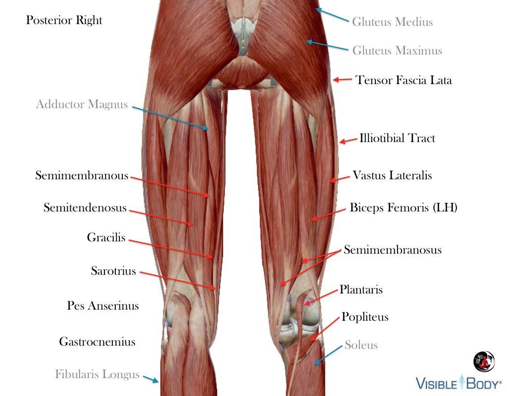
11 Posterior muscles of the knee joint are also mono- and biarticulating. Muscles indirectly impacting the knee are indicated with blue arrows.
12
Joints of the Lower Limb II
 Joints of the Lower Limb II Lecture Objectives Describe the components of the knee and ankle joint. List the ligaments associated with these joints and their attachments. List the muscles acting on these
The Knee. Prof. Oluwadiya Kehinde
 The Knee Prof. Oluwadiya Kehinde www.oluwadiya.sitesled.com The Knee: Introduction 3 bones: femur, tibia and patella 2 separate joints: tibiofemoral and patellofemoral. Function: i. Primarily a hinge joint,
The Knee Joint By Prof. Dr. Muhammad Imran Qureshi
 The Knee Joint By Prof. Dr. Muhammad Imran Qureshi Structurally, it is the Largest and the most complex joint in the body because of the functions that it performs: Allows mobility (flexion/extension)
To describe he knee joint, ligaments, structure & To list the main features of other lower limb joints
 To describe he knee joint, ligaments, structure & neurovascular supply To demonstrate the ankle joint anatomy To list the main features of other lower limb joints To list main groups of lymph nodes in
The Knee. Tibio-Femoral
 The Knee Tibio-Femoral Osteology Distal Femur with Proximal Tibia Largest Joint Cavity in the Body A modified hinge joint with significant passive rotation Technically, one degree of freedom (Flexion/Extension)
Hip joint Type: Articulating bones:
 Ana (242 ) Hip joint Type: Synovial, ball & socket Articulating bones: Formed between head of femur and lunate surface of acetabulum of hip bone. Capsule: it is a strong fibrous sleeve connecting the articulating
The Dance Hall by Vincent van Gogh,1888
 The Dance Hall by Vincent van Gogh,1888 Articulations of the pelvic girdle Lumbosacral joints, sacroiliac joints & pubic symphysis The remaining joints of the lower limb Hip joint Knee joint Tibiofibular
UNIT 7 JOINTS. Knee and Ankle Joints DR. ABDEL-MONEM A. HEGAZY
 UNIT 7 JOINTS Knee and Ankle Joints BY DR. ABDEL-MONEM A. HEGAZY (Degree in Bachelor of Medicine and Surgery with honor 1983, Dipl."Gynaecology and Obstetrics "1989, Master "Anatomy and Embryology "1994,
and K n e e J o i n t Is the most complicated joint in the body!!!!
 K n e e J o i n t K n e e J o i n t Is the most complicated joint in the body!!!! 1-Consists of two condylar joints between: A-The medial and lateral condyles of the femur and The condyles of the tibia
Articulations. Articulation. Joint between bones. Does not mean movement! Some joints are immovable; sutures.
 Articulations Joint between bones Articulation Does not mean movement Some joints are immovable; sutures. Classification of joints Two questions about joints: 1- How does it move? - functional 2- How is
A Patient s Guide to Knee Anatomy. Stephanie E. Siegrist, MD, LLC
 A Patient s Guide to Knee Anatomy Hands, shoulders, knees and toes (and elbows and ankles, too!) Most bone and joint conditions have several treatment options. The best treatment for you is based on your
Knee Joint Assessment and General View
 Knee Joint Assessment and General View Done by; Mshari S. Alghadier BSc Physical Therapy RHPT 366 m.alghadier@sau.edu.sa http://faculty.sau.edu.sa/m.alghadier/ Functional anatomy The knee is the largest
A Patient s Guide to Knee Anatomy
 A Patient s Guide to Knee Anatomy 15195 Heathcote Blvd Suite 334 Haymarket, VA 20169 Phone: 703-369-9070 Fax: 703-369-9240 DISCLAIMER: The information in this booklet is compiled from a variety of sources.
The Knee. Clarification of Terms. Osteology of the Knee 7/28/2013. The knee consists of: The tibiofemoral joint Patellofemoral joint
 The Knee Clarification of Terms The knee consists of: The tibiofemoral joint Patellofemoral joint Mansfield, p273 Osteology of the Knee Distal Femur Proximal tibia and fibula Patella 1 Osteology of the
Exercise 13. Articulations and Body Movements
 Exercise 13 Articulations and Body Movements Articulations Articulations, or joints, are points where a bone is connected to one or more other bones. Articulations hold the skeleton together. Articulations
CLASSIFICATION OF JOINTS STRUCTURAL VS FUNCTIONAL
 CHAPTER 8 JOINTS CLASSIFICATION OF JOINTS STRUCTURAL VS FUNCTIONAL The most moveable type of joint is a 1) Synarthrosis 2) Amphiarthrosis 3) Diarthrosis FIBROUS JOINTS Figure 8.1 Fibrous joints. (a) Suture
Joints: Part B 10/30/14. Classification of Synovial Joints. Six types, based on shape of articular surfaces: Plane Joints
 PowerPoint Lecture Slides prepared by Janice Meeking, Mount Royal College C H A P T E R 8 Joints: Part B Classification of Synovial Joints Six types, based on shape of articular surfaces: Plane Hinge Pivot
Joints Outline 8.1 Joints are classified into three structural and three functional categories (p. 251; Table 8.1) A. Joints are classified by
 A. Joints are classified by") Joints Outline 8.1 Joints are classified into three structural and three functional categories (p. 251; Table 8.1) A. Joints are classified by structure and by function: Structural classification focuses
Joints Outline 8.1 Joints are classified into three structural and three functional categories (p. 251; Table 8.1) A. Joints are classified by structure and by function: Structural classification focuses
Pelvic Girdle
 ARTICULATIONS OF LOWER EXTREMITY Pages 429-437 Pelvic Girdle formed by connection of the hip bones and the sacrum Sacroiliac Joints compound joints synovial joint - anterior, between the auricular surfaces
In the name of god. Knee. By: Tofigh Bahraminia Graduate Student of the Pathology Sports and corrective actions. Heat: Dr. Babakhani. Nov.
 In the name of god Knee By: Tofigh Bahraminia Graduate Student of the Pathology Sports and corrective actions Heat: Dr. Babakhani Nov. 2014 1 Anatomy-Bones Bones Femur Medial/lateral femoral condyles articulate
Copyright 2003 Pearson Education, Inc. publishing as Benjamin Cummings. Dr. Nabil Khouri MD, MSc, Ph.D
 Dr. Nabil Khouri MD, MSc, Ph.D Pelvic Girdle (Hip) Organization of the Lower Limb It is divided into: The Gluteal region The thigh The knee The leg The ankle The foot The thigh and the leg have compartments
UNIT 2 - CHAPTER 8: JOINTS OF THE SKELETAL SYSTEM LEARNING OUTCOMES:
 LEARNING OUTCOMES: 8.1 Introduction 1. List the functions of joints. 2. Explain how joints can be classified according to the type of tissue that binds the bones together and the degree of movement possible
UNIT 2 - CHAPTER 8: JOINTS OF THE SKELETAL SYSTEM LEARNING OUTCOMES:
 LEARNING OUTCOMES: 8.1 Types of Joints 1. Explain how joints can be classified according to the type of tissue that binds the bones together and the degree of movement possible at the joint. (p. 268) 2.
Joints Dr. Ali Ebneshahidi
 Joints Dr. Ali Ebneshahidi Function of Joints 1. Serve as functional junctions between bones. 2. Bind bones, strokes, and other related tissues together. 3. Allow bone growth to occur. 4. Permit certain
CHAPTER 8: THE BIOMECHANICS OF THE HUMAN LOWER EXTREMITY
 CHAPTER 8: THE BIOMECHANICS OF THE HUMAN LOWER EXTREMITY _ 1. The hip joint is the articulation between the and the. A. femur, acetabulum B. femur, spine C. femur, tibia _ 2. Which of the following is
Objectives. The BIG Joint. Case 1. Boney Architecture. Presenter Disclosure Information. Common Knee Problems
 3:30 4:15 pm Common Knee Problems SPEAKER Christopher J. Visco, MD Presenter Disclosure Information The following relationships exist related to this presentation: Christopher J. Visco, MD: Speaker s Bureau
Biology 325 Fall 2003
 Name: pre-lab exercise due at beginning of your lab session Matching a. fibrous joints b. cartilaginous joints c. synovial joints 1. exhibit a joint cavity 2. types are sutures and syndesmoses 3. bones
The Knee. Two Joints: Tibiofemoral. Patellofemoral
 Evaluating the Knee The Knee Two Joints: Tibiofemoral Patellofemoral HISTORY Remember the questions from lecture #2? Girth OBSERVATION TibioFemoral Alignment What are the consequences of faulty alignment?
Anatomy and Physiology 1 Chapter 9 self quiz Pro, Dima Darwish,MD.
 Anatomy and Physiology 1 Chapter 9 self quiz Pro, Dima Darwish,MD. 1) Joints can be classified structurally as A) bony. B) fibrous. C) cartilaginous. D) synovial. E) All of the answers are correct. 2)
BONES JOINTS MUSCLES OF THE LOWER LIMB
 BONES JOINTS MUSCLES OF THE LOWER LIMB LOWER LIMB: BONES LOWER LIMB GLUTEAL REGION consists of 6 major segments: FEMORAL REGION (THIGH) KNEE REGION LEG REGION TALOCRURAL REGION (ANKLE) FOOT REGION LOWER
TOTAL KNEE ARTHROPLASTY (TKA)
") TOTAL KNEE ARTHROPLASTY (TKA) 1 Anatomy, Biomechanics, and Design 2 Femur Medial and lateral condyles Convex, asymmetric Medial larger than lateral 3 Tibia Tibial plateau Medial tibial condyle: concave
TOTAL KNEE ARTHROPLASTY (TKA) 1 Anatomy, Biomechanics, and Design 2 Femur Medial and lateral condyles Convex, asymmetric Medial larger than lateral 3 Tibia Tibial plateau Medial tibial condyle: concave
Knee Anatomy Introduction Welcome to BodyZone Physiotherapy's patient resource about Knee problems.
 Knee Anatomy Introduction Welcome to BodyZone Physiotherapy's patient resource about Knee problems. To better understand how knee problems occur, it is important to understand some of the anatomy of thee
8.2: Fibrous Joints. There are three (3) types of fibrous joints (synarthroses): Syndesmosis Suture Gomphosis. Interosseus membrane of leg.
 types of fibrous joints (synarthroses): Syndesmosis Suture Gomphosis. Interosseus membrane of leg.") 8.1: Introduction Are known as articulations Functional junctions between bones Bind parts of skeletal system together Make bone growth possible Permit parts of the skeleton to change shape during childbirth
8.1: Introduction Are known as articulations Functional junctions between bones Bind parts of skeletal system together Make bone growth possible Permit parts of the skeleton to change shape during childbirth
It is formed by fusion of 3 bones: I. Ilium (superior bone). II. Pubis (antero-inferior bone). III. Ischium (postero-inferior bone).
. II. Pubis (antero-inferior bone). III. Ischium (postero-inferior bone).") It is formed by fusion of 3 bones: I. Ilium (superior bone). II. Pubis (antero-inferior bone). III. Ischium (postero-inferior bone). Pubis Acetabulum Ana (242 ) The three constituent of bones of the hip
It is formed by fusion of 3 bones: I. Ilium (superior bone). II. Pubis (antero-inferior bone). III. Ischium (postero-inferior bone). Pubis Acetabulum Ana (242 ) The three constituent of bones of the hip
Knee Injury Assessment
 Knee Injury Assessment Clinical Anatomy p. 186 Femur Medial condyle Lateral condyle Femoral trochlea Tibia Intercondylar notch Tibial tuberosity Tibial plateau Fibula Fibular head Patella Clinical Anatomy
Joints. Judi Laprade. Illustrations from: Essential Clinical Anatomy 3 rd ed. (ECA3) Moore, K. and Agur, A. Lippincott Williams and Wilkins, 2007
 Moore, K. and Agur, A. Lippincott Williams and Wilkins, 2007") Slide 1 Joints Judi Laprade Illustrations from: Essential Clinical Anatomy 3 rd ed. (ECA3) Moore, K. and Agur, A. Lippincott Williams and Wilkins, 2007 Grant s Atlas of Anatomy 12 th ed. (GA12) Agur, A.
Slide 1 Joints Judi Laprade Illustrations from: Essential Clinical Anatomy 3 rd ed. (ECA3) Moore, K. and Agur, A. Lippincott Williams and Wilkins, 2007 Grant s Atlas of Anatomy 12 th ed. (GA12) Agur, A.
The hip joint is a multiaxial synovial ball-and-socket joint between the head of the femur and the acetabulum of the
 NfW Hip Joint The hip joint is a multiaxial synovial ball-and-socket joint between the head of the femur and the acetabulum of the pelvic bone. Unlike the ball-and-socket shoulder joint, the hip joint
ChiroCredit.com Presents Biomechanics: Focus on
 ChiroCredit.com Presents Biomechanics: Focus on the Knee Presented by: Ivo Waerlop, DC Shawn Allen, DC 1 Focus on The Knee 2 Pertinent Anatomy Femur Tibia Fibula Patella Prepatellar bursa Infrapatellar
Bones of Lower Limb. Dr. Heba Kalbouneh Associate Professor of Anatomy and Histology
 Bones of Lower Limb Dr. Heba Kalbouneh Associate Professor of Anatomy and Histology Bones of the lower limb Hip Bone Made up of 3 bones: 1) Ilium (flat), superior in position 2) Ischium (L), postero-inferior
Practical 1 Worksheet
 Practical 1 Worksheet ANATOMICAL TERMS 1. Use the word bank to fill in the missing words. reference side stand body arms palms anatomical forward All anatomical terms have a(n) point which is called the
Topic 7: Hip and pelvis. Parts of the hip. Parts of the femur
 Topic 7: Hip and pelvis Parts of the hip Parts of the femur Classifying the hip joint Ball and socket Synovial Multiaxial Movements of the hip: Abduction/adduction Flexion/extension Medial/lateral rotation
I. Introduction. Unit Two. of the Skeletal System. II. Classification of Joints. URLs for this chapter:
 8 URLs for this chapter: http://www.vh.org/adult/provider/radiology/joint Fluoro/JointFluoroHP.html of the Skeletal System Karen Webb Smith Unit Two http://www.science.ubc.ca/~biomania/tutorial/bonejt/
To classify the joints relative to structure & shape
 To classify the joints relative to structure & shape To describe the anatomy of the hip joint To describe the ankle joint To memorize their blood & nerve supply JOINTS: Joints are sites where skeletal
The Lower Limb II. Anatomy RHS 241 Lecture 3 Dr. Einas Al-Eisa
 The Lower Limb II Anatomy RHS 241 Lecture 3 Dr. Einas Al-Eisa Tibia The larger & medial bone of the leg Functions: Attachment of muscles Transfer of weight from femur to skeleton of the foot Articulations
Introduction. Fibrous Joints. 8.1: Types of Joints. Cartilaginous Joints. Fibrous Joints 12/14/2016. Chapter 08 Lecture Outline
 Introduction Chapter 08 Lecture Outline See separate PowerPoint slides for all figures and tables preinserted into PowerPoint without notes. Joints (Articulations): Functional junctions between bones Bind
CHAPTER 9 LECTURE OUTLINE INTRODUCTION
 CHAPTER 9 LECTURE OUTLINE INTRODUCTION I. A joint (articulation or arthrosis) is a point of contact between two or more bones, between cartilage and bones, or between teeth and bones. A. The scientific
Joints. Agenda. Joints. Structural and Functional Classification of Articulations
 Joints Structural and Functional Classification of Articulations Agenda Joint Basics Classification Structural Joint Details Joint Stability Movements of Synovial Joints Shape Classification of Synovial
Exercise Science Section 4: Joint Mechanics and Joint Injuries
 Exercise Science Section 4: Joint Mechanics and Joint Injuries An Introduction to Health and Physical Education Ted Temertzoglou Paul Challen ISBN 1-55077-132-9 Types of Joints Fibrous joint Cartilaginous
9.1 Joints. Objectives Describe the structural and functional classifications of joints
 Joints 9.1 Joints Describe the structural and functional classifications of joints Joints have both structural and functional classifications: The criteria for classifying joints structurally are anatomical
Lecture 9: Arthrology
 Lecture 9: Arthrology M/O Chapter 9 45. Classify joints based on the degree of movement allowed and give examples of each classification. 46. Classify joints based on anatomical structure and give examples
Definition: A joint or articulation is a place in the body where two bones come together.
 Definition: A joint or articulation is a place in the body where two bones come together. CLASSES OF JOINTS. 1. Joints are classified according to how the bones are held together. 2. The three types of
ARTICULATIONS and MUSCULAR SYSTEM
 ARTICULATIONS and MUSCULAR SYSTEM PART #1 ARTICULATIONS 1. Introduction A. Articulation C. Kinesiology B. Arthrology D. Rheumatology 2. Structural Classifications for Joints A. Fibrous Joints i. Suture
Biomechanics of the Knee. Valerie Nuñez SpR Frimley Park Hospital
 Biomechanics of the Knee Valerie Nuñez SpR Frimley Park Hospital Knee Biomechanics Kinematics Range of Motion Joint Motion Kinetics Knee Stabilisers Joint Forces Axes The Mechanical Stresses to which
The Lower Limb. Anatomy RHS 241 Lecture 2 Dr. Einas Al-Eisa
 The Lower Limb Anatomy RHS 241 Lecture 2 Dr. Einas Al-Eisa The bony pelvis Protective osseofibrous ring for the pelvic viscera Transfer of forces to: acetabulum & head of femur (when standing) ischial
MRI KNEE WHAT TO SEE. Dr. SHEKHAR SRIVASTAV. Sr.Consultant KNEE & SHOULDER ARTHROSCOPY
 MRI KNEE WHAT TO SEE Dr. SHEKHAR SRIVASTAV Sr.Consultant KNEE & SHOULDER ARTHROSCOPY MRI KNEE - WHAT TO SEE MRI is the most accurate and frequently used diagnostic tool for evaluation of internal derangement
The Hip (Iliofemoral) Joint. Presented by: Rob, Rachel, Alina and Lisa
 Joint. Presented by: Rob, Rachel, Alina and Lisa") The Hip (Iliofemoral) Joint Presented by: Rob, Rachel, Alina and Lisa Surface Anatomy: Posterior Surface Anatomy: Anterior Bones: Os Coxae Consists of 3 Portions: Ilium Ischium Pubis Bones: Pubis Portion
The Hip (Iliofemoral) Joint Presented by: Rob, Rachel, Alina and Lisa Surface Anatomy: Posterior Surface Anatomy: Anterior Bones: Os Coxae Consists of 3 Portions: Ilium Ischium Pubis Bones: Pubis Portion
SHOULDER JOINT ANATOMY AND KINESIOLOGY
 SHOULDER JOINT ANATOMY AND KINESIOLOGY SHOULDER JOINT ANATOMY AND KINESIOLOGY The shoulder joint, also called the glenohumeral joint, consists of the scapula and humerus. The motions of the shoulder joint
HOW DO WE DIAGNOSE LAMENESS IN YOUR HORSE?
 HOW DO WE DIAGNOSE LAMENESS IN YOUR HORSE? To help horse owners better understand the tools we routinely use at VetweRx to evaluate their horse s soundness, the following section of this website reviews
Amy Warenda Czura, Ph.D. 1 SCCC BIO130 Lab 7 Appendicular Skeleton & Articulations
 The Skeletal System II: Appendicular Skeleton and Articulations Exercises 11, 13 (begins: page 145 in 9 th and 10 th editions) Exercises 10, 11 (begins: page 147 in 11 th edition, page 149 in 12 th edition)
Understanding Leg Anatomy and Function THE UPPER LEG
 Understanding Leg Anatomy and Function THE UPPER LEG The long thigh bone is the femur. It connects to the pelvis to form the hip joint and then extends down to meet the tibia (shin bone) at the knee joint.
A NEW CONCEPT IN FUNCTIONAL ORTHOSES
 A NEW CONCEPT IN FUNCTIONAL ORTHOSES THE KNEE in movement! Climbing stairs, walking and running are everyday actions that we can perform thanks to our knees. The knee joint is one of the most exposed and
PowerPoint Lecture Slides prepared by Janice Meeking, Mount Royal College C H A P T E R. Joints: Part A. Copyright 2010 Pearson Education, Inc.
 PowerPoint Lecture Slides prepared by Janice Meeking, Mount Royal College C H A P T E R 8 Joints: Part A Warm Up 11/28/16 Happy Thanksgiving welcome back! J (be ready to share something fun you did over
Human Anatomy & Physiology I Dr. Sullivan Unit IX Arthrology (joints) - Chapter 9
 - Chapter 9") Human Anatomy & Physiology I Dr. Sullivan Unit IX Arthrology (joints) - Chapter 9 I. Joints: aka Articulations a) Joints are points of contact between two or more bones. Joints may be moveable or may not
Human Anatomy & Physiology I Dr. Sullivan Unit IX Arthrology (joints) - Chapter 9 I. Joints: aka Articulations a) Joints are points of contact between two or more bones. Joints may be moveable or may not
KNEE INJURIES IN FEMALE SOCCER PLAYERS: A FOCUS ON THE ACL
 KNEE INJURIES IN FEMALE SOCCER PLAYERS: A FOCUS ON THE ACL Item Type text; Electronic Thesis Authors PEÑA, VANESSA NICOLE Publisher The University of Arizona. Rights Copyright is held by the author. Digital
Knee Disarticulation Amputation
 Knee Disarticulation Amputation Pre-Op 64 year old man, previous spinal cord injury, diabetes, renal failure, and a history of spasticity with dynamic knee flexion contracture. He had an open left ankle
The Articular System OBJECTIVES ACTIVITIES. A. Completion
 C H A P T E R 8 The Articular System OBJECTIVES After studying this chapter, you should be able to: 1. Name and describe the three types of joints. 2. Name the two types of synarthroses joints. 3. Name
Chapter 9 Articulations Articulations joints where two bones interconnect. Two classification methods are used to categorize joints:
 Chapter 9 Articulations Articulations joints where two bones interconnect Two classification methods are used to categorize joints: Functional classification Structural classification Functional classification
Unicompartmental Knee Resurfacing
 Disclaimer This movie is an educational resource only and should not be used to manage knee pain. All decisions about the management of knee pain must be made in conjunction with your Physician or a licensed
Figure 7: Bones of the lower limb
 BONES OF THE APPENDICULAR SKELETON The appendicular skeleton is composed of the 126 bones of the appendages and the pectoral and pelvic girdles, which attach the limbs to the axial skeleton. Although the
Gluteal region DR. GITANJALI KHORWAL
 Gluteal region DR. GITANJALI KHORWAL Gluteal region The transitional area between the trunk and the lower extremity. The gluteal region includes the rounded, posterior buttocks and the laterally placed
Joints of the lower limb
 Joints of the lower limb 1-Type: Hip joint Synovial ball-and-socket joint 2-Articular surfaces: a- head of femur b- lunate surface of acetabulum Which is deepened by the fibrocartilaginous labrum acetabulare
Ligamentous and Meniscal Injuries: Diagnosis and Management
 Ligamentous and Meniscal Injuries: Diagnosis and Management Daniel K Williams, MD Franciscan Physician Network Orthopedic Specialists September 29, 2017 No Financial Disclosures INTRODUCTION Overview of
FACTORS AFFECTING PATELLAR TRACKING IN TOTAL KNEE ARTHROPLASTY
 FACTORS AFFECTING PATELLAR TRACKING IN TOTAL KNEE ARTHROPLASTY Essay submitted in partial fulfillment of M.Sc. Degree in Orthopaedic surgery Cairo University By Ahmed Ali Ahmed Ali Elsayed M.B.B.Ch Supervised
The Lower Limb. Sevda LAFCI FAHRİOĞLU, MD.PhD.
 The Lower Limb Sevda LAFCI FAHRİOĞLU, MD.PhD. The Lower Limb The bones of the lower limb form the inferior part of the appendicular skeleton the organ of locomotion for bearing the weight of body stronger
ABSTRACT 2 Key Words: meniscus, tibiofemoral joint, osteoarthritis
 ABSTRACT In the United States, meniscal lesions represent the most common intra-articular knee injury. 1 In fact, the mean annual incidence of meniscal tears is approximately 60 to 70 per 100,000 patients.
The Skeletal System. Dr. Naim Kittana Dr. Suhaib Hattab. Faculty of Medicine & Health Sciences An-Najah National University
 The Skeletal System Dr. Naim Kittana Dr. Suhaib Hattab Faculty of Medicine & Health Sciences An-Najah National University 1 Declaration The content and the figures of this seminar were directly adopted
3/15/15. Chapter 8: Joints. Classification of Joints. Classification of Joints. } Objectives. } Functional Classifications
 Chapter 8: Joints Classification of Joints } Objectives } Define Joint or Articulation } Classify Joints by Structure and by Function } Describe the general structure, know the properties of, and provide
ACL RECONSTRUCTION HAMSTRING METHOD. Presents ACL RECONSTRUCTION HAMSTRING METHOD. Multimedia Health Education
 HAMSTRING METHOD Presents HAMSTRING METHOD Multimedia Health Education Disclaimer Stephen J. Incavo MD This movie is an educational resource only and should not be used to make a decision on Anterior Cruciate
Partial Knee Replacement
 Partial Knee Replacement A partial knee replacement removes damaged cartilage from the knee and replaces it with prosthetic implants. Unlike a total knee replacement, which removes all of the cartilage,
Chapter 7: Skeletal System: Gross Anatomy
 Chapter 7: Skeletal System: Gross Anatomy I. General Considerations A. How many bones in an average adult skeleton? B. Anatomic features of bones are based on II. Axial Skeleton A. Skull 1. Functionally
Ch. 8 Joints of the Skeletal System
 Ch. 8 Joints of the Skeletal System Part 1: Classifying Joints & Joint Movements Interactive pages 269-278 Types of Joints (AKA: Articulations) Structural Classification (type of tissue that binds the
PRE & POST OPERATIVE RADIOLOGICAL ASSESSMENT IN TOTAL KNEE REPLACEMENT. Dr. Divya Rani K 2 nd Year Resident Dept. of Radiology
 PRE & POST OPERATIVE RADIOLOGICAL ASSESSMENT IN TOTAL KNEE REPLACEMENT Dr. Divya Rani K 2 nd Year Resident Dept. of Radiology PRE OPERATIVE ASSESSMENT RADIOGRAPHS Radiographs are used for assessment and
PARTIAL KNEE REPLACEMENT
 PARTIAL KNEE REPLACEMENT A partial knee replacement removes damaged cartilage from the knee and replaces it with prosthetic implants. Unlike a total knee replacement, which removes all of the cartilage,
Biology 218 Human Anatomy
 Chapter 9 Adapted form Tortora 10 th ed. LECTURE OUTLINE A. Introduction (p. 229) 1. A joint or articulation or arthrosis is a point of contact between neighboring bones, between cartilage and bones, or
Lab Activity 9. Appendicular Skeleton Martini Chapter 8. Portland Community College BI 231
 Lab Activity 9 Appendicular Skeleton Martini Chapter 8 Portland Community College BI 231 Appendicular Skeleton Upper & Lower extremities Shoulder Girdle Pelvic Girdle 2 Humerus 3 Humerus: Proximal End
The Appendicular Skeleton
 8 The Appendicular Skeleton PowerPoint Lecture Presentations prepared by Jason LaPres Lone Star College North Harris 8-1 The Pectoral Girdle The Pectoral Girdle Also called shoulder girdle Connects the
The Skeletal System. Dr. Naim Kittana. Faculty of Medicine & Health Sciences An-Najah National University
 The Skeletal System Dr. Naim Kittana Faculty of Medicine & Health Sciences An-Najah National University 1 Declaration The content and the figures of this seminar were directly adopted from the text book
Pelvic cavity. Gross anatomy of the lower limb. Walking. Sándor Katz M.D.,Ph.D.
 Pelvic cavity. Gross anatomy of the lower limb. Walking. Sándor Katz M.D.,Ph.D. Lower limb Pelvic girdle Free lower extremity Hip bone Definitive fusion of the Y- shaped growth plate occurs 16th -18th
Pre-Op Planning for your knee replacement surgery
 Pre-Op Planning for your knee replacement surgery Are You Considering Knee Replacement Surgery? Knee pain can be the result of injury, biomechanical problems, or disease. When stiffness and pain in your
FUNCTIONAL ANATOMY OF SHOULDER JOINT
 FUNCTIONAL ANATOMY OF SHOULDER JOINT ARTICULATION Articulation is between: The rounded head of the Glenoid cavity humerus and The shallow, pear-shaped glenoid cavity of the scapula. 2 The articular surfaces
Knee Ultrasonography step by step
 Knee Ultrasonography step by step Poster No.: C-2809 Congress: ECR 2018 Type: Educational Exhibit Authors: J. A. Torres de Abreu Macedo, N. Pereira da Silva, A. I. Aguiar, F. Alves, F. Caseiro Alves; Coimbra/PT
Mr. S. Tanweer Ashraf MS, MRCS (Ed), FRCS (Eng). FRCS (Tr&Orth) MSc Ortho Engineering (Cardiff),
, FRCS (Eng). FRCS (Tr&Orth) MSc Ortho Engineering (Cardiff),") Mr. S. Tanweer Ashraf MS, MRCS (Ed), FRCS (Eng). FRCS (Tr&Orth) MSc Ortho Engineering (Cardiff), Consultant Knee Surgeon NHS: The Royal Orthopaedic Hospital & Queen Elizabeth Hospital, Birmingham MRI Knee:
Mr. S. Tanweer Ashraf MS, MRCS (Ed), FRCS (Eng). FRCS (Tr&Orth) MSc Ortho Engineering (Cardiff), Consultant Knee Surgeon NHS: The Royal Orthopaedic Hospital & Queen Elizabeth Hospital, Birmingham MRI Knee:
Biomechanics of. Knee Replacement. Mujda Hakime, Paul Malcolm
 Biomechanics of Knee Replacement Mujda Hakime, Paul Malcolm 1 Table of contents Knee Anatomy Movements of the Knee Knee conditions leading to knee replacement Materials Alignment and Joint Loading Knee
NHS Training for Physiotherapy Support Workers. Workbook 11 The articular system
 NHS Training for Physiotherapy Support Workers Workbook 11 The articular system Contents Workbook 11 The articular system 1 11.1 Aim 3 11.2 Learning outcomes 3 11.3 The articular system 4 11.4 Individual
Imaging the Athlete s Knee. Peter Lowry, MD Musculoskeletal Radiology University of Colorado
 Imaging the Athlete s Knee Peter Lowry, MD Musculoskeletal Radiology University of Colorado None Disclosures Knee Imaging: Radiographs Can be performed weight-bearing or non-weight-bearing View options
* Articular system I
 *Articular system I *Articular system=syndesmology (Systema articulare) System of joints Joint occurs, where 2 bones meet Combine bones of skeleton into a single unit Provide mobility *Classification
Torn ACL - Anatomic Footprint ACL Reconstruction
 Torn ACL - Anatomic Footprint ACL Reconstruction The anterior cruciate ligament (ACL) is one of four ligaments that are crucial to the stability of your knee. It is a strong fibrous tissue that connects
Prevention and Treatment of Injuries. Anatomy. Anatomy. Chapter 20 The Knee Westfield High School Houston, Texas
 Prevention and Treatment of Injuries Chapter 20 The Knee Westfield High School Houston, Texas Anatomy MCL, Medial Collateral Ligament LCL, Lateral Collateral Ligament PCL, Posterior Cruciate Ligament ACL,