Cardiac Masses. Dennis A. Tighe, MD, FASE. University of Massachusetts Medical School Worcester, MA
|
|
|
- Erik Whitehead
- 5 years ago
- Views:
Transcription
1 Cardiac Masses Dennis A. Tighe, MD, FASE University of Massachusetts Medical School Worcester, MA
2 Cardiac Masses: Considerations Definition of the mass Nature Location Benign or malignant Presentation Incidental finding Obstruction Direct myocardial involvement Embolization Constitutional or systemic symptoms Echocardiography remains 1 o imaging modality Multimodality imaging may be required for characterization
3 Cardiac Masses: Differential Diagnosis Anatomical variants Implanted devices Thrombus Vegetations Tumors Primary Metastatic Artifacts

4 Anatomical Structures Moderator band Trabeculation False tendons Hypertrophy Papillary m. Trabeculation Chiari network Crista terminalis Eustachian valve ASA LHIAS Pectinate m. Q-tip Aorta Effusions Hiatus hernia Pericardial cyst

5 Anatomical Variants LAA Th RA
6 Implanted Devices Pacemaker leads Cardioverter-defibrillator leads Right heart catheters Occluder devices Prosthetic valves/clips Foreign bodies

7 Implanted Devices LA RA CT


8 Implanted Devices LA RA wire
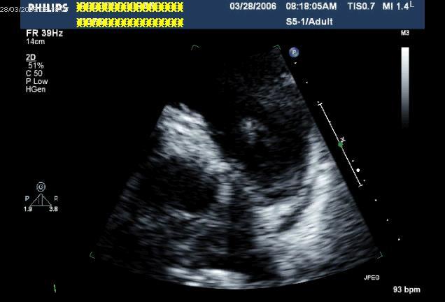
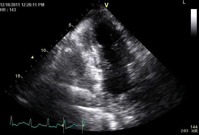
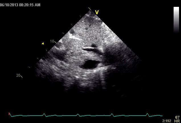
9 Thrombus Most commonly encountered intra-cardiac mass Often associated cardiac pathology LV thrombus Apex most common Acute MI» Estimated 4-15% patients with anterior MI Dilated cardiomyopathy» DDx: false tendons, trabeculations, artifacts, apical hypertrophy, tumors, non-compaction, HES LA thrombus Appendage Body Right heart thrombus Catheter-related Pulmonary embolism Appendage RV apical area

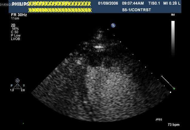
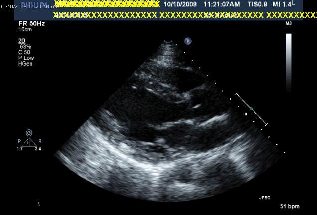
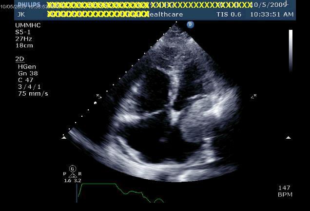
10 LV Thrombus
11

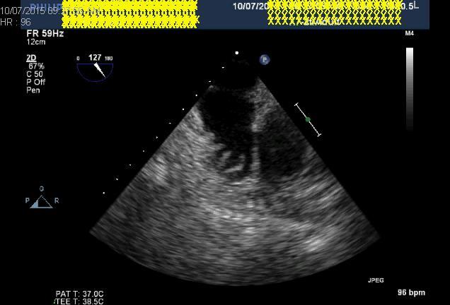
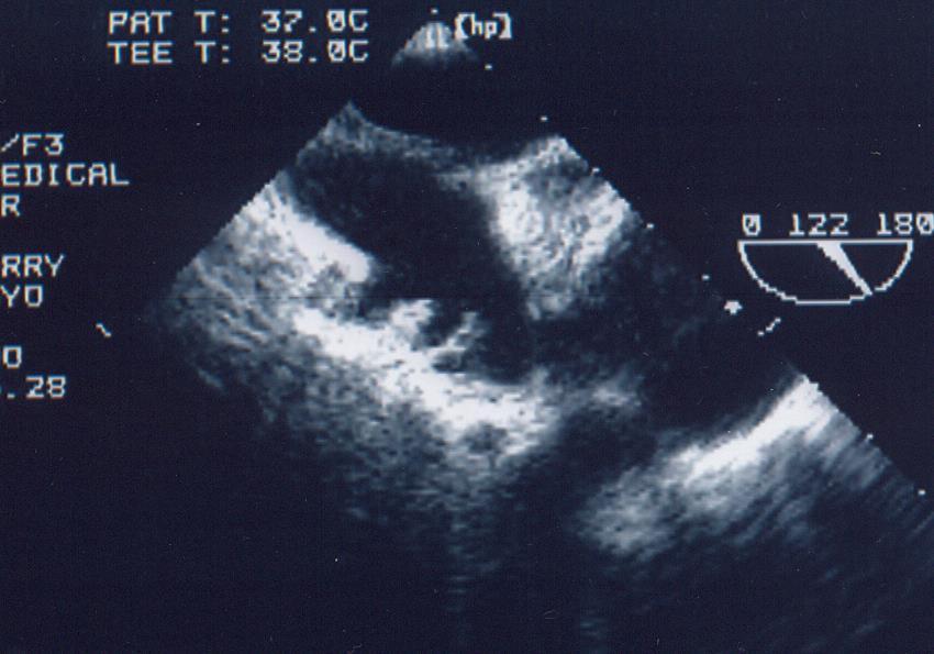
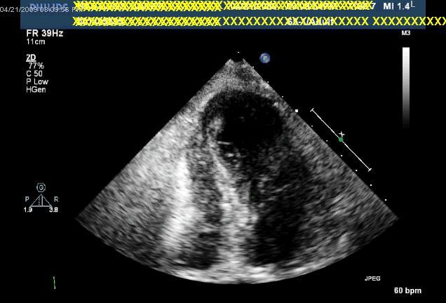
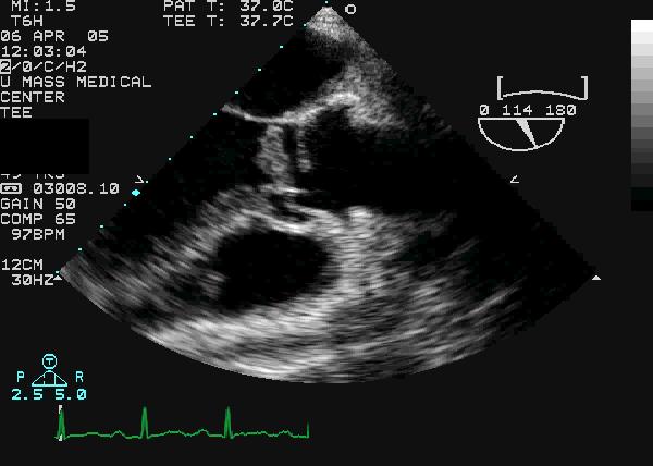
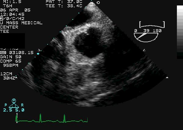
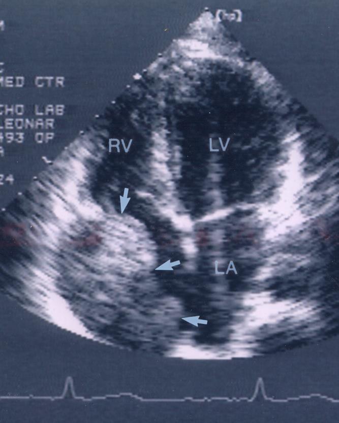
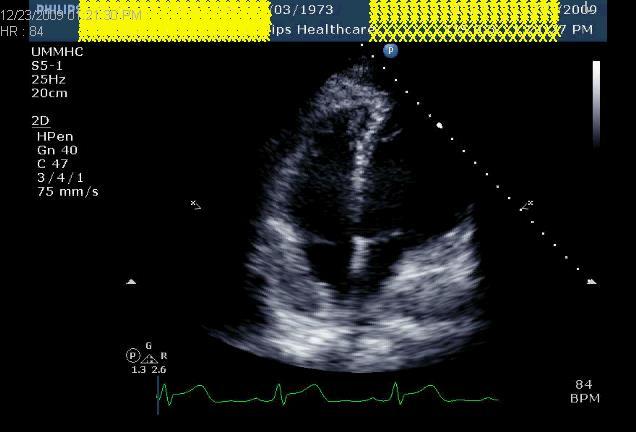
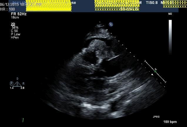
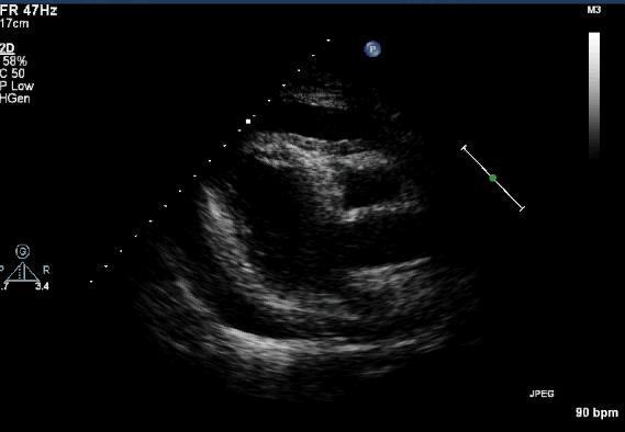
12 LA LAA Thrombus

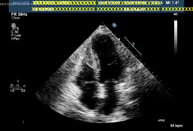
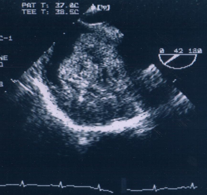
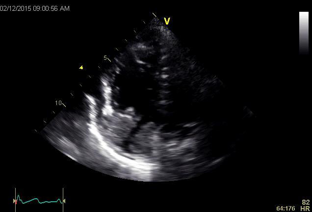
13 LA Thrombus


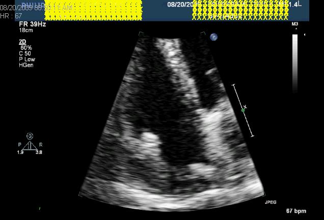
14 Right Heart Thrombus Pulmonary Embolism Catheter-related
15 Vegetations Locations: Valve surfaces, areas of endocardium opposite intra-cardiac shunts, or prosthetic materials Atrial surface mitral valve Ventricular surface of aortic valve Characteristics: Mobile, oscillating Tissue density differing from surrounding tissue May calcify if chronic/healed Valve dysfunction may occur Valvular regurgitation Valvular stenosis (if large enough) Infective or non-infective
16
17
18 Cardiac Tumors Primary Rare 0.017% to 0.033% of autopsies Benign vs. malignant Secondary (Metastatic)
19 Primary Cardiac Tumors Benign (80%) Myxoma Fibroelastoma Rhabdomyoma Fibroma Lipoma Hemangioma Teratoma Paraganglioma Malignant (20%) Sarcoma Lymphoma Mesothelioma
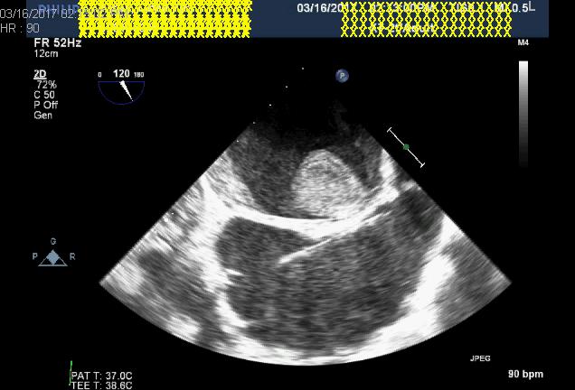
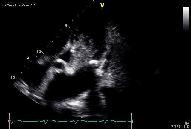
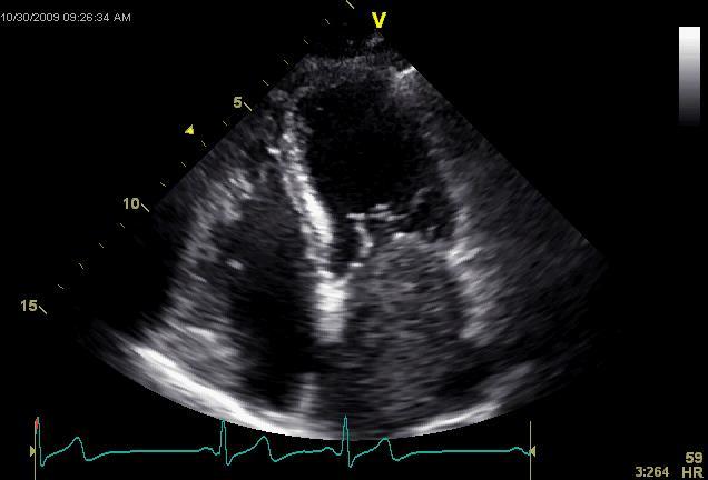
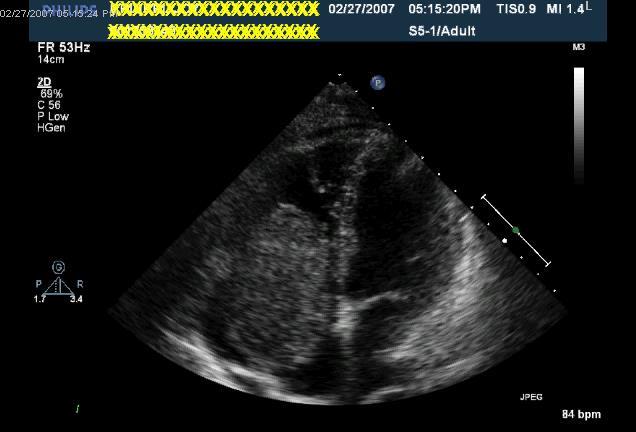
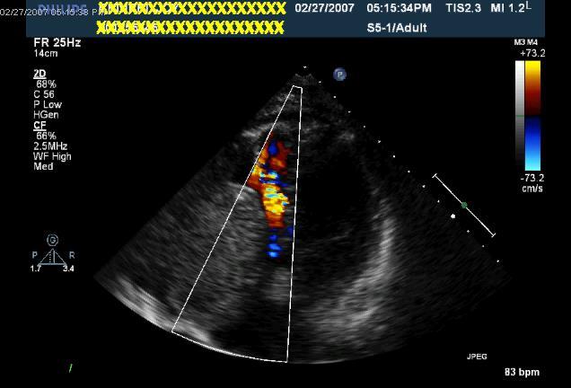
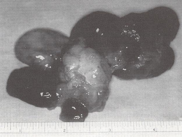
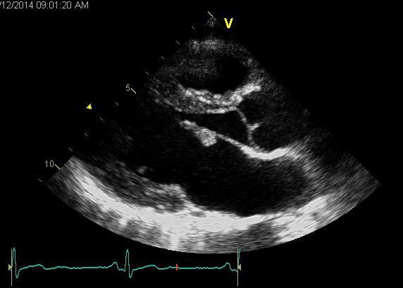
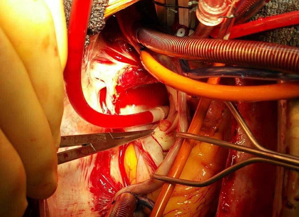
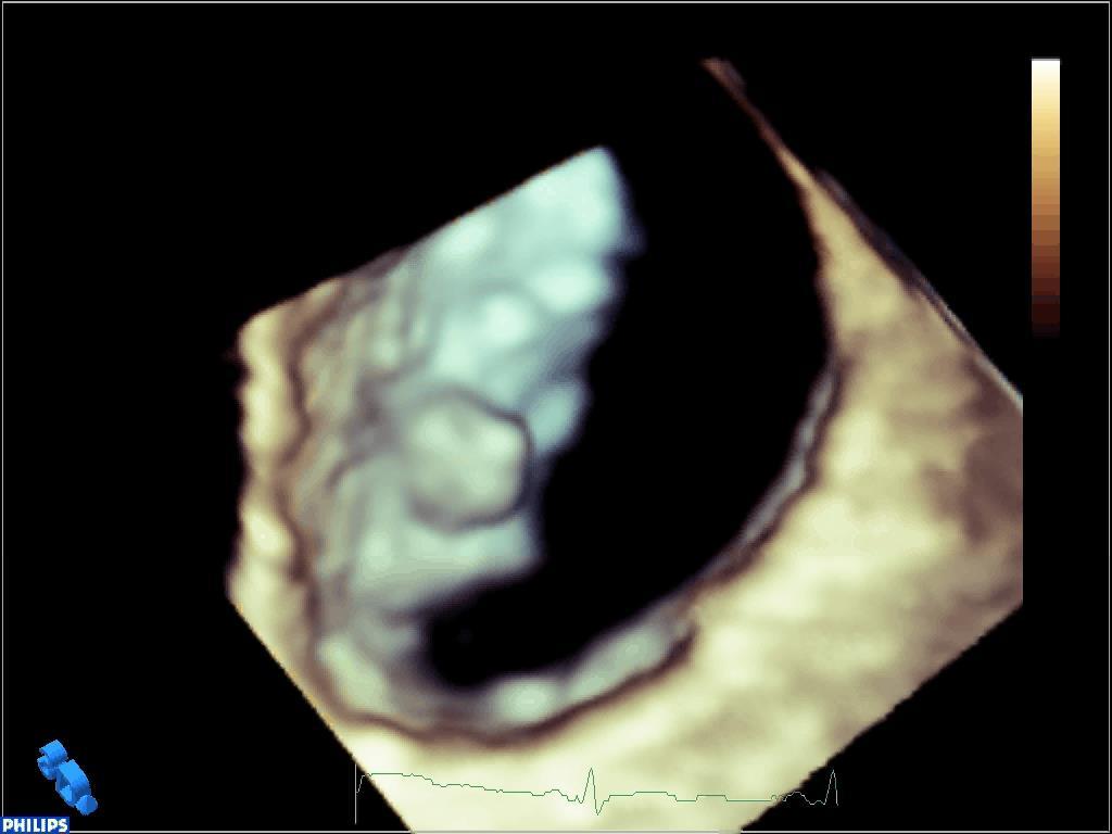
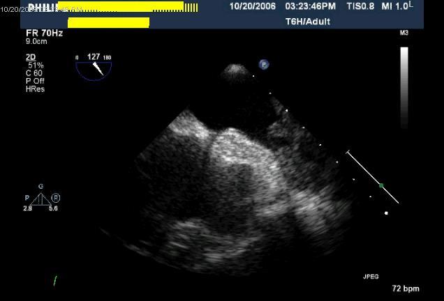
20 Cardiac Myxoma Most common primary cardiac tumor Majority are sporadic 10% familial Can recur and may be multi-centric Most frequently discovered 3 rd to 6 th decades Female preponderance (60-70%) Can arise anywhere within the heart About 75% occur in the left atrium near fossa ovalis Stalk Clinical presentation Constitutional, embolic or obstructive symptoms Many detected asymptomatically


21 LA Myxoma LA RV LV IAS RA RA LA RV
22
23 RA Myxoma

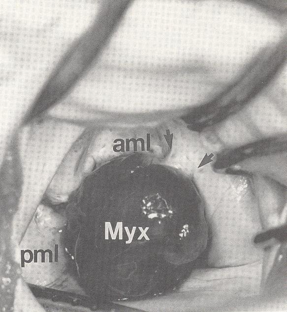
24 Valvular Myxoma LA RA LV RV
25 Papillary Fibroelastoma Second most prevalent adult 1 o cardiac tumor Commonly involves cardiac valves Aortic valve most common location Both surfaces show equal prevalence Other cardiac structures/chambers (15-25%) Majority found in left heart Pathology: Avascular, papillary fronds, pedunculated Mid-portion of the valve Usually do not cause valvular dysfunction Significant embolic potential recognized


26 N=611 Gowda RM et al. Am Heart J 2003;146:404. Weems WB et al. J Am Soc Echocardiogr 2002;15:382. Klarich KW et al. J Am Coll Cardiol 1997;30:784.
27

28

29 Multiple tumors Right-sided
30 Lambl s Excresences Common Found in 70-80% adults Pathology: Linear, filiform fronds Multiple Located at closure lines Ventricular surface of semilunar valves Atrial surface of mitral valve Do not interfere with valve function
31
32 Other Benign Primary Tumors Rhabdomyoma Most common tumor in pediatric age group Muscular May protrude into cavity Association with tuberous sclerosis Spontaneous regression Fibroma Lipoma Teratoma Angioma Paraganglioma Blood-filled cyst


33 Other Benign Primary Tumors Rhabdomyomas Fibroma
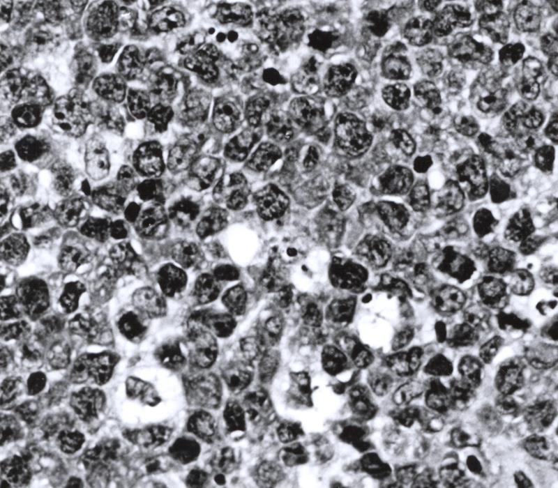
34 Malignant Primary Cardiac Tumors Sarcomas (80%) Angiosarcoma Usually found in right atrium Highly invasive Lung metastases common Other types (left atrium more common) Undifferentiated sarcoma Rhabdomyosarcoma Fibrosarcoma Leiomyosaraoma Osteosarcoma (calification) Mesotheliomas (10%) Arise from pericardium Rarely may involve conduction system Lymphomas (3-5%) Paragangliomas
35 Angiosarcoma LA RA
36

37 Primary Cardiac Lymphoma
38
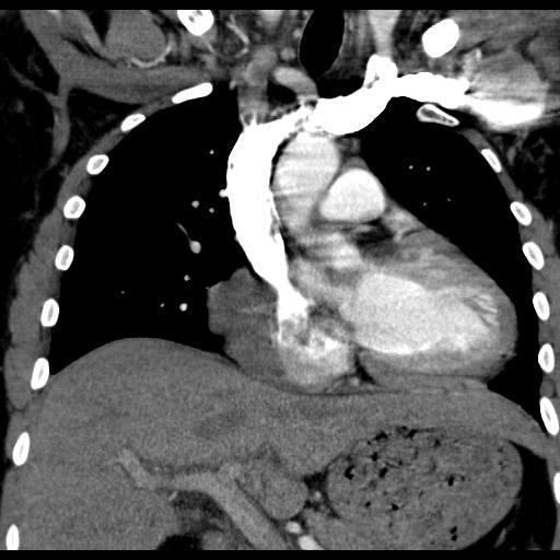
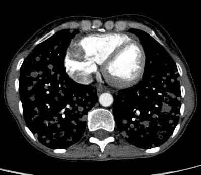
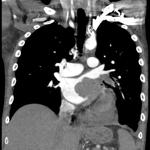
39 Secondary (Metastatic) Cardiac Tumors At least 20-to-40 times more common than primary cardiac tumors 5-12% cancer patients Consider with known malignancy and occurrence of new CV symptoms Breast and lung cancer and heme malignancy encountered most commonly Malignant melanoma has highest propensity for metastasis to the heart
40 Secondary (Metastatic) Cardiac Tumors Pericardial involvement Most common Pericardium > Myocardium > Endocardium Hematogenous/Lymphatic spread Melanoma, lymphoma, breast Direct extension Lung, breast, esophageal Invasion via venous structures Vena cava Renal, Hepatocellular, Uterine Pulmonary veins Lung, breast, thyroid
41
42
43
44 LLPV
45 Burke A et al. Heart 2008;94:
46 Extra-cardiac Masses
47

48
49 Thank you for your attention
Cardiac Masses. Cardiac Masses: Considerations. Dennis A. Tighe, MD, FASE. University of Massachusetts Medical School Worcester, MA 4/16/2018
 Cardiac Masses Dennis A. Tighe, MD, FASE University of Massachusetts Medical School Worcester, MA Cardiac Masses: Considerations Definition of the mass Nature Location Benign or malignant Presentation
Cardiac Mass and Mass-like Structures
 KSE 2017 Basic Echo Review Course (4) Nov 26, 2017 Cardiac Mass and Mass-like Structures Sun Hwa Lee, MD, PhD Chonbuk National University Hospital & Medical School Introduction Although cardiac tumors
Cardiac Tumors Sharon S. Brouha, MD
 Cardiac Tumors Sharon S. Brouha, MD CARDIAC TUMORS Imaging techniques Sharon Sudarshan Brouha, MD, MPH Assistant Clinical Professor Cardiothoracic Imaging Section University of California San Diego Cardiac
Malignant Cardiac Tumors Rad-Path Correlation
 Malignant Cardiac Tumors Rad-Path Correlation Vincent B. Ho, M.D., M.B.A. 1 Jean Jeudy, M.D. 2 Aletta Ann Frazier, M.D. 2 1 Uniformed Services University of the Health Sciences 2 University of Maryland
Adult Echocardiography Examination Content Outline
 Adult Echocardiography Examination Content Outline (Outline Summary) # Domain Subdomain Percentage 1 2 3 4 5 Anatomy and Physiology Pathology Clinical Care and Safety Measurement Techniques, Maneuvers,
Blank DISCLOSURES INTRACARDIAC MASSES MULTI-MODALITY IMAGING ECHO HAWAII JANUARY 2017 NONE
 Blank INTRACARDIAC MASSES MULTI-MODALITY IMAGING ECHO HAWAII JANUARY 2017 David A. Orsinelli, MD, FACC, FASE Director, Structural Heart Imaging Professor, Internal Medicine The Ohio State University Division
Index. radiologic.theclinics.com. Note: Page numbers of article titles are in boldface type.
 Index Note: Page numbers of article titles are in boldface type. A ALCAPA. See Anomalous left coronary artery from the pulmonary artery. Angiosarcoma computed tomographic assessment of, 809 811 Anomalous
CHAPTER VIII - Primary and Secondary Cardiac Tumours - Marian GASPAR
 CHAPTER VIII - Primary and Secondary Cardiac Tumours - Marian GASPAR 8. 1. Introduction - History Although cardiac tumours have been described on anatomical parts by doctors since the 17th century, their
ACUTE CENTRAL PERIFERALEMBOLISM
 EAE TEACHING COURSE 2010 Belgrade, Serbia October 22-23, 2010 ACUTE CENTRAL and PERIFERALEMBOLISM Maria João Andrade Lisbon, PT BACKGROUND Stroke is a leading cause of mortality and long-term disability
Welcome, Intro & Goals
 Welcome, Intro & Goals PP16 Imaging Conference Bicol Hospital, Legaspi City, Philippines July 2016 David Adams, ACS, RCS, RDCS, FASE Duke University Medical Center Echocardiography The Anatomy Lesson of
the Cardiovascular System I
 the Cardiovascular System I By: Dr. Nabil A Khouri MD, MsC, Ph.D MEDIASTINUM 1. Superior Mediastinum 2. inferior Mediastinum Anterior mediastinum. Middle mediastinum. Posterior mediastinum Anatomy of
Normal TTE Examination, Doppler Echocardiography and Normal Antegrade Flow Patterns
 Normal TTE Examination, Doppler Echocardiography and Normal Antegrade Flow Patterns Pravin Patil, MD FACC FASE Associate Professor of Medicine Director, Cardiovascular Disease Training Program Lewis Katz
Looking Outside the Box: Incidental Extracardiac Finding in Echo
 Looking Outside the Box: Incidental Extracardiac Finding in Echo Dr. Aijaz Shah Head of Division, Adult Echocardiography Laboratory Prince Sultan Cardiac Centre Riyadh Case 1 17 year old boy presented
The Normal Echocardiogram
 The Normal Echocardiogram Pravin V. Patil, MD FACC Lewis Katz School of Medicine at Temple University Acknowledgments Dr. Susan Wiegers Dr. Martin Keane Temple Cardiac Sonographers Disclosures No relevant
The earliest description of cardiac
 CE Directed Peer Reading Review Computed Tomography of Cardiac Malignancies Bryant Furlow, BA Primary cardiac neoplasms tumors originating in heart tissue are rare. Most are benign, but even these can
Image Library Case Listing:
 Image Library Case Listing: 1. Giant left atrial myxoma with mitral valve damage 2. Type A aortic dissection 3. Primum ASD 4. Aortic Transection from motor vehicle accident 5. Snake thrombus in right atrium
EAE RECOMMENDATIONS FOR TRANSESOPHAGEAL ECHO. Cardiac Sources of Embolism. Luigi P. Badano, MD, FESC
 EAE RECOMMENDATIONS FOR TRANSESOPHAGEAL ECHO. Cardiac Sources of Embolism Luigi P. Badano, MD, FESC Background Stroke is the 3 cause of death in several industrial countries; Embolism accounts for 15-30%
Echocardiography after stroke - where to look
 Echocardiography after stroke - where to look Vuyisile T. Nkomo, MD,MPH, FACC, FASE Joint Cardiac Imaging Society of South Africa/Mayo Clinic Echocardiography Workshop 2017 2016 MFMER slide-1 Disclosures
Giant Right Atrial Myxoma: The Importance of Transesophageal Echocardiography during Diagnosis, Evaluation, and Resection
 Elizabeth Ungerman, Wendy Haft CASE REPORT 10.5005/jp-journals-10034-1059 Giant Right Atrial Myxoma: The Importance of Transesophageal Echocardiography during Diagnosis, Evaluation, and Resection 1 Elizabeth
ADVANCED CARDIOVASCULAR IMAGING. Medical Knowledge. Goals and Objectives PF EF MF LF Aspirational
 Medical Knowledge Goals and Objectives PF EF MF LF Aspirational Know the basic principles of magnetic resonance imaging (MRI) including the role of the magnetic fields and gradient coil systems, generation
Semiology of the Heart in the 21 st century
 Semiology of the Heart in the 21 st century Workshop Rodrigo Salgado Dept of Radiology Antwerp University Hospital - Belgium Question The cardiothoracic index a. Is something I always mention, because
Neoplasms of the Heart, Pericardium, and Great Vessels Histopathology Reporting Guide
 Neoplasms of the Heart, Pericardium, and Great Vessels Histopathology Reporting Guide Family/Last name Gender Male Female Given name(s) Date of birth DD MM YYYY Patient identifiers Date of request Accession/Laboratory
Cardiac Masses on Cardiac CT: A Review
 Curr Cardiovasc Imaging Rep (2014) 7:9281 DOI 10.1007/s12410-014-9281-1 CARDIAC COMPUTED TOMOGRAPHY (S ACHENBACH AND T VILLINES, SECTION EDITOR) Cardiac Masses on Cardiac CT: A Review David Kassop & Michael
JOINT MEETING 2 Tricuspid club Chairpersons: G. Athanassopoulos, A. Avgeropoulou, M. Khoury, G. Stavridis
 JOINT MEETING 2 Tricuspid club Chairpersons: G. Athanassopoulos, A. Avgeropoulou, M. Khoury, G. Stavridis Similarities and differences in Tricuspid vs. Mitral Valve Anatomy and Imaging. Echo evaluation
Cardiac Radiography. Jared D. Christensen, M.D.
 Cardiac Radiography Jared D. Christensen, M.D. Cardiac radiography Jared D. Christensen, M.D. Overview Basic Concepts Technique Normal anatomy Cases Technique 3 Standard Views Posterior-Anterior (PA) Anterior-Posterior
Heart and Lungs. LUNG Coronal section demonstrates relationship of pulmonary parenchyma to heart and chest wall.
 Heart and Lungs Normal Sonographic Anatomy THORAX Axial and coronal sections demonstrate integrity of thorax, fetal breathing movements, and overall size and shape. LUNG Coronal section demonstrates relationship
The Heart. Happy Friday! #takeoutyournotes #testnotgradedyet
 The Heart Happy Friday! #takeoutyournotes #testnotgradedyet Introduction Cardiovascular system distributes blood Pump (heart) Distribution areas (capillaries) Heart has 4 compartments 2 receive blood (atria)
Heart Anatomy. 7/5/02 Stephen G Davenport 1
 Heart Anatomy Copyright 1999, Stephen G. Davenport, No part of this publication may be reproduced, stored in a retrieval system, or transmitted, in any form without prior written permission. 7/5/02 Stephen
Pediatric Echocardiography Examination Content Outline
 Pediatric Echocardiography Examination Content Outline (Outline Summary) # Domain Subdomain Percentage 1 Anatomy and Physiology Normal Anatomy and Physiology 10% 2 Abnormal Pathology and Pathophysiology
PRINCIPLES OF ENDOCARDITIS
 015 // Endocarditis CONTENTS 140 Principles of Endocarditis 141 Native Valve Endocarditis 143 Complications of Native Valve Endocarditis 145 Right Heart Endocarditis 145 Prosthetic Valve Endocarditis 146
human anatomy 2016 lecture thirteen Dr meethak ali ahmed neurosurgeon
 Heart The heart is a hollow muscular organ that is somewhat pyramid shaped and lies within the pericardium in the mediastinum. It is connected at its base to the great blood vessels but otherwise lies
Anatomy of the Heart. Figure 20 2c
 Anatomy of the Heart Figure 20 2c Pericardium & Myocardium Remember, the heart sits in it s own cavity, known as the mediastinum. The heart is surrounded by the Pericardium, a double lining of the pericardial
HISTORY. Question: What category of heart disease is suggested by this history? CHIEF COMPLAINT: Heart murmur present since early infancy.
 HISTORY 18-year-old man. CHIEF COMPLAINT: Heart murmur present since early infancy. PRESENT ILLNESS: Although normal at birth, a heart murmur was heard at the six week check-up and has persisted since
Cardiac MRI in ACHD What We. ACHD Patients
 Cardiac MRI in ACHD What We Have Learned to Apply to ACHD Patients Faris Al Mousily, MBChB, FAAC, FACC Consultant, Pediatric Cardiology, KFSH&RC/Jeddah Adjunct Faculty, Division of Pediatric Cardiology
THE HEART. A. The Pericardium - a double sac of serous membrane surrounding the heart
 THE HEART I. Size and Location: A. Fist-size weighing less than a pound (250 to 350 grams). B. Located in the mediastinum between the 2 nd rib and the 5 th intercostal space. 1. Tipped to the left, resting
THE CARDIOVASCULAR SYSTEM. Part 1
 THE CARDIOVASCULAR SYSTEM Part 1 CARDIOVASCULAR SYSTEM Blood Heart Blood vessels What is the function of this system? What other systems does it affect? CARDIOVASCULAR SYSTEM Functions Transport gases,
Middle mediastinum---- heart & pericardium. Dep. of Human Anatomy Zhou Hongying
 Middle mediastinum---- heart & pericardium Dep. of Human Anatomy Zhou Hongying eaglezhyxzy@163.com Subdivisions of the mediastinum Contents of Middle mediastinum Heart Pericardium: a serous sac enclosing
Echocardiography Conference
 Echocardiography Conference David Stultz, MD Cardiology Fellow, PGY-6 September 20, 2005 Atrial Septal Aneurysm Bulging of Fossa Ovalis Associated commonly with Atrial septal defect or small perforations
ECHO SYMPOSIUM. August 16, 2012 UP Techno Hub
 ECHO SYMPOSIUM August 16, 2012 UP Techno Hub OBJECTIVES 1. To present a case of right atrial mass 2. To discuss the epidemiology, clinical presentation, approach to diagnosis, treatment and prognosis of
Valve Disease Board Review Questions
 Valve Disease Board Review Questions Dennis A. Tighe, MD, FASE University of Massachusetts Medical School Worcester, MA Case 1 History A 61 year-old man Presents to hospital with worsening shortness of
pulmonary valve on, 107 pulmonary valve vegetations on, 113
 INDEX Adriamycin-induced cardiomyopathy, 176 Amyloidosis, 160-161 echocardiographic abnormalities in, 160 intra-mural tumors similar to, 294 myocardial involvement in, 160-161 two-dimensional echocardiography
Echocardiographic Evaluation of the Cardiomyopathies. Stephanie Coulter, MD, FACC, FASE April, 2016
 Echocardiographic Evaluation of the Cardiomyopathies Stephanie Coulter, MD, FACC, FASE April, 2016 Cardiomyopathies (CMP) primary disease intrinsic to cardiac muscle Dilated CMP Hypertrophic CMP Infiltrative
Case 47 Clinical Presentation
 93 Case 47 C Clinical Presentation 45-year-old man presents with chest pain and new onset of a murmur. Echocardiography shows severe aortic insufficiency. 94 RadCases Cardiac Imaging Imaging Findings C
Mitral Valve Disease, When to Intervene
 Mitral Valve Disease, When to Intervene Swedish Heart and Vascular Institute Ming Zhang MD PhD Interventional Cardiology Structure Heart Disease Conflict of Interest None Current ACC/AHA guideline Stages
Clinical classification of cardiovascular tumors and tumor-like lesions, and its incidences
 Gen Thorac Cardiovasc Surg (2013) 61:435 447 DOI 10.1007/s11748-013-0214-8 CURRENT TOPICS REVIEW ARTICLE Clinical classification of cardiovascular tumors and tumor-like lesions, and its incidences Jun
ATRIAL SEPTAL CLOSURE AND LEFT ATRIAL APPENDAGE OCCLUSION: INDICATIONS AND GUIDANCE ECHOCARDIOGRAPHY IN INTERVENTIONAL CARDIOLOGY
 ATRIAL SEPTAL CLOSURE AND LEFT ATRIAL APPENDAGE OCCLUSION: INDICATIONS AND GUIDANCE Aristides G. Panlilio, MD, FPCP, FPCC,FPSE, FASE Philippine Heart Center Chinese General Hospital and Medical Center
Primitive Heart Undifferenciated Sarcoma: A case Report and Literature Review
 Article ID: WMC003579 ISSN 2046-1690 Primitive Heart Undifferenciated Sarcoma: A case Report and Literature Review Corresponding Author: Dr. Hind El Yacoubi, Doctor, Medical Oncology Departement National
Multimodality Imaging of Right-Sided (Tricuspid Valve) Papillary Fibroelastoma: Recognition of a Surgically Remediable Disease
 Papillary Fibroelastoma: Recognition of a Surgically Remediable Disease") Published online: September 21, 2013 1662 6575/13/0063 0485$38.00/0 This is an Open Access article licensed under the terms of the Creative Commons Attribution-NonCommercial 3.0 Unported license (CC BY-NC)
Published online: September 21, 2013 1662 6575/13/0063 0485$38.00/0 This is an Open Access article licensed under the terms of the Creative Commons Attribution-NonCommercial 3.0 Unported license (CC BY-NC)
Atlas of Practical Cardiac Applications of MRI
 Atlas of Practical Cardiac Applications of MRI Atlas of Practical Cardiac Applications of MRI Guillcm Pons-LIado, MD. Director, Cardiac Imaging Unit, Cardiology Department, Hospital de la Santa Creu i
The heart is also there! : Unexpected cardiac findings on chest CT
 The heart is also there! : Unexpected cardiac findings on chest CT Poster No.: C-1585 Congress: ECR 2013 Type: Educational Exhibit Authors: J. Castillo de Juan, E. Alcalde Odriozola, S. Cisneros, E. Larrazabal,
Diseases of the Conduction System
 4 CHAPTER 4 Diseases of the Conduction System Diseases of the conduction system are numerous and varied. The authors have selected a few representative entities for this section: complete heart block as
Echocardiography as a diagnostic and management tool in medical emergencies
 Echocardiography as a diagnostic and management tool in medical emergencies Frank van der Heusen MD Department of Anesthesia and perioperative Care UCSF Medical Center Objective of this presentation Indications
MI Acute occlusion of the proximal left anterior descending (LAD) artery is the cause of 40% to 50% of all MIs. *
 artery is the cause of 40% to 50% of all MIs. *") MI *33% -50% die before hospital lethal arrhythmia Sudden Cardiac Death. * Arrhythmias are caused by electrical abnormalities of the ischemic myocardium and conduction system. *Acute occlusion of the proximal
MI *33% -50% die before hospital lethal arrhythmia Sudden Cardiac Death. * Arrhythmias are caused by electrical abnormalities of the ischemic myocardium and conduction system. *Acute occlusion of the proximal
Breakout Session: Transesophageal Echocardiography
 Breakout Session: Transesophageal Echocardiography Doris Ockert, MD Andrew Schroeder, MD University of Wisconsin School of Medicine and Public Health Jutta Novalija, MD, PhD Medical College of Wisconsin
Until the 1950s, cardiac tumours were
 Heart 2001;85:218 222 GENERAL CARDIOLOGY Epidemiology and presentation 218 Cardiac tumours: diagnosis and management Leonard M Shapiro Department of Cardiology, Papworth Hospital, Cambridge, UK Correspondence
Cardiac Imaging in abnormal rhythm Role of MDCT
 Cardiac Imaging in abnormal rhythm Role of MDCT Cardiac Imaging in abnormal rhythm Role of MDCT Scope of the problem CT in Atrial Fibrillation CT and pacing Ventricular arrhythmia Other applications 1
CARDIOVASCULAR SYSTEM
 CARDIOVASCULAR SYSTEM Overview Heart and Vessels 2 Major Divisions Pulmonary Circuit Systemic Circuit Closed and Continuous Loop Location Aorta Superior vena cava Right lung Pulmonary trunk Base of heart
The Heart. The Heart A muscular double pump. The Pulmonary and Systemic Circuits
 C H A P T E R 19 The Heart The Heart A muscular double pump circuit takes blood to and from the lungs Systemic circuit vessels transport blood to and from body tissues Atria receive blood from the pulmonary
Making the Black Box of the Heart More Transparent!
 Making the Black Box of the Heart More Transparent! Elena Peña MD Assistant Professor of Radiology University of Ottawa Cardiothoracic Radiologist The Ottawa Hospital 79th CAR Annual Scientific Meeting,
Echocardiographic Evaluation of Mitral Valve Prostheses
 Echocardiographic Evaluation of Mitral Valve Prostheses Dennis A. Tighe, M.D., FACC, FACP, FASE Cardiovascular Medicine University of Massachusetts Medical School Worcester, MA www.asecho.org 1 Nishimura
Section V Cardiac Radiology
 Section V Cardiac Radiology Figure 1 89. Based on the diagram (Figure 1), which of the following vessels typically supplies the anterolateral cardiac segment? A. Left anterior descending B. Circumflex
Clinical Indications for Echocardiography
 Clinical Indications for Echocardiography Echocardiography is widely utilised and potential applications are increasing with advances in technology. The aim of this document is two-fold: 1) To define clinical
Emergency Intraoperative Echocardiography
 Emergency Intraoperative Echocardiography Justiaan Swanevelder Department of Anaesthesia, Glenfield Hospital University Hospitals of Leicester NHS Trust, UK Carl Gustav Jung (1875-1961) Your vision will
2. right heart = pulmonary pump takes blood to lungs to pick up oxygen and get rid of carbon dioxide
 A. location in thorax, in inferior mediastinum posterior to sternum medial to lungs superior to diaphragm anterior to vertebrae orientation - oblique apex points down and to the left 2/3 of mass on left
Radiology of the respiratory/cardiac diseases (part 2)
") Cardiology Cycle - Lecture 6 436 Teams Radiology of the respiratory/cardiac diseases (part 2) Objectives Done By Team Leaders: Khalid Alshehri Hanin Bashaikh Team Members: Leena Alwakeel Aroob Alhuthail
Cardiology Cycle - Lecture 6 436 Teams Radiology of the respiratory/cardiac diseases (part 2) Objectives Done By Team Leaders: Khalid Alshehri Hanin Bashaikh Team Members: Leena Alwakeel Aroob Alhuthail
Data Collected: June 17, Reported: June 30, Survey Dates 05/24/ /07/2010
 Job Task Analysis for ARDMS Pediatric Echocardiography Data Collected: June 17, 2010 Reported: Analysis Summary For: Pediatric Echocardiography Exam Survey Dates 05/24/2010-06/07/2010 Invited Respondents
8/31/2016. Mitraclip in Matthew Johnson, MD
 Mitraclip in 2016 Matthew Johnson, MD 1 Abnormal Valve Function Valve Stenosis Obstruction to valve flow during that phase of the cardiac cycle when the valve is normally open. Hemodynamic hallmark - pressure
AORTIC VALVE CASES. Richard L. Hallett, MD
 AORTIC VALVE CASES Richard L. Hallett, MD Section Chief, Cardiovascular Imaging Northwest Radiology Network Indianapolis, IN Adjunct Assistant Professor of Radiology Stanford University Hospital and Clinics
IRM cardiaque en cancérologie: le rôle du radiologue
 IRM cardiaque en cancérologie: le rôle du radiologue Laurent MACRON Centre Cardiologique du Nord (CCN) Saint Denis Centre Cardiologique du Nord - Saint Denis - France CMR in oncology Characterisation of
The Cardiovascular System Part I: Heart Outline of class lecture After studying part I of this chapter you should be able to:
 The Cardiovascular System Part I: Heart Outline of class lecture After studying part I of this chapter you should be able to: 1. Describe the functions of the heart 2. Describe the location of the heart,
Watchman and Structural update..the next frontier. Ari Chanda, MD Cardiology Associates of Fredericksburg
 Watchman and Structural update..the next frontier Ari Chanda, MD Cardiology Associates of Fredericksburg Different Left Atrial Appendage (LAA) morphologies Watchman (the device) Fabric Anchors Device structure
Certificate in Clinician Performed Ultrasound (CCPU) Syllabus. Rapid Cardiac Echo (RCE)
 Syllabus. Rapid Cardiac Echo (RCE)") Certificate in Clinician Performed Ultrasound (CCPU) Syllabus Rapid Cardiac Echo (RCE) Purpose: Rapid Cardiac Echocardiography (RCE) This unit is designed to cover the theoretical and practical curriculum
Certificate in Clinician Performed Ultrasound (CCPU) Syllabus Rapid Cardiac Echo (RCE) Purpose: Rapid Cardiac Echocardiography (RCE) This unit is designed to cover the theoretical and practical curriculum
Human Anatomy and Physiology Chapter 19 Worksheet 1- The Heart
 Human Anatomy and Physiology Chapter 19 Worksheet 1- The Heart Name Date Period 1. The "double pump" function of the heart includes the right side, which serves as the circuit pump, while the left side
ARTIFACTS: THEORY AND ILLUSTRATIVE EXAMPLES
 ARTIFACTS: THEORY AND ILLUSTRATIVE EXAMPLES Robert A. Levine, M.D. Marielle Scherrer-Crosbie, M.D. Eric M. Isselbacher, M.D. No conflicts of interest Philippe Bertrand, Pieter Vendervoort, Hasselt and
Ins and Outs of the Atria
 Ins and Outs of the Atria San Antonio Echocardiography Society July 2007 Joe M. Moody, Jr, MD UTHSCSA and STVAHCS Atrial Echo: The Ins and Outs Atrial Anatomic review Atrial size Atrial function by echo
Anatomy lab -1- Imp note: papillary muscle Trabeculae Carneae chordae tendineae
 Anatomy lab -1- Imp note: the arrangement of this sheet is different than the lab recording, it has been arranged in a certain way to make it easier to study. When you open the left ventricle you can see
Read Chapters 21 & 22, McKinley et al
 ACTIVITY 9: BLOOD AND HEART OBJECTIVES: 1) How to get ready: Read Chapters 21 & 22, McKinley et al., Human Anatomy, 5e. All text references are for this textbook. Read dissection instructions BEFORE YOU
CJ Shuster A&P2 Lab Addenum Beef Heart Dissection 1. Heart Dissection. (taken from Johnson, Weipz and Savage Lab Book)
") CJ Shuster A&P2 Lab Addenum Beef Heart Dissection 1 Heart Dissection. (taken from Johnson, Weipz and Savage Lab Book) Introduction When you have finished examining the model, you are ready to begin your
CJ Shuster A&P2 Lab Addenum Beef Heart Dissection 1 Heart Dissection. (taken from Johnson, Weipz and Savage Lab Book) Introduction When you have finished examining the model, you are ready to begin your
Normal TTE/TEE Examinations
 Normal TTE/TEE Examinations Geoffrey A. Rose, MD FACC FASE Sanger Heart & Vascular Institute Before you begin imaging... Obtain the patient s Height Weight BP PLAX View PLAX View Is apex @ 9-10 o clock?
Left atrial function. Aliakbar Arvandi MD
 In the clinic Left atrial function Abstract The left atrium (LA) is a left posterior cardiac chamber which is located adjacent to the esophagus. It is separated from the right atrium by the inter-atrial
Cardiovascular System
 Cardiovascular System The Heart Cardiovascular System The Heart Overview What does the heart do? By timed muscular contractions creates pressure gradients blood moves then from high pressure to low pressure
Chapter 14. Circulatory System Images. VT-122 Anatomy & Physiology II
 Chapter 14 Circulatory System Images VT-122 Anatomy & Physiology II The mediastinum Dog heart Dog heart Cat heart Dog heart ultrasound Can see pericardium as distinct bright line Pericardial effusion Fluid
ARRHYTHMIAS AND DEVICE THERAPY
 Topic List A BASICS 1 History of Cardiology 2 Clinical Skills 2.1 History Taking 2.2 Physical Examination 2.3 Electrocardiography 2.99 Clinical Skills - Other B IMAGING 3 Imaging 3.1 Echocardiography 3.2
Atrial Septal Defects
 Supplementary ACHD Echo Acquisition Protocol for Atrial Septal Defects The following protocol for echo in adult patients with atrial septal defects (ASDs) is a guide for performing a comprehensive assessment
/b O. Figure 4.1 Tracing of a normal dorsoventral angiocardiogram. The. veins (PV) enter the left atrium (LA) well within the
 enter the left atrium (LA) well within the") /b O " Figure 4.1 Tracing of a normal dorsoventral angiocardiogram. The pu~nonary veins (PV) enter the left atrium (LA) well within the limits of the cardiac silhouette; the left atri~m does not contribute
/b O " Figure 4.1 Tracing of a normal dorsoventral angiocardiogram. The pu~nonary veins (PV) enter the left atrium (LA) well within the limits of the cardiac silhouette; the left atri~m does not contribute
Pericardial Diseases. Smonporn Boonyaratavej, MD. Division of Cardiology, Department of Medicine Chulalongkorn University
 Pericardial Diseases Smonporn Boonyaratavej, MD Division of Cardiology, Department of Medicine Chulalongkorn University Cardiac Center, King Chulalongkorn Memorial Hospital 21 AUGUST 2016 Pericardial
Value of echocardiography in chronic dyspnea
 Value of echocardiography in chronic dyspnea Jahrestagung Schweizerische Gesellschaft für /Schweizerische Gesellschaft für Pneumologie B. Kaufmann 16.06.2016 Chronic dyspnea Shortness of breath lasting
Ch 19: Cardiovascular System - The Heart -
 Ch 19: Cardiovascular System - The Heart - Give a detailed description of the superficial and internal anatomy of the heart, including the pericardium, the myocardium, and the cardiac muscle. Trace the
April 16, 09:00-09:15 중앙대학교 윤신원
 April 16, 09:00-09:15 중앙대학교 윤신원 When to perform Echocardiography in IE? Vegetations?(pathologic Whatever the level hallmark) of suspicion Intracardiac abscess? Confirm or R/O at the Earliest opportunity.
INTRODUCTORY REMARKS:
 INTRODUCTORY REMARKS: The circulatory system provides a way for the blood to be transported throughout the body. This provides nutrients to the cells and allows wastes to be removed. Open vs. Closed Circulatory
HYPERTROPHY: Behind the curtain. V. Yotova St. Radboud Medical University Center, Nijmegen
 HYPERTROPHY: Behind the curtain V. Yotova St. Radboud Medical University Center, Nijmegen Disclosure of interest: none Relative wall thickness (cm) M 0.22 0.42 0.43 0.47 0.48 0.52 0.53 F 0.24 0.42 0.43
Chapter 20 (1) The Heart
 The Heart") Chapter 20 (1) The Heart Learning Objectives Describe the location and structure of the heart Describe the path of a drop of blood from the superior vena cava or inferior vena cava through the heart out
Chapter 20 (1) The Heart Learning Objectives Describe the location and structure of the heart Describe the path of a drop of blood from the superior vena cava or inferior vena cava through the heart out
PRACTICAL ECHOCARDIOGRAPHY IN THE ADULT with Doppler and color-doppler flow imaging
 PRACTICAL ECHOCARDIOGRAPHY IN THE ADULT with Doppler and color-doppler flow imaging PRACTICAL ECHOCARDIOGRAPHY IN THE ADULT with Doppler and color-doppler flow imaging by J.P.M. HAMER Thoraxcentre, Department
Cardiac Mass in a 15-Year-Old Boy
 Cardiac Mass in a 15-Year-Old Boy Echocardiographic Case Report Hortensia Vuçini Department of Cardiology and Cardiac Surgery UHC Mother Theresa Tirana, Albania October 20, 2007 Case Presentation 15 year-old
The Cardiovascular System
 The Cardiovascular System The Manila Times College of Subic Prepared by: Stevens B. Badar, RN, MANc THE HEART Anatomy of the Heart Location and Size approx. the size of a person s fist, hollow and cone-shaped,
ΚΑΡΔΙΟΛΟΓΟΣ EUROPEAN ACCREDITATION IN TRANSTHORACIC AND TRANSESOPHAGEAL ECHOCARDIOGRAPHY
 1 ΚΑΡΔΙΟΛΟΓΟΣ EUROPEAN ACCREDITATION IN TRANSTHORACIC AND TRANSESOPHAGEAL ECHOCARDIOGRAPHY 2 Constrictive pericarditis (CP) is characterized by impaired ventricular filling due to a stiffened or noncompliant
Cardiovascular manifestations of HIV
 Cardiovascular manifestations of HIV Prabhakar Rajiah, MBBS, MD, FRCR Associate Professor of Radiology Associate Director, Cardiac CT and MRI University of Texas Southwestern Medical Center, Dallas, USA
AP2 Lab 1 - Blood & Heart
 AP2 Lab 1 - Blood & Heart Project 1 - Formed Elements Identification & Recognition See fig. 17.10 and Table 17.2. Instructor may also provide other images. Note: See Fig. 17.11 All formed elements are
2D/3D in Evaluation of Atrial Septum
 2D/3D in Evaluation of Atrial Septum Roberto M Lang, MD OSTIUM SECUNDUM ASD: 2D AND 3D TNSESOPHAGEAL ECHO 1 Biplane views 90 0 3D Acquisi on Acquire 3D volume Lang RM et al. JASE 2012;25:3 46. Right atrial
Index. Note: Page numbers of article titles are in boldface type.
 Index Note: Page numbers of article titles are in boldface type. A Acute coronary syndrome(s), anticoagulant therapy in, 706, 707 antiplatelet therapy in, 702 ß-blockers in, 703 cardiac biomarkers in,