Reporting Ultrasound Findings and Diagnosis
|
|
|
- Anna Powell
- 5 years ago
- Views:
Transcription

1 Reporting Ultrasound Findings and Diagnosis Rodina Nestorova MD Rheumatology Centre St. Irina, Sofia Bulgarian MSUS Society Basic MSU Course Jan 2016 Plovdiv, Bulgaria

2 ULTRASOUND REPORT COLLECTION DESCRIPTION RECORDING INTERPRETATION
3 US REPORT GENERAL COMMENTS The ultrasound report should be written and issued by the sonographer undertaking the ultrasound examination and viewed as an integral part of the whole examination

4 MSUS report Steps Before US examination During US examination After US examination
5 Before US examination А clinical history should be taken prior to any examination Clinical question or motivation to be addressed should be indicated at the beginning of the report Bene diagnoscitur, bene curatur. Well diagnosed, well cured.
6 . 1.For a patient with articular pain, swelling, or mechanical symptoms, without definitive diagnosis on clinical examination, it is reasonable to use MSUS to further elucidate the diagnosis at the following joints: glenohumeral, acromioclavicular, sternoclavicular, elbow, wrist, metacarpophalangeal, interphalangeal, hip, knee, ankle, midfoot, and metatarsophalangeal. However, performing MSUS at the TMJ and costochondral joints will not add to the clinical assessment. 2. For a patient with mono- or oligoarthralgia, current or historical, without definitive diagnosis on clinical examination, it is reasonable to use MSUS to evaluate for evidence of subclinical inflammatory arthritis or enthesitis at the following asymptomatic joints or regions: glenohumeral, acromioclavicular, sternoclavicular, elbow, wrist, metacarpophalangeal, interphalangeal, hip, knee, ankle, midfoot, and Metatarsophalangeal. 3. For a patient with diagnosed inflammatory arthritis and new or ongoing symptoms without definitive diagnosis on clinical examination, it is reasonable to use MSUS to evaluate for inflammatory disease activity, structural damage, or emergence of an alternate cause at the following sites: glenohumeral, acromioclavicular, elbow, wrist, metacarpophalangeal, interphalangeal, hip, knee, ankle, midfoot and metatarsophalangeal, and entheseal. 4. For a patient with pain or mechanical symptoms of the hip region without definitive diagnosis on clinical examination, it is reasonable to use MSUS to evaluate effusion, intraarticular and periarticular lesions, and adjacent regional soft tissue structures. 5. For a patient with periarticular pain without definitive diagnosis on clinical examination, it is reasonable to use MSUS to evaluate tendon and soft tissue pathologies and the nature and localization of adjacent swelling at the shoulder, elbow, hand, hip, knee, ankle, and forefoot. 7. For a patient with shoulder pain or mechanical symptoms, without definitive diagnosis on clinical examination, it is reasonable to use MSUS to evaluate underlying structural disorders, but not for adhesive capsulitis or as preparation for surgical intervention. 8. For a patient with regional mechanical symptoms, without definitive diagnosis on clinical examination, it is reasonable to use MSUS to evaluate for inflammation, tendon, and soft tissue pathologies at the regions: shoulder, elbow, hand, wrist, hip, knee, ankle, and foot. 9. It is reasonable to use MSUS to evaluate the parotid and submandibular glands in a patient being evaluated for Sjogren s disease to determine whether they have typical changes. 10. For a patient with symptoms in the region of a joint whose evaluation is obfuscated by adipose or other local derangements of soft tissue, it is reasonable to use MSUS to facilitate clinical assessment at the glenohumeral, acromioclavicular, elbow, wrist, hand, metacarpophalangeal, interphalangeal, hip, knee, ankle/foot, and metatarsophalangeal joints. 11. For a patient with regional neuropathic pain without definitive diagnosis on clinical examination, it is reasonable to use MSUS to diagnose entrapment of the median nerve at the carpal tunnel, the ulnar nerve at the cubital tunnel, and the posterior tibial nerve at the tarsal tunnel. 12. It is reasonable to use MSUS to guide articular and periarticular aspiration or injection at sites that include the synovial, tenosynovial, bursal, peritendinous, and perientheseal areas. 13. Use of MSUS may be reasonable for guidance during synovial biopsy procedures. 14. It is reasonable to use MSUS to monitor disease activity and structural progression at the glenohumeral, acromioclavicular, elbow, wrist, hand, metacarpophalangeal, interphalangeal, hip, ACR Report on Reasonable Use of MSUS in Rheumatology Practice knee, 2012ankle, foot, and metatarsophalangeal sites in patients with
7 During US examination 3. Interpretation of US findings, US diagnosis and conclusions 2. Description of US findings (terminology) 1. Examination method
8 During US examination 1. Examination method Equipment US mode List of anatomic areas examined Scanning technique Scanning Guidelines followed /EULAR/
9 During US examination 1. Examination method For each position and structure: Patient position Probe placement Bony landmarks Sonographic image Naredo E, Joint ultrasonography. Sonoanatomy and examination technique. 2007, Euromedice
10 During US examination 2. Detailed description of US findings: checklist Echogenicity Hyperechoic Hypoechoic Anechoic Location Related to: skin, bones, adjacent anatomical structures Shape & Contour Well defined or not Regular, irregular

11 During US examination 2. Detailed description of US findings: checklist Internal echotexture Homogeneous Heterogeneous Reaction to compression Displaceable Non Doppler signal Presence or not Inside/Around
12 During US examination 2. Detailed description of US findings: checklist Artifacts Posterior acoustic shadowing Posterior acoustic enhancement
13 During US examination 2. Detailed description of US findings: checklist Quantification /measurement/ Semiquantitative scale Quantitative scale

14 During examination 2. Detailed description of US findings International standardised terminology and definitions The J. Rheumatology 2005;32(12):2485-7
15 During examination 2. Detailed description of US findings RA Bone Erosion Synovial Fluid Synovial Hypertrophy An intraarticular discontinuity of the bone surface that is visible in 2 perpendicular planes Abnormal hypoechoic or anechoic (relative to subdermal fat, but sometimes may be isoechoic or hyperechoic) intraarticular material that is displaceable and compressible, but does not exhibit Doppler signal Abnormal hypoechoic (relative to subdermal fat, but sometimes may be isoechoic or hyperechoic) intraarticular tissue that is nondisplaceable and poorly compressible and which may exhibit Doppler signal The J. Rheumatology 2005;32(12):2485-7
16 During examination 2. Detailed description of US findings Tenosynovitis Hypoechoic or anechoic thickened tissue with or without fluid within the tendon sheath, which is seen in 2 perpendicular planes and which may exhibit Doppler signal. The J. Rheumatology 2005;32(12):2485-7
17 During examination 2. Detailed description of US findings Enthesitis Abnormally hypoechoic (loss of normal fibrillar architecture) and/or thickened tendon or ligament at its bony attachment (may occasionally contain hyperechoic foci consistent with calcification), seen in two perpendicular planes that may exhibit Doppler signal and/or bony changes, including enthesophytes, erosions, or irregularity. The J. Rheumatology 2005;32(12):2485-7
18 During examination 2. Detailed description of US findings Tenosynovitis on B-mode Tenosynovitis on Doppler mode Abnormal anechoic and/or hypoechoic (relative to tendon fibers) tendon sheath widening which can be related both to the presence of tenosynovial abnormal fluid and/or hypertrophy Presence of peri-tendinous Doppler signal within the synovial sheath, seen in two perpendicular planes, excluding normal nutrient vessels in mesotenon or vinculae, only if the tendon shows peri-tendinous synovial sheath widening on B-mode
19 During examination 2. Detailed description of US findings Tendinosis Focal or global thickening and hypoechogenicity of the tendon with abnormal heterogeneous echotexture (hypoechoic areas) which may exhibit a hypervascular pattern at CD/PD (neovascularization)
20 During examination 2. Detailed description of US findings Paratenonitis Abnormal hypoechoic halo (transverse) or line (longitudinal) surrounding a tendon without synovial sheath (and contour irregularities)
21 During examination 2. Detailed description of US findings Tendon tear Old complete tear Partial or fullthickness tendon defect/ interruption of the tendon fibres An absence of the tendon at its anatomic location
22 During examination Some examples

23 SSP Hum. head How to describe the SSP tendon? Normal: the supraspinatus tendon shows normal shape and internal echotexture OR Abnormal: the supraspinatus tendon shows a hypoechoic defect.

24 Additional pathology in this case?

25 How to describe the Peronei tendons? Normal: the peronei tendons show normal shape and internal echotexture OR Abnormal: the peronei tendons show a hypoechoic thickened tissue within the tendon sheath with or without fluid, visible in 2 perpendicular planes (may exhibit Doppler signal)
26 Any limitations for the examination should be stated and the reason indicated Eg During examination Description of all examined structures (i.e. normal or abnormal) should appear in the report The thick subcutaneous tissue of the patient prevented the examination of the anterior recess of the hip (transducer frequency MHz) The impossibility of shoulder internal rotation and adduction prevented complete visualization of the supraspinatus tendon
27 During examination Description of all examined structures (i.e. normal or abnormal) should appear in the report Anatomic structures that cannot be properly or entirely visualized (e.g. knee menisci, wrist triangular fibricartilage, ) should not be reported as normal If present, pathological findings should be reported

28 How to describe the Medial meniscus? Normal: the outer part of medial meniscus shows normal shape and internal echotexture OR Abnormal: hypoechoic defect in the peripheral aspect of the medial meniscus
29 During examination 2. Detailed description of US findings Quantification/Measurement Semiquantitative Quantitative
30 During examination Scoring the degree of pathology Effusion/synovial hypertrophy Doppler signal Bone erosions Nerve thickness Tendon and muscle tears When a follow up is planned
31 During examination 2. Detailed description of US findings Quantification/Measurement Quantitative A quantitative measure of the lesion can be provided, accompanied by the normal values according to the literature and the clinical relevance of the measure, if applicable Qvistgaard ARD 2001, Terslev Acta Rad 2003, Versaminidis Med Biol 2005
32 During examination 2. Detailed description of US findings Quantification/Measurement Quantitative A numeric measure is not strictly necessary in many MSUS examinations High anatomical variability! Quantitative measurement is recommended/mandatory: tendon tears, muscle ruptures, nerve thickness, cortical erosions Sometimes quantitative measures are useless for diagnosis but may be useful for monitoring


33 How to describe the Median nerve? Normal: the Median nerve shows normal cross section and internal echotexture OR Abnormal: the Median nerve shows abnormal cross section and internal echotexture
34 During examination 2. Detailed description of US findings Quantification/Measurement Semiquantitative A semiquantitative grading of the pathological findings can be provided, if applicable and described in the literature Szkudlarek A&R 2001


35 A semiquantitative score for Doppler synovitis OMERACT: semiquantitative score 0-3 Newman, Szkudlarek, Filippucci, Naredo, Iagnocco, Brown


36 A semiquantitative score for Doppler tenosynovitis Grade 0 Grade 1 Grade 2 Grade 3 Naredo E, et al. Reliability of a consensus based US score for tenosynovitis in RA. Ann Rheum Dis. 2013;72(8):


37 Bone erosions Long Trans Very small < 1 mm Small 1-2 mm, Moderate 2 4 mm Large >4 mm Always evaluate all the bone surface available, not only with standardized scans The J. Rheumatology 2005;32(12):2485-7
38 3. Interpretation of US findings, US diagnosis and conclusions Do not make ambiguous US diagnosis. If you are unsure of the diagnosis: Be honest and accept this Ask a colleague for opinion/help Explain patient-, disease- or machinedependent limitations Suggest alternative diagnostic methods
39 3. Interpretation of US findings, US diagnosis and conclusions Some examples


40 3. Interpretation of US findings, US diagnosis and conclusions 42 yrs old male; Pain, swelling, (no redness) in the posterior elbow/right/. US examination, Diagnosis, Therapy?


41 3. Interpretation of US findings, US diagnosis and conclusions 55 yrs old female; Pain, stiffness, swelling in some small joints of the hands. US examination, Diagnosis, Therapy? Long III PIP Long V PIP MCP II Long II PIP


42 3. Interpretation of US findings, US diagnosis and conclusions 55 yrs old female; Pain, stiffness, swelling in some small joints of the hands. US examination, Diagnosis, Therapy? Long Long

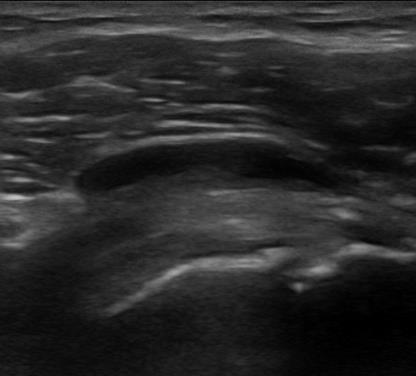
43 3. Interpretation of US findings, US diagnosis and conclusions 34 yrs old female; 15 months history of RA. MTX during the last 6 months (20 mg weekly) US, NewTherapy?
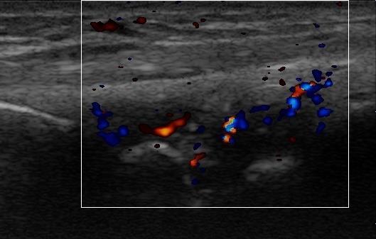
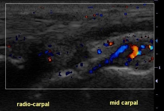
44 Long Long


45 II MCP R Long II MCP L Long


46 Long MSUS is my tool to make a decision! IV PIP Long I PIP Long

47 Third step Write the report! The responsibility for the accuracy of the report is yours!
48 US REPORT All images should have the following information correctly recorded on them Obligatory data 1. Patient identification 2. Date of examination 3. Hospital/department identification 4. The name and status of the sonographer issuing the report
49 US REPORT GENERAL COMMENTS Write the report as soon as possible after the examination Overwrite in all documented US images: 1. The anatomic area 2. The scan (transverse, longitudinal, oblique) 3. The name of the visible bones
50 US REPORT Documentation and Image recording Таке a proper number of scans with normal and pathological findings (jpeg images, videoclips) Store images as: * printed images, * on a tape or * electronic medium
51 US REPORT Documentation and Image recording All written reports and recorded images of ultrasound studies keep in the patient's record Take every opportunity to maintain and develop your knowledge, skills and competence
52 Conclusions Use standardized terminology and Take images of definitions Show of well correctly scanned pathology documented HELP images structures THE CLINICIAN and TO DIAGNOSE make very AND accurate TAKE THERAPEUTIC reports DECISIONS!
53 NEVER FORGET Your report is a teaching YOUR tool REPORT for yоunger IS YOUR BUSNESS sonographers! CARD!
54 Acknowledgement The author wishes to acknowledge the contribution of Esperanza Naredo, Nemanja Damjanov and Hilde Berner Hammer in this presentation.

55
Ultrasound in Rheumatology
 Arthritis Research UK Primary Care Centre Winner of a Queen s Anniversary Prize For Higher and Further Education 2009 Ultrasound in Rheumatology Alison Hall Consultant MSK Sonographer/Research Fellow Primary
ELENI ANDIPA General Hospital of Athens G. Gennimatas
 ELENI ANDIPA General Hospital of Athens G. Gennimatas Technological advances over the last years have caused a dramatic improvement in ultrasound quality and resolution An established imaging modality
Ultrasound in Rheumatology
 Ultrasound in Rheumatology Alison Hall Consultant MSK Sonographer Research Institute for Primary Care & Health Sciences, Keele University Department of Rheumatology, Cannock Hospital, Royal Wolverhampton
Bulgarian Association for Musculoskeletal Ultrasound
 MUSCULOSKELETAL ULTRASOUND COURSE INTERMEDIATE LEVEL ORGANIZATION AND COMMITTEE Scientific Organiser Prof. Annamaria Iagnocco Local Organisers Prof Anastas Batalov Dr Rodina Nestorova Dr Plamen Todorov
International Musculoskeletal Ultrasound Course MITOS
 Basic and Intermediate Levels th International Musculoskeletal Ultrasound Course MITOS Final Course Program 30 November - 2 December 2017 Wyndham Grand Athens Hotel Athens Greece www.synthesispco.com/mitoscourse2017
Sonographic appearance of chronic inflammatory rheumatism
 Sonographic appearance of chronic inflammatory rheumatism Poster No.: C-2237 Congress: ECR 2013 Type: Educational Exhibit Authors: H. Elfattach, F. Houari, O. Addou, M. Maaroufi, S. Tizniti ; 1 1 1 1 2
"EULAR endorsed course"
 MUSCULOSKELTAL SONOGRAPHY COURSE InterMediate Level 15 17 September, 2017 Dubai, UAE Organizing Office: Compass Conferences "EULAR endorsed course" Organizers Ingrid Möller (Scientific Organizer) Esperanza
Scoring and Grading B-Mode Synovitis and Doppler findings in pediatric MSKUS. Johannes Roth MD PhD FRCPC RhMSUS
 Scoring and Grading B-Mode Synovitis and Doppler findings in pediatric MSKUS Johannes Roth MD PhD FRCPC RhMSUS Pathology - Definition Synovitis Synovitis on ultrasonography in children B-mode and Doppler
David Bong (Spain), MD Faculty member of the EULAR MSUS courses since 2011.
, MD Faculty member of the EULAR MSUS courses since 2011.") MUSCULOSKELTAL SONOGRAPHY COURSE Intermediate Level Sept 30 & Oct 1 & 2, 2016 Dubai, United Arab Emirates Organizing Office: Compass Conferences ENDORSED Organizers Ingrid Möller (Scientific Organizer)
MUSCULOSKELTAL SONOGRAPHY COURSE Intermediate Level Sept 30 & Oct 1 & 2, 2016 Dubai, United Arab Emirates Organizing Office: Compass Conferences ENDORSED Organizers Ingrid Möller (Scientific Organizer)
Pragmatic ultrasound in the diagnosis of soft tissue rheumatic pain. Plamen Todorov
 Pragmatic ultrasound in the diagnosis of soft tissue rheumatic pain Plamen Todorov INTRODUCTION Soft tissue rheumatism: nonsystemic, focal pathological syndromes involving the periarticular structures.
Clinical Practice Guideline. Ultrasound in Rheumatological Settings. Version
 Clinical Practice Guideline Ultrasound in Rheumatological Settings Version 1.1.2017 November 2017 Table of Contents Abbreviations...3 Introduction...4 Diagnostic Musculoskeletal Ultrasound...6 Definition
MUSCULOSKELTAL SONOGRAPHY COURSE Basic Level 4 6 November, 2016 Manama, Kingdom of Bahrain Organizing Office: Compass Conferences ENDORSED
 MUSCULOSKELTAL SONOGRAPHY COURSE Basic Level 4 6 November, 2016 Manama, Kingdom of Bahrain Organizing Office: Compass Conferences ENDORSED Organizers Möller (Scientific Organizer) Ayman Fahim (Organizer)
EULAR 25th Jubilee EULAR Sonography Course Basic, Intermediate and Advanced Musculoskeletal Ultrasound (MSUS) in Rheumatology
 in Rheumatology") EULAR 25th Jubilee EULAR Sonography Course Basic, Intermediate and Advanced Musculoskeletal Ultrasound (MSUS) in Rheumatology Sunday 10th Wednesday 13th June 2018 Amsterdam, Netherlands ORGANISATION AND
EULAR 25th Jubilee EULAR Sonography Course Basic, Intermediate and Advanced Musculoskeletal Ultrasound (MSUS) in Rheumatology Sunday 10th Wednesday 13th June 2018 Amsterdam, Netherlands ORGANISATION AND
US finding of the shoulder (with live demonstration) 인제의대상계백병원 안재기
 인제의대상계백병원 안재기") US finding of the shoulder (with live demonstration) 인제의대상계백병원 안재기 Shoulder US Biceps tendon & Rotator Cuff Long Head of Biceps Tendon Subscapularis tendon Supraspinatus tendon Infraspinatus tendon Teres
US finding of the shoulder (with live demonstration) 인제의대상계백병원 안재기 Shoulder US Biceps tendon & Rotator Cuff Long Head of Biceps Tendon Subscapularis tendon Supraspinatus tendon Infraspinatus tendon Teres
Table of contents. Foreword. Preface. 1 Introduction Historical Perspective 00
 Table of contents Foreword Preface 1 Introduction 00 1.1 Historical Perspective 00 2 Fundamentals of musculoskeletal ultrasound 00 2.1 Frequency and wavelength 00 2.2 Generating ultrasound waves 00 2.3
EULAR 25th Jubilee EULAR Sonography Course Basic, Intermediate and Advanced Musculoskeletal Ultrasound (MSUS) in Rheumatology
 in Rheumatology") EULAR 25th Jubilee EULAR Sonography Course Basic, Intermediate and Advanced Musculoskeletal Ultrasound (MSUS) in Rheumatology Sunday 10th Wednesday 13th June 2018 Amsterdam, Netherlands ORGANISATION AND
EULAR 25th Jubilee EULAR Sonography Course Basic, Intermediate and Advanced Musculoskeletal Ultrasound (MSUS) in Rheumatology Sunday 10th Wednesday 13th June 2018 Amsterdam, Netherlands ORGANISATION AND
EULAR 24th EULAR Sonography Course Basic, Intermediate and Advanced Musculoskeletal Ultrasound (MSUS) in Rheumatology
 in Rheumatology") EULAR 24th EULAR Sonography Course Basic, Intermediate and Advanced Musculoskeletal Ultrasound (MSUS) in Rheumatology Sunday 11th Wednesday 14th June 2017 Madrid, Spain ORGANISATION AND COMMITTEE Scientific
EULAR 24th EULAR Sonography Course Basic, Intermediate and Advanced Musculoskeletal Ultrasound (MSUS) in Rheumatology Sunday 11th Wednesday 14th June 2017 Madrid, Spain ORGANISATION AND COMMITTEE Scientific
Principles of Ultrasound. Cara C. Prideaux, M.D. University of Utah PM&R Sports Medicine Fellow March 14, 2012
 Principles of Ultrasound Cara C. Prideaux, M.D. University of Utah PM&R Sports Medicine Fellow March 14, 2012 None Disclosures Outline Introduction Benefits and Limitations of US Ultrasound (US) Physics
EULAR 24th EULAR Sonography Course Basic, Intermediate and Advanced Musculoskeletal Ultrasound (MSUS) in Rheumatology
 in Rheumatology") EULAR 24th EULAR Sonography Course Basic, Intermediate and Advanced Musculoskeletal Ultrasound (MSUS) in Rheumatology Sunday 11th Wednesday 14th June 2017 Madrid, Spain ORGANISATION AND COMMITTEE Scientific
EULAR 24th EULAR Sonography Course Basic, Intermediate and Advanced Musculoskeletal Ultrasound (MSUS) in Rheumatology Sunday 11th Wednesday 14th June 2017 Madrid, Spain ORGANISATION AND COMMITTEE Scientific
The Role of Ultrasonography in the Assessment of Rheumatic Diseases. Current and Potential Role of Ultrasonography in Rheumatology
 The Role of Ultrasonography in the Assessment of Rheumatic Diseases Karina D. Torralba, MD, MACM, CCD, Assistant Professor of Medicine, Keck School of Medicine, University of Southern California Objective:
3rd MUSCULOSKELETAL SONOGRAPHY COURSE FOR RHEUMATOLOGISTS - INTERMEDIATE LEVEL -
 3rd MUSCULOSKELETAL SONOGRAPHY COURSE FOR RHEUMATOLOGISTS - INTERMEDIATE LEVEL - Bucharest, Romania October, 13th-15th 2016 Organizer: Romanian Society of Rheumatology Prof. Dr. Ruxandra Ionescu Course
"EULAR endorsed course"
 MUSCULOSKELTAL SONOGRAPHY COURSE Basic Level 20 22 July, 2018 Abu Dhabi, United Arab Emirates Organizing Office: Compass Conferences "EULAR endorsed course" Organizers Möller (Scientific Organizer) Esperanza
International Musculoskeletal Ultrasound Course MITOS
 Basic and Intermediate Levels th International Musculoskeletal Ultrasound Course MITOS General information and Course program 30 November - 2 December 2017 Wyndham Grand Athens Hotel Athens Greece www.synthesispco.com/mitoscourse2017
Introduction to Ultrasound Guided Shoulder Injections. Alison Hall Consultant Sonographer Keele University Cannock Chase Hospital
 Introduction to Ultrasound Guided Shoulder Injections Alison Hall Consultant Sonographer Keele University Cannock Chase Hospital Safe Robust Aim: to provide a service that is Cost effective To enable patients
6 th MUSCULOSKELETAL SONOGRAPHY COURSE. Belgrade, 5 th 7 th October, 2017 INTERMEDIATE LEVEL COURSE
 6 th MUSCULOSKELETAL SONOGRAPHY COURSE INTERMEDIATE LEVEL COURSE Belgrade, 5 th 7 th October, 2017 ORGANIZER Rheumatology Association of Serbia www.belgrade-course.org SCIENTIFICALLY ENDORSED BY eular
EDUCATIONAL COURSE. MUSCULOSKELETAL ULTRASOUND in RHEUMATOLOGY BASIC COURSE. Course Coordinator Annamaria Iagnocco
 EDUCATIONAL COURSE 2019 MUSCULOSKELETAL ULTRASOUND in RHEUMATOLOGY BASIC COURSE Rome, March 21-23, 2019 Course Coordinator Annamaria Iagnocco COURSE PRESENTATION Musculoskeletal ultrasound in rheumatology
26 th Sonography Course MSUS Intermediate
 26 th Sonography Course MSUS Intermediate Madrid, Spain Sunday, 9 th June Wednesday, 12 th June 2019 The 26 th EULAR Sonography Course will be run simultaneously at three levels, basic, intermediate and
Focused Musculoskeletal Ultrasound
 Focused Musculoskeletal Ultrasound David Lewis Consultant Emergency Medicine Ipswich (Club Doctor, Ipswich Town FC) Advanced Emergency Ultrasound Objectives! General principles! Musculoskeletal anatomy!
Case study # 6 Sharon P
 Patient is a morbidly obese 70 year old female presenting with left shoulder pain after a severe fall. Patient is in moderate to severe pain with extremely limited range of motion due to extensive shoulder
Lateral Elbow Pathology
 Lateral Elbow Pathology Jon A. Jacobson, M.D. Professor of adiology Director, Division of Musculoskeletal adiology University of Michigan Disclosures: Consultant: Bioclinica Advisory Board: GE, Philips
Ultrasound of Shoulder Pathology and Intervention 서울대학교병원재활의학과 김기원
 Ultrasound of Shoulder Pathology and Intervention 서울대학교병원재활의학과 김기원 Ultrasound for Shoulder Disorder Advantage Dynamic evaluation Immediate clinical correlation + Intervention Weakness Diagnostic accuracy?
UltraSound Course. International Musculoskeletal MITOS. Athens, Greece. Basic and Intermediate Levels - Course Program. 29 Nov - 1 Dec 2018
 Basic and Intermediate Levels - Course Program th International Musculoskeletal UltraSound Course 29 Nov - 1 Dec 2018 Wyndham Grand Athens Hotel Athens, Greece MITOS Musculosceletal Imaging Techniques
Ultrasound of the Shoulder
 Ultrasound of the Shoulder Patrick Battaglia, DC, DACBR Logan University, Department of Radiology Outline Review ultrasound appearance of NMSK tissues Present indications for ultrasound of the shoulder.
Rotator Cuff and Biceps Pathology
 Rotator Cuff and Biceps Pathology Jon A. Jacobson, M.D. Professor of Radiology Director, Division of Musculoskeletal Radiology University of Michigan Disclosures: Consultant: Bioclinica Advisory Board:
Scientific Programme
 Scientific Programme (preliminary version) Musculoskeletal Sonography Course for Rheumatologists Warszawa 2010 th st September 28 - October 01, 2010 Warszawa, Poland under scientific patronage of the European
9/18/18. Welcome- MSK Ultrasound Workshop. Introduction to Musculoskeletal Ultrasound. Acknowledgement of Country. The Workshop.
 Acknowledgement of Country Welcome- MSK Ultrasound Workshop I would like to acknowledge that this meeting is being held on the traditional lands of the Wurundjeri and Boonwurrung people and pay my respect
3 rd MUSCULOSKELETAL SONOGRAPHY COURSE FOR RHEUMATOLOGISTS
 3 rd MUSCULOSKELETAL SONOGRAPHY COURSE FOR RHEUMATOLOGISTS UNDER SCIENTIFIC ENDORSEMENT OF THE EUROPEAN LEAGUE AGAINST RHEUMATISM BASIC AND INTERMEDIATE LEVEL Belgrade, March 29th-31st, 2012 G E N E R
The Egyptian Journal of Hospital Medicine (October 2017) Vol. 69 (4), Page
 Vol. 69 (4), Page") The Egyptian Journal of Hospital Medicine (October 2017) Vol. 69 (4), Page 2294-2300 Role of Magnetic Resonance Imaging and Ultrasonography in Diagnosis and Follow Up Rheumatoid Arthritis in Hand and Wrist
The Egyptian Journal of Hospital Medicine (October 2017) Vol. 69 (4), Page 2294-2300 Role of Magnetic Resonance Imaging and Ultrasonography in Diagnosis and Follow Up Rheumatoid Arthritis in Hand and Wrist
The Essentials Tissue Characterization and Knobology
 The Essentials Tissue Characterization and Knobology Randy E. Moore, DC, RDMS RMSK No relevant financial relationships Ultrasound The New Standard of Care Musculoskeletal sonography has become the standard
Index. Note: Page numbers of article titles are in boldface type.
 Note: Page numbers of article titles are in boldface type. A ACJ. See Acromioclavicular joint (ACJ) Acromioclavicular joint (ACJ) procedures of, 557 559 Ankle and foot procedures of, 649 671 (See also
Ultrasound Evaluation of Masses
 Ultrasound Evaluation of Masses Jon A. Jacobson, M.D. Professor of Radiology Director, Division of Musculoskeletal Radiology University of Michigan Disclosures: Consultant: Bioclinica Advisory Panel: GE,
Introduction to Musculoskeletal Ultrasound. Disclosures. Evidence Based Medicine Key References 8/30/2017
 Introduction to Musculoskeletal Ultrasound Johannes Roth MD, PhD, FRCPC, RhMSUS Professor of Pediatrics University of Ottawa Gurjit S Kaeley MBBS, MRCP, RhMSUS Professor of Medicine Division Chief Director
Musculoskeletal Examination
 Musculoskeletal Examination Statement of Goals Know how to perform a complete musculoskeletal examination. Learning Objectives A. Describe the anatomy of the musculoskeletal system including the bony structures,
Sonographic assessment of adult and juvenile rheumatoid arthritis
 Sonographic assessment of adult and juvenile rheumatoid arthritis Poster No.: C-1485 Congress: ECR 2013 Type: Educational Exhibit Authors: C. A. S. Ruano, P. L. Pegado, J. M. G. Lourenco, P. Alves, L.
Ultrasound in Rheumatological Conditions:
 Ultrasound in Rheumatological Conditions: Status and Perspectives Nancy A. Chauvin, MD Assistant Professor of Radiology Director of Musculoskeletal Imaging The Children s Hospital of Philadelphia University
Why? Ultrasound of the Foot. Ultrasound of the Foot. General Rules. Plantar Fascia. Plantar Fasciitis 18/09/2018
 Ultrasound of the Foot Why? Ultrasound of the Foot Plantar fasciitis Plantar fascia fibromatosis Morton s neuroma Intermetatarsal bursitis Adventitial bursitis Plantar plate tears MTP joint synovitis Ganglia
The Shoulder. Systematically scanning the shoulder provides extremely useful diagnostic information. The Shoulder
 1 ! The most ACCESSIBLE to sonographic exam! The most MOBILE and VULNERABLE extremity AND Systematically scanning the shoulder provides extremely useful diagnostic information! The Goal for this section
Shoulder Elbow Wrist/Hand
 Shoulder Elbow Wrist/Hand Randy E. Moore DC RDMS RMSK General Musculoskeletal Imaging, Inc. 1 Shoulder Tendinosis : 3 key Ultrasound Findings 1. Increased cellularity thickened and ACR inhomogeneous CLV
Ultrasound assessment of most frequent shoulder disorders
 Ultrasound assessment of most frequent shoulder disorders Poster No.: C-2026 Congress: ECR 2014 Type: Educational Exhibit Authors: S. P. Ivanoski; Ohrid/MK Keywords: Trauma, Athletic injuries, Arthritides,
Ultrasound Evaluation of Posteromedial Ankle Pathology. Andrew C Cordle, M.D., Ph.D. 9/21/2018
 Ultrasound Evaluation of Posteromedial Ankle Pathology Andrew C Cordle, M.D., Ph.D. 9/21/2018 Overview: Pathology of the Posteromedial Ankle Flexor Tendon Pathology Accessory Navicular Bone Pathology Tarsal
General information and Course programme of the. 4 MUSCULOSKELETAL SONOGRAPHY COURSE FOR RHEUMATOLOGISTS - BASIC LEVEL - st
 General information and Course programme of the th 4 MUSCULOSKELETAL SONOGRAPHY COURSE FOR RHEUMATOLOGISTS - BASIC LEVEL - st 1 MUSCULOSKELETAL SONOGRAPHY COURSE FOR RHEUMATOLOGISTS - INTERMEDIATE LEVEL
Ultras ono graphic Evaluation of Rotator Cuff Tendons in Patients with Rheumatoid Arthritis
 Med. J. Cairo Univ., Vol. 83, No. 1, June: 395-399, 215 www.medicaljournalofcairouniversity.net Ultras ono graphic Evaluation of Rotator Cuff Tendons in Patients with Rheumatoid Arthritis HALA I. ELGENDY,
Crystal Deposition Disease and Psoriatic Arthritis
 74 Crystal Deposition Disease and Psoriatic Arthritis Philip J. O Connor, MRCP, FRCR, FFSEM (UK) 1,2 1 Department of Radiology, Leeds Teaching Hospitals, Chapel Allerton Hospital, Leeds, United Kingdom
Musculoskeletal Ultrasound Fundamentals
 Fundamentals Benjamin D. Levine, M.D. Associate Professor of Radiology Musculoskeletal Imaging Dept. of Radiological Sciences UCLA Health System I. Image Optimization II. Image Interpretation Artifacts
Comparative study of high resolusion ultrasonography and magnetic resonance imaging in diagnosing traumatic knee injuries & pathologies
 Original article: Comparative study of high resolusion ultrasonography and magnetic resonance imaging in diagnosing traumatic knee injuries & pathologies Dr. Rakesh Gujjar*, Dr. R. P. Bansal, Dr. Sandeep
2 MUSCULOSKELETAL SONOGRAPHY COURSE FOR RHEUMATOLOGISTS - INTERMEDIATE LEVEL -
 General information and Course programme of the nd 2 MUSCULOSKELETAL SONOGRAPHY COURSE FOR RHEUMATOLOGISTS - INTERMEDIATE LEVEL - rd th Zagreb, December 3 December 5, 2018. This course has been scientifically
MUSCULOSKELETAL ULTRASOUND COURSE - BASIC LEVEL- Graz, November 2 nd 4 th, 2016
 MUSCULOSKELETAL ULTRASOUND COURSE - BASIC LEVEL- Graz, November 2 nd 4 th, 2016 GENERAL INFORMATION Course opening Wednesday, Novemeber 2 nd, 2016 - h 17.00 Course closing Friday, November 4 th, 2012 -
Common Applications for Sonography and Guided Intervention: Shoulder
 Common Applications for Sonography and Guided Intervention: Shoulder Jon A. Jacobson, M.D. Professor of Radiology Director, Division of Musculoskeletal Radiology University of Michigan Disclosures: Consultant:
APPROPRIATE USE GUIDELINES
 APPROPRIATE USE GUIDELINES Appropriateness of Advanced Imaging Procedures (MRI, CT, Bone Scan/PET) in Patients with Shoulder Pain CDI QUALITY INSTITUTE: PROVIDER LED ENTITY (PLE) Compiled by Rob Liddell,
Graz, October 3 rd 5 th, 2013
 MUSCULOSKELETAL SONOGRAPHY COURSE IN RHEUMATOLOGY INTERMEDIATE LEVEL Graz, October 3 rd 5 th, 2013 This course has been scientifically endorsed by: GENERAL INFORMATION Course opening: Thursday, October
This course is endorsed by
 MUSCULOSKELETAL ULTRASOUND COURSE FOR RHEUMATOLOGISTS BASIC LEVEL BUCHAREST, ROMANIA 27-29.APRIL.2017 COURSE DIRECTORS: Annamaria Iagnocco Mihai Bojinca VENUE AND DATES: Double Tree by Hilton Nerva Traian
The Elbow 3/5/2015. The Elbow Scanning Sequence. * Anterior Joint (The anterior Pyramid ) * Lateral Epicondyle * Medial Epicondyle * Posterior Joint
 * Lateral Epicondyle * Medial Epicondyle * Posterior Joint") Scanning Sequence * Anterior Joint (The anterior Pyramid ) * Lateral Epicondyle * Medial Epicondyle * Posterior Joint Anterior Elbow Pyramid Courtesy of Jay Smith, MD. Vice chair PMR Mayo Clinic Rochester,
Scanning Sequence * Anterior Joint (The anterior Pyramid ) * Lateral Epicondyle * Medial Epicondyle * Posterior Joint Anterior Elbow Pyramid Courtesy of Jay Smith, MD. Vice chair PMR Mayo Clinic Rochester,
Urgent Cases and Foreign Bodies
 Urgent Cases and Foreign Bodies Catherine J. Brandon, MD, MS University of Michigan Ann Arbor, MI, USA Introduction: Patients added on to the schedule from the emergency department or as urgent add-on
MUSCULOSKELETAL DISORDERS: THE BIGGEST JOB SAFETY PROBLEM. What Are Musculoskeletal Disorders
 MUSCULOSKELETAL DISORDERS: THE BIGGEST JOB SAFETY PROBLEM What Are Musculoskeletal Disorders Every year more than 1.8 million workers in the United States suffer painful back and repetitive strain injuries,
Ultrasonographic Evaluation of Painful Shoulder joint in rural population
 Original article: Ultrasonographic Evaluation of Painful Shoulder joint in rural population Dr. Pankaj Garg*, Dr. V.N. Marathe, Dr. S. G. Gandage, Dr.S.G.Kachewar Department of Radiology, Rural Medical
Pragmatic use of US in intraarticular and periarticular procedures
 Pragmatic use of US in intraarticular and periarticular procedures Plovdiv, 13. January 2018 Dr. med. Giorgio Tamborrini-Schütz www.irheuma.com member of the EULAR Network of Imaging Training Centres Ulrasound
Paediatric rheumatology
 Paediatric rheumatology Ultrasonography vs. clinical examination in children with suspected arthritis. Does it make sense to use poliarticular ultrasonographic screening? G. Filippou, L. Cantarini, I.
2 nd MUSCULOSKELETAL SONOGRAPHY COURSE FOR RHEUMATOLOGISTS BASIC LEVEL. Athens, December 17 th - 19 th, 2015
 MITOS 2 nd MUSCULOSKELETAL SONOGRAPHY COURSE FOR RHEUMATOLOGISTS BASIC LEVEL Athens, December 17 th - 19 th, 2015 Musculoskeletal sonography course for rheumatologists, Athens 2015 intends to be a combination
15 th EULAR SONOGRAPHY COURSE. June 08 th - 11 th, 2008 Paris, France. Basic, Intermediate and Advanced Musculoskeletal Ultrasound in Rheumatology
 15 th EULAR SONOGRAPHY COURSE Basic, Intermediate and Advanced Musculoskeletal Ultrasound in Rheumatology June 08 th - 11 th, 2008 Paris, France Societé française de rhumatologie Université de Versailles
Ultrasound of the Knee
 Ultrasound of the Knee Jon A. Jacobson, M.D. Professor of Radiology Director, Division of Musculoskeletal Radiology University of Michigan Disclosures: Consultant: Bioclinica Book Royalties: Elsevier Advisory
MUSCULOSKELETAL ULTRASOUND in RHEUMATOLOGY
 EDUCATIONAL COURSE 2018 MUSCULOSKELETAL ULTRASOUND in RHEUMATOLOGY BASIC COURSE This course is endorsed by Rome, March 15-17, 2018 Course Coordinator Annamaria Iagnocco COURSE PRESENTATION Musculoskeletal
Musculoskeletal Ultrasound
 EFSUMB Course Book Student Edition Editors: Jan Tuma, Radu Badea, Christoph F. Dietrich Musculoskeletal Ultrasound Giorgio Tamborrini Ultrasound Center, Switzerland Corresponding author: KD Dr. med. Giorgio
Soft Tissue Rheumatism. Elinor Mody, MD Chief, Division of Rheumatology Reliant Medical Group
 Soft Tissue Rheumatism Elinor Mody, MD Chief, Division of Rheumatology Reliant Medical Group Some problems are difficult, but diagnosing and treating most causes of joint pain are not! Common areas of
Musculoskeletal Ultrasound: Basics, Utility, and Clinical Applications
 Musculoskeletal Ultrasound: Basics, Utility, and Clinical Applications Andrew Lavigne, MD, FRCPC Physical Medicine and Rehabilitation CSCN Diplomat (EMG) Dip Sport Medicine Eugene Maida, MD, PGY-4 Resident
Ultrasound Guided Injections
 Ultrasound Guided Injection Technique More accurate injections Better Results! 1 Benefits: Increased Level of Certainty ie : really know how accurate PRP/Prolotherapy Avoid damage to articular cartilage
Point of Care Ultrasound on the Field of Play K AT I E N ANOS, MD
 Point of Care Ultrasound on the Field of Play K AT I E N ANOS, MD H I GH P ERFORMANCE S PORTS MEDICINE P HYSI ATRIST, P R ACTICING S PORTS MEDI CINE No disclosures No disclosures Who am I? Objectives Over
MUSCULOSKELETAL SONOGRAPHY COURSE FOR RHEUMATOLOGISTS - BASIC LEVEL - June 27 th 29 th, 2013 Innsbruck, Austria
 MUSCULOSKELETAL SONOGRAPHY COURSE FOR RHEUMATOLOGISTS - BASIC LEVEL - June 27 th 29 th, 2013 Innsbruck, Austria GENERAL INFORMATION Course opening: Thursday, June 27 th, 2013 - h 12.00 Course closing:
3/20/2017. Disclosures. Ultrasound Fundamentals. Ultrasound Fundamentals. Bone Anatomy. Tissue Characteristics
 Disclosures Images of ultrasound equipment in this presentation are not an endorsement Fundamentals of Musculoskeletal Ultrasound Physics and Knobology Shane A. Shapiro, M.D. Assistant Professor Orthopedic
Ultrasound of Mid and Hindfoot Pathology
 Ultrasound of Mid and Hindfoot Pathology Levon N. Nazarian, M.D. Professor of Radiology Thomas Jefferson University Hospital Disclosures None relevant to this presentation Educational Objective Following
Professor Lisa Stamp
 Professor Lisa Stamp Rheumatologist University of Otago, Christchurch 8:30-9:25 WS #65: Joint Injection Techniques 9:35-10:30 WS #75: Joint Injection Techniques (Repeated) Joint/soft tissue corticosteroid
Ultrasound of the Knee Joint. Jun Sung Park,M.D. Bundang General Hospital Dept. of Rehabilitation Medicine
 Ultrasound of the Knee Joint Jun Sung Park,M.D. Bundang General Hospital Dept. of Rehabilitation Medicine Clinical History and P/E Chronic or Acute Symptoms Chronic Sx. : possible of systemic articular
Ultrasound of the Hip: Anatomy, Pathology, and Procedures
 Ultrasound of the Hip: Anatomy, Pathology, and Procedures Jon A. Jacobson, M.D. Professor of Radiology Director, Division of Musculoskeletal Radiology University of Michigan Outline Hip Joint Native hip
Gout. Crystal deposition disease: Imaging perspectives. Crystal associated arthropathies. Clinical Stages of Gout 07/06/60
 Crystal associated arthropathies Crystal deposition disease: Imaging perspectives Warapat Virayavanich, MD Ramathibodi hospital, Mahidol University Commonly seen arthropathy MSU (gout) CPPD HADD Uncommon
2015 ARDMS Musculoskeletal Sonographer Job Task Analysis Summary Report
 P a g e 1 2015 ARDMS Musculoskeletal Sonographer Job Task Analysis Summary Report American Registry for Diagnostic Medical Sonography (ARDMS) P a g e 2 Table of Contents ABOUT THE REPORT... 3 METHODOLOGY...
MUSCULOSKELETAL SONOGRAPHY COURSE IN RHEUMATOLOGY - ADVANCED LEVELGraz, November 7th 9th, 2018
 MUSCULOSKELETAL SONOGRAPHY COURSE IN RHEUMATOLOGY - ADVANCED LEVELGraz, November 7th 9th, 2018 Dear colleague, This course has been scientifically endorsed by: ultrasound is increasingly used in rheumatology
June 26 th 28 th, 2014 Innsbruck, Austria
 MUSCULOSKELETAL SONOGRAPHY COURSE FOR RHEUMATOLOGISTS - INTERMEDIATE LEVEL - June 26 th 28 th, 2014 Innsbruck, Austria This course is scientifically endorsed by: GENERAL INFORMATION Course opening: Thursday,
Anatomy of Peripheral Nerve 가톨릭대학교 재활의학과 김재민
 Anatomy of Peripheral Nerve 가톨릭대학교 재활의학과 김재민 Contents US appearance of nerves Scanning technique Peripheral nerve pathology Nerves of arm Nerves of leg US Appearance of Nerve Multiple longitudinal hypoechoic
Introduction to Ultrasound Examination of the Hand and upper
 Introduction to Ultrasound Examination of the Hand and upper Emil Dionysian, M.D. Ultrasound of upper ext. Upside Convenient Opens another exam dimension Can be like a stethoscope Helps 3-D D visualization
Psoriatic arthritis: early ultrasound findings
 Psoriatic arthritis: early ultrasound findings Poster No.: C-0399 Congress: ECR 2014 Type: Educational Exhibit Authors: R. Persechino 1, L. Cristiano 1, A. Bartoloni 1, C. Cantone 2, A. Keywords: DOI:
3rd MUSCULOSKELETAL SONOGRAPHY COURSE FOR RHEUMATOLOGISTS - BASIC LEVEL Zagreb, May 29th - May 31st, 2014.
 General information and Course programme of the 3rd MUSCULOSKELETAL SONOGRAPHY COURSE FOR RHEUMATOLOGISTS - BASIC LEVEL Zagreb, May 29th - May 31st, 2014. This course has been scientifically endorsed by
Of the many advances that have come into the field of rheumatology today is the art
 36 Journal of the association of physicians of india vol 62 october, 2014 review article Musculo-skeletal Ultrasound in Rheumatology Practice Ved Chaturvedi * Abstract In rheumatology ultrasound is a relatively
September 14 th 16 th, 2017 Innsbruck, Austria
 MUSCULOSKELETAL SONOGRAPHY COURSE FOR RHEUMATOLOGISTS - BASIC LEVEL - September 14 th 16 th, 2017 Innsbruck, Austria This course is scientifically endorsed by: GENERAL INFORMATION Course opening: Thursday,
I-A-1) Non-specific thickening of synovial membrane
 Non-specific thickening of synovial membrane") I-A-1) Non-specific thickening of synovial membrane Grayscale Metatarsal Power Doppler Dorsal aspect of metatarsophalangeal joint in right 1 st toe, longitudinal view Asterisks indicate non-specific thickening
I-A-1) Non-specific thickening of synovial membrane Grayscale Metatarsal Power Doppler Dorsal aspect of metatarsophalangeal joint in right 1 st toe, longitudinal view Asterisks indicate non-specific thickening
MUSCULOSKELETAL ULTRASOUND
 MUSCULOSKELETAL ULTRASOUND A brief overview of diagnostic and therapeutic applications in musculoskeletal medicine. By Elmer G. Pinzon, MD, MPH and Randy E. Moore, DC, RDMS The use of musculoskeletal ultrasound
September 5 th 7 th, 2018 Innsbruck, Austria
 MUSCULOSKELETAL SONOGRAPHY COURSE FOR RHEUMATOLOGISTS - INTERMEDIATE LEVEL - September 5 th 7 th, 2018 Innsbruck, Austria This course is scientifically endorsed by: GENERAL INFORMATION Course opening:
Ultrasound in Peripheral Nerve Interventions
 Ultrasound in Peripheral Nerve Interventions John L. Lin, M.D. Shepherd Center Assistant Clinical Professor Emory University, School of Medicine Outline Ultrasound basics Nerve blocks in physiatric setting
Ultrasonography of the Neck as an Adjunct to FNA. Nicole Massoll M.D.
 Ultrasonography of the Neck as an Adjunct to FNA Nicole Massoll M.D. Basic Features of Head and Neck Ultrasound and Anatomy Nicole Massoll M.D. University of Arkansas for Medical Sciences, Little Rock
Terminology Tissue Appearance
 By Marc Nielsen, MD Advantages/Disadvantages Generation of Image Ultrasound Machine/Transducer selection Modes of Ultrasound Terminology Tissue Appearance Scanning Technique Real-time Portable No ionizing