Ultrasound in Rheumatological Conditions:
|
|
|
- Scot Shaw
- 5 years ago
- Views:
Transcription


1 Ultrasound in Rheumatological Conditions: Status and Perspectives Nancy A. Chauvin, MD Assistant Professor of Radiology Director of Musculoskeletal Imaging The Children s Hospital of Philadelphia University of Pennsylvania, Perelman School of Medicine SPR 2016 Pediatric Ultrasound Course Orlando, Florida
2 No disclosures
3 Goals Briefly review pathophysiology of JIA Review US techniques to optimize imaging & interpretation Identify the appearance of normal MSK structures in children Discuss the current role of US in JIA Future directions of imaging

4 Juvenile Idiopathic Arthritis Most common chronic rheumatologic disease of childhood Definition : Arthritis that begins before age 16y, continues for approximately 6 weeks and involves 1 or multiple joints 1/1,000 children in North America Medicalobsever.com Ording et al. Insights Imaging. 2015; 6(3):
5 Juvenile Idiopathic Arthritis Historically, two main classification systems: European Juvenile Chronic Arthritis (JCA) American Juvenile Rheumatoid Arthritis (JRA) ILAR (International League of Associations for Rheumatology) 1995; Juvenile Idiopathic Arthritis (JIA) Classification updated in major subtypes umbrella term Petty RE et al. J Rheumatol. 2004; 31(2):
6 7 Major Subtypes Subtype Frequency Findings Systemic Arthritis 4-17% Systemic symptoms (fever, rash, organ) Sex Ratio F=M Oligoarthritis 27-56% Up to 4 joints in 6mo F>>>M RF + Polyarthritis 2-7% 5 or more joints F>>M RF Polyarthritis 11-15% 5 or more joints F>>M Enthesitis-Related Arthritis 15-20% Insertion sites of tendons and ligaments & axial disease M>>F Psoriatic arthritis 2-11% Typical skin and nail findings F>M Undifferentiated arthritis 11-15% Daldrup-Link HE et al. Magn Reson Imaging Clin N Am. Aug 2009;17(3): , vi. Juvenile Spondyloarthritis


7 JIA Pathophysiology Hallmark: Synovial thickening & inflammation Ording et al. Insights Imaging. 2015; 6(3):
8 Role of Imaging JIA diagnosis based on clinical & laboratory findings Imaging may detect early manifestations of arthritis Synovitis, cartilage abnormalities, erosions Evaluate the extent of disease/joint involvement Monitor disease progression & treatment response Imaging can more accurately distinguish between arthritis and tenosynovitis than clinical examination alone and in some cases, it can help define the subtype of JIA Provides opportunity to implement therapy at an early stage
9 Role of Imaging Radiographs traditional method of evaluating joints Poor sensitivity in depicting active arthritis Rarely show erosive disease until late in disease course Increase use of US & MRI Ultrasound has a higher sensitivity and specificity for the presence of synovitis than clinical assessment along and is the most sensitive method for detection of tenosynovitis Ording et al. Insights Imaging 2015; 6(3):
10 Imaging Obstacles in Children Sparsity of normative data To correctly diagnose pathology, need to be familiar with normal agedependent changes of the growing skeleton Large amount of data in adults apply to children? Unique features of the growing skeleton Physiologic features of recently ossified bones can be misinterpreted as cortical erosions Appearance, thickness & vascularity of the epiphyseal cartilage varies with skeletal maturation Suboptimal reader agreement on validation studies Damasio et al. Pediatr Radiol. Jun 2010; 40(6): Chauvin NA et al. Pediatr Radiol. Aug 2015; 45(9):
11 Ultrasound Rapid & inexpensive evaluation Survey multiple joints advantage over MRI No sedation or exposure to radiation Uses: Synovial proliferation, joint fluid, cartilage thickness, erosions, tenosynovitis Color & Power Doppler synovial vascularity & hyperemia US contrast agents may further improve evaluation of synovial hyperemia Sheybani et al. Radiographics. Sep-Oct 2013; 33(5): Johnson et al. Pediatr Radiol. Aug 2006; 36(8): Doria AS et al. Pediatr Radiol. Jul 2001; 31(7):
12 US Limitations No validated US scoring scales for JIA Highly operator dependent and lack of normative data Unable to assess for bone marrow edema Deeper portions of joints are unassessable by ultrasound TMJ, sacroiliac joints Poor at evaluating subtalar disease 2,3 US is not reliable in the assessment of active TMJ arthritis & MRI should be performed 1 No established imaging protocols 1 Muller et al. Rheumatology (Oxford). Jun 2009;48(6): Janow et al. J Rheumatol. Dec 2011;38(12): Petty et al. J Rheumatol. Feb 2004;31(2):
13 *1 radiologist in group to do US exams ** A few radiologists like to do US exams Protocol for JIA Institution A Ultrasound Approach Targeted US exams when recommended by radiologist in rheum-rad case conference Established Protocols Radiology Specialization Rheumatology POC US Yes Yes* No B Targeted US exams In progress Yes Yes C Typically MRI but for multi-joint US screening & targeted US exams No Yes Beginning phase D US for looking for an effusion No Yes Not sure E Targeted US exams No Yes No F Targeted US exams Yes Yes Yes G Targeted US exams No No** Yes H Mostly MR, few targeted US exams No Yes Not sure

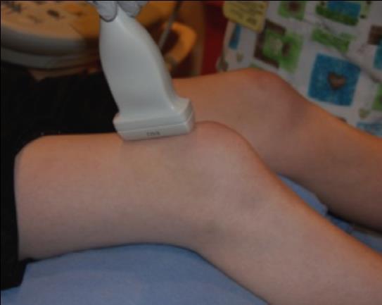
14 Imaging Techniques Achilles tendon Stand-off pad Good contact between the transducer and skin: Use of coupling gel or standoff pad


15 Water Bath Techniques - Poor contact with curved contours of the hands and feet - Compression of clinically relevant superficial structures - Patient discomfort caused by transducer contact Shah SH, Callahan MJ. Pediatr Radiol 2013; 43 Suppl 1:S23-40.


16 Balloon Technique Right third toe Long


17 Knob-ology Make use of machine presets Achilles Tendon Focal zones point of interest Keger s Fat Calcaneus

18 Contralateral Side for Comparison Helps differentiate normal from abnormal Particularly useful in pediatric population with changing appearance of the growing skeleton Right side Left side Be cautious of bilateral pathology, especially in subclinical disease wrist, PIP, subtalar and foot joints Magni-Manzoni et al. Arthritis Rheum. 2009; 61:

19 Enthesis Organ Components: Tendon Various portions of fibrocartilage Sub-tendinous bursae Fat Insertion of the fibers into the bone through the fibrocartilage Weiss PF, Chauvin NA, Roth J. Current Rheumatology Reports, In press.


20 Tendons Achilles Tendon, Long Well defined structure. Echogenic compared to the adjacent fat and muscle Longitudinal: Parallel lines, densely packed fibrillary pattern Transverse: Fiber cable wire appearance Achilles Tendon, Transverse


:1344-1354 Grechenig et al J Clin")
21 Enthesis Long, Achilles tendon calcaneus 5- year old calcaneus 11- year old Surrounded by a connective tissue called paratenon elastic sleeve for the tendon - Lined on its inner surface by synovial cells 16- year old calcaneus Other tendons have synovial sheaths - parietal and visceral sheets - lined by synovial cells Chauvin et al. Pediatr Radiol. Aug 2015;45(9): Grechenig et al J Clin Ultrasound 2004; 32(7):
")


22 Anisotropy Property of all tendons (direction dependent) Occurs when the US beam is not at 90 degrees Demonstrates false hypoechogenicity within the tendon Heel to Toe maneuver


23 Two Imaging Planes * * * * Transverse Quadriceps Tendon Longitudinal


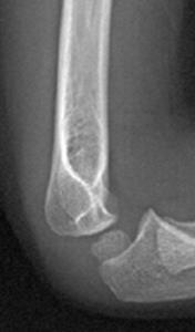
24 Common Flexor Tendon Medial Trochlear Elbow, 5 year-old girl

 in the region of the plantar")
25 Plantar Fascia * Calcaneal tuberosity 14 year old girl. There is a thin rim of cartilage (*) in the region of the plantar fascia insertion

26 Lateral Elbow Normal PDI CET Longitudinal PDI of the common extensor tendon in a healthy 5-year old
27

:1344-1354. Roth et al. Arthritis Care Res (Hoboken). Jan 2015;67(1):136-142. Collado et al. Clin Exp Rheumatol.")
28 Joint effusion OMERACT definitions Abnormal hypoechoic or anechoic (relative to subdermal fat), or in some cases isoechoic or hyperechoic, intra-articular material that is displaceable * and compressible but does not demonstrate Doppler signal TIBIA Physiologic fluid is common in children and it can be difficult distinguishing between normal amounts of joint fluid from a joint effusions TALUS Chauvin et al. Pediatr Radiol. Aug 2015;45(9): Roth et al. Arthritis Care Res (Hoboken). Jan 2015;67(1): Collado et al. Clin Exp Rheumatol. Nov-Dec 2007;25(6): Anterior ankle joint


29 Ankle Anterior ankle; at the dome of the talus Talus Anterior Ankle Trans


30 Elbow Hum Cap Crab position Olecranon


31 Elbow Effusion Anterior Fat Pad Sign


32 Synovial thickening OMERACT definitions Solid, non-compressible, abnormally hypoechoic tissue associated with joint lines or surrounding tendons Can be difficult to discern from adjacent hypoechoic epiphyseal cartilage T2wFS Images courtesy of Monica Epelman, MD


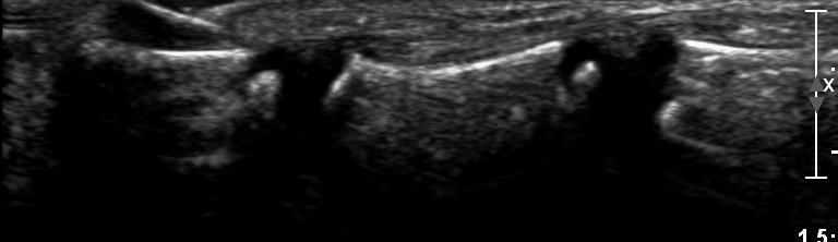
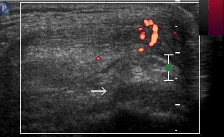
33 Synovitis OMERACT definitions Synovial thickening may not represent ongoing disease Doppler ultrasound to depict increased synovial blood flow Healthy children may exhibit some Doppler signal due to physiologically enhanced blood flow...need for better knowledge of normal LONG dorsal aspect of the wrist Magni-Manzoni et al. Nat Rev Rheumatol. Jun 2012;8(6):


34 6 year old girl with pauciarticular JIA and knee pain. Transverse images of suprapatellar region
35 Ultrasound - Synovitis US assessment of disease activity can be more informative than clinical examination Janow et al J Rheumatol. Dec 2011;38(12): Laurell et al. Clin Exp Rheumatol. Jan-Feb 2013;31(1): Subclinical synovitis is frequently observed by US, particularly within the hands and feet Magni-Manzoni et al. Arthritis Rheum. Nov ;61(11): Haslam et al. Rheumatology (Oxford). Jan 2010;49(1): Filippou et al. Clin Exp Rheumatol. Mar-Apr 2011;29(2): Pascoli et al. J Rheumatol. Nov 2010;37(11): A semi-quantitative system for grading synovial thickening is used in adult rheumatology but no such system has been validated in JIA Mandl et al. J Rheumatol 2011; 38(9):


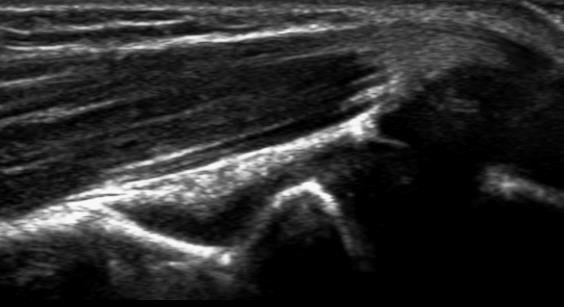
36 Bursitis 15-year old girl with JIA and shoulder pain

:1344-1354. Roth et al. Arthritis Care Res (Hoboken). Jan 2015;67(1):136-142.")
37 Tenosynovitis - OMERACT definitions Hypoechoic or anechoic thickened tissue with or without fluid in the tendon sheath which may or may not exhibit Doppler signal In children, tenosynovitis is most commonly seen around the ankle joint and along the extensor tendons of the wrist Chauvin et al. Pediatr Radiol. Aug 2015;45(9): Roth et al. Arthritis Care Res (Hoboken). Jan 2015;67(1): T2wFS 2 year-old with pauciarticular JIA


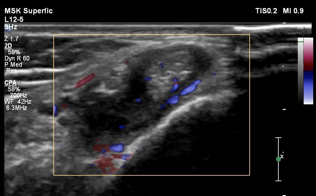
38 12-year old boy with JIA and medial left ankle swelling

39 Enthesitis OMERACT definitions Inflammation at the tendinous or ligamentous insertion - abnormally hypoechoic foci and/or thickened tendon or ligament at its bony attachment that is seen in two perpendicular planes May exhibit abnormal Doppler signal and/or bony changes such as enthesophytes, erosions or cortical irregularities, however these features are less commonly seen in children Within the tendon, perientheseal and cartilage Roth J et al. Arthritis Care Res (Hoboken). Jan 2015;67(1): Chauvin et al. Pediatr Radiol. Aug 2015;45(9): Weiss PF, Chauvin NA, Roth J. Current Rheumatology Reports, In press.


40 Enthesitis Lateral epicondyle Longitudinal image of the CET in a 13 year old girl Low flow settings with a low PFR and low WF Gain adjusted to allow maximum sensitivity without creating artifacts
41 Ultrasound - Enthesitis US can detect subclinical enthesitis Dopper-US revelaed enthesitis in 50% of clinically normal entheses Jousse-Joulin et al. Arthritis Care Res (Hoboken). Jun 2011;63(6): Standard dolorimeter examination for the detection of enthesitis in children with ERA and found that dolorimeter testing had poor accuracy and reliability, as compared with power Doppler US Weiss et al. Arthritis Rheumatol. Jan 2014;66(1): Prognostic significance of subclinical inflammation still needs to be determined

:329-336.")
42 Bone erosion OMERACT definitions Bone erosion is defined as discontinuity of the bone surface visible in two perpendicular planes Long, Volar aspect of the carpus (trapezium) Magni-Manzoni et al. Nat Rev Rheumatol. Jun 2012;8(6): Long, Volar aspect of the proximal phalanx 7 year-old girl, JIA


43 13 year-old, without arthritis 11 year-old, polyarticular JIA Assessment of erosive changes in children is challenging - physiologic irregularities in recently ossified bone that can be misinterpreted as cortical erosions, highlighting the need for further knowledge of normal bone anatomy throughout pediatric age groups Avenarius DF et al. Pediatr Radiol 2016; 46(3):
44 Ultrasound - Cartilage Age and gender normal US reference standards for cartilage thickness of the knee, ankle, wrist, MCP and PIP joints in children Spannow AH, Pfeiffer-Jensen M, Andersen NT, Herlin T, Stenbog E. Ultrasonographic measurements of joint cartilage thickness in healthy children: age- and sex-related standard reference values. J Rheumatol. Dec 2010;37(12): Further validated by demonstrating good agreement between MRI & US for measurement of cartilage thickness in healthy children Spannow AH, Stenboeg E, Pfeiffer-Jensen M, et al. Ultrasound and MRI measurements of joint cartilage in healthy children: a validation study. Ultraschall Med. Jan 2011;32 Suppl 1:S
45 Ultrasound - Cartilage It has been shown that patients with JIA have reduced cartilage thickness when compared to age- and gender-matched controls, although interestingly, this is observed in both clinically affected and non-affected joints Pradsgaard DO, Spannow AH, Heuck C, Herlin T. Decreased cartilage thickness in juvenile idiopathic arthritis assessed by ultrasonography. J Rheumatol. Sep 2013;40(9): Work in progress


46 Nail Disease - Psoriasis Long, Nail bed- Subject NP Long, DIP joint in affected nail disease DP DIPJ Long, Nail bed- Control Aydin et al. Dermatology 2012; 225: NP nail plate, DP distal phalanx DIPJ distal interphalangeal joint
47 Current Status No defined imaging recommendations for JIA OMERACT Outcome Measures in Rheumatology Health-e-Child Radiology Group Timing & ultilization of imaging in JIA is tailored to the individual patient Established adult protocols and standardized scoring systems must be modified before adopted for children Large, long-term innovative research must be established by collaboration with rheumatologists & radiologists Sheybani et al. Radiographics. Sep-Oct 2013;33(5): D'Agostino et al. J Rheumatol. Aug 2009;36(8):
48 Lingering Questions Which US findings are preferred for establishing a definite diagnosis? Can ultrasound predict/evaluate remission? How frequently do US scans have to be repeated? Which joints need to be screened? What is a sufficient protocol?
49 Ultrasound - Summary US is a powerful tool that may allow differentiation between synovial, tendinous and entheseal inflammation Prognostic signficance of subclinical information depicted by US still needs to be determined Subclinical disease may potentially alert the physician towards more aggressive treatment & close monitoring of the patient

50 THANK YOU! Nancy A. Chauvin, MD
ELENI ANDIPA General Hospital of Athens G. Gennimatas
 ELENI ANDIPA General Hospital of Athens G. Gennimatas Technological advances over the last years have caused a dramatic improvement in ultrasound quality and resolution An established imaging modality
Ultrasound in Rheumatology
 Arthritis Research UK Primary Care Centre Winner of a Queen s Anniversary Prize For Higher and Further Education 2009 Ultrasound in Rheumatology Alison Hall Consultant MSK Sonographer/Research Fellow Primary
8/29/2012. Outline Juvenile idiopathic arthritis. 1. Classification-ILAR. 1. Classification-clinical diagnosis. 1. JIA classification
 Outline Juvenile idiopathic arthritis 1. Classification and symptoms (ILAR-International league of Associations for Rheumatology) 2. Imaging J. Herman Kan, M.D. Section chief, musculoskeletal imaging Edward
Ultrasound in Rheumatology
 Ultrasound in Rheumatology Alison Hall Consultant MSK Sonographer Research Institute for Primary Care & Health Sciences, Keele University Department of Rheumatology, Cannock Hospital, Royal Wolverhampton
Scoring and Grading B-Mode Synovitis and Doppler findings in pediatric MSKUS. Johannes Roth MD PhD FRCPC RhMSUS
 Scoring and Grading B-Mode Synovitis and Doppler findings in pediatric MSKUS Johannes Roth MD PhD FRCPC RhMSUS Pathology - Definition Synovitis Synovitis on ultrasonography in children B-mode and Doppler
Sonographic assessment of adult and juvenile rheumatoid arthritis
 Sonographic assessment of adult and juvenile rheumatoid arthritis Poster No.: C-1485 Congress: ECR 2013 Type: Educational Exhibit Authors: C. A. S. Ruano, P. L. Pegado, J. M. G. Lourenco, P. Alves, L.
Reporting Ultrasound Findings and Diagnosis
 Reporting Ultrasound Findings and Diagnosis Rodina Nestorova MD Rheumatology Centre St. Irina, Sofia Bulgarian MSUS Society Basic MSU Course 14-16 Jan 2016 Plovdiv, Bulgaria ULTRASOUND REPORT COLLECTION
Sonographic appearance of chronic inflammatory rheumatism
 Sonographic appearance of chronic inflammatory rheumatism Poster No.: C-2237 Congress: ECR 2013 Type: Educational Exhibit Authors: H. Elfattach, F. Houari, O. Addou, M. Maaroufi, S. Tizniti ; 1 1 1 1 2
Psoriatic arthritis: early ultrasound findings
 Psoriatic arthritis: early ultrasound findings Poster No.: C-0399 Congress: ECR 2014 Type: Educational Exhibit Authors: R. Persechino 1, L. Cristiano 1, A. Bartoloni 1, C. Cantone 2, A. Keywords: DOI:
Current Status in Pediatric Musculoskeletal Ultrasonography. Johannes Roth, MD PhD FRCPC RhMSUS
 1 Current Status in Pediatric Musculoskeletal Ultrasonography Johannes Roth, MD PhD FRCPC RhMSUS 2 Objectives Provide a rationale for the use of imaging and ultrasonography in particular in pediatric rheumatology
Pragmatic ultrasound in the diagnosis of soft tissue rheumatic pain. Plamen Todorov
 Pragmatic ultrasound in the diagnosis of soft tissue rheumatic pain Plamen Todorov INTRODUCTION Soft tissue rheumatism: nonsystemic, focal pathological syndromes involving the periarticular structures.
Ultrasound Evaluation of Masses
 Ultrasound Evaluation of Masses Jon A. Jacobson, M.D. Professor of Radiology Director, Division of Musculoskeletal Radiology University of Michigan Disclosures: Consultant: Bioclinica Advisory Panel: GE,
Running head: Radiological Modalities and Childhood Arthritis 1
 Running head: Radiological Modalities and Childhood Arthritis 1 Contemporary Radiological Modalities in the Diagnosis of Childhood Arthritis November 15 th, 2011 Radiological Modalities and Childhood Arthritis
Paediatric rheumatology
 Paediatric rheumatology Ultrasonography vs. clinical examination in children with suspected arthritis. Does it make sense to use poliarticular ultrasonographic screening? G. Filippou, L. Cantarini, I.
Table of contents. Foreword. Preface. 1 Introduction Historical Perspective 00
 Table of contents Foreword Preface 1 Introduction 00 1.1 Historical Perspective 00 2 Fundamentals of musculoskeletal ultrasound 00 2.1 Frequency and wavelength 00 2.2 Generating ultrasound waves 00 2.3
Knee, Ankle, and Foot: Normal and Abnormal Features with MRI and Ultrasound Correlation. Disclosures. Outline. Joint Effusion. Suprapatellar recess
 Knee, Ankle, and Foot: Normal and Abnormal Features with MRI and Ultrasound Correlation Jon A. Jacobson, M.D. Professor of Radiology Director, Division of Musculoskeletal Radiology University of Michigan
Introduction to Musculoskeletal Ultrasound. Disclosures. Evidence Based Medicine Key References 8/30/2017
 Introduction to Musculoskeletal Ultrasound Johannes Roth MD, PhD, FRCPC, RhMSUS Professor of Pediatrics University of Ottawa Gurjit S Kaeley MBBS, MRCP, RhMSUS Professor of Medicine Division Chief Director
Juvenile Idiopathic Arthritis (JIA)
") Juvenile Idiopathic Arthritis (JIA) Kaveh Ardalan, MD, MS Division of Rheumatology Ann & Robert H. Lurie Children s Hospital of Chicago Assistant Professor, Pediatrics and Medical Social Sciences Northwestern
Juvenile Idiopathic Arthritis (JIA) Kaveh Ardalan, MD, MS Division of Rheumatology Ann & Robert H. Lurie Children s Hospital of Chicago Assistant Professor, Pediatrics and Medical Social Sciences Northwestern
Principles of Ultrasound. Cara C. Prideaux, M.D. University of Utah PM&R Sports Medicine Fellow March 14, 2012
 Principles of Ultrasound Cara C. Prideaux, M.D. University of Utah PM&R Sports Medicine Fellow March 14, 2012 None Disclosures Outline Introduction Benefits and Limitations of US Ultrasound (US) Physics
The Essentials Tissue Characterization and Knobology
 The Essentials Tissue Characterization and Knobology Randy E. Moore, DC, RDMS RMSK No relevant financial relationships Ultrasound The New Standard of Care Musculoskeletal sonography has become the standard
Review. Role of musculoskeletal ultrasound in juvenile idiopathic arthritis
 Role of musculoskeletal ultrasound in juvenile idiopathic arthritis Juvenile idiopathic arthritis is a serious autoimmune childhood disease that encompasses several types of chronic arthritis. Diagnosis
The Elbow 3/5/2015. The Elbow Scanning Sequence. * Anterior Joint (The anterior Pyramid ) * Lateral Epicondyle * Medial Epicondyle * Posterior Joint
 * Lateral Epicondyle * Medial Epicondyle * Posterior Joint") Scanning Sequence * Anterior Joint (The anterior Pyramid ) * Lateral Epicondyle * Medial Epicondyle * Posterior Joint Anterior Elbow Pyramid Courtesy of Jay Smith, MD. Vice chair PMR Mayo Clinic Rochester,
Scanning Sequence * Anterior Joint (The anterior Pyramid ) * Lateral Epicondyle * Medial Epicondyle * Posterior Joint Anterior Elbow Pyramid Courtesy of Jay Smith, MD. Vice chair PMR Mayo Clinic Rochester,
Ultrasound in juvenile idiopathic arthritis
 Magni-Manzoni Pediatric Rheumatology (2016) 14:33 DOI 10.1186/s12969-016-0096-2 REVIEW Open Access Ultrasound in juvenile idiopathic arthritis Silvia Magni-Manzoni Abstract Background: In the recent years,
I-A-1) Non-specific thickening of synovial membrane
 Non-specific thickening of synovial membrane") I-A-1) Non-specific thickening of synovial membrane Grayscale Metatarsal Power Doppler Dorsal aspect of metatarsophalangeal joint in right 1 st toe, longitudinal view Asterisks indicate non-specific thickening
I-A-1) Non-specific thickening of synovial membrane Grayscale Metatarsal Power Doppler Dorsal aspect of metatarsophalangeal joint in right 1 st toe, longitudinal view Asterisks indicate non-specific thickening
Why? Ultrasound of the Foot. Ultrasound of the Foot. General Rules. Plantar Fascia. Plantar Fasciitis 18/09/2018
 Ultrasound of the Foot Why? Ultrasound of the Foot Plantar fasciitis Plantar fascia fibromatosis Morton s neuroma Intermetatarsal bursitis Adventitial bursitis Plantar plate tears MTP joint synovitis Ganglia
Sacroiliac Joint Imaging
 Sacroiliac Joint Imaging Jacob Jaremko, MD, PhD Edmonton, Canada SPR, May 2017 Longview, Alberta Overview SI joint anatomy Sacroiliitis pathophysiology Sacroiliitis imaging Disease features Imaging protocols
Ultrasound of the Knee
 Ultrasound of the Knee Jon A. Jacobson, M.D. Professor of Radiology Director, Division of Musculoskeletal Radiology University of Michigan Disclosures: Consultant: Bioclinica Book Royalties: Elsevier Advisory
Introduction to Ultrasound Examination of the Hand and upper
 Introduction to Ultrasound Examination of the Hand and upper Emil Dionysian, M.D. Ultrasound of upper ext. Upside Convenient Opens another exam dimension Can be like a stethoscope Helps 3-D D visualization
Juvenile Spondyloarthritis / Enthesitis Related Arthritis (SpA-ERA)
") www.printo.it/pediatric-rheumatology/gb/intro Juvenile Spondyloarthritis / Enthesitis Related Arthritis (SpA-ERA) Version of 2016 1. WHAT IS JUVENILE SPONDYLOARTHRITIS/ENTHESITIS- RELATED ARTHRITIS (SpA-ERA)
www.printo.it/pediatric-rheumatology/gb/intro Juvenile Spondyloarthritis / Enthesitis Related Arthritis (SpA-ERA) Version of 2016 1. WHAT IS JUVENILE SPONDYLOARTHRITIS/ENTHESITIS- RELATED ARTHRITIS (SpA-ERA)
Imaging of Ankle and Foot pain
 Imaging of Ankle and Foot pain Pramot Tanutit, M.D. Department of Radiology Faculty of Medicine, Prince of Songkla University 1 Outlines Plain film: anatomy Common causes of ankle and foot pain Exclude:
Ultrasound of Mid and Hindfoot Pathology
 Ultrasound of Mid and Hindfoot Pathology Levon N. Nazarian, M.D. Professor of Radiology Thomas Jefferson University Hospital Disclosures None relevant to this presentation Educational Objective Following
EARLY INFLAMMATORY ARTHRITIS. Cristina Tacu Consultant Rheumatologist Brighton and Sussex University Hospital
 EARLY INFLAMMATORY ARTHRITIS Cristina Tacu Consultant Rheumatologist Brighton and Sussex University Hospital EIA: Introduction National priority Preventable cause of disability Very common condition High
Clinical Practice Guideline. Ultrasound in Rheumatological Settings. Version
 Clinical Practice Guideline Ultrasound in Rheumatological Settings Version 1.1.2017 November 2017 Table of Contents Abbreviations...3 Introduction...4 Diagnostic Musculoskeletal Ultrasound...6 Definition
Rotator Cuff and Biceps Pathology
 Rotator Cuff and Biceps Pathology Jon A. Jacobson, M.D. Professor of Radiology Director, Division of Musculoskeletal Radiology University of Michigan Disclosures: Consultant: Bioclinica Advisory Board:
Shane A. Shapiro, M.D. Assistant Professor, Orthopedic Surgery Mayo Clinic 2012 MFMER slide MFMER slide-3
 Ultrasound Foot and Ankle Pathology Disclosures None relevant Shane A. Shapiro, M.D. Assistant Professor, Orthopedic Surgery Mayo Clinic Florida @ShaneShapiroMD 2012 MFMER slide-2 Foot and Ankle Fundamentals
2019 RHEUMATOLOGIC ULTRASOUND (RhUS) CURRICULUM SUPPLEMENT TO THE AMERICAN COLLEGE OF RHEUMATOLOGY 2015 CORE CURRICULUM OUTLINE
 CURRICULUM SUPPLEMENT TO THE AMERICAN COLLEGE OF RHEUMATOLOGY 2015 CORE CURRICULUM OUTLINE") 2019 RHEUMATOLOGIC ULTRASOUND (RhUS) CURRICULUM SUPPLEMENT TO THE AMERICAN COLLEGE OF RHEUMATOLOGY 2015 CORE CURRICULUM OUTLINE TABLE OF CONTENTS I. MEDICAL KNOWLEDGE II. PATIENT CARE III. PRACTICE-BASED
2019 RHEUMATOLOGIC ULTRASOUND (RhUS) CURRICULUM SUPPLEMENT TO THE AMERICAN COLLEGE OF RHEUMATOLOGY 2015 CORE CURRICULUM OUTLINE TABLE OF CONTENTS I. MEDICAL KNOWLEDGE II. PATIENT CARE III. PRACTICE-BASED
The Egyptian Journal of Hospital Medicine (October 2017) Vol. 69 (4), Page
 Vol. 69 (4), Page") The Egyptian Journal of Hospital Medicine (October 2017) Vol. 69 (4), Page 2294-2300 Role of Magnetic Resonance Imaging and Ultrasonography in Diagnosis and Follow Up Rheumatoid Arthritis in Hand and Wrist
The Egyptian Journal of Hospital Medicine (October 2017) Vol. 69 (4), Page 2294-2300 Role of Magnetic Resonance Imaging and Ultrasonography in Diagnosis and Follow Up Rheumatoid Arthritis in Hand and Wrist
Ultrasound Guided Injections
 Ultrasound Guided Injection Technique More accurate injections Better Results! 1 Benefits: Increased Level of Certainty ie : really know how accurate PRP/Prolotherapy Avoid damage to articular cartilage
Ultrasound of the Shoulder
 Ultrasound of the Shoulder Patrick Battaglia, DC, DACBR Logan University, Department of Radiology Outline Review ultrasound appearance of NMSK tissues Present indications for ultrasound of the shoulder.
9/18/18. Welcome- MSK Ultrasound Workshop. Introduction to Musculoskeletal Ultrasound. Acknowledgement of Country. The Workshop.
 Acknowledgement of Country Welcome- MSK Ultrasound Workshop I would like to acknowledge that this meeting is being held on the traditional lands of the Wurundjeri and Boonwurrung people and pay my respect
Rheumatoid Arthritis 2. Inflammatory Diseases. Definition. Imaging Signs
 Rheumatoid Arthritis 2 Definition " Epidemiology Affects 2% of the population Peak incidence (diagnosis) in 4th and 5th decades Women affected 3 4 times more often than men Increased familial incidence
Lateral Elbow Pathology
 Lateral Elbow Pathology Jon A. Jacobson, M.D. Professor of adiology Director, Division of Musculoskeletal adiology University of Michigan Disclosures: Consultant: Bioclinica Advisory Board: GE, Philips
Urgent Cases and Foreign Bodies
 Urgent Cases and Foreign Bodies Catherine J. Brandon, MD, MS University of Michigan Ann Arbor, MI, USA Introduction: Patients added on to the schedule from the emergency department or as urgent add-on
A Patient s Guide to Psoriatic Arthritis
 A Patient s Guide to Psoriatic Arthritis Glendale Adventist Medical Center 1509 Wilson Terrace Glendale, CA 91206 Phone: (818) 409-8000 DISCLAIMER: The information in this booklet is compiled from a variety
Imaging the musculoskeletal system. An Introduction
 Imaging the musculoskeletal system An Introduction Objectives Discuss: commonly used imaging modalities in the musculoskeletal system normal imaging anatomy in the extremities fracture description Imaging
Disclosure. Pre-Procedural Considerations. Transducer Selection. Sterile Procedure. Sterile Procedure. Ultrasound Guided Foot and Ankle Injections
 Ultrasound Guided Foot and Ankle Injections Disclosure No relevant financial relationships exist Shane A. Shapiro, M.D. Assistant Professor, Orthopedic Surgery Mayo Clinic Florida @ShaneShapiroMD 2012
Early Rheumatoid Arthritis: AReview of MRI and Sonographic Findings
 outry et al. MRI and Sonography of Rheumatoid rthritis Musculoskeletal Imaging Pictorial Essay Nathalie outry 1 Mélanie Morel 1 René-Marc Flipo 2 Xavier Demondion 1,3 nne Cotten 1 outry N, Morel M, Flipo
Superficial Lumps and Bumps: Ultrasound Assessment
 Posterior knee Superficial Lumps and Bumps: Ultrasound Assessment Walter Mak, MD Department of Medical Imaging St. Michael s Hospital SM SM MGas MGas MGas MGas Synovial lined Synovial cyst: extrusion of
Ultrasound Evaluation of Posteromedial Ankle Pathology. Andrew C Cordle, M.D., Ph.D. 9/21/2018
 Ultrasound Evaluation of Posteromedial Ankle Pathology Andrew C Cordle, M.D., Ph.D. 9/21/2018 Overview: Pathology of the Posteromedial Ankle Flexor Tendon Pathology Accessory Navicular Bone Pathology Tarsal
Update - Imaging of the Spondyloarthropathies. Spondyloarthropathies. Spondyloarthropathies
 Update - Imaging of the Spondyloarthropathies Donald J. Flemming, M.D. Dept of Radiology Penn State Hershey Medical Center Spondyloarthropathies Family of inflammatory arthritides of synovium and entheses
Topics. Musculoskeletal Infection Extremities. Detection of Infection. Role of Imaging in Extremity Infection. Detection of Infection
 Topics Musculoskeletal Infection Extremities Nuttaya Pattamapaspong M.D. Department of Radiology, Faculty of Medicine, Chiang Mai University, Chiang Mai, Thailand Role of imaging in extremity infection
Case Studies. A. Kent Allen, DVM LAMENESS AND IMAGING IN THE SPORT HORSE
 Case Studies A. Kent Allen, DVM Author s address: Virginia Equine Imaging, 2716 Landmark School Road, The Plains, VA 20198; e-mail: vaequine@aol.com. 2007 AAEP. 1. Case Study #1: Medial Collateral Desmitis
Ultrasound of the Hip: Anatomy, Pathology, and Procedures
 Ultrasound of the Hip: Anatomy, Pathology, and Procedures Jon A. Jacobson, M.D. Professor of Radiology Director, Division of Musculoskeletal Radiology University of Michigan Outline Hip Joint Native hip
Ultrasound of Shoulder Pathology and Intervention 서울대학교병원재활의학과 김기원
 Ultrasound of Shoulder Pathology and Intervention 서울대학교병원재활의학과 김기원 Ultrasound for Shoulder Disorder Advantage Dynamic evaluation Immediate clinical correlation + Intervention Weakness Diagnostic accuracy?
Spondyloarthritis Physical Exam Measures. Axial SpA Measures. Ear Anatomy (Wikipedia) 5/4/2018. Tragus to Wall and Occiput to Wall Measurement
 5/4/2018. Tragus to Wall and Occiput to Wall Measurement") AxSpA Measures in Clinical Trials Spondyloarthritis Physical Exam Measures Philip Mease MD, MACR Director, Rheumatology Research, Swedish-Providence-St. Joseph Health Systems Clinical Professor, University
AxSpA Measures in Clinical Trials Spondyloarthritis Physical Exam Measures Philip Mease MD, MACR Director, Rheumatology Research, Swedish-Providence-St. Joseph Health Systems Clinical Professor, University
3/20/2017. Disclosures. Ultrasound Fundamentals. Ultrasound Fundamentals. Bone Anatomy. Tissue Characteristics
 Disclosures Images of ultrasound equipment in this presentation are not an endorsement Fundamentals of Musculoskeletal Ultrasound Physics and Knobology Shane A. Shapiro, M.D. Assistant Professor Orthopedic
Terminology Tissue Appearance
 By Marc Nielsen, MD Advantages/Disadvantages Generation of Image Ultrasound Machine/Transducer selection Modes of Ultrasound Terminology Tissue Appearance Scanning Technique Real-time Portable No ionizing
Podiatry Ultrasound Report Templates
 Podiatry Ultrasound Report Templates 1 st Edition Compiled exclusively for the clients of Fisher Biomedical Inc. Podiatric Ultrasound Report Templates Welcome to our first edition of sample podiatric ultrasound
Proceedings of the 55th Annual Convention of the American Association of Equine Practitioners
 Close this window to return to IVIS www.ivis.org Proceedings of the 55th Annual Convention of the American Association of Equine Practitioners December 5 9, 2009, Las Vegas, Nevada Program Chair : Nathaniel
Chapter 8 The Skeletal System: The Appendicular Skeleton. Copyright 2009 John Wiley & Sons, Inc.
 Chapter 8 The Skeletal System: The Appendicular Skeleton Appendicular Skeleton It includes bones of the upper and lower limbs Girdles attach the limbs to the axial skeleton The pectoral girdle consists
Common Applications for Sonography and Guided Intervention: Shoulder
 Common Applications for Sonography and Guided Intervention: Shoulder Jon A. Jacobson, M.D. Professor of Radiology Director, Division of Musculoskeletal Radiology University of Michigan Disclosures: Consultant:
AOS 3: Rheumatoid Arthritis
 AOS 3: Rheumatoid Arthritis Arthritis (General) = inflamed joint - NOT a single disease: covers >100 types - Involves disability + decreased quality of life o Can also occur in young people (not just the
Pediatric Musculoskeletal Ultrasound: Cases reviewed and lessons learned
 Pediatric Musculoskeletal Ultrasound: Cases reviewed and lessons learned Jessica Leschied, MD Sections of Pediatric and Musculoskeletal Radiology C.S. Mott Children s Hospital University of Michigan Ann
A Comparative Study of Ultrasonographic Findings with Clinical and Radiological Findings of Painful Osteoarthritis of the Knee Joint
 Med. J. Cairo Univ., Vol. 84, No. 3, December: 97-, www.medicaljournalofcairouniversity.net A Comparative Study of Ultrasonographic Findings with Clinical and Radiological Findings of Painful Osteoarthritis
Answers to Pre-Lab Quiz (p. 171) Answers to Activity Questions
 Answers to Activity Questions") Answers to Pre-Lab Quiz (p. 171) 1. Holds bones together; allows the rigid skeleton some flexibility so that gross body movements can occur 2. c, amount of movement allowed by the joint 3. synovial 4.
Answers to Pre-Lab Quiz (p. 171) 1. Holds bones together; allows the rigid skeleton some flexibility so that gross body movements can occur 2. c, amount of movement allowed by the joint 3. synovial 4.
Chapter 5 The Skeletal System
 Chapter 5 The Skeletal System The Skeletal System Parts of the skeletal system Bones (skeleton) Joints Cartilages Ligaments (bone to bone)(tendon=bone to muscle) Divided into two divisions Axial skeleton:
Ultrasound assessment of most frequent shoulder disorders
 Ultrasound assessment of most frequent shoulder disorders Poster No.: C-2026 Congress: ECR 2014 Type: Educational Exhibit Authors: S. P. Ivanoski; Ohrid/MK Keywords: Trauma, Athletic injuries, Arthritides,
Enthesophytes are analogous to osteophytes of
 A Case Report & Literature Review Open Fracture as a Rare Complication of Olecranon Enthesophyte in a Patient With Gout Rafid Kakel, M, and Joseph Tumilty, M Abstract Enthesophytes are analogous to osteophytes
ORTHOPAEDIC INJECTION AND ASPIRATION TECHNIQUES
 ORTHOPAEDIC INJECTION AND ASPIRATION TECHNIQUES OAAPN October 20, 2016 David H. Sohn, JD MD Chief, Shoulder and Sports Medicine University of Toledo Medical Center When to aspirate? To rule out infection
Crystal Deposition Disease and Psoriatic Arthritis
 74 Crystal Deposition Disease and Psoriatic Arthritis Philip J. O Connor, MRCP, FRCR, FFSEM (UK) 1,2 1 Department of Radiology, Leeds Teaching Hospitals, Chapel Allerton Hospital, Leeds, United Kingdom
Wrist and Ankle MRI of Patients With Juvenile Idiopathic Arthritis: Identification of Unsuspected Multicompartmental Tenosynovitis and Arthritis
 Pediatric Imaging Original Research Javadi et al. Wrist and nkle MRI of Patients With Juvenile Idiopathic rthritis Pediatric Imaging Original Research Sanaz Javadi 1 J. Herman Kan 1 Robert C. Orth 1 Marietta
Professor Lisa Stamp
 Professor Lisa Stamp Rheumatologist University of Otago, Christchurch 8:30-9:25 WS #65: Joint Injection Techniques 9:35-10:30 WS #75: Joint Injection Techniques (Repeated) Joint/soft tissue corticosteroid
Comparative study of high resolusion ultrasonography and magnetic resonance imaging in diagnosing traumatic knee injuries & pathologies
 Original article: Comparative study of high resolusion ultrasonography and magnetic resonance imaging in diagnosing traumatic knee injuries & pathologies Dr. Rakesh Gujjar*, Dr. R. P. Bansal, Dr. Sandeep
Student Objectives. When you have completed the exercises in this chapter, you will have accomplished the following objectives:
 Student Objectives When you have completed the exercises in this chapter, you will have accomplished the following objectives: Classification of Joints 1. Define joint or articulation. 2. Classify joints
MRI KNEE WHAT TO SEE. Dr. SHEKHAR SRIVASTAV. Sr.Consultant KNEE & SHOULDER ARTHROSCOPY
 MRI KNEE WHAT TO SEE Dr. SHEKHAR SRIVASTAV Sr.Consultant KNEE & SHOULDER ARTHROSCOPY MRI KNEE - WHAT TO SEE MRI is the most accurate and frequently used diagnostic tool for evaluation of internal derangement
Equine Diagnostic Radiography & Ultrasonography
 Equine Diagnostic adiography & Ultrasonography Dr. ussell Tucker DACV Prof Emeritus, WSU 90 ~30 DLPMO ~ projects medial forward lateral backward ~30 PL to DM projection 1 EQUINE DISTAL LIMB TENDONS & LIGAMENTS
Sonographic Evaluation of Subclinical Entheseal Involvement in Patients With Behçet Disease
 Musculoskeletal Imaging Original Research Ozkan et al. Ultrasound of Enthesopathy in ehçet Disease Musculoskeletal Imaging Original Research Fuat Ozkan 1 Gozde Yildirim Cetin 2 etul akan 3 Ali Murat Kalender
Peripheral Nerve Ultrasound
 Peripheral Nerve Ultrasound Jon A. Jacobson, M.D. Professor of Radiology Director, Division of Musculoskeletal Radiology University of Michigan Normal Peripheral Nerve Ultrasound appearance: Hypoechoic
The use of musculoskeletal ultrasound (MSKUS) as a. Musculoskeletal Ultrasound as a Diagnostic and Prognostic Tool in Rheumatoid Arthritis
 as a. Musculoskeletal Ultrasound as a Diagnostic and Prognostic Tool in Rheumatoid Arthritis") 215 Musculoskeletal Ultrasound as a Diagnostic and Prognostic Tool in Rheumatoid Arthritis Manish Jain, M.D., and Jonathan Samuels, M.D. Abstract The use of musculoskeletal ultrasound (MSKUS) has increased
215 Musculoskeletal Ultrasound as a Diagnostic and Prognostic Tool in Rheumatoid Arthritis Manish Jain, M.D., and Jonathan Samuels, M.D. Abstract The use of musculoskeletal ultrasound (MSKUS) has increased
KAPA 2017 Musculoskeletal Aspiration and Injection Workshop. W. Scott Black, MD Physician Assistant Studies Program University of Kentucky
 KAPA 2017 Musculoskeletal Aspiration and Injection Workshop W. Scott Black, MD Physician Assistant Studies Program University of Kentucky Aspiration Relatively quick and inexpensive Can be performed in
Proceedings of the 59th Annual Convention of the American Association of Equine Practitioners - AAEP -
 http://www.ivis.org Proceedings of the 59th Annual Convention of the American Association of Equine Practitioners - AAEP - December 7-11, 2013 Nashville, TN, USA Next Meeting : Dec. 6-10, 2014 - Salt Lake
Grey-scale ultrasound findings of lower extremity entheses in healthy children
 Lin et al. Pediatric Rheumatology (2015)13:14 DOI 10.1186/s12969-015-0012-1 RESEARCH ARTICLE Open Access Grey-scale ultrasound findings of lower extremity entheses in healthy children Clara Lin 1*, Mohammad
US finding of the shoulder (with live demonstration) 인제의대상계백병원 안재기
 인제의대상계백병원 안재기") US finding of the shoulder (with live demonstration) 인제의대상계백병원 안재기 Shoulder US Biceps tendon & Rotator Cuff Long Head of Biceps Tendon Subscapularis tendon Supraspinatus tendon Infraspinatus tendon Teres
US finding of the shoulder (with live demonstration) 인제의대상계백병원 안재기 Shoulder US Biceps tendon & Rotator Cuff Long Head of Biceps Tendon Subscapularis tendon Supraspinatus tendon Infraspinatus tendon Teres
Cover Page. The handle holds various files of this Leiden University dissertation.
 Cover Page The handle http://hdl.handle.net/1887/29578 holds various files of this Leiden University dissertation. Author: Krabben, Annemarie Title: Predictive factors for the development and disease course
Clarification of Terms
 Clarification of Terms The plantar aspect of the foot refers to the role or its bottom The dorsal aspect refers to the top or its superior portion The ankle and foot perform three main functions: 1. shock
Musculoskeletal Ultrasound Fundamentals
 Fundamentals Benjamin D. Levine, M.D. Associate Professor of Radiology Musculoskeletal Imaging Dept. of Radiological Sciences UCLA Health System I. Image Optimization II. Image Interpretation Artifacts
Case study #12 Left knee
 The patient is a 55 year old female who presents with bilateral knee pain. Patient is a collegiate softball coach and has a very active lifestyle and career that is hampered by her chronic knee pain. She
Chapter 2 Pitfalls in Musculoskeletal Ultrasound
 Chapter 2 Pitfalls in Musculoskeletal Ultrasound Violeta Maria Vlad MD, PhD Introduction Taking a good ultrasound (US) picture is an art. Interpreting it is a science. This is in fact everything US is
Evaluate The Ultrasound Enthesis Score in Patients Suffering From Psoriasis to Detect Subclinical Enthesopathy
 IOSR Journal of Dental and Medical Sciences (IOSR-JDMS) e-issn: 2279-0853, p-issn: 2279-0861.Volume 16, Issue 7 Ver. VI (July. 2017), PP 40-45 www.iosrjournals.org Evaluate The Ultrasound Enthesis Score
Musculoskeletal Ultrasound
 EFSUMB Course Book Student Edition Editors: Jan Tuma, Radu Badea, Christoph F. Dietrich Musculoskeletal Ultrasound Giorgio Tamborrini Ultrasound Center, Switzerland Corresponding author: KD Dr. med. Giorgio
Sonography of Knee and Calf Pain: the differential considerations
 Sonography of Knee and Calf Pain: the differential considerations Dr. Lisa L. S.Wong Consultant Radiologist St Paul s Hospital Outline Ultrasound techniques Common pathologies in calf and posterior knee
Natasha M. Werpy, DVM, Diplomate ACVR; Betsy Charles, DVM; Norm Rantanen, DVM, MS, Diplomate ACVR IMAGING
 Should I Throw Away My Ultrasound Machine Now That MRI Is Here? A Review of Ultrasound and MRI for the Diagnosis of Musculoskeletal Injury in the Equine Patient Natasha M. Werpy, DVM, Diplomate ACVR; Betsy
17/10/2017. Foot and Ankle
 17/10/2017 Alicia M. Yochum RN, DC, DACBR, RMSK Foot and Ankle Plantar Fasciitis Hallux Valgus Deformity Achilles Tendinosis Posterior Tibialis Tendon tendinopathy Stress Fracture Ligamentous tearing Turf
A. Incorrect! The appendicular skeleton includes bones of the shoulder, arm, hand, pelvis, leg and foot.
 Anatomy and Physiology - Problem Drill 08: The Skeletal System III No. 1 of 10 1. Which of the following statements about the appendicular skeleton is correct? A. The appendicular skeleton includes bones
The Shoulder. Systematically scanning the shoulder provides extremely useful diagnostic information. The Shoulder
 1 ! The most ACCESSIBLE to sonographic exam! The most MOBILE and VULNERABLE extremity AND Systematically scanning the shoulder provides extremely useful diagnostic information! The Goal for this section
JUVENILE IDIOPATHIC ARTHRITIS A case based study. By: Michael Zhanel. Home for the Summer Program July to August, 2018.
 JUVENILE IDIOPATHIC ARTHRITIS A case based study By: Michael Zhanel Home for the Summer Program July to August, 2018 Oakbank, Manitoba Supervisor: Dr. Nader Shenouda Abstract Juvenile idiopathic arthritis
Ultrasonography of Peripheral Nerve -upper extremity
 Ultrasonography of Peripheral Nerve -upper extremity Department of Physical Medicine and Rehabilitation Korea University Guro Hospital Korea University College of Medicine Yoon Joon Shik Normal median
Imaging of the knee in juvenile idiopathic arthritis
 Pediatr Radiol (2018) 48:818 827 https://doi.org/10.1007/s00247-017-4015-6 MINISYMPOSIUM: JUVENILE IDIOPATHIC ARTHRITIS Imaging of the knee in juvenile idiopathic arthritis Robert Hemke 1 & Nikolay Tzaribachev
Rheumatology Cases for the Internist
 Rheumatology Cases for the Internist Marc C. Hochberg, MD, MPH Professor of Medicine Head, Division of Rheumatology and Clinical Immunology Vice Chair, Department of Medicine University of Maryland School
Juvenile Chronic Arthritis
 Juvenile Chronic Arthritis Dr. Christa Visser MBChB MMed (Med Phys) Diploma Musculoskeletal Medicine (UK), Member Society of Orthopaedic Medicine (UK) Childhood Arthritis JCA/JIA/JRA Remember Acute rheumatic