SCPA602 Respiratory System
|
|
|
- Brook Rodgers
- 5 years ago
- Views:
Transcription


1 SCPA602 Respiratory System Associate Professor Dr. Wannee Jiraungkoorskul Department of Pathobiology, Faculty of Science, Mahidol University Tel: , 1
2 Objectives Describe the anatomy and histology of respiratory system Describe the interrelationships and functions of the different parts of the respiratory system. 2
3 Respiratory system The respiratory system includes the lungs and a branching system of tubes that link the sites of gas exchange with the external environment. Air is moved through the lungs by a ventilating mechanism, consisting of the thoracic cage, intercostal muscles, diaphragm, and elastic components of the lung tissue. The respiratory system is divided anatomically into structures of the upper and lower respiratory tracts. 3
4 4

5 5

6 6

7 7
8 8

9 Anatomically, the respiratory tract has upper and lower parts. Histologically and functionally, the respiratory system has a conducting portion, which consists of all the components that condition air and bring it into the lungs, and a respiratory portion, where gas exchange actually occurs, consisting of respiratory bronchioles, alveolar ducts, and alveoli in the lungs. Portions of two sets of paranasal sinuses are shown here. Respiratory system 9
10 Respiratory system Functionally, these structures make up the system's conducting portion, which consists of the nasal cavities, nasopharynx, larynx, trachea, bronchi (Gr. bronchos,windpipe), bronchioles, and terminal bronchioles; and a respiratory portion (where gas exchange takes place), consisting of respiratory bronchioles, alveolar ducts, and alveoli. Alveoli are saclike structures that make up the greater part of the lungs. They are the main sites for the principal function of the lungs the exchange of O 2 and CO 2 between inspired air and blood. The conducting portion serves two main functions: to provide a conduit through which air moves to and from the lungs and to condition the inspired air. To ensure an uninterrupted supply of air, a combination of cartilage, elastic and collagen fibers, and smooth muscle provides the conducting portion with rigid structural support and the necessary flexibility and extensibility. 10
11 Respiratory Epithelium Most of the conducting portion is lined with ciliated pseudostratified columnar epithelium known as respiratory epithelium. This epithelium has at least five cell types, all of which touch the thick basement membrane: 1. Ciliated columnar cells are the most abundant, each with about 300 cilia on its apical surface. 2. Goblet cells are also abundant in some areas of the respiratory epithelium, filled in their apical portions with granules of mucin glycoproteins. 11
12 Respiratory Epithelium 3. Brush cells are a much more sparsely scattered and less easily found, columnar cell type, which has a small apical surface bearing a tuft of many short, blunt microvilli. Brush cells express some signal transduction components like those of gustatory cells and have afferent nerve endings on their basal surfaces and are considered to be chemosensory receptors. Small granule cells are also difficult to distinguish in routine preparations, but possess numerous dense core granules nm in diameter. Like brush cells, they represent about 3% of the total cells and are part of the diffuse neuroendocrine system. Basal cells, small rounded cells on the basement membrane and not extending to the luminal surface, are stem cells that give rise to the other cell types. 12

13 Respiratory Epithelium (a): Details of its structure vary in different regions of the respiratory tract, but it usually rests on a very thick basement membrane (BM) and has several cell types, some columnar, some basal and all contacting the basement membrane. Ciliated columnar cells are the most abundant, with hundreds of long robust cilia (C) on each of their bulging apical ends which provide a lush cover of cilia on the luminal surface. Most of the small rounded cells at the basement membrane are stem cells and their differentiating progeny, which together make up about 30% of the epithelium. Intraepithelial lymphocytes and dendritic cells are also present in respiratory epithelium. Mucus-secreting goblet cells (G) are also present. The lamina propria is wellvascularized (V). X400. Mallory trichrome. 13
 among the numerous ciliated cells. X2500.")
14 Respiratory Epithelium (b): SEM shows the luminal surface of goblet cells (G) among the numerous ciliated cells. X

15 Respiratory Epithelium (c): As shown by SEM of another region, goblet cells (G) predominate in some areas, with subsurface accumulations of mucus evident in some (arrows). The film of mucus traps most airborne dust particles and microorganisms and the ciliary movements continuously propel the sheet of mucus toward the esophagus for elimination. Other columnar cells, representing only about 3% of the cells in respiratory epithelium, are brush cells (B) with small apical surfaces bearing a tuft of short, blunt microvilli. Brush cells have features of chemosensory receptors but their physiological significance is highly uncertain. X
16 Trachea The trachea is cm long and lined with a typical respiratory mucosa. In the lamina propria numerous seromucous glands produce watery mucus and in the submucosa C-shaped rings of hyaline cartilage keep the tracheal lumen open. The open ends of the cartilage rings are on the posterior surface, against the esophagus, and are bridged by a bundle of smooth muscle (trachealis muscle) and a sheet of fibroelastic tissue attached to the perichondrium. The entire organ is surrounded by adventitia. 16
17 The wall of the trachea is lined by typical respiratory epithelium (E) underlain by connective tissue (CT) and seromucous glands (G) in the lamina propria. The submucosa contains C-shaped rings of hyaline cartilage (C) covered by perichondrium (P). The watery mucous fluid produced by goblet cells and by the glands forms a layer that permits the ciliary movement to propel foreign particles continuously out of the respiratory system in the mucociliary escalator. Trachea 17

18 The openings in the cartilage rings are on the posterior surface, against the esophagus, and contain smooth muscle and elastic tissue. These allow distention of the tracheal lumen when large pieces of food pass through the esophagus. The trachealis muscle in the opening of the C also contracts during the cough reflex to narrow the tracheal lumen and produce stronger expulsion of air and dislodged mucus in the air passages. X50. H&E. Trachea 18
: Within each lung bronchi subdivide further to form the bronchial tree, the last component of the air conducting system.")
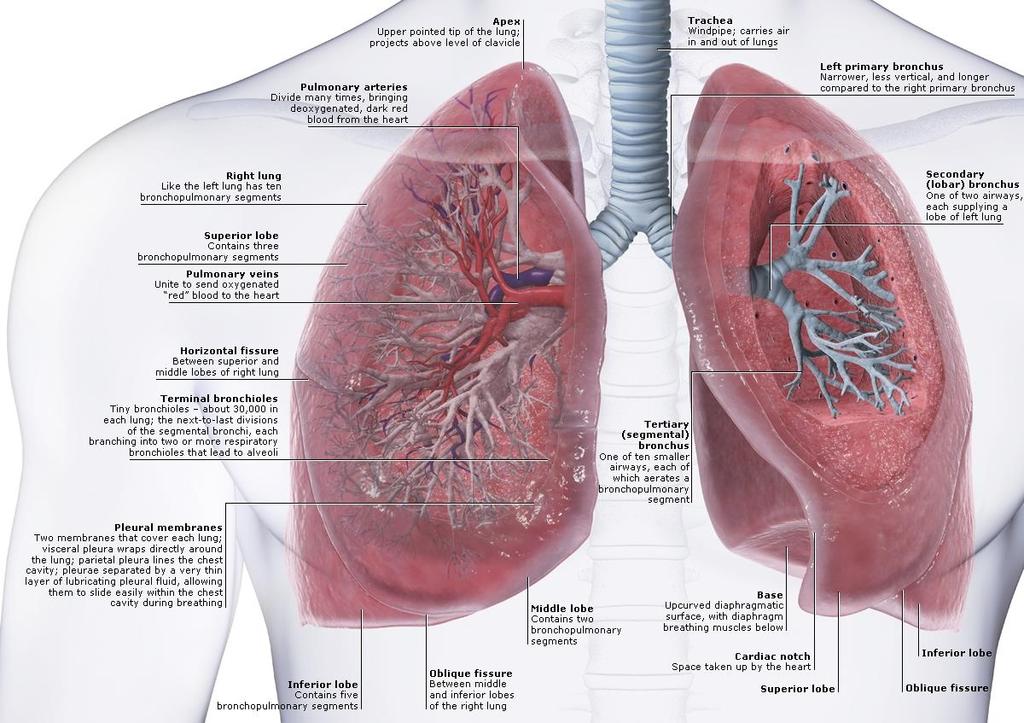
19 The trachea bifurcates as right and left primary bronchi that enter the hilum on the posterior side of each lung along with the pulmonary vessels, lymphatics, and nerves. (a): Within each lung bronchi subdivide further to form the bronchial tree, the last component of the air conducting system. (b): Diagram shows colorcoding of the major branches of the bronchial tree. Bronchial tree 19
 and contains many seromucous glands (G) in the submucosa which drain into the lumen.")
20 Tertiary (segmental) bronchus In a cross-section of a large bronchus the lining of respiratory epithelium (E) and the mucosa are folded due to contraction of its smooth muscle (SM). At this stage in the bronchial tree, the wall is also surrounded by many pieces of hyaline cartilage (C) and contains many seromucous glands (G) in the submucosa which drain into the lumen. In the connective tissue surrounding the bronchi can be seen arteries and veins (V), which are also branching as smaller and smaller vessels in the approach to the respiratory bronchioles. All bronchi are surrounded by distinctive lung tissue (LT) showing the many empty spaces of pulmonary alveoli. X56. H&E. 20
 contains the distinct layer of smooth muscle (SM) surrounding the entire bronchus.")
21 (a): A higher power view of the bronchus shows the epithelium (E) of mainly pseudostratified ciliated columnar cells with a few goblet cells. The lamina propria (LP) contains the distinct layer of smooth muscle (SM) surrounding the entire bronchus. The submucosa is the site of the supporting cartilage (C) and the adventitia includes blood vessels (V) and nerves (N). Lung tissue (LT) directly surrounds the adventitia of bronchi. X140. H&E Bronchial wall 21
 and small serous glands (G) near cartilage (C). X400. H&E.")
22 (b): This micrograph shows the epithelium of a smaller bronchus, in which the epithelium is primarily of columnar cells with cilia (arrows), with fewer goblet cells. The lamina propria has both smooth muscle (SM) and small serous glands (G) near cartilage (C). X400. H&E. Bronchial wall 22
: A large bronchiole has the characteristically folded respiratory epithelium (E) and prominent smooth muscle (arrows), but is supported only by fibrous")
23 Bronchial branches less than about 5 mm in diameter lack supporting cartilage and are called bronchioles. (a): A large bronchiole has the characteristically folded respiratory epithelium (E) and prominent smooth muscle (arrows), but is supported only by fibrous connective tissue (C) with no glands. X140. H&E. Bronchioles 23
 nearby and to a lesser extent in the accompanying venule (V).")
24 (b): Staining for elastic fibers reveals the high elastic content of the smooth muscle (arrowhead) associated with the muscle of a smaller bronchiole (B) in which the epithelium is simple columnar. Darkly stained elastic fibers are also present in the tunica media of a large arteriole (A) nearby and to a lesser extent in the accompanying venule (V). The connective tissue includes many lymphocytes (L) of MALT and lymphoid nodules are also common at this level. X180. Elastic stain. Bronchioles 24
 comprise a high proportion of the wall. X300. H&E.")
25 (c): In very small bronchioles the epithelium (E) is reduced to simple low columnar and the several layers of smooth muscle cells (arrows) comprise a high proportion of the wall. X300. H&E. Bronchioles 25
: Cross-section shows that a terminal bronchiole has only one or two layers of smooth muscle cells. The epithelium contains ciliated cuboidal cells and many low columnar nonciliated cells.")
26 Terminal bronchiole and Clara cells The last parts of the air conducting system before the sites of gas exchange appear are called the terminal bronchioles, which generally have diameters of one to two mm. (a): Cross-section shows that a terminal bronchiole has only one or two layers of smooth muscle cells. The epithelium contains ciliated cuboidal cells and many low columnar nonciliated cells. X300. PT 26

27 Terminal bronchiole and Clara cells (b): The nonciliated Clara cells with bulging domes of apical cytoplasm contain granules, as seen better in a plastic section. Named for Dr. Max Clara, the histologist who first described them in 1937, these cells have several important functions. They secrete components of surfactant which reduces surface tension and helps prevent collapse of the bronchioles. In addition, Clara cells produce enzymes that help break down mucus locally. The P450 enzyme system of their smooth ER detoxifies potentially harmful compounds in air. In other defensive functions, Clara cells also produce the secretory component for the transfer of IgA into the bronchiolar lumen; lysozyme and other enzymes active against bacteria and viruses; and several cytokines that regulate local inflammatory responses. Mitotically active cells are also present and include the stem cells for the bronchiolar epithelium. X500. PT. 27
28 Terminal bronchioles, respiratory bronchioles, and alveoli Terminal bronchioles branch into respiratory bronchioles, which then branch further into alveolar ducts and individual alveoli. Respiratory bronchioles are similar in most respects to terminal bronchioles except for the presence of scattered alveoli along their length. 28

29 Terminal bronchioles (a): Diagram shows the branching relationship, as well as the pulmonary blood vessels that travel with the bronchioles and the dense layer of branching capillaries that surrounds each alveolus for gas exchange between blood and air 29

30 Terminal bronchioles (b):the micrograph shows the branching nature of the bronchioles in two dimensions. X60. H&E. 30

31 Terminal bronchioles (c): SEM shows in three dimensions the relationship of alveoli to terminal and respiratory bronchioles. X
32 Respiratory bronchioles, alveolar ducts, and alveoli Lung tissue has a spongy structure because of the abundant air passages and pockets called alveoli. (a): Typical section of lung tissue including many bronchioles, some of which are respiratory bronchioles (RB) cut lengthwise, and showing the branching continuity with alveolar ducts (AD) and sacs (AS). Respiratory bronchioles still have a layer of smooth muscle and some regions of cuboidal epithelium, but alveolar ducts have only sparse strands of smooth muscle and an epithelium consisting of only a series of neighboring alveoli. The smooth muscle fibers are sphincter-like and appear as knobs between adjacent alveoli. Individual alveoli (A) all open to the sacs or ducts. The respiratory bronchiole runs along a thin-walled branch of the pulmonary artery (PA), which has a relatively thin wall, while branches of the pulmonary vein (V) course elsewhere in the parenchyma. X14. H&E. 32

33 Respiratory bronchioles, alveolar ducts, and alveoli 33
.")
34 Respiratory bronchioles, alveolar ducts, and alveoli (b): Higher magnification shows the relationship of the many rounded, thinwalled alveoli (A) to alveolar ducts (AD). Alveolar ducts end in two or more clusters of alveoli called alveolar sacs (AS). Those alveoli shown here that do not show openings to the ducts or the sacs have their connections in adjacent planes of other sections. X140. H&E. 34
35 Alveoli and the blood-air barrier Gas exchange between air and blood occurs at a membranous barrier between each alveolus and the capillaries surrounding it. The total area of this air-blood barrier in each lung has been calculated at approximately 70 m 2. (a): Diagram shows the relationship between capillaries and two or more saclike alveoli. (b): The air-blood barrier consists of an alveolar type I cell, a capillary endothelial cell, and their fused basement membranes. Oxygen diffuses from alveolar air into capillary blood and carbon dioxide moves in the opposite direction. The inner lining of alveoli is covered by a layer of surfactant, not depicted here, which lowers fluid surface tension and helps prevent collapse of alveoli. 35

36 Alveoli and the blood-air barrier 36
, which line almost the entire alveolus surface and across which gas exchange occurs.")
37 The wall between alveoli (A) contains several cell types. As seen here the capillaries (C) contain erythrocytes and leukocytes. The alveoli are lined mainly by squamous type I alveolar cells (I), which line almost the entire alveolus surface and across which gas exchange occurs. Type II alveolar cells line a bit of each alveolus and are large rounded cells, often bulging into the alveolus (II). These type II cells have many functions of Clara cells, including production of surfactant. Also present are alveolar macrophages (M), sometimes called dust cells, which may be in the alveoli or in the interalveolar septa. Alveolar walls 37
: Diagram illustrates the parietal pleura lining the inner surface of the thoracic cavity and the visceral pleura covering the outer surface of the lung.")
38 The pleura are serous membranes (serosa) associated with each lung and thoracic cavity. (a): Diagram illustrates the parietal pleura lining the inner surface of the thoracic cavity and the visceral pleura covering the outer surface of the lung. Between these layers is the narrow space of the pleural cavity. Pleura 38
. The connective tissue is rich in both collagen and elastic fibers and contains both blood vessels (V) and lymphatics (L). X140.")
39 (b): Both layers are similar histogically and consist of a simple squamous mesothelium (M) on a thin layer of connective tissue, as shown here for visceral pleura covering alveoli (A). The connective tissue is rich in both collagen and elastic fibers and contains both blood vessels (V) and lymphatics (L). X140. Pleura 39


40 References Kierszenbaum AL, Tres L. Histology and Cell Biology: An Introduction to Pathology. 4 th ed., Sounders. 2015, 752pp. Moore KL, Agur AMR and Dalley AF Essential Clinical Anatomy. 5 nd Edition. Wolters Kluwer. 686pp. 40
Organs Histology D. Sahar AL-Sharqi. Respiratory system
 Respiratory system The respiratory system provides for exchange of O2 and CO2 to and from the blood. Respiratory organs include the lungs and a branching system of bronchial tubes that link the sites of
HISTOLOGY OF THE RESPIRATORY SYSTEM I. Introduction A. The respiratory system provides for gas exchange between the environment and the blood. B.
 HISTOLOGY OF THE RESPIRATORY SYSTEM I. Introduction A. The respiratory system provides for gas exchange between the environment and the blood. B. The human respiratory system may be subdivided into two
The Respiratory System. Prof. Dr.Mohammed Hisham Al-Muhtaseb
 The Respiratory System Prof. Dr.Mohammed Hisham Al-Muhtaseb Objectives (lecture + practical) 1. Identify the conduction part of the respiratory tract and analyze the function of each segment 2. Identify
Lower Respiratory Tract (Trachea, Bronchi, Bronchioles) & the Lung
 & the Lung") Lower Respiratory Tract (Trachea, Bronchi, Bronchioles) & the Lung Color code: Important Extra & Doctor notes Editing file Objectives: By the end of this lecture, the student should be able to describe:
Lower Respiratory Tract (Trachea, Bronchi, Bronchioles) & the Lung Color code: Important Extra & Doctor notes Editing file Objectives: By the end of this lecture, the student should be able to describe:
Lec #2 histology. Bronchioles:
 Lec #2 histology. Last lecture we talked about the upper respiratory tract histology, this one is about the lower part histology. We will discuss the histology of: -bronchioles -respiratory bronchioles
The Respiratory System
 The Respiratory System Cells continually use O2 & release CO2 Respiratory system designed for gas exchange Cardiovascular system transports gases in blood Failure of either system rapid cell death from
THE RESPIRATORY SYSTEM
 THE RESPIRATORY SYSTEM Functions of the Respiratory System Provides extensive gas exchange surface area between air and circulating blood Moves air to and from exchange surfaces of lungs Protects respiratory
The Respiratory System. Dr. Ali Ebneshahidi
 The Respiratory System Dr. Ali Ebneshahidi Functions of The Respiratory System To allow gases from the environment to enter the bronchial tree through inspiration by expanding the thoracic volume. To allow
Includes : - the lung - a system of tube
 FYH - ERDS 1 Includes : - the lung - a system of tube Divided into 2 principal regions : - conducting portion : nasal cavity, nasopharynx, larynx, trachea, bronchi, bronchioles & terminal bronchioles -
Respiratory System. Functional Anatomy of the Respiratory System
 Respiratory System Overview of the Respiratory System s Job Major Duty Respiration Other important aspects ph control Vocalization Processing incoming air Protection Metabolism (ACE) What structures allow
Histology and development of the respiratory system
 Histology and development of the respiratory system Árpád Dobolyi Semmelweis University, Department of Anatomy, Histology and Embryology Outline of the lecture 1. Structure of the trachea 2. Histology
Organs of the Respiratory System Laboratory Exercise 52
 Organs of the Respiratory System Laboratory Exercise 52 Background The organs of the respiratory system include the nose, nasal cavity, sinuses, pharynx, larynx, trachea, bronchial tree, and lungs. They
CHAPTER 22 RESPIRATORY
 pulmonary ventilation move air external respiration exchange gases transportation of gases internal respiration exchange gases CHAPTER 22 RESPIRATORY in / out lungs air - blood blood - cells cell respiration
MH1001 Larynx, Monkey, H&E
 MH1001 Larynx, Monkey, H&E - False vocal fold : ciliated pseuodostratified columnar epithelium - True vocal fold : nonkeratinized stratified squamous epithelium vocalis muscle, vocal ligament - Thyroid
Bio 322 Human Anatomy Objectives for the laboratory exercise Respiratory System
 Bio 322 Human Anatomy Objectives for the laboratory exercise Respiratory System Required reading before beginning this lab: Saladin, KS: Human Anatomy 5 th ed (2017) Chapter 23 For this lab you will use
Chapter 16. Respiratory System
 Chapter 16 Respiratory System Introduction Respiration = the entire process of exchanging gases between the atmosphere and body cells 1. Ventilation 2. Gas exchange 3. Gas transport : 4. Cellular respiration
Ch16: Respiratory System
 Ch16: Respiratory System Function: - O2 in and CO2 out of the blood vessels in the lungs - O2 out and CO2 into the blood vessels around the cells - Gas exchange happens in - Other organs purify, humidify,
NURSE-UP RESPIRATORY SYSTEM
 NURSE-UP RESPIRATORY SYSTEM FUNCTIONS OF THE RESPIRATORY SYSTEM Pulmonary Ventilation - Breathing Gas exchanger External Respiration between lungs and bloodstream Internal Respiration between bloodstream
Tissues. tissue = many cells w/ same structure and function. cell shape aids its function tissue shape aids its function
 Tissues tissue = many cells w/ same structure and function cell shape aids its function tissue shape aids its function Histology = study of tissues 4 types of tissues Epithelial coverings contact openings
The Respiratory System
 13 PART A The Respiratory System PowerPoint Lecture Slide Presentation by Jerry L. Cook, Sam Houston University ESSENTIALS OF HUMAN ANATOMY & PHYSIOLOGY EIGHTH EDITION ELAINE N. MARIEB Organs of the Respiratory
7/12/2012. Respiratory system. Respiratory Response to Toxic Injury (Lung) Ninth Industrial Toxicology and Pathology Short Course.
 Ninth Industrial Toxicology and Pathology Short Course.") Ninth Industrial Toxicology and Pathology Short Course 23 27 July, 2012 Contemporary Concepts in Target Organ Toxicologic Pathology Respiratory system Respiratory Response to Toxic Injury (Lung) Eric Wheeldon
Ninth Industrial Toxicology and Pathology Short Course 23 27 July, 2012 Contemporary Concepts in Target Organ Toxicologic Pathology Respiratory system Respiratory Response to Toxic Injury (Lung) Eric Wheeldon
Lecture Overview. Respiratory System. Martini s Visual Anatomy and Physiology First Edition. Chapter 20 - Respiratory System Lecture 11
 Martini s Visual Anatomy and Physiology First Edition Martini Ober Chapter 20 - Respiratory System Lecture 11 1 Lecture Overview Overview of respiration Functions of breathing Organs of the respiratory
General Human Histology. The Respiratory System
 General Human Histology Lecture 5 Assist. Prof. Ahmed Anwar Albir The Respiratory System The respiratory system includes the lungs and a system of tubes that link the sites of gas exchange with the external
Respiratory System. Organization of the Respiratory System
 Respiratory System In addition to the provision of oxygen and elimination of carbon dioxide, the respiratory system serves other functions, as listed in (Table 15 1). Respiration has two quite different
Bronchioles. Alveoli. Type I alveolar cells are very thin simple squamous epithelial cells and form most of the lining of an alveolus.
 276 Bronchioles Bronchioles continue on to form bronchi. The primary identifying feature is the loss of hyaline cartilage. The epithelium has become simple ciliated columnar, and there is a complete ring
Chapter 23 The Respiratory System
 Chapter 23 The Respiratory System Cells continually use O 2 & release CO 2 Respiratory System designed for gas exchange Cardiovascular system transports gases in blood Failure of either system rapid cell
Lab Activity 27. Anatomy of the Respiratory System. Portland Community College BI 233
 Lab Activity 27 Anatomy of the Respiratory System Portland Community College BI 233 1 Terminology Pulmonary Ventilation: aka breathing, is the movement of air into and out of the lungs External Respiration:
Endeavour College of Natural Health endeavour.edu.au
 Endeavour College of Natural Health endeavour.edu.au BIOH122 Human Biological Science 2 Session 10 Respiratory System 1 Anatomy & Physiology Bioscience Department Endeavour College of Natural Health endeavour.edu.au
B. Correct! As air travels through the nasal cavities, it is warmed and humidified.
 Human Anatomy - Problem Drill 20: The Respiratory System Question No. 1 of 10 1. Which of the following statements about the portion of the respiratory system labeled in the image below is correct? Question
Tissue: The Living Fabric: Part A
 PowerPoint Lecture Slides prepared by Janice Meeking, Mount Royal College C H A P T E R 4 Tissue: The Living Fabric: Part A Tissues Groups of cells similar in structure and function Types of tissues Epithelial
Cell and Tissue Types. Epithelial, Connective, Muscle, Nerve
 Cell and Tissue Types Epithelial, Connective, Muscle, Nerve Objectives Explain the major stages of the cell cycle and cellular division (mitosis). Describe specific events occurring in each of the phases
The Respiratory System:
 The Respiratory System: Respiration Involves both the respiratory and the circulatory systems Four processes that supply the body with O 2 and dispose of CO 2 Respiration Pulmonary ventilation (breathing):
The Respiratory System
 PowerPoint Lecture Slide Presentation by Vince Austin Human Anatomy & Physiology FIFTH EDITION Elaine N. Marieb The Respiratory System Dr Nabil Khouri. MD, Ph.D Respiratory System Consists of a conducting
SCPA602 Cardiovascular System
 SCPA602 Cardiovascular System Associate Professor Dr. Wannee Jiraungkoorskul Department of Pathobiology, Faculty of Science, Mahidol University Tel: 02-201-5563, E-mail: wannee.jir@mahidol.ac.th 1 Objectives
LUNGS. Requirements of a Respiratory System
 Respiratory System Requirements of a Respiratory System Gas exchange is the physical method that organisms use to obtain oxygen from their surroundings and remove carbon dioxide. Oxygen is needed for aerobic
2402 : Anatomy/Physiology
 Dr. Chris Doumen Lecture 1 2402 : Anatomy/Physiology RESPIRATORY SYSTEM I nt r oduc t i on TextBook Readings Pages 830 through 845. Make use of the figures in your textbook ; a picture is worth a thousand
The respiratory system has multiple organs, we will begin with the nose and nasal cavity.
 Respiratory System (Peer reviewed and edited) Slide 1: Respiratory System Slide 2: Functions Functions of respiratory system include gas exchange, communication, olfaction, and ph regulation. Gas exchange
The Respiratory System
 C h a p t e r 24 The Respiratory System PowerPoint Lecture Slides prepared by Jason LaPres North Harris College Houston, Texas Copyright 2009 Pearson Education, Inc., publishing as Pearson Benjamin Cummings
Tissues. tissue = many cells w/ same structure and function. cell shape aids function tissue shape aids function. Histology = study of tissues
 Tissues tissue = many cells w/ same structure and function cell shape aids function tissue shape aids function Histology = study of tissues 4 types of tissues Epithelial coverings contact openings Connective
The Anatomy and Physiology of the Respiratory System
 CHAPTER 1 The Anatomy and Physiology of the Respiratory System Sagittal Section of Upper Airway Fig. 1-1. Sagittal section of upper airway. Structure of the Nose Fig. 1-2. Structure of the nose. Sagittal
This is not a required assignment but it is recommended.
 SU 12 Name: This is not a required assignment but it is recommended. BIO 116 - Anatomy & Physiology II Practice Assignment 2 - The Respiratory and Cardiovascular Systems 1. The exchange of oxygen and carbon
Histology = the study of tissues. Tissue = a complex of cells that have a common function
 { EPITHELIAL TISSUE Histology = the study of tissues Tissue = a complex of cells that have a common function The Four Primary Tissue Types: Epithelium (epithelial tissue) covers body surfaces, lines body
Respiratory System Functions. Respiratory System Organization. Respiratory System Organization
 Respiratory System Functions Functions of Respiratory System Gas exchange between blood and air Move air to and from exchange surfaces Protect exchange surfaces from environmental variations and pathogens
Anatomy of the Lungs. Dr. Gondo Gozali Department of anatomy
 Anatomy of the Lungs Dr. Gondo Gozali Department of anatomy 1 Pulmonary Function Ventilation and Respiration Ventilation is the movement of air in and out of the lungs Respiration is the process of gas
Upper Respiratory Histology
 Upper Respiratory Histology - Today we ll discuss the histology of larynx, trachea, primary, secondary, and tertiary bronchus. *First: The Larynx: -The picture below represents a section in the larynx,
2. List seven functions performed by the respiratory system?
 The Respiratory System C23 Study Guide Tortora and Derrickson 1. In physiology we recognize that the word respiration has three meanings. What are the three different meanings of the word respiration as
This is the first histology lecture for the respiratory tract system.
 This is the first histology lecture for the respiratory tract system. Respiratory tract starts with nose and ends with lungs, the respiratory tract is divided into upper part ( conducting ) like the nose,
Karachi King s College of Nursing
 Karachi King s College of Nursing Badil Dass Lecturer Respiratory system Respiratory System Respiratory system consist of: Nose Pharynx (Throat) Larynx (Voice Box) Trachea (Wind Pipe) Bronchi Bronchioles
I. Anatomy of the Respiratory System A. Upper Respiratory System Structures 1. Nose a. External Nares (Nostrils) 1) Vestibule Stratified Squamous
 1) Vestibule Stratified Squamous") I. Anatomy of the Respiratory System A. Upper Respiratory System Structures 1. Nose a. External Nares (Nostrils) 1) Vestibule Stratified Squamous Epithelium b. Nasal Cartilages 1) Nasal Cavity Pseudostratified
I. Anatomy of the Respiratory System A. Upper Respiratory System Structures 1. Nose a. External Nares (Nostrils) 1) Vestibule Stratified Squamous Epithelium b. Nasal Cartilages 1) Nasal Cavity Pseudostratified
Glandular Epithelium. Dr. Heba Kalbouneh Associate Professor of Anatomy and Histology
 Glandular Epithelium Dr. Heba Kalbouneh Associate Professor of Anatomy and Histology Glands Glandular epithelia are tissues formed by cells specialized to produce secretion. Secretion: if substances produced
Basic Histology. By Mrs. Bailey
 Basic Histology By Mrs. Bailey Primary Tissues 1. Epithelial Tissue 2. Connective Tissue 3. Muscle Tissue 4. Nervous Tissue Very cellular Supported by underlying connective tissue Epithelial & connective
The peripheral (secondary) lymphoid tissues
 lymphoid tissues") The peripheral (secondary) lymphoid tissues The peripheral (secondary) lymphoid tissues : are the lymph nodes, spleen, Mucosal associated lymphoid tissue (MALT). All secondary lymphoid organs have one
The peripheral (secondary) lymphoid tissues The peripheral (secondary) lymphoid tissues : are the lymph nodes, spleen, Mucosal associated lymphoid tissue (MALT). All secondary lymphoid organs have one
Epithelium. Four primary tissue types:
 Epithelium Four primary tissue types: Epithelial (covering) Connective (support) Nervous (control) Muscular (movement) Smooth muscle Cardiac muscle Skeletal muscle 1 Epithelial Tissue Features Epithelial
Tissues 10/21/2016. Epithelial Tissue
 Tissues This is a generalized cell diagram. It shows the anatomy of a cell, but most cells do not actually look like this. Cells can have a wide variety of shapes and sizes, depending on their function.
NAME PER DATE. membrane
 NAME PER DATE Chapter 9, Section 1 Review Matching: 1. alveolar capillary membrane 2. alveoli 3. bronchioles 4. cardiopulmonary system 5. conchae 6. epiglottis 7. larynx 8. mediastinum 9. nares 10. olfactory
General Structure of Digestive Tract
 Dr. Nabil Khouri General Structure of Digestive Tract Common Characteristics: Hollow tube composed of a lumen whose diameter varies. Surrounded by a wall made up of 4 principal layers: Mucosa Epithelial
Unit Nine - The Respiratory System
 Unit Nine - The Respiratory System I. Introduction A. Definition: the respiratory system consists of the nose, nasal cavity, (throat), (voice box), (windpipe), bronchi and lungs (which contain the alveoli).
Tissue: The Living Fabric
 PowerPoint Lecture Slide Presentation by Vince Austin Human Anatomy & Physiology FIFTH EDITION Elaine N. Marieb Chapter 4 Tissue: The Living Fabric Part A Tissues Groups of cells similar in structure and
The Respiratory System. Supplies body with oxygen Disposes of carbon dioxide Four processes in respiration
 C H A P T E R 22 The Respiratory System The Respiratory System Supplies body with oxygen Disposes of carbon dioxide Four processes in respiration Pulmonary ventilation External respiration Transport of
Prelab #4 BLOOD; BONE MARROW; RESPIRATORY; INTEGUEMENT Page 1
 Prelab #4 BLOOD; BONE MARROW; RESPIRATORY; INTEGUEMENT Page 1 Blood Slide 101 This a classic slide of blood cells using a Wright stain. Inspect red blood cells and their appearance. Note the approximate
The Respiratory System
 Essentials of Human Anatomy & Physiology Elaine N. Marieb Seventh Edition Chapter 13 The Respiratory System Slides 13.1 13.30 Lecture Slides in PowerPoint by Jerry L. Cook Copyright 2003 Pearson Education,
CHAPTER 7.1 STRUCTURES OF THE RESPIRATORY SYSTEM
 CHAPTER 7.1 STRUCTURES OF THE RESPIRATORY SYSTEM Pages 244-247 DO NOW What structures, do you think, are active participating in the breathing process? 2 WHAT ARE WE DOING IN TODAY S CLASS Finishing Digestion
Glandular Epithelium. Dr. Heba Kalbouneh Assistant Professor of Anatomy and Histology
 Glandular Epithelium Dr. Heba Kalbouneh Assistant Professor of Anatomy and Histology Glands Gla dular epithelia are tissues for ed y ells spe ialized to produ e se retio. Secretion: if substances produced
Human Anatomy and Physiology - Problem Drill 20: Immunity and the Lymphatic System
 Human Anatomy and Physiology - Problem Drill 20: Immunity and the Lymphatic System Question No. 1 of 10 The lymphatic system is formed early during human development. Which of the following statements
Epithelium tissue system
 Epithelium tissue system Histology : is the study of the microscopic anatomy (microanatomy) of cells and tissues of plants and animals. It is commonly performed by examining cells and tissues under a light
Activity 1: Respiratory System Lab
 Activity 1: Respiratory System Lab Launch Human Anatomy Atlas. Navigate to Quizzes/Lab Activities, find the Respiratory Lab section. Don t have AR? Select view 1. Respiratory System. 2. Fill in the blanks.
EPITHELIUM 3/12/2018 د. درويش بدران د. ماهر الحديدي د.امجد الشطرات و احسان العمري
 EPITHELIUM 1 2 3 1- SIMPLE SQUAMOUS EPITHELIUM It is a single layer of flat cells that resembles a tiled floor when viewed from apical surface; centrally located nucleus that is flattened and oval or spherical
Epithelial Tissue. Functions include: 1. Protection 4. Absorption 2. Secretion 5. Filtration 3. Sensory reception
 Tissues There are 4 primary tissue types in the human body: 1. Epithelial (covering/lining) 2. Connective (support) 3. Muscle (movement) 4. Nervous (control) Epithelium Epithelial Tissue Covers the surface
Tuesday, December 13, 16. Respiratory System
 Respiratory System Trivia Time... What is the fastest sneeze speed? What is the surface area of the lungs? (hint... think of how large the small intestine was) How many breaths does the average person
Epithelial Lecture Test Questions
 Epithelial Lecture Test Questions 1. Which of the following free surfaces lack(s) epithelia: a. lung alveoli (air sacs) b. hard palate c. joint cavities d. abdominal cavity e. salivary gland ducts 2. Which
Chapter 11 The Respiratory System
 Biology 12 Name: Respiratory System Per: Date: Chapter 11 The Respiratory System Complete using BC Biology 12, page 342-371 11.1 The Respiratory System pages 346-350 1. Distinguish between A. ventilation:
Basic Tissue Types and Functions
 Tissues Histology Basic Tissue Types and Functions 1) Epithelial tissue covering 2) Connective tissue support 3) Muscle tissue movement 4) Nervous tissue control Epithelial Tissue 1) Covers a body surface
Chapter 10 Lecture Outline
 Chapter 10 Lecture Outline See separate PowerPoint slides for all figures and tables preinserted into PowerPoint without notes. Copyright 2016 McGraw-Hill Education. Permission required for reproduction
Development of Respiratory System. Dr. Sanaa Alshaarawy& Dr. Saeed Vohra
 Development of Respiratory System Dr. Sanaa Alshaarawy& Dr. Saeed Vohra OBJECTIVES At the end of the lecture the students should be able to: Identify the development of the laryngeotracheal (respiratory)
Function: to supply blood with, and to rid the body of
 1 2 3 4 5 Bio 1102 Lec. 7 (guided): Chapter 10 The Respiratory System Respiratory System Function: to supply blood with, and to rid the body of Oxygen: needed by cells to break down food in cellular respiration
Chapter 13. The Respiratory System.
 Chapter 13 The Respiratory System https://www.youtube.com/watch?v=hc1ytxc_84a https://www.youtube.com/watch?v=9fxm85fy4sq http://ed.ted.com/lessons/what-do-the-lungs-do-emma-bryce Primary Function of Breathing
Notes to complete gas exchange in mammals
 Notes to complete gas exchange in mammals Mass flow of air to respiratory surface this is achieved through the mechanics of ventilation (breathing). This ensures a regular supply of air into and out of
Study of different tissues Abnormal cells and tissues can be compared to normal tissues to identify disease, such as cancer Being able to know and
 CHAPTER 4 Study of different tissues Abnormal cells and tissues can be compared to normal tissues to identify disease, such as cancer Being able to know and recognize normal tissues under the microscope
Unit I Problem 9 Histology: Basic Tissues of The Body
 Unit I Problem 9 Histology: Basic Tissues of The Body - What is the difference between cytology and histology? Cytology: it is the study of the structure and functions of cells and their contents. Histology:
The Human Respiration System
 The Human Respiration System Nasal Passage Overall function is to filter, warm and moisten air as it enters the body. The nasal passages are the primary site of air movement we tend to be nose breathers.
THE GOOFY ANATOMIST QUIZZES
 THE GOOFY ANATOMIST QUIZZES 7. LUNGS Q1. Fill in the blanks: the lung has lobes and fissures. A. Right, three, two. B. Right, two, one. C. Left, three, two. D. Left, two, three. Q2. The base of the lung
A. cells that perform related functions and are similar in structure. B. extracellular material - made by cells and secreted into interstitial space
 I. tissue components A. cells that perform related functions and are similar in structure B. extracellular material - made by cells and secreted into interstitial space II. tissue types A. epithelium (e.)
Tongue In the buccal cavity of the digestive system
 Tongue In the buccal cavity of the digestive system same layers as those of tubular organs Mucosa, submucosa, and muscularis muscularis = the muscularis externa no muscularis mucosa 1 Tongue ling = tongue
Syllabus: 6 pages (Page 6 lists corresponding figures for Grant's Atlas 11 th & 12 th Eds.)
") PLEURAL CAVITY AND LUNGS Dr. Milton M. Sholley SELF STUDY RESOURCES Essential Clinical Anatomy 3 rd ed. (ECA): pp. 70 81 Syllabus: 6 pages (Page 6 lists corresponding figures for Grant's Atlas 11 th &
PLEURAL CAVITY AND LUNGS Dr. Milton M. Sholley SELF STUDY RESOURCES Essential Clinical Anatomy 3 rd ed. (ECA): pp. 70 81 Syllabus: 6 pages (Page 6 lists corresponding figures for Grant's Atlas 11 th &
Chapter 10 Respiration
 1 Chapter 10 Respiration Introduction/Importance of the Respiratory System All eukaryotic organisms need oxygen to perform cellular respiration (production of ATP), either aerobically or anaerobically.
Lecture Overview. Marieb s Human Anatomy and Physiology. Chapter 4 Tissues: The Living Fabric Epithelial Tissues Lecture 9. Introduction to Tissues
 Marieb s Human Anatomy and Physiology Marieb Hoehn Chapter 4 Tissues: The Living Fabric Epithelial Tissues Lecture 9 Lecture Overview Introduction to Tissues Epithelial Tissues Location General characteristics
Unit 14: The Respiratory System
 Unit 14: The Respiratory System See what you already know! 1. Fill in the diagram on your own 2. Collaborate with your partner The Respiratory System The major function of the respiratory system is gas
The respiratory system structure and function
 Name: Class: Date: Active reading 11A + Biology Gr11A The respiratory system structure and function The function of the respiratory system is to bring oxygen into the body and eliminate carbon dioxide
Lecture Overview. Chapter 4 Epithelial Tissues Lecture 9. Introduction to Tissues. Epithelial Tissues. Glandular Epithelium
 Visual Anatomy & Physiology First Edition Martini & Ober Chapter 4 Lecture 9 Lecture Overview Introduction to Tissues Location General characteristics Functions Classification Glandular Epithelium 2 Where
Small intestine. Small intestine
 General features Tubular organ longest part; 5-6 m most of chemical digestion absorption of nutrients reabsorption of H2O occurs. Two structural features; maximize the lumenal surface area villi microvilli
Body Tissues Pearson Education, Inc.
 Body Tissues Tissues Groups of cells with similar structure and function Four primary types: Epithelial tissue (epithelium).1 Connective tissue.2 Muscle tissue.3 Nervous tissue.4 Epithelial Tissues Locations:
Respiratory System. BSC 2086 A&P 2 Professor Tcherina Duncombe Palm Beach State College
 Respiratory System BSC 2086 A&P 2 Professor Tcherina Duncombe Palm Beach State College Respiration Ventilation of lungs Gas exchange between air/bld and bld/tissue Use of oxygen in cellular respiration
Respiratory System. All I need is the air that I breathe
 Respiratory System All I need is the air that I breathe Men go abroad to wonder at the heights of mountains, at the huge waves of the sea, at the long courses of the rivers, at the vast compass of the
Unit 9. Respiratory System 16-1
 Unit 9 Respiratory System 16-1 Works together with the circulatory system Exchange of gases between atmosphere, blood, and cells If respiratory system and/or circulatory system fails, death will occur
For more information about how to cite these materials visit
 Author(s): Michael Hortsch, Ph.D., 2009 License: Unless otherwise noted, this material is made available under the terms of the Creative Commons Attribution Non-commercial Share Alike 3.0 License: http://creativecommons.org/licenses/by-nc-sa/3.0/
Slide 154: Pancreas, H&E
 Slide 154: Pancreas, H&E the pancreas, located adjacent to the duodenum, is a mixed exocrine and endocrine gland; it is usually readily identifiable by the presence of the interspersed endocrine pancreatic
Tissues Chapter 5...Tissue - a group or mass of similar cells working together to perform certain common functions
 Tissues Chapter 5...Tissue - a group or mass of similar cells working together to perform certain common functions There are 4 major types of tissue Epithelial Connective Muscle Nervous 1. Epithelial Tissue
CHAPTER 24. Respiratory System
 CHAPTER 24 Respiratory System RESPIRATION INCLUDES Air moves in and out of lungs Continuous replacement of gases in alveoli (air sacs) Gas exchange between blood and air at alveoli Transport of respiratory
MASTERY TEST. 3. Carbon dioxide combines with water to form. An excess of COz will cause the blood ph to (increaseldecrease).
.") MASTERY TEST Now take the mastery test. Do not guess. Some questions may have more than one correct answer. As soon as you complete the test, correct it. Note your successes and failures so that you can
MASTERY TEST Now take the mastery test. Do not guess. Some questions may have more than one correct answer. As soon as you complete the test, correct it. Note your successes and failures so that you can
Respiratory System. Introduction. Atmosphere. Some Properties of Gases. Human Respiratory System. Introduction
 Introduction Respiratory System Energy that we consume in our food is temporarily stored in the bonds of ATP (adenosine triphosphate) before being used by the cell. Cells use ATP for movement and to drive