Stefano A. Pileri. Chair of Pathology Haematopathology Unit Department L. and A. Seràgnoli. Bologna University Anatomic Theatre
|
|
|
- Jasper Green
- 5 years ago
- Views:
Transcription

1 Histiocytic and dendritic cell tumours Stefano A. Pileri Chair of Pathology Haematopathology Unit Department L. and A. Seràgnoli gnoli Bologna University Anatomic Theatre

")
2 Ilya Ilyich Mechnikov CD163+ Macrophage phagocytosing Treponema pallidum Phagocytosis in innate immunity against infection Nobel Prize in Physiology or Medicine 1908 Some pathological processes and neoplasms derived from true histiocytes of relevance in diagnostic haematopathology Haemophagocytic Syndromes Rosai-Dorfman Disease Xanthogranuloma family Cutaneous and/or systemic (including Erdheim-Chester disease) Histiocytic Sarcoma


3 In addition to mononuclear phagocytes, granulocytes, and lymphocytes, we noticed a large stellate cell with distinct properties from the former cell types. The term dendritic cell would thus be appropriate for this particular cells type. J Exp Med 1973;137: Paul Langerhans Nobel Prize in Physiology or Medicine
. Shortman K and Liu Y-J. Nat Rev Immunol 2002; 2:151")
4 Dendritic Cells Dendritic cells (DC) are a sparsely distributed, migratory group of elements specialized for the uptake, transport, processing and presentation of antigens to T and B cells (adaptative immunity). Shortman K and Liu Y-J. Nat Rev Immunol 2002; 2:151
5 Dendritic Cells IN VITRO (EX VIVO) Myeloid DC (MDC) Monocyte-derived DC (Mo- DC) Plasmacytoid DC (PDC) IN VIVO Langerhans cells Interdigitating DC Interstitial DC One cell with many names
6


7 Langerhans Cells Epithelia (skin, lung, mucosa) Interstitial DC Interstitium (dermis, heart, kidney, ) Zaba LC, et al. J Clin Invest 2007; 117; Ochoa MT, et al. J Invest Dermatol 2008; 128; Langerhans DC Langerin CD1a Langerin/CD207 S100 E-cadherin CD163 F-XIIIa CD11c CD68 MMR/CD206 DCSIGN/CD209 FXIII-a Macrophage CD1a Interstitial DC (CD1a) CD1c CD11c CD68 MMR/CD206 DCSIGN/CD209
8 Secondary Lymphoid Organs Lymph maturation and migration Interstitial DC Langerhans DC Cytokines,CD40L DNA, RNA, LPS Peripheral Tissues


9 Interdigitating Dendritic Cells CD1a S-100 CD1a S-100 RECENTLY IMMIGRATED LONG STANDING Langerin Langerin


10 Langerhans DC: DCLAMP - IDC: DCLAMP +

11 DC-SIGN+ in Lymph node parenchyma Interstitial-like DC Steady-state migration B T
12

13 FDC CB FDC FDC

 KiM4p, NGFR Fascin")
14 Other DC-like cells Mesenchymal Stem cells Follicular Dendritic Cells FDC CD21+35 Primary and secondary B follicles Long-living, antigen-carrying cells, involved in GC physiology DRC-1, CD21, CD23, CD35 Clusterin, Podoplanin CXCL13 (S100) KiM4p, NGFR Fascin CD23

15 Other DC-like cells Mesenchymal Stem cells Fibroblastic Reticulum Cells FRC Traffic area of the lymph node Delimitate conduits for immune cell migration and contacts Actins, Desmin, Fascin CD68 VCAM-1 SM ACTIN CK8/18 (subset)

16


17 Composite nodule HEV Lennert & Remmele, 1958 Interfollicular lymphoblasts Lennert, 1978 T-associated plasma cells

18 Nature Med 1999; 5: Anti-IFNa


 Pos PARAFFIN")


19 PDC/IPC in peripheral blood and tissues 0.01~0.05% CD3, CD19, CD20, CD11c, CD13, CD14, CD33, CD16, CD56, CD4, CD36, CD68, CD123, BDCA2, ILT3, TCL1, Neg (Lin - ) Pos PARAFFIN CD68 CD123 (IL3Ra) CLA/Heca452 BCL11a TCL1 CD2AP Granzyme B Cella M, et al. Nat Med 1999; 5:919 CD2AP CD123 BDCA-2 CD123 FROZEN CD303/BDCA2 CD123 (IL3Ra) Chem-R23
20 GM-CSF IL-4 CD34+ GM-CSF TNF-a GM-CSF IL-4 TGF-b CD14- CD11c+ CD1a+CLA+ FLT3L TPO PDC CD14+ CD11c+ CD1a-CLAmonocyte CD14- CD11c- CD1a- BDCA2+ CD123+ Progenitors Blood Precursors Interstitial DC Langerhans DC Plasmacytoid DC IL3 CD40L Bacteria Viruses Immature DC Mature DC

21

22

23 W Burgdorf JKC Chan G Delsol F Facchetti CDM Fletcher TM Grogan KL Grogg ES Jaffe RJ Jaffe DM Jones SA Pileri LM Weiss
24
25 Histiocytic sarcoma Median age: 42.4 years (6 months - 75 years) M/F = 2.6/1 Presentation: nodal (27%) extranodal (55%) (skin, intestine, bone) systemic (18%) [malignant histiocytosis] Stage III-IV (78%) Hepato/splenomegaly (22%) Pancytopenia (18%) Possible association with other tumors (18%)
. Finely-moderately dispersed chromatin.")

26 Large cells (>20μm) with round-oval eccentric nuclei (indented or convoluted). Finely-moderately dispersed chromatin. One or more small but distinct nucleoli. Abundant cytoplasm mitotic figures/10 HPF. More often, moderate degree of pleomorphism.


27 Degree of pleomorphism more pronounced in some cases, with frequent spindle and/or multinucleated cells. Growth pattern: diffuse and cohesive.


28 Erythrophagocytosis: absent or inconspicuous. Lymphophagocytosis: exceptional. Foamy/xanthomatous appearance: occasional.
 100% CD163 100%")

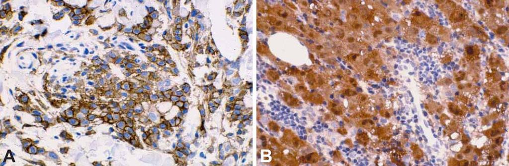
29 Phenotype: CD68 + (KP1 = PG-M1) 100% CD % Lysozyme + 90% CD % S % CD4 + 50% CD68/KP1 CD68/PG-M1


30
 Negativity for CD1a, CD21, CD35, CNA.")
, as well as for EBER at")
31 Phenotype: Ki % (mean: 18.8%) Negativity for CD1a, CD21, CD35, CNA.42, CD34, MPO, B- and T-cell markers, and CD30 (with minor exceptions), as well as for EBER at ISH. Lysozyme MIB-1/Ki-67

32 Electron microscopy Lysosomes + RER + mitochondria Birbeck granules - Junctions - Desmosomes - Projections -

33 Molecular biology Lineage promiscuity? Dual genotype? Cross-contamination? Small clonal-like lymphoid populations?
34 Behaviour Aggressive tumour. 50% of the patients die of progressive disease. 30% of the patients achieve CR, but relapse. Only 20% patients alive and in CR.

35 Blood. 2008; 112: ALK CD163 1/3: TPM3/ALK
 with the exception of Erdheim-Chester disease.")
. S-100 in 20% of cases.")
36 Disseminated Juvenile Xanthogranuloma Proliferation of histiocytes similar to those of the dermal JXG with a foamy (xanthomatous) component and Touton-type giant cells. Occurring by age 10 years (50% within the 1 st year) with the exception of Erdheim-Chester disease. Association with NF1 (and JMML and LCH). Skin, mucosa, CNS, eye, liver, lymph node, lung and BM. Positivities for CD14, CD68, CD163, Stabilin-1, Factor XIIIa and fascin (dermal /interstitial DC). S-100 in 20% of cases. CD1a and Langerin negative. Benign though CNS lesions can cause local consequences and even death.
37 Langerhans cell tumours Langerhans cell histiocytosis Langerhans cell sarcoma


: a clonal")
38 Langerhans cell Histiocytosis Clonal proliferation of Langerhans cells Single or multiorgan disorder Indolent aggressive disease (stage-dependent) Willman CL, et al. Langerhans'cell histiocytosis (histiocytosis X): a clonal proliferative disease. N Engl J Med 1994;331:154-60


39 LCs predominant and admixed with varying amounts of eosinophils, histiocytes, neutrophils and lymphocytes. Large size. Benign-looking grooved nuclei with inconspicuous nucleoli. Wide rim of acidophilic cytoplasm mitotic figures/10 HPF.


40 At times, a certain degree of atypia is observed. These cases show more prominent nucleoli, a higher pleomorphism and a lower content of eosinophils mitotic figures/10 HPF. Such finding is, however, irrelevant on prognostic grounds.

")


41 Phenotype: CD1a + S Langerin + CD68 +/- Lys +/- CD45 -/+ CD1a Ki-67: 2-25% (10%) S-100 Langerin Negativity for CD21, CD35*, CD30, B and T-cell markers, MPO, CD34, and EBER by ISH.

42 Electron microscopy Lysosomes -/+ Birbeck granules + Junctions - Desmosomes - Projections -


43 CD1a

44 Association of LCH with other neoplasms The two diseases are totally independent LCH is clonal The LCH is a focal reaction to the malignant neoplasm
45 Willman CL, et al. Langerhans'cell histiocytosis (histiocytosis X): a clonal proliferative disease. N Engl J Med 1994;331: Clonal (9/10 cases) Yousem SA, et al. Pulmonary Langerhans' cell histiocytosis: molecular analysis of clonality. Am J Surg Pathol 2001;25:630-6 Twenty-four nodules in 13 female patients 7 (29%) clonal, 17 (71%) nonclonal Six cases with multiple discrete nodules, three (50%) showed a nonclonal LCH cell population In contrast to systemic LCH, pulmonary LCH appears to be primarily a reactive process in which non-lethal, nonmalignant clonal evolution of LCH cells may arise in the setting of non-clonal LCH cell hyperplasia.
46 Langerhans cell tumours Langerhans cell histiocytosis Langerhans cell sarcoma

47 Frankly malignant cytology. Nuclei with prominent nucleoli and rare grooves. Occasional eosinophils. > 50 mitotic figures/10 HPF.


48 Marked distension of the sinuses: frequent.


 and occasional EBER")
49 The same phenotypic profile as LCH, with a few exceptions: the higher MIB-1/Ki-67 marking (10-60%; mean = 22.5%) and occasional EBER and/or focal CD35 positivity. S-100 CD68 Ki-67
50 Behaviour Aggressive tumour. 50% of the patients died of progressive disease. 40% of the patients achieved CR, but some relapsed. Only a few patients alive and in complete remission. Occasional association with mediastinal germ cell tumour, shizophrenia or fibromyalgia.
51 Follicular dendritic cell and interdigitating dendritic cell sarcomas
 with a fusiform or ovoid shape.")

52 Common morphologic features. Clear-cut distinction possible by IHC and EM. Large cells (>20μm) with a fusiform or ovoid shape. Oval central nucleus with delicate nuclear membrane, finely dispersed chromatin and a small, but distinct nucleolus. Abundant cytoplasm acidophilic on HE and grey on Giemsa. Indistinct cell borders.
.")

53 Small amount of lymphocytes intermingled. Rare mitotic figures (1-5/ 10HPF). Whorled, fascicular or storiform pattern, at time reminiscent of the 360 pattern of meningioma. In rare cases, paracortical location (IDC sarcoma). In a few cases, residual HV Castleman disease.

54 In some cases, numerous bizarre, often atypical multinucleated cells, with higher mitotic activity and coagulative necrosis.
55 from follicular dendritic cells sarcomas from interdigitating dendritic cells


 EGFR / Chr.")

56 CD23 Podoplanin FDC sarcoma immunophenotype EGFR EGFR / Chr. 7 CD21, 23, 35, CNA.42 KiM4p, NGFR, EGFR Clusterin, Podoplanin, Claudin 4 + (use more than 1!) EGFR / Chr. 7 CD68 +/- CD1a - S100 -/+ CD117 - CD45RB, CK, CD20 exceptionally +


57 CD35 CD21


58


59 Electron microscopy Lysosomes e Birbeck granules - Junctions + Desmosomes + Projections +
60 Age: median 58.6 years (40-90 years). M/F = 1/1. Localised presentation (stage I): nodal (50%) or extra-nodal (mesentery, skin, liver, spleen, pharynx, oral cavity, etc.). Widespread presentation (stage IV): rare. Association with HV Castleman disease and schizophrenia in some cases. Occasional previous history of malignant tumour. Indolent tumour (P% of deaths for disease: 10%). Local recurrences in 40% of cases. Adverse prognostic factors: intra-abdominal disease larger than 6 cm, cytological atypia, necrosis, and high mitotic rate.


61 HV Castleman s disease HV Castleman Genetic alterations FDC dysplasia/overgrowth Further genetic alterations? FDC sarcoma (From: Chan JKC, Adv Anat Pathol 1997)


62 Inflammatory pseudotumour-like FDC sarcoma
63 from follicular dendritic cells sarcomas from interdigitating dendritic cells


64 S-100 Phenotype: S CD68 + or Lys + CD45 -/+ IDCS Negativity for CD1a, CD21, CD35, CNA.42, CD3, CD20, CD30, CD79a, MPO, CD34, and EBER by ISH. Ki-67: 10-15% CD68

65 Electron microscopy Lysosomes -/+ Birbeck granules - Junctions + Desmosomes - Projections +
66 Age: median 72 years (60-83 years). M/F = 1/1. Presentation: nodal (75%) or extra-nodal (skin). Stage: I (75%) IV (25%). Collection of more numerous cases needed in order to draw any conclusions on the behaviour of the disease.
67 Rare dendritic cell tumours - Indeterminate dendritic cell tumour - Fibroblastic reticular cell tumour - Dendritic cell tumour, NOS

68 CD1a S-100


69 Andriko et al. Am J Surg Pathol 1998, 2:1048 (SMA +, des + ) Ylagan et al. Diagn Cytopathol 2003, 28:278 (ker + ) Schuerfeld et al. Histopathology 2003, 43:491 (ker + ) S-100 Ker


70 PP Piccaluga, E Sabattini, F Bacci, C Agostinelli, M Rossi, C Sagramoso, S Righi, A Gazzola,, T Sista, M Piccioli, MR Sapienza, C Mannu,, F Fuligni,, F Sandri,, P Artioli, G De Biase,, G Da Pozzo, C Tigrini,, M Monari and D Bignami
3/24/2017 DENDRITIC CELL NEOPLASMS: HISTOLOGY, IMMUNOHISTOCHEMISTRY, AND MOLECULAR GENETICS. Disclosure of Relevant Financial Relationships
 DENDRITIC CELL NEOPLASMS: HISTOLOGY, IMMUNOHISTOCHEMISTRY, AND MOLECULAR GENETICS Jason L. Hornick, M.D., Ph.D. Director of Surgical Pathology and Immunohistochemistry Brigham and Women s Hospital Professor
Leukemia (2007) 21, Cytoplasmic mutated nucleophosmin (NPM) defines the molecular status of a significant fraction of myeloid sarcomas
 21, Cytoplasmic mutated nucleophosmin (NPM) defines the molecular status of a significant fraction of myeloid sarcomas") Leukemia (2007) 21, 1566-1570 Cytoplasmic mutated nucleophosmin (NPM) defines the molecular status of a significant fraction of myeloid sarcomas Clinical presentation Mean age: 55.8 years (range: 16-87).
Leukemia (2007) 21, 1566-1570 Cytoplasmic mutated nucleophosmin (NPM) defines the molecular status of a significant fraction of myeloid sarcomas Clinical presentation Mean age: 55.8 years (range: 16-87).
Case Presentation. Maha Akkawi, MD, Fatima Obeidat, MD, Tariq Aladily, MD. Department of Pathology Jordan University Hospital Amman, Jordan
 Case Presentation Maha Akkawi, MD, Fatima Obeidat, MD, Tariq Aladily, MD Department of Pathology Jordan University Hospital Amman, Jordan The 25th Annual Congress of the ADIAP The 8/11/2013 1 5th International
Spectrum of clinical presentations
 Spectrum of clinical presentations Case History A 7-day-old male patient born full-term via uncomplicated vaginal delivery was seen for multiple erythematous red-brown purpuric lesions that were present
Follicular dendritic cell sarcoma of inguinal lymph node A case report
 Malaysian J Pathol 2008; 30(2) : 115 119 CASE REPORT Follicular dendritic cell sarcoma of inguinal lymph node A case report Jayalakshmi PAILOOR, MPath, FRCPath, Krishnan R IYENGAR, MD, DNB, CHAN KS, MPath*
Differential diagnosis of hematolymphoid tumors composed of medium-sized cells. Brian Skinnider B.C. Cancer Agency, Vancouver General Hospital
 Differential diagnosis of hematolymphoid tumors composed of medium-sized cells Brian Skinnider B.C. Cancer Agency, Vancouver General Hospital Lymphoma classification Lymphoma diagnosis starts with morphologic
MULTISYSTEMIC LANGERHANS CELL HISTIOCYTOSIS IN ADULT AN UNCOMMON INCIDENCE POSING A DIAGNOSTIC CHALLENGE. Monal Trisal
 Pathology Case Report International Journal of Clinical And Diagnostic Research ISSN 2395-3403 Volume 4, Issue 4, July-Aug 2016. Glorigin Lifesciences Private Limited. MULTISYSTEMIC LANGERHANS CELL HISTIOCYTOSIS
Anaplastic Large Cell Lymphoma (of T cell lineage)
") Anaplastic Large Cell Lymphoma (of T cell lineage) Definition T-cell lymphoma comprised of large cells with abundant cytoplasm and pleomorphic, often horseshoe-shaped nuclei CD30+ Most express cytotoxic
Anaplastic Large Cell Lymphoma (of T cell lineage) Definition T-cell lymphoma comprised of large cells with abundant cytoplasm and pleomorphic, often horseshoe-shaped nuclei CD30+ Most express cytotoxic
SPECIALISED ACCESSORY CELLS OF LYMPHOID TISSUES. Interdigitating dendritic cell (IDC) of lymph node T-zone
 of lymph node T-zone") KRISTIN HENRY IMPERIAL COLLEGE LONDON at CHARING CROSS HOSPITAL 23 rd ADIAP INTERNATIONAL CONGRESS Beirut November 29 - December 3, 2011 Dendritic Cell Associated Diseases: neoplastic and non-neoplastic
KRISTIN HENRY IMPERIAL COLLEGE LONDON at CHARING CROSS HOSPITAL 23 rd ADIAP INTERNATIONAL CONGRESS Beirut November 29 - December 3, 2011 Dendritic Cell Associated Diseases: neoplastic and non-neoplastic
Contents. vii. Preface... Acknowledgments... v xiii
 Contents Preface... Acknowledgments... v xiii SECTION I 1. Introduction... 3 Knowledge-Based Diagnosis... 4 Systematic Examination of the Lymph Node... 7 Cell Type Identification... 9 Cell Size and Cellularity...
Case 3. Ann T. Moriarty,MD
 Case 3 Ann T. Moriarty,MD Case 3 59 year old male with asymptomatic cervical lymphadenopathy. These images are from a fine needle biopsy of a left cervical lymph node. Image 1 Papanicolaou Stained smear,100x.
Question 1. Kupffer cells, microglial cells and osteoclasts are all examples of what type of immune system cell?
 Abbas Chapter 2: Sarah Spriet February 8, 2015 Question 1. Kupffer cells, microglial cells and osteoclasts are all examples of what type of immune system cell? a. Dendritic cells b. Macrophages c. Monocytes
الفتوي الاصفر الحبيبوم = Xanthogranuloma_Juvenile JUVENILE XANTHOGRANULOMA 1 / 9
 JUVENILE XANTHOGRANULOMA 1 / 9 Clinical Findings CUTANEOUS LESIONS JXG is a benign, self-healing disorder that is characterized by asymptomatic yellowish papulonodular lesions of the skin and other organs
Primary Cutaneous CD30-Positive T-cell Lymphoproliferative Disorders
 Primary Cutaneous CD30-Positive T-cell Lymphoproliferative Disorders Definition A spectrum of related conditions originating from transformed or activated CD30-positive T-lymphocytes May coexist in individual
Hemangioendothelioma with a Prominent Lymphoid Infiltrate Mimicking Follicular Dendritic Cell Tumor: Report of a Case
 Journal of Cancer Research Updates, 2013, 2, 135-139 135 Hemangioendothelioma with a Prominent Lymphoid Infiltrate Mimicking Follicular Dendritic Cell Tumor: Report of a Case Justin Kerstetter 1, Mia Perez
The Relevance of Cytologic Atypia in Cutaneous Neural Tumors
 The Relevance of Cytologic Atypia in Cutaneous Neural Tumors Recent Findings - New Developments New Problems Zsolt B. Argenyi, M.D. Professor of Pathology & Dermatology Director of Dermatopathology Department
PHYSIOLOGY AND MANAGEMENT OF HISTIOCYTIC DISEASE. Brant Ward, MD, PhD Division of Rheumatology, Allergy, and Immunology
 PHYSIOLOGY AND MANAGEMENT OF HISTIOCYTIC DISEASE Brant Ward, MD, PhD Division of Rheumatology, Allergy, and Immunology What do histiocytes do? Apoptotic body removal Phagocytosis Antigen presentation Types
4. TEXTBOOK: ABUL K. ABBAS. ANDREW H. LICHTMAN. CELLULAR AND MOLECULAR IMMUNOLOGY. 5 TH EDITION. Chapter 2. pg
 LECTURE: 03 Title: CELLS INVOLVED IN THE IMMUNE RESPONSE LEARNING OBJECTIVES: The student should be able to: Identify the organs where the process of the blood formation occurs. Identify the main cell
Almost any suspected tumor can be aspirated easily and safely. Some masses are more risky to aspirate including:
 DOES THIS PATIENT HAVE CANCER? USING IN-HOUSE CYTOLOGY TO HELP YOU MAKE THIS DIAGNOSIS. Joyce Obradovich, DVM, Diplomate, ACVIM (Oncology) Animal Cancer & Imaging Center, Canton, Michigan Almost every
Title. CitationPathology International, 59(11): Issue Date Doc URL. Rights. Type. File Information.
: Issue Date Doc URL. Rights. Type. File Information.") Title Follicular dendritic cell sarcoma of small intestine Yamada, Yosuke; Haga, Hironori; Hernandez, Mako; Kub Author(s) Yoshihiro CitationPathology International, 59(11): 809-812 Issue Date 2009-11 Doc
Title Follicular dendritic cell sarcoma of small intestine Yamada, Yosuke; Haga, Hironori; Hernandez, Mako; Kub Author(s) Yoshihiro CitationPathology International, 59(11): 809-812 Issue Date 2009-11 Doc
Histiocytic Neoplasms of the Dog and Cat
 Histiocytic Neoplasms of the Dog and Cat V.E. Valli DVM Histiocytic and Dendritic Cell Populations Both lineages are bone marrow derived. Macrophages are part of the innate immune system that are phagocytic
Non-Hodgkin lymphomas (NHLs) Hodgkin lymphoma )HL)
 Hodgkin lymphoma )HL)") Non-Hodgkin lymphomas (NHLs) Hodgkin lymphoma )HL) Lymphoid Neoplasms: 1- non-hodgkin lymphomas (NHLs) 2- Hodgkin lymphoma 3- plasma cell neoplasms Non-Hodgkin lymphomas (NHLs) Acute Lymphoblastic Leukemia/Lymphoma
Non-Hodgkin lymphomas (NHLs) Hodgkin lymphoma )HL) Lymphoid Neoplasms: 1- non-hodgkin lymphomas (NHLs) 2- Hodgkin lymphoma 3- plasma cell neoplasms Non-Hodgkin lymphomas (NHLs) Acute Lymphoblastic Leukemia/Lymphoma
PET AND BIOMARKERS. Third international workshop on
 Third international workshop on interim-pet in lymphoma PET AND BIOMARKERS Stefano A. Pileri Claudio Agostinelli Pier Paolo Piccaluga Simona Righi Lisa Argnani Pier Luigi Zinzani Andrea Gallamini Luisa
Cytology of Follicular Dendritic Cell Sarcoma on Intraoperative Touch Imprint Smears
 The Korean Journal of Pathology 2009; 43: 589-93 DOI: 10.4132/KoreanJPathol.2009.43.6.589 Cytology of Follicular Dendritic Cell Sarcoma on Intraoperative Touch Imprint Smears - Case Report - Ju Young Song
Individual cells Extracellular matrix
 Connective Tissue Connective Tissue Elements Individual cells Extracellular matrix»fibers» Collagen» Elastic» Reticular»Ground Substance» PG (proteoglycans)» GAG (glycosaminoglycan)» GP (glycoprotein)
Hematopathology Specialty Conference Case #1
 Hematopathology Specialty Conference Case #1 Robert (Bob) Ohgami, MD, PhD Assistant Professor Stanford University Disclosure of Relevant Financial Relationships Disclosure of Relevant Financial Relationships
FNA of Thyroid. Toward a Uniform Terminology With Management Guidelines. NCI NCI Thyroid FNA State of the Science Conference
 FNA of Thyroid NCI NCI Thyroid FNA State of the Science Conference Toward a Uniform Terminology With Management Guidelines Thyroid Thyroid FNA Cytomorphology NCI Thyroid FNA State of the Science Conference
LYMPHOID ORGANS. Dr. Iram Tassaduq
 LYMPHOID ORGANS Dr. Iram Tassaduq COMPONENTS OF IMMUNE SYSTEM Lymphocytes Diffuse Lymphatic Tissue Lymphatic Nodules Lymph node Spleen Bone marrow Thymus Functions of Immune System Has the ability to distinguish
, , 2011 HODGKIN LYMPHOMA
 European Federation of Cytology Societies 4tu Annual Tutorial in Cytopathology Trieste, June 6-10, 2011 HODGKIN LYMPHOMA Classification The World Health Organization Classification of Lymphomas (2001)
Primer of Immunohistochemistry (Leukocytic)
") Primer of Immunohistochemistry (Leukocytic) Paul K. Shitabata, M.D. Dermatopathology Institute Torrance, CA BENIGN LYMPHOID SKIN LESIONS CAPABLE OF SIMULATING LYMPHOMA -Jessner s lymphoid infiltrate -Dermal-subcutaneous
Primer of Immunohistochemistry (Leukocytic) Paul K. Shitabata, M.D. Dermatopathology Institute Torrance, CA BENIGN LYMPHOID SKIN LESIONS CAPABLE OF SIMULATING LYMPHOMA -Jessner s lymphoid infiltrate -Dermal-subcutaneous
HODGKIN LYMPHOMA DR. ALEJANDRA ZARATE OSORNO HOSPITAL ESPAÑOL DE MEXICO
 HODGKIN LYMPHOMA DR. ALEJANDRA ZARATE OSORNO HOSPITAL ESPAÑOL DE MEXICO HODGKIN LYMPHOMA CLASSIFICATION Lukes & Butler Rye WHO-2016 Linphocytic and/or histiocytic Nodular & diffuse Nodular Sclerosis Lymphocyte
Combinations of morphology codes of haematological malignancies (HM) referring to the same tumour or to a potential transformation
 referring to the same tumour or to a potential transformation") Major subgroups according to the World Health Organisation (WHO) Classification Myeloproliferative neoplasms (MPN) Myeloid and lymphoid neoplasms with eosinophilia and abnormalities of PDGFRA, PDGFRB or
Major subgroups according to the World Health Organisation (WHO) Classification Myeloproliferative neoplasms (MPN) Myeloid and lymphoid neoplasms with eosinophilia and abnormalities of PDGFRA, PDGFRB or
Formation of Blood Cells
 Hematopoiesis Lecture Objectives Name organs responsible for hematopoiesis in the fetus. List the developmental stages of hematopoiesis both prenatally and postnatally. Outline the major steps of post
Cellular Neurothekeoma
 Cellular Neurothekeoma Scott W Binder, MD Pritzker Professor of Pathology & Dermatology Sr. Vice Chair Director, Pathology Clinical Services Chief, Dermatopathology Geffen/UCLA School of Medicine Clinical
Immunopathology of Lymphoma
 Immunopathology of Lymphoma Noraidah Masir MBBCh, M.Med (Pathology), D.Phil. Department of Pathology Faculty of Medicine Universiti Kebangsaan Malaysia Lymphoma classification has been challenging to pathologists.
Burkitt lymphoma. Sporadic Endemic in Africa associated with EBV Translocations involving MYC gene on chromosome 8
 Heme 8 Burkitt lymphoma Sporadic Endemic in Africa associated with EBV Translocations involving MYC gene on chromosome 8 Most common is t(8;14) Believed to be the fastest growing tumor in humans!!!! Morphology
Sheet #7. Dr. Heba Kalbouneh. Dr. Heba Kalbouneh. Dr. Heba Kalbouneh
 Sheet #7 Dr. Heba Kalbouneh Dr. Heba Kalbouneh Dr. Heba Kalbouneh Connective tissue The differences between epithelial and connective tissue - Epithelial cells are tightly packed (no or minimal spaces
Evening specialty conference: Liver
 Evening specialty conference: Liver Joseph Misdraji, M.D. Disclosure of Relevant Financial Relationships Disclosure of Relevant Financial Relationships USCAP requires that all planners (Education Committee)
Lymphoma Update: Lymphoma Update: What s Likely to be New in the New WHO. Patrick Treseler, MD, PhD University of California San Francisco
 Lymphoma Update: What s Likely to be New in the New WHO Blood 127:2375; 2016 Patrick Treseler, MD, PhD University of California San Francisco Lymphoma Update: What IS New in the New WHO! Patrick Treseler,
PRECURSOR LYMHPOID NEOPLASMS. B lymphoblastic leukaemia/lymphoma T lymphoblastic leukaemia/lymphoma
 PRECURSOR LYMHPOID NEOPLASMS B lymphoblastic leukaemia/lymphoma T lymphoblastic leukaemia/lymphoma B lymphoblastic leukaemia/lymphoma Definition: B lymphoblastic leukaemia/lymphoma is a neoplasm of precursor
VETERINARY HEMATOLOGY ATLAS OF COMMON DOMESTIC AND NON-DOMESTIC SPECIES COPYRIGHTED MATERIAL SECOND EDITION
 VETERINARY HEMATOLOGY ATLAS OF COMMON DOMESTIC AND NON-DOMESTIC SPECIES SECOND EDITION COPYRIGHTED MATERIAL CHAPTER ONE HEMATOPOIESIS GENERAL FEATURES All blood cells have a finite life span, but in normal
By Dr. Mohamed Saad Daoud
 By Dr. Mohamed Saad Daoud Part I Introduction Types of White Blood Cells Genesis of the White Blood Cells Life Span of the White Blood Cells Dr. Mohamed Saad Daoud 2 Leucocytes Introduction: Infectious
ALCL was firstly described by Stein et al. (Blood, 66:848-58) in 1985.
 in 1985.") ALCL was firstly described by Stein et al. (Blood, 66:848-58) in 1985. The tumour previously often misdiagnosed as malignant histiocytosis or metastatic involvement by occult carcinoma was characterised
ALCL was firstly described by Stein et al. (Blood, 66:848-58) in 1985. The tumour previously often misdiagnosed as malignant histiocytosis or metastatic involvement by occult carcinoma was characterised
A case of giant cell tumour of soft parts in a horse Francesco Cian 1, Sarah Whiteoak 2, Jennifer Stewart 1
 A case of giant cell tumour of soft parts in a horse Francesco Cian 1, Sarah Whiteoak 2, Jennifer Stewart 1 1 Animal Health Trust, Newmarket, UK 2 608 Equine and Farm Vets, Rowington, UK Signalment: Horse,
PET Imaging in Langerhans Cell Histiocytosis
 PET Imaging in Langerhans Cell Histiocytosis Christiane Franzius Bremen, Germany Histiocytosis Histiocytosis Idiopathic proliferation of histiocytes Two types of histiocytes macrophages: antigen processing
ACCME/Disclosures ALK FUSION-POSITIVE MESENCHYMAL TUMORS. Tumor types with ALK rearrangements. Anaplastic Lymphoma Kinase. Jason L.
 Companion Meeting of the International Society of Bone and Soft Tissue Pathology The Evolving Concept of Mesenchymal Tumors ALK FUSION-POSITIVE MESENCHYMAL TUMORS Jason L. Hornick, MD, PhD March 13, 2016
Mimics of Lymphoma in Routine Biopsies. Mixed follicular and paracortical hyperplasia. Types of Lymphoid Hyperplasia
 Mimics of Lymphoma in Routine Biopsies Patrick Treseler, MD, PhD Professor of Pathology University of California San Francisco Types of Lymphoid Hyperplasia Follicular hyperplasia (B-cells) Paracortical
Desmoplastic Melanoma R/O BCC. Clinical Information. 74 y.o. man with lesion on left side of neck r/o BCC
 R/O BCC Sabine Kohler, M.D. Professor of Pathology and Dermatology Dermatopathology Service Stanford University School of Medicine Clinical Information 74 y.o. man with lesion on left side of neck r/o
Atypical Palisaded Myofibroblastoma of Lymph Node: Report of a rare case.
 ISPUB.COM The Internet Journal of Pathology Volume 10 Number 1 Atypical Palisaded Myofibroblastoma of Lymph Node: Report of a rare case. V Kinnera, R Nandyala, M Yootla, K Mandyam Citation V Kinnera, R
SH/EAHP Workshop 2011 Los Angeles, California, USA. October 27-29, Session 5. Other Lymphohistiocytic Malignancies of the Skin
 SH/EAHP Workshop 2011 Los Angeles, California, USA October 27-29, 2011 Session 5 Other Lymphohistiocytic Malignancies of the Skin Dr. Patty Jansen Dr. Leticia Quintanilla-Fend Submitted cases session
DEPARTMENT OF PHYSIOLOGY
 UNIVERSITY OF MEDICAL SCIENCES, ONDO DEPARTMENT OF PHYSIOLOGY BLOOD AND BODY FLUID PHYSIOLOGY LECTURER: MR A.O. AKINOLA OBJECTIVES Leukopoiesis Thrombopoiesis Leukopoiesis and Lymphopoiesis White blood
Mimics of Lymphoma in Routine Biopsies. I have nothing to disclose regarding the information to be reported in this talk.
 Mimics of Lymphoma in Routine Biopsies Patrick Treseler, MD, PhD Professor of Pathology University of California San Francisco I have nothing to disclose regarding the information to be reported in this
Int J Clin Exp Pathol 2018;11(5): /ISSN: /IJCEP Zhi Xin 1, Di Kong 2. Introduction. Materials and methods
: /ISSN: /IJCEP Zhi Xin 1, Di Kong 2. Introduction. Materials and methods") Int J Clin Exp Pathol 2018;11(5):2372-2376 www.ijcep.com /ISSN:1936-2625/IJCEP0072600 Original Article Clinicopathologic profile of extranodal follicular dendritic cell sarcoma in the mesentery of small
Int J Clin Exp Pathol 2018;11(5):2372-2376 www.ijcep.com /ISSN:1936-2625/IJCEP0072600 Original Article Clinicopathologic profile of extranodal follicular dendritic cell sarcoma in the mesentery of small
Intrapelvic Bulky Tumor as an Unusual Presentation of Erdheim-Chester Disease
 CASE REPORT Intrapelvic Bulky Tumor as an Unusual Presentation of Erdheim-Chester Disease Satoru Taguchi 1,2, Yukiko Kishida 3, Koichi Tamura 3, Yorito Nose 2, Toshikazu Sato 1,2, Akira Ishikawa 1,2, Yukio
Case year female. Routine Pap smear
 Case 1 57 year female Routine Pap smear Diagnosis? 1. Atypical glandular cells of unknown significance (AGUS) 2. Endocervical AIS 3. Endocervical adenocarcinoma 4. Endometrial adenocarcinoma 5. Adenocarcinoma
DETERMINATION OF A LYMPHOID PROCESS
 Chapter 2 Applications of Touch Preparation Cytology to Intraoperative Consultations: Lymph Nodes and Extranodal Tissues for Evaluation of Hematolymphoid Disorders INTRODUCTION As discussed in Chap. 1,
A 25 year old female with a palpable mass in the right lower quadrant of her abdomen
 May 2016 A 25 year old female with a palpable mass in the right lower quadrant of her abdomen Contributed by: Paul Ndekwe, MD, Resident Physician, Indiana University School of Department of Pathology and
LYMPH GLAND. By : Group 1
 LYMPH GLAND By : Group 1 ANATOMY LYMPH NODE Lymphatic Organs Red bone marrow Thymus gland Lymph nodes Lymph nodules Spleen Primary organs Secondary organs Lymph Nodes Firm, smooth-surfaced, bean-shaped
Dermatopathology. Dr. Rafael Botella Estrada. Hospital La Fe de Valencia
 Dermatopathology Dr. Rafael Botella Estrada. Hospital La Fe de Valencia DERMATOPATHOLOGY CASE CHALLENGE: RECOGNIZING MIMIS AND MASQUERADERS Rosalie Elenitsas. University of Pennsylvania Spectrum Lupus
Diplomate of the American Board of Pathology in Anatomic and Clinical Pathology
 A 33-year-old male with a left lower leg mass. Contributed by Shaoxiong Chen, MD, PhD Assistant Professor Indiana University School of Medicine/ IU Health Partners Department of Pathology and Laboratory
Hematolymphoid lesions of the skin Part II Myeloid neoplastic proliferations Houston Society of Clinical Pathologists Symposium April 14, 2018
 Hematolymphoid lesions of the skin Part II Myeloid neoplastic proliferations Houston Society of Clinical Pathologists Symposium April 14, 2018 Carlos A. Torres-Cabala, MD Associate Professor Chief, Dermatopathology
Isolated Juvenile Xanthogranuloma in Thoracic Spine: Intraoperative Cytological Diagnosis of a Rare Presentation
 J Interdiscipl Histopathol 2014; 2(3): 158-162 ISSN: 2146-8362 Case Report Isolated Juvenile Xanthogranuloma in Thoracic Spine: Intraoperative Cytological Diagnosis of a Rare Presentation Shashi Singhvi,
MACROPHAGE "MONOCYTES" SURFACE RECEPTORS
 LECTURE: 13 Title: MACROPHAGE "MONOCYTES" SURFACE RECEPTORS LEARNING OBJECTIVES: The student should be able to: Describe the blood monocytes (size, and shape of nucleus). Enumerate some of the monocytes
Blood Cell Identification Graded
 BCP-21 Blood Cell Identification Graded Case History The patient is a 37-year-old female with a history of multiple sickle cell crises. She now presents with avascular necrosis of the left hip. Laboratory
Synonyms. Mast cell disease Mast cell proliferative disease
 Mastocytosis Definition Mastocytosis is a proliferation of mast cells and their subsequent accumulation in one or more organ systems. Mast cells are derived from hematopoietic progenitors, thus mastocytosis
Thyroid Nodules: Understanding FNA Cytology (The Bethesda System for Reporting of Thyroid Cytopathology) Shamlal Mangray, MB, BS
 Shamlal Mangray, MB, BS") Thyroid Nodules: Understanding FNA Cytology (The Bethesda System for Reporting of Thyroid Cytopathology) Shamlal Mangray, MB, BS Attending Pathologist Rhode Island Hospital, Providence, RI DISCLOSURE:
Thyroid Nodules: Understanding FNA Cytology (The Bethesda System for Reporting of Thyroid Cytopathology) Shamlal Mangray, MB, BS Attending Pathologist Rhode Island Hospital, Providence, RI DISCLOSURE:
2010 Hematopoietic and Lymphoid ICD-O Codes - Alphabetical List THIS TABLE REPLACES ALL ICD-O-3 Codes
 Acute basophilic leukemia 9870/3 Acute biphenotypic leukemia [OBS] 9805/3 Acute erythroid leukemia 9840/3 Acute megakaryoblastic leukemia 9910/3 Acute monoblastic and monocytic leukemia 9891/3 Acute myeloid
2012 Hematopoietic and Lymphoid ICD-O Codes - Numerical List THIS TABLE REPLACES ALL ICD-O-3 Codes
 Malignant lymphoma, NOS 9590/3 Non-Hodgkin lymphoma, NOS 9591/3 B-cell lymphoma, unclassifiable, with features intermediate between diffuse large B-cell lymphoma and classical Hodgkin lymphoma 9596/3 Primary
Division of Pathology
 Case 38 Adult woman with a 35mm right breast lump at the 10 o clock position. Excision performed. (Case contributed by Dr Mihir Gudi, KKH) Division of Pathology Merlion, One Fullerton Singapore Diagnosis
MORPHOLOGY OF BONE MARROW ASPIRATES. Dr.Prasanna N Kumar Head Department of Pathology, Oman Medical College, Oman
 MORPHOLOGY OF BONE MARROW ASPIRATES Dr.Prasanna N Kumar Head Department of Pathology, Oman Medical College, Oman BONE MARROW ASPIRATION Sites Sternum Anterior or posterior iliac spines Aspiration from
Classifications of lymphomas
 Classifications of lymphomas Lukes and Collins Kiel classification Working formulation REAL classification (1994) WHO classification (2000) WHO CLASSIFICATIONF OF NEOPLASMS HAEMATOPETIC AND LYMPHOID TISSUES
9/25/2017. Disclosure. I have nothing to disclose. Young S. Kim MD Dept. of Pathology
 Disclosure MAST CELLNEOPLASM I have nothing to disclose. Young S. Kim MD Dept. of Pathology 1 Objectives What is mast cell lineage? Changes in updated WHO 2016 mastocytosis Issues of Mastocytosis CD30
Lymphoma and Pseudolymphoma
 Lymphoma and Pseudolymphoma Laura B. Pincus, MD Co-Director, Cutaneous Lymphoma Clinic Associate Professor Dermatology and Pathology University of California, San Francisco I HAVE NO RELEVANT RELATIONSHIPS
Langerhans Cell Sarcoma Arising in a Lymph Node
 The Korean Journal of Pathology 2011; 45: 101-105 DOI: 10.4132/KoreanJPathol.2011.45.1.101 Langerhans Cell Sarcoma Arising in a Lymph Node - A Case Report and Review of the Literature - Dong-Wook Kang
CASE year old male with a PET avid nodule in the left adrenal gland
 CASE 1 55 year old male with a PET avid nodule in the left adrenal gland Case 1 Adrenal gland parenchyma partly replaced by a spindle cell tumour with mild nuclear pleomorphism Atypical mitoses present
Plasma cell myeloma (multiple myeloma)
") Plasma cell myeloma (multiple myeloma) Common lymphoid neoplasm, present at old age (70 years average) Remember: plasma cells are terminally differentiated B-lymphocytes that produces antibodies. B-cells
Plasma cell myeloma (multiple myeloma) Common lymphoid neoplasm, present at old age (70 years average) Remember: plasma cells are terminally differentiated B-lymphocytes that produces antibodies. B-cells
SH/EAHP WORKSHOP 2017 CASE 210 PRESENTATION
 SH/EAHP WORKSHOP 2017 CASE 210 PRESENTATION Jonathon H Gralewski DO, MS, Ginell R Post MD, PhD, Youzhong Yuan MD September 9, 2017 Clinical History 60 year old male with history of c-maf high-risk IgG
EDUCATIONAL COMMENTARY MORPHOLOGIC ABNORMALITIES IN LEUKOCYTES
 EDUCATIONAL COMMENTARY MORPHOLOGIC ABNORMALITIES IN LEUKOCYTES Educational commentary is provided through our affiliation with the American Society for Clinical Pathology (ASCP). To obtain FREE CME/CMLE
Gastrointestinal stromal tumor
 Gastrointestinal stromal tumor 영남의대병리학교실 최준혁 Classification of gastrointestinal mesenchymal tumor Gastrointestinal stromal tumor(gist) Smooth muscle tumors : leiomyoma, leiomyosarcoma Neurogenic tumors
FOLLICULARITY in LYMPHOMA
 FOLLICULARITY in LYMPHOMA Reactive Follicular Hyperplasia Follicular Hyperplasia irregular follicles Follicular Hyperplasia dark and light zones Light Zone Dark Zone Follicular hyperplasia MIB1 Follicular
Case 4 Diagnosis 2/21/2011 TGB
 Case 4 22 year old female presented with asymmetric enlargement of the left lobe of the thyroid gland. No information available relative to a prior fine needle aspiration biopsy. A left lobectomy was performed.
Case year old female presented with asymmetric enlargement of the left lobe of the thyroid
 Case 4 22 year old female presented with asymmetric enlargement of the left lobe of the thyroid gland. No information available relative to a prior fine needle aspiration biopsy. A left lobectomy was performed.
No financial or other disclosures
 Case 2014-5 Esther N. Bit-Ivan, DO Northwestern University Jason Wang, MD Jason Park, MD Korgun Koral, MD Children s Medical Center Charles Timmons, MD Veena Rajaram, MD No financial or other disclosures
Case Report A case of interdigitating dendritic cell sarcoma/ histiocytic sarcoma a diagnostic pitfall
 Int J Clin Exp Pathol 2014;7(1):378-385 www.ijcep.com /ISSN:1936-2625/IJCEP1310033 Case Report A case of interdigitating dendritic cell sarcoma/ histiocytic sarcoma a diagnostic pitfall Rebecca L Johnson
Case Report Follicular dendritic cell sarcoma of the tonsil with extensive amyloid deposits and pseudoangiomatous clefts: a case report
 Int J Clin Exp Pathol 2016;9(3):4055-4060 www.ijcep.com /ISSN:1936-2625/IJCEP0021087 Case Report Follicular dendritic cell sarcoma of the tonsil with extensive amyloid deposits and pseudoangiomatous clefts:
World Journal of Surgical, Medical and Radiation Oncology
 World Journal of Surgical, Medical and Radiation Oncology Case Report Open Access Multimodal Treatment of Mediastinal Follicular Dendritic Cell Sarcoma: A case report successfully treated Stefano Sanna
Diagnostic Cytology of Cancer Cases
 Diagnostic Cytology of Cancer Cases Somporn Techangamsuwan Companion Animal Cancer Research Unit (CAC-RU) Department of Pathology, Faculty of Veterinary Science, Chulalongkorn University 1 Tumor or Non-tumor
Blood and Haemopoiesis
 Blood and Haemopoiesis Li Shulei lishulei@tom.com Department of Histology & Embryology Connective Tissue Connective tissue proper Connective tissue with special properties Loose connective tissue Dense
What is New in the 2015 WHO Lung Cancer Classification? Zhaolin Xu, MD, FRCPC, FCAP
 What is New in the 2015 WHO Lung Cancer Classification? Zhaolin Xu, MD, FRCPC, FCAP Professor, Dept of Pathology, Dalhousie University, Canada Pulmonary Pathologist and Cytopathologist, QEII HSC Senior
T cell lymphoma diagnostics and differential diagnosis to Hodgkin lymphoma
 T cell lymphoma diagnostics and differential diagnosis to Hodgkin lymphoma Sylvia Hartmann Dr. Senckenberg Institute of Pathology Goethe University Frankfurt Overview Borderline ALCL classical HL Borderline
Neuroendocrine Lung Tumors Myers
 Diagnosis and Classification of Neuroendocrine Lung Tumors Jeffrey L. Myers, M.D. A. James French Professor Director, Anatomic Pathology & MLabs University of Michigan, Ann Arbor, MI myerjeff@umich.edu
Acute Myeloid Leukemia with Recurrent Cytogenetic Abnormalities
 Acute Myeloid Leukemia with Recurrent Cytogenetic Abnormalities Acute Myeloid Leukemia with recurrent cytogenetic Abnormalities -t(8;21)(q22;q22)(aml/eto) -inv(16) or t(16;16) -t(15;17) -11q23 Acute Myeloid
Myxo-inflammatory Fibroblastic sarcoma
 AKA Myxo-inflammatory Fibroblastic sarcoma Acral Myxoinflammatory fibroblastic sarcomaam.j.surg.path1998; 22; 911-924 Inflammatory myxoid tumour of soft parts with bizarre giant cells [Pathol.Res.Pract.
Hematopathology Lab. Third year medical students
 Hematopathology Lab Third year medical students Objectives Identify the lesion Know the specific name of the lesion Know associated disease Know relevant pathologic background Spherocytes: appear small,
88-year-old Female with Lymphadenopathy. Faizi Ali, MD
 88-year-old Female with Lymphadenopathy Faizi Ali, MD Clinical History A 88-year-old caucasian female presented to our hospital with the complaints of nausea, vomiting,diarrhea, shortness of breath and
Pearls and pitfalls in interpretation of lymphoid lesions in needle biopsies
 Pearls and pitfalls in interpretation of lymphoid lesions in needle biopsies Megan S. Lim MD PhD University of Pennsylvania October 8, 2018 Objectives To understand how the trend toward less invasive lymph
Diagnostic problems in uterine smooth muscle tumors
 Diagnostic problems in uterine smooth muscle tumors Marina Kos Ljudevit Jurak Clinical Department of Pathology, Clinical Hospital Center Sestre milosrdnice, Zagreb Institute of Pathology, University of
Many of the hematolymphoid disorders are derived
 REVIEW ARTICLE Practical Immunohistochemistry in Hematopathology: A Review of Useful Antibodies for Diagnosis Ji Lu, MD and Karen L. Chang, MD Abstract: This review article offers some useful panels of