Orbital Tumor and Tumorlike. Thiparom Sananmuang, MD. Neuroradiologist Ramathibodi hospital, Mahidol University
|
|
|
- Adam Williams
- 5 years ago
- Views:
Transcription


1 Orbital Tumor and Tumorlike lesion Thiparom Sananmuang, MD. Neuroradiologist Ramathibodi hospital, Mahidol University

2 Outline Principle of orbital imaging Case-based approach tumor & tumor-like lesion Take home message

3 Principle of orbital Imaging Compartment-based approach is essential CT scan & MRI play key role in work-up Don t forget clinical! Age : Adult VS Ped Symptom and underlying disease

4 Orbital compartment Globe Preseptal Postseptal Conal Intraconal + optic nerve complex Extraconal + Lacrimal gl. Bone and Sinus

5 medial lateral Axial T1FS+Gd


6 Superior Superior oblique Lacrimal gland Lateral Medial Inferior Coronal T1FS+Gd

7 Optic nerve have BBB Enhanced normal size Normal optic nerve Acute optic neuritis

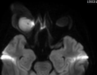
8 Case 1 4Y girl left eye blurring 4 M

9 Possible : Optic N. complex disease Optic nerve complex tumor Optic neuritis Optic neuropathy


10 T2W T1FS+Gd Enhancing enlarged optic nerve : Optic nerve glioma

11 Optic nerve glioma Nearly all Juvenile pilocytic astrocytoma (WHO gr I) Who? : Children, associate with NF 1 Symptom : Slow progressive decreased visual acuity Imaging: CT : Fusiform enlarged optic nerve, posterior extension along optic tract MRI : Enhanced, hypert2, **Watch for intracranial extension


12 Optic nerve Glioma VS Meningioma Enlarged optic nerve Wrap around optic nerve Tram-Track
13 Case 2 42 F, Painless proptosis


14 42 F, Painless proptosis T1W T2W T1FS+Gd


15 DDX Adult well-circumscribed orbital mass Cavernous Malformation Hemangiopericytoma Schwannoma Key! Congenital vascular malform M/C benign orbital mass in adult Well-circumscribed oval mass Lateral intraconal Iso-T1/Hyper-T2 Progressive filling in enhancement Key! Rare tumor originate from pericyte Well-circumscribed oval mass Extraconal adj PNS Iso-T1/ Iso-T2 Vivid arterial enhance & early wash-out Key! Benign prolif of Schwann cell (CN 3-5) Well-circumscribed tubular/fusiform mass Heterogeneous Hyper-T2 & Enhance Abuts optic aperture


16 T1FS+Gd Patho: Cavernous Malformation

17 Case 3 24 M, Painless proptosis right eye 1 Y Prior CT found lacrimal mass


18 NCCT + C Dermoid cyst
 Imaging Extraconal & Well circumscribed CT : Cystic or Fat density MRI : Hyper-T1/T2, Fat suppressed +/- Fat-fluid level or")
19 Dermoid M/C congenital orbital lesion Arise from epithelial sequestration (zygomaticofrontal and frontoethmoidal sutures) Symptoms : Slow growing mass or Mimic acute inflammation (ruptured) Imaging Extraconal & Well circumscribed CT : Cystic or Fat density MRI : Hyper-T1/T2, Fat suppressed +/- Fat-fluid level or Calcification


20 Epidermoid DDX Extraconal cystic lesion Subperiosteal abscess Encephalocele Key! Ectodermal rest Heterogeneous cystic Restricted diffusion +/-HyperT1 : high protein Key! Paranasal sinus inflammation Fat reticulation Key! Track leading to cranial fossa Korean J Radiol 2007;8:
21 Case 4 58 F, Painless lacrimal mass


22 58 F, painless proptosis T1W T2W Benign mixed tumor T1FS+Gd
23 Lacrimal mass Epithelial Benign mixed tumours (BMT)* Adenoid cystic carcinoma (ACC)$ Carcinoma ex-pleomorphic adenoma Adenocarcinoma Mucoepidermoid carcinoma Squamous cell CA * M/C benign $ M/C malignant Non-epithelial Lymphoproliferative Inflammatory (Dacryocystitis) Non-carcinomatous metastases

24 Benign mixed tumour (BMT) M/C benign epithelial tumor of lacrimal gl. Who?: 4 th -5 th decade of life Symptom: Painless, slow-growing mass Imaging : CT: Well-circumscribed lacrimal mass, cystic degeneration, calcifications MRI: Heterogeneous signal T2 & enhancement

25 Malignant ACC VS BMT ACC Painful (perineural spread) Painless BMT Bone erosion More calcification Bone remodeling: Smooth concave * Imaging of early ACC may be identical to BMT


26 72 F, Painful proptosis : Adenoid cystic carcinoma T2W T1FS+Gd T1FS+Gd
27 Case 5 35 M, Right eye proptosis 1 M
28 T1FS+Gd

29 DDX Conal lesion Thyroid opthalmopathy M/C Malignancy : lymphoma, leukemia, metastases Infectious myositis Vascular congestion : DAVF at cavernous sinus


30 Imaging characteristic : TAO Fusiform enlargement Orderly involvement I m Slow Spare myotendinous junction Fat proliferation Bilateral > Unilateral May precede the onset of abnormal thyroid function


31 Idiopathic orbital inflammatory disease


32 Conal lesion alert: Not TAO!!! Disproportionate fat reticulation Unusual order EOM involvement Involvement of Myotendinous junction Other associated findings: Abnormal vessel, other organ involvement

33 Case 6 67 M, Painless proptosis both eyes 1 Y

34 T2W T1FS+Gd

35 DDX: Multicompartment lesions (Adult) Venolymphatic malformation Lymphoproliferative disorder Metastasis Infection


36 Venolymphatic malformation Metastasis Lymphoproliferative disorder Key! Vascular malformation with lymphoid & venous element Ped > Adult Cystic Fluid-fluid level +/- Phelobolith Key! Various imaging features Hx of underlying CA Key! Diffuse, Homogeneous enhance soft tissue Mold structure VariableT2 Hyperdense in NCCT, +/- restricted diffusion Other organ involvement (lacrimal upto 40%) Bilateral lesion 24%


37 NCCT DWI ADC Patho: B-Cell lymphoma
38 Lymphoproliferative disorder M/C in older Adult (>60 yrs) Lymphoid hyperplasia Idiopathic orbital inflammation Reactive lymphoid hyperplasia IgG4 Atypical lymphoid hyperplasia Lymphoma NHL (MALT) is M/C


39 Benign lymphoproliferative VS Lymphoma Benign lymphoproliferative Painful More common Iso-T2/ Hypo-T2* Higher ADC Lymphoma Painless Hyper-T2 Lower ADC * IgG4 due to fibrosis


40 IgG4 : Myositis IgG4 : Tolosa hunt Lymphoma : Tram-Track IgG4 mimic infection


41 IgG4 : Lacrimal Lymphoma : Lacrimal Clues! : Bilateral & Other organ involvement!!
42 Case 7 68 M, visual field defect


43 T1FS T2FS Subtract T1FS+Gd
 Choroidal hemangioma")
44 Occular mass Melanocytic tumor (Adult) Retinoblastoma (Ped) Choroidal hemangioma Metastasis

 HypointenseT2 Collar button")
45 Adult HyperintenseT1 occular lesion Uveal Melanoma Choroidal hemangioma Key! M/C intraocular malignancy in adult (6 th decade) Moderate enhance HyperintenseT1 (20% amelanoic mimic metas) HypointenseT2 Collar button shape Key! Strong & Early enhance Iso/HyperintesenT1 HyperintenseT2
46 Our case T1FS T2FS Subtract T1FS+Gd Patho: NSCLC metastasis


47 Orbital metastasis Breast, Prostate, Melanoma, Lung Various imaging features CA breast: Scirrhous met Carcinoid tumor: Nodular EOM mass CA Prostate: Bone met
48 Case 8 25 F, Intermittent proptosis right eye
 Recurrent orbital hemorrhage Venous congestion during menstruation or intense muscular effort")
49 Intermittent Proptosis Orbital varix (90%) Highly vascular neoplasms with liable congestion (eg. Angioma, lymphangioma) Recurrent orbital hemorrhage Venous congestion during menstruation or intense muscular effort Recurrent emphysema of orbit

50 What is orbital Varix? Congenital weakness of postcapillary venous wall dilatation of the valveless orbital veins Symptoms: Stress proptosis, Spontaneous intraorbital hemorrhage Imaging: Normal/ mild enlarged vein distend during increased venous pressure, phlebolith

 Orbital")
51 Resting Valsava NCCT: Resting Irregular soft tissue Extraconal space Phleboliths Contrast CT: Val Increased size Enhance match venous structure (SOV) Orbital venous Varix
52 Case 9 7 Y boy, Rapid Proptosis right eye


53 T1W T2W T1FS+Gd DWI Patho: Rhabdomyosarcoma

54 DDX Pediatric orbital mass (Non-Globe) Venolymphatic malformation Optic nerve glioma Rhabdomyosarcoma Infantile hemangioma Plexiform neurofibroma Metastasis (Neuroblastoma)
 Well-circumscribed")

55 Rhabdomyosarcoma (RMS) Metastasis Neuroblastoma Key! M/C extraocular tumor in Ped (6-8 yrs) Well-circumscribed Extraconal/Conal+Extraconal Homogeneous Marked enhance Restricted diffusion Key! Skull metastasis with Sun-ray periosteal reaction

 Infiltrative & Extraconal lesion Thin septa or")

56 Infantile Hemangioma Orbital plexiform neurofibroma (OPNF) Key! M/C vasculogenic tumor in infancy Strawberry-like skin mass Grow and then involute with time (start at 1 yr of age) Infiltrative & Extraconal lesion Thin septa or intralesion/perilesion flow void Key! Target sign on T2W Orbital and periorbital infiltrative soft tissue masses Diagnostic for NF1

57 57

58 Take home message Compartmental base approach is essential Don t forget clinical: Age, Symptoms, Underlying Look for other organ involvement


59 What we saw today Intraconal Conal Extraconal Occular Multicompartment ONG Dermoid HyperT1 met Lymphoma Look for other causes!!! Myotenidinous jc Disp fat reticulation Abn vascular Unusual order EOM Cavernous malforn Lacrimal tumor **Lymphoproliferative disorder & Metastasis can have many features


60 Special thanks to my colleagues and fellows at Ramathibodi for their support!!!


61 References Tina D. Tailor, MD Divakar Gupta, MD Roberta W et.al, Orbital Neoplasms in Adults: Clinical, Radiologic, and Pathologic Review. RadioGraphics 2013; 33: Bela S. Purohit, Maria Isabel Vargas, Angeliki Ailianou et al. Orbital tumours and tumour-like lesions: exploring the armamentarium of multiparametric imaging. Insights Imaging (2016) 7:43 68 Ellen M. Chung, James G. Smirniotopoulos, MD, Charles S. Specht, MD, Pediatric Orbit Tumors and Tumorlike Lesions: Nonosseous Lesions of the Extraocular Orbit, RadioGraphics 2007; 27:
13/02/1440 بسم ا هلل ا لرحمن ا لر حيم
 بسم ا هلل ا لرحمن ا لر حيم 1 Slowly progressive versus rapidly progressive proptosis by Ali M ISMAIL professor of ophthalmology @SOHAG U H Occuloplastic fellow @NNUH Occuloplastic fellow @Cambridge UH
C. Douglas Phillips MD FACR Director of Head and Neck Imaging Weill Cornell Medical College/NewYork-Presbyterian Hospital
 C. Douglas Phillips MD FACR Director of Head and Neck Imaging Weill Cornell Medical College/NewYork-Presbyterian Hospital Disclosures Neither I nor any family members have any pertinent financial relations
Case Studies In the Orbit
 Case Studies In the Orbit Ethan Neufeld MD Department of Radiology and Imaging Sciences University of Utah Health Sciences Center Salt Lake City, Utah, USA Case Studies In the Orbit Learning objectives:
Orbital Tumors - A Clinico Pathological Study
 Orbital Tumors - A Clinico Pathological Study Radha. J. DO, Ani Sreedhar. MS. Little Flower Hospital, Angamaly, Kerala ORIGINAL ARTICLES Abstract: Aim. To study the clinical and histopathological profiles
Is there a role of CT in the evaluation of Proptosis
 International Journal of scientific research and management (IJSRM) Volume 3 Issue 4 Pages 2662-2666 2015 \ Website: www.ijsrm.in ISSN (e): 2321-3418 Is there a role of CT in the evaluation of Proptosis
Orbital facia. Periororbital facia Orbital septum Bulbar facia Muscular facia
 Anatomy Orbital facia Periororbital facia Orbital septum Bulbar facia Muscular facia Physiology of symptoms 1) Proptosis ( exophthalmos) Pseudoproptosis Axial Non axial Pulsating Positional Intermittent
The Radiological Spectrum of Orbital Pathologies that Involve the Lacrimal Gland and the Lacrimal Fossa
 The Radiological Spectrum of Orbital Pathologies that Involve the Lacrimal Gland and the Lacrimal Fossa Won Sang Jung, MD 1 Kook Jin Ahn, MD 1 Mi Ra Park, MD 2 Ji Young Kim, MD 1 Jae Jeong Choi, MD 1 Bum
Advances In Orbital Neuropathology
 Advances In Orbital Neuropathology Charles G. Eberhart, MD PhD Associate Professor of Pathology, Ophthalmology and Oncology Johns Hopkins University School of Medicine Overview Non-neoplastic lesions Microphthalmos/pseudoglioma
PITUITARY PARASELLAR LESIONS. Kim Learned, MD
 PITUITARY PARASELLAR LESIONS Kim Learned, MD DIFFERENTIALS Pituitary Sella Clivus, Sphenoid Sinus Suprasellar Optic chiasm, Hypothalamus, Circle of Willis Parasellar Cavernous Sinus Case 1 17 YEAR-OLD
Orbital Tumors And Pseudotumors
 Orbital Tumors And Pseudotumors Poster No.: C-2412 Congress: ECR 2015 Type: Educational Exhibit Authors: M. Limeme, N. Benzina, A. BelKhiria, H. Zaghouani, S. Majdoub, N. Mallat, H. Amara, D. Bakir, C.
Imaging features of orbital neoplasm developed in pediatrics
 Imaging features of orbital neoplasm developed in pediatrics Poster No.: C-1119 Congress: ECR 2015 Type: Educational Exhibit Authors: J. H. Yoo; Seoul/KR Keywords: Eyes, Head and neck, Paediatric, CT,
CT and MRI Imaging Spectrum of Orbital Masses: A Pictorial Essay.
 CT and MRI Imaging Spectrum of Orbital Masses: A Pictorial Essay. Poster No.: C-1556 Congress: ECR 2014 Type: Educational Exhibit Authors: I. Chouchene, S. MAJDOUB, A. Achour, H. Zaghouani, M. 1 1 1 1
Orbital lesions in pediatrics.
 Orbital lesions in pediatrics. Poster No.: C-1912 Congress: ECR 2012 Type: Educational Exhibit Authors: J. H. Yoo, J. V. HUNTER ; Seoul/KR, HOUSTON, TX/US Keywords: Head and neck, CT, MR, Computer Applications-Detection,
Diagnostic Role of CT in the Evaluation of Proptosis
 IOSR Journal of Dental and Medical Sciences (IOSR-JDMS) e-issn: 2279-0853, p-issn: 2279-0861.Volume 14, Issue 4 Ver. IX (Apr. 2015), PP 25-31 www.iosrjournals.org Diagnostic Role of CT in the Evaluation
CTA/MRA of Pediatric Hepatic Masses Radiology-Pathology Correlation
 Acta Radiológica Portuguesa, Vol.XVIII, nº70, pág. 41-50, Abr.-Jun., 2006 CTA/MRA of Pediatric Hepatic Masses Radiology-Pathology Correlation Marilyn J. Siegel Mallinckrodt Institute of Radiology, Washington
Case Studies in the Skull Base
 Case Studies in the Skull Base Amy C Tsai, MD Neuroradiology Fellow Department of Radiology and Imaging Sciences University of Utah Health Sciences Center Salt Lake City, Utah, USA No disclosures related
Dr. T. Venkat Kishan Asst. Prof Department of Radiodiagnosis
 Dr. T. Venkat Kishan Asst. Prof Department of Radiodiagnosis Schwannomas (also called neurinomas or neurilemmomas) constitute the most common primary cranial nerve tumors. They are benign slow-growing
The Investigation of Proptosis in Paediatric Practice.
 The Investigation of Proptosis in Paediatric Practice. Ms Sayantani Ghosh 1, Mr Saugat Dey 1 1 Bankura Sammilani Medical College and Hospital, 136, Dr. Meghnad Saha Road, PRATYASHA APARTMENT Flat-4C, KOLKATA-
Contents. Basic Ultrasound Principles and Terminology. Ultrasound Nodule Characteristics
 Contents Basic Ultrasound Principles and Terminology Basic Ultrasound Principles... 1 Ultrasound System... 2 Linear Transducer for Superficial Images and Ultrasound-Guided FNA... 3 Scanning Planes... 4
Imaging the Spinal Cord & Intradural Disease
 Department of Radiology University of California San Diego Imaging the Spinal Cord & Intradural Disease John R. Hesselink, M.D. Spinal Cord Diseases Tumors Syringohydromyelia Trauma Ischemia / Infarction
Imaging of Hearing Loss
 Contemporary Imaging of Sensorineural Hearing Loss Imaging of Hearing Loss Discussion Outline (SNHL) Imaging Approaches Anatomic Relationships Lesions: SNHL KL Salzman, MD University of Utah School of
The many faces of extranodal lymphoma
 The many faces of extranodal lymphoma Frank Pameijer Departments of Radiology and Radiation Oncology University Medical Center Utrecht Special thanks to Ilona M Schmalfuss, MD University of Florida Gainesville,
A Case of Carotid-Cavernous Fistula
 A Case of Carotid-Cavernous Fistula By : Mohamed Elkhawaga 2 nd Year Resident of Ophthalmology Alexandria University A 19 year old male patient came to our outpatient clinic, complaining of : -Severe conjunctival
objectives Pitfalls and Pearls in PET/CT imaging Kevin Robinson, DO Assistant Professor Department of Radiology Michigan State University
 objectives Pitfalls and Pearls in PET/CT imaging Kevin Robinson, DO Assistant Professor Department of Radiology Michigan State University To determine the regions of physiologic activity To understand
OVERVIEW OF ORBITAL TUMORS AND APROACH TO MANAGEMENT
 OVERVIEW OF ORBITAL TUMORS AND APROACH TO MANAGEMENT Dr E M Nyenze University of Nairobi Work up for patient with proptosis/orbital tumor History Age Pain Progression Periorbital changes History of other
Metastasis. 57 year old with progressive Headache and Right Sided Visual Loss
 Metastasis 1% of sellar/parasellar masses Usually occurs with known primary Can involve third ventricle, hypothalamus, infundibular stalk May be both supra-, intrasellar 57 year old with progressive Headache
JMSCR Vol 3 Issue 11 Page November 2015
 www.jmscr.igmpublication.org Impact Factor 3.79 Index Copernicus Value: 5.88 ISSN (e)-2347-176x ISSN (p) 2455-0450 DOI: http://dx.doi.org/10.18535/jmscr/v3i11.37 Accuracy of CT in Diagnosis of Orbital
RADIOLOGY TEACHING CONFERENCE
 RADIOLOGY TEACHING CONFERENCE John Athas, MD Monica Tadros, MD Columbia University, College of Physicians & Surgeons Department of Otolaryngology- Head & Neck Surgery September 27, 2007 CT SCAN IMAGING
Sonography of soft-tissue vascular lesions
 Sonography of soft-tissue vascular lesions Oscar M. Navarro Associate Professor, University of Toronto Dept. of Diagnostic Imaging, The Hospital for Sick Children Toronto, Canada Declaration of Disclosure
Differential Diagnosis of Oral Masses. Palatal Lesions
 Differential Diagnosis of Oral Masses Palatal Lesions Palatal Masses Periapical Abscess Torus Palatinus Mucocele Lymphoid Hyperplasia Adenomatous Hyperplasia Benign Salivary Neoplasms Malignant Salivary
Clinical Ophthalmic Echography
 Roger P. Harrie Cynthia J. Kendall Clinical Ophthalmic Echography A Case Study Approach Second Edition DVD-VIDEO INCLUDED 123 Clinical Ophthalmic Echography Roger P. Harrie Cynthia J. Kendall Clinical
DISORDERS OF THE SALIVARY GLANDS Neoplasms Dr.M.Baskaran Selvapathy S IV
 DISORDERS OF THE SALIVARY GLANDS Neoplasms Dr.M.Baskaran Selvapathy S IV NEOPLASMS A) Epithelial I. Benign Pleomorphic adenoma( Mixed tumour) Adenolymphoma (Warthin s tumour) Oxyphil adenoma (Oncocytoma)
Role of imaging in RCC. Ultrasonography. Solid lesion. Cystic RCC. Solid RCC 31/08/60. From Diagnosis to Treatment: the Radiologist Perspective
 Role of imaging in RCC From Diagnosis to Treatment: the Radiologist Perspective Diagnosis Staging Follow up Imaging modalities Limitations and pitfalls Duangkamon Prapruttam, MD Department of Therapeutic
Year 2003 Paper two: Questions supplied by Tricia
 question 43 A 42-year-old man presents with a two-year history of increasing right facial numbness. He has a history of intermittent unsteadiness, mild hearing loss and vertigo but has otherwise been well.
Small lesions involving scalp and skull in pediatric age.
 Small lesions involving scalp and skull in pediatric age. Poster No.: C-1149 Congress: ECR 2013 Type: Educational Exhibit Authors: M. J. Yi, J. H. Yoo; Seoul/KR Keywords: Education and training, Education,
INFECTION. HIV Infection DWI
 HIV Infection INFECTION DWI Fig Axial CT and MRI images show multiple enlarged lymph nodes in the neck as well as in the parotid gland bilaterally. These nodes were suppurative with positive diffusion.
Pediatric Ocular Sonography
 Pediatric Ocular Sonography Cicero J Torres A Silva, MD Associate Professor of Radiology 2016 SPR Pediatric Ultrasound Course Yale University School of Medicine None Disclosures Objectives of Presentation
Pictorial review of extraconal and osseous orbital pathology - what can be found 'around' the orbits?
 Pictorial review of extraconal and osseous orbital pathology - what can be found 'around' the orbits? Poster No.: C-2011 Congress: ECR 2013 Type: Educational Exhibit Authors: M. Meissnitzer, T. Meissnitzer,
Small lesions involving scalp and skull in pediatric age.
 Small lesions involving scalp and skull in pediatric age. Poster No.: C-1149 Congress: ECR 2013 Type: Educational Exhibit Authors: M. J. Yi, J. H. Yoo; Seoul/ Keywords: Education and training, Education,
Frequency and Morphological Patterns of Malignant Intra Orbital Tumors in Various age Groups
 Original Article Frequency and Morphological Patterns of Intra Orbital Tumors in Various age Groups Tahira Soomro, Shahnaz Imdad Kehar, Mohammad Anwar Pak J Ophthalmol 2011, Vol. 27 No. 4.....................................................................................................
Orbit Deformities in Craniofacial Neurofibromatosis Type 1
 AJNR Am J Neuroradiol 24:1678 1682, September 2003 Orbit Deformities in Craniofacial Neurofibromatosis Type 1 Claude Jacquemin, Thomas M. Bosley, and Helena Svedberg BACKGROUND AND PURPOSE: The possible
Pediatric Spine Tumors (and other masses)
") Pediatric Spine Tumors (and other masses) Francisco A Perez, MD, PhD Assistant Professor Neuroradiology and Pediatric Radiology Seattle Children s Hospital University of Washington, Seattle Commercial
Pediatric Spine Tumors (and other masses) Francisco A Perez, MD, PhD Assistant Professor Neuroradiology and Pediatric Radiology Seattle Children s Hospital University of Washington, Seattle Commercial
Musculoskeletal Sarcomas
 Musculoskeletal Sarcomas Robert C. Orth, M.D., Ph.D. Edward B. Singleton Department of Pediatric Radiology Texas Children s Hospital Page 0 xxx00.#####.ppt 9/23/2012 9:01:18 AM No disclosures Page 1 xxx00.#####.ppt
Lacrimal Gland Masses
 Residents Section Pattern of the Month Gao et al. Lacrimal Gland Masses Residents Section Pattern of the Month ownloaded from www.ajronline.org by 148.251.232.83 on 03/30/18 from IP address 148.251.232.83.
Radiology of hepatobiliary diseases
 GI cycle - Lecture 14 436 Teams Radiology of hepatobiliary diseases Objectives 1. To Interpret plan x-ray radiograph of abdomen with common pathologies. 2. To know the common pathologies presentation.
Neckmasses in infancy and childhood: Clinical and radiological classification and imaging approaches M. Mearadji
 Neckmasses in infancy and childhood: Clinical and radiological classification and imaging approaches M. Mearadji International Foundation for Pediatric Imaging Aid Introduction Neck masses are a frequent
NEURORADIOLOGY DIL part 5
 NEURORADIOLOGY DIL part 5 Masses and tumors K. Agyem MD, G. Hall MD, D. Palathinkal MD, Alexandre Menard March/April 2015 OVERVIEW Introduction to Neuroimaging - DIL part 1 Basic Brain Anatomy - DIL part
MR Imaging of Orbital and Ocular Disease
 259 MR Imaging of Orbital and Ocular Disease David F. Sobel 1 William Kelly 1 Bent O. Kjos1 Devron Char 2 Michael Brant-Zawadzki1 David Norman 1 Magnetic resonance (MR) images of 27 patients with ocular
OPHTHALMIC PATHOLOGY SPECIALTY CONFERENCE
 DISCLOSURE STATEMENT OPHTHALMIC PATHOLOGY SPECIALTY CONFERENCE No financial disclosures No off-label usage Diva Regina Salomão, M.D. Professor of Laboratory Medicine and Pathology Mayo Clinic College of
Anatomy of the orbit. Lay-out. Imaging technique. 3 x 3. brief overview of the basic anatomy of the orbit and its structures
 Anatomy of the orbit Prof. Pia C Sundgren MD, PhD Department of Diagnostic Radiology, Clinical Sciences, Lund University, Sweden Lay-out brief overview of the basic anatomy of the orbit and its structures
CT and MR imaging findings of tumors and tumor-like conditions of the Lacrimal sac
 CT and MR imaging findings of tumors and tumor-like conditions of the Lacrimal sac Award: Certificate of Merit Poster No.: C-1926 Congress: ECR 2011 Type: Educational Exhibit Authors: J. W. Choi, H.-J.
Perineural Tumor Spread. In Head & Neck Cancer
 Head and Neck Imaging Conference University of Perineural Tumor Spread In Head & Neck Cancer Philip Chapman MD University of Alabama, Birmingham OBJECTIVES: 1. Define (PNTS) 2. Distinguish from pathologic
Secondary Tumors After Hereditary Retinoblastoma: A Case of Orbital Leiomyosarcoma 50 Years After Initial Enucleation and Radiation Therapy
 Secondary Tumors After Hereditary Retinoblastoma: A Case of Orbital Leiomyosarcoma 50 Years After Initial Enucleation and Radiation Therapy John J Chen, MD, PhD and Richard C Allen, MD, PhD November 28,
CT & MRI Evaluation of Brain Tumour & Tumour like Conditions
 CT & MRI Evaluation of Brain Tumour & Tumour like Conditions Dr. Anjana Trivedi 1, Dr. Jay Thakkar 2, Dr. Maulik Jethva 3, Dr. Ishita Virda 4 1 M.D. Radiology, Professor and Head, P.D.U. Medical College
Diagnostic Imaging in Patients with Orbital Cellulitis and Inflammatory Pseudotumor. Fumiyuki Uehara, M.D. Norio Ohba, M.D.
 Diagnostic Imaging in Patients with Orbital Cellulitis and Inflammatory Pseudotumor Fumiyuki Uehara, M.D. Norio Ohba, M.D. We have had the opportunity, at an eye clinic, to examine and treat a patient
4Ps LUMPS AND BUMPS B.L.&T. BUMPS, LUMPS, AND TATTOOS. Most Common BUMP in the oral cavity Fibroma INTERDENTAL PAPILLAE LESIONS
 B.L.&T. BUMPS, LUMPS, AND TATTOOS LUMPS AND BUMPS DIFFERENTIAL DIAGNOSIS FOR LUMPS AND BUMPS Traumatic Fibroma Papilloma Epulis Fissuratum Inflammatory Papillary Hyperplasia Lesions of Attached Gingiva
CNS TUMORS. D r. Ali Eltayb ( U. of Omdurman. I ). M. Path (U. of Alexandria)
. M. Path (U. of Alexandria)") CNS TUMORS D r. Ali Eltayb ( U. of Omdurman. I ). M. Path (U. of Alexandria) CNS TUMORS The annual incidence of intracranial tumors of the CNS ISmore than intraspinal tumors May be Primary or Secondary
CNS TUMORS D r. Ali Eltayb ( U. of Omdurman. I ). M. Path (U. of Alexandria) CNS TUMORS The annual incidence of intracranial tumors of the CNS ISmore than intraspinal tumors May be Primary or Secondary
Rethinking Orbital Imaging
 Rethinking Orbital Imaging Establishing Guidelines for Interpreting Orbital Imaging Studies and Evaluating Their Predictive Value in Patients with Orbital Tumors Guy J. Ben Simon, MD, 1 Christine C. Annunziata,
Orbital Tumors October 2001
 TITLE: Orbital Tumors SOURCE: Grand Rounds Presentation, UTMB, Dept. of Otolaryngology DATE: October 31, 2001 RESIDENT PHYSICIAN: Michael Underbrink, MD FACULTY ADVISOR: Shawn Newlands, MD SERIES EDITORS:
Paranasal Sinuses: Neoplastic Lesions
 Pravin Mundada Department of Radiology, Geneva University Hospital, Switzerland Paranasal Sinuses: Neoplastic Lesions ESHNR 2017 Lisbon, Portugal Layout of the presentation Clinical & imaging features
A Journey Down The Canal
 A Journey Down The Canal Radiological Assessment of Spinal Cord Masses John Berry-Candelario HMS III Gillian Lieberman, MD BIDMC Objectives Patient review Anatomy of the spine Imaging techniques Classification
Role of Computed Tomography and Magnetic Resonance Imaging in Orbital Tumors with Histopathological Correlation
 ORIGINAL RESEARCH www.ijcmr.com in Orbital Tumors with Histopathological Correlation P.Sree Hari 1, Praveen Kumar Alle 2 ABSTRACT Introduction: CT and MRI when performed separately or in combination, will
MR Atlas of Orbital Pathologies
 MR Atlas of Orbital Pathologies Poster No.: C-1212 Congress: ECR 2015 Type: Educational Exhibit Authors: D. S. Baviskar; Abu Dhabi/AE Keywords: Head and neck, Eyes, Neuroradiology peripheral nerve, MR,
PEDIATRIC ORBITAL TUMORS RADIOTHERAPY PLANNING
 PEDIATRIC ORBITAL TUMORS RADIOTHERAPY PLANNING ANATOMY ANATOMY CONT ANATOMY CONT. ANATOMY CONT. EYE OF A CHILD Normal tissue tolerance doses (in conventional #) TD 5/5 TD 50/5 Endpoint Gy Gy Optic nerve
Neuroradiology Case of the Day
 Neuroradiology Case of the Day 76 th CAR Annual Meeting, Montreal, Quebec April 27, 2013 Eugene Yu, MD Assistant Professor of Radiology and Otolaryngology-Head and Neck Surgery Head and Neck Imaging Princess
ORBITAL TUMORS IN CHILDREN: A DESCRIPTIVE STUDY AT TERTIARY CARE CENTRE
 ORIGINAL ARTICLE ORBITAL TUMORS IN CHILDREN: A DESCRIPTIVE STUDY AT TERTIARY CARE CENTRE Palak J Modi 1, Nayana A Shah 1, Jignasa N Bhalodia 2, R N Gonsai 3 Authors Affiliation: 1 Tutor; 2 Associate Professor,
! Women greater than men (4:1)» Typical of other autoimmune diseases
» Typical of other autoimmune diseases") 1 2 3 4 : Overview and Diagnosis Suzanne K. Freitag, M.D. Director, Ophthalmic Plastic Surgery Massachusetts Eye and Ear Infirmary Harvard Medical School! I have no financial disclosures. Learning Objectives!
1 2 3 4 : Overview and Diagnosis Suzanne K. Freitag, M.D. Director, Ophthalmic Plastic Surgery Massachusetts Eye and Ear Infirmary Harvard Medical School! I have no financial disclosures. Learning Objectives!
Ocular Neoplasia What s Common? What s New? Richard R Dubielzig
 Ocular Neoplasia What s Common? What s New? Richard R Dubielzig Orbit 288 6% Tumors of the globe make up 3225 out of 6110 total neoplasms = 53%. Tumors of the conjunctiva make up 1192 out of 6110 total
Posterior fossa tumors: clues to differential diagnosis with case-based review
 Posterior fossa tumors: clues to differential diagnosis with case-based review Poster No.: C-0323 Congress: ECR 2017 Type: Educational Exhibit Authors: H. A. Aboughalia, M. Abdelhady; Doha/QA Keywords:
Pediatric Orbital Tumours in Upper Egypt: A 3-year Retrospective Analysis at a University Hospital.
 Commentary http://www.alliedacademies.org/clinical-ophthalmology-and-vision-science/ Pediatric Orbital Tumours in Upper Egypt: A 3-year Retrospective Analysis at a University Hospital. Ahmad Mostafa Abdallah
Evaluation of Liver Mass Lesions. American College of Gastroenterology 2013 Regional Postgraduate Course
 Evaluation of Liver Mass Lesions American College of Gastroenterology 2013 Regional Postgraduate Course Lewis R. Roberts, MB ChB, PhD Division of Gastroenterology and Hepatology Mayo Clinic College of
Clinical Ophthalmic Echography
 Clinical Ophthalmic Echography Roger P. Harrie Cynthia J. Kendall Clinical Ophthalmic Echography A Case Study Approach Second Edition Roger P. Harrie Moran Eye Center University of Utah Salt Lake City,
Dispelling Rumors about Tumors. Case
 Dispelling Rumors about Tumors Jesse L. Berry, MD Arizona Ophthalmology Society 2017 Associate Director, Ocular Oncology Service Associate Program Director USC/CHLA, Keck School of Medicine Case 65 year
Essentials of Clinical MR, 2 nd edition. 51. Primary Neoplasms
 51. Primary Neoplasms As with spinal central canal neoplasms in other regions, those of the lumbar spine may be classified as extradural, intradural extramedullary, and medullary. If an extradural lesion
Case 9087 Retropharyngeal nodular fasciitis
 Case 9087 Retropharyngeal nodular fasciitis Santiago I 1; Cavalheiro F 2; Noruégas MJ 3; Sanches MC3 1 Hospital Infante D. Pedro, Aveiro, Portugal 2 Hospitais da Universidade de Coimbra, Portugal 3 Hospital
Spine. Neuroradiology. Spine. Spine Pathology. Distribution of fractures. Radiological algorithm. Role of radiology 18/11/2015
 Spine Neuroradiology Spine Prof.Dr.Nail Bulakbaşı X Ray: AP/L/Oblique Vertebra & disc spaces CT & CTA Vertebra, discs, vessels MRI & MRA Vertebra, disc, vessels, meninges Spinal cord & nerves Myelography
Ranbir Singh Sandhu Gillian Lieberman, MD. Orbital Lymphoma. Ranbir Singh Sandhu, University College London Medical School Gillian Lieberman MD, BIDMC
 Orbital Lymphoma Ranbir Singh Sandhu, University College London Medical School Gillian Lieberman MD, BIDMC 1 Our Patient Clinical Picture This topic will be presented in the context of our patient. She
Unknown Cases from the Participants
 Unknown Cases from the Participants Case 1: 1 Case 1: Case 1: DDX? Answer on next slide Case 1: MS V5 Neuropathy Case 2: Case 2: 76 year old woman Ultrasound for multinodular goiter finds suspicious nodule
Pathology of Mediastinal Tumors
 SAMO Meeting Lucerne 2009 Pathology of Mediastinal Tumors Alex Soltermann Most common lesions (adults) Clinical presentation 50% of the patients are asymptomatic, lesion discovered incidentally Symptoms
Cryptogenic Enlargement Of Bilateral Superior Ophthalmic Veins
 ISPUB.COM The Internet Journal of Radiology Volume 18 Number 1 Cryptogenic Enlargement Of Bilateral Superior Ophthalmic Veins K Kragha Citation K Kragha. Cryptogenic Enlargement Of Bilateral Superior Ophthalmic
Pediatric CNS Tumors. Disclosures. Acknowledgements. Introduction. Introduction. Posterior Fossa Tumors. Whitney Finke, MD
 Pediatric CNS Tumors Disclosures Whitney Finke, MD Neuroradiology Fellow PGY-6 University of Utah Health Sciences Center Salt Lake City, Utah None Acknowledgements Introduction Nicholas A. Koontz, MD Luke
Pediatric Abdominal Masses. Andrew Phelps MD Assistant Professor of Pediatric Radiology UCSF Benioff Children's Hospital
 Pediatric Abdominal Masses Andrew Phelps MD Assistant Professor of Pediatric Radiology UCSF Benioff Children's Hospital No Disclosures Take Home Message All you need to remember are the 5 common masses
CLINICAL PEARLS IN OCULAR ONCOLOGY
 CLINICAL PEARLS IN OCULAR ONCOLOGY IRIS NEVUS - Two kinds circumscribed and diffuse - Photodocumentation important to monitor growth - Risk Factors for iris nevus growth to melanoma (ABCDEF) A Age (young),
Solitary Orbital Metastasis Presenting as Extraocular Muscle Mass in Lung Cancer Patient: A Case Report 외안근비대의형태로나타난폐암환자의고립성안와전이 : 증례보고
 Case Report pissn 1738-2637 / eissn 2288-2928 https://doi.org/10.3348/jksr.2017.77.3.143 Solitary Orbital Metastasis Presenting as Extraocular Muscle Mass in Lung Cancer Patient: A Case Report 외안근비대의형태로나타난폐암환자의고립성안와전이
Renal masses - the role of diagnostic imaging
 Renal masses - the role of diagnostic imaging Poster No.: C-2471 Congress: ECR 2015 Type: Educational Exhibit Authors: V. Rai#; Bjelovar/HR Keywords: Cysts, Cancer, Structured reporting, Ultrasound, MR,
RINGS N THINGS: Imaging Patterns in Differential Diagnosis. Anne G. Osborn, M.D.
 RINGS N THINGS: Imaging Patterns in Differential Diagnosis Anne G. Osborn, M.D. ExpDDxs: Intra-axial (Parenchymal) Lesions Ring-enhancing lesions, solitary 1 Ring-enhancing lesion crossing corpus callosum
Chapter 2 Nonmalignant Tumors of the Orbit
 Chapter 2 Nonmalignant Tumors of the Orbit Eric M. Hink and Vikram Durairaj Abstract Most orbital tumors are nonmalignant. Nonmalignant orbital tumors can arise from any of the structures within the orbit,
CT 101 :Pancreas and Spleen
 CT 101 :Pancreas and Spleen Shikha Khullar,, MD, MPH Division of Radiology University of South Alabama The Pancreas Normal Pancreas 3 Phase Pancreatic CT Non contrast Arterial phase : 30-35 35 second
Vision Health: Conditions, Disorders & Treatments EYELID DISORDERS
 Vision Health: Conditions, Disorders & Treatments EYELID DISORDERS There are a number of disorders that can affect the eyelid. Entropion Entropion is an inward turning of the eyelid and lashes toward the
ADRENAL MEDULLARY DISORDERS: PHAEOCHROMOCYTOMAS AND MORE
 ADRENAL MEDULLARY DISORDERS: PHAEOCHROMOCYTOMAS AND MORE DR ANJU SAHDEV READER AND CONSULTANT RADIOLOGIST QUEEN MARY UNIVERSITY AND ST BARTHOLOMEW S HOSPITAL BARTS HEALTH, LONDON, UK DISCLOSURE OF CONFLICT
Salivary ultrasound. Dr T J Beale Royal National Throat Nose & Ear and UCLH Hospitals London UK
 Salivary ultrasound Dr T J Beale Royal National Throat Nose & Ear and UCLH Hospitals London UK Two main groups of patients with presenting symptoms of: Obstructive or chronic inflammatory symptoms (salivary
Normal Sonographic Anatomy
 hapter 2:The Liver DUNSTAN ABRAHAM Normal Sonographic Anatomy Homogeneous, echogenic texture (Figure 2-1) Measures approximately 15 cm in length and 10 12.5 cm anterior to posterior; measurement taken
Visual pathways in the chiasm
 Visual pathways in the chiasm Intracranial relationships of the optic nerve Fixation of the chiasm Chiasmatic pathologies The function of the optic chiasm may be altered by the presence of : 4) Artero
Optic Pathway Gliomas, Germinomas, Spinal Cord Tumours. Colin Kennedy March 2015
 Optic Pathway Gliomas, Germinomas, Spinal Cord Tumours Colin Kennedy March 2015 Glioma of the optic chiasm. T1-weighted MRI with gadolinium enhancement, showing intense irregular uptake of contrast. The
EXPERT DIFFERENTIAL DIAGNOSIS:
 EXPERT DIFFERENTIAL DIAGNOSIS: Sellar Region Anne G. Osborn, M.D. DISCLOSURE: Published RSNA 2008 SELLA, PITUITARY: Normal Gross, 3T Anatomy SELLA, PITUITARY: Anatomically-Based Differential Diagnoses
Benign brain lesions
 Benign brain lesions Diagnostic and Interventional Radiology Hung-Wen Kao Department of Radiology, Tri-Service General Hospital, National Defense Medical Center Computed tomography Hounsfield unit (HU)
Ultrasound of soft-tissue vascular anomalies
 Ultrasound of soft-tissue vascular anomalies Oscar M. Navarro Associate Professor, University of Toronto Dept. of Diagnostic Imaging, The Hospital for Sick Children Toronto, Canada Declaration of Disclosure
Case Presentation 主治醫師 : 宋文鑫日期 :
 Case Presentation 主治醫師 : 宋文鑫日期 : 2015-2-28 General Data Name:OOO Chart Number:OOOOOOO Date of Admission:2014 年 08 月 04 日 Age: 33 y/o Sex:female Occupation : 會計 Chief Complaint Palpable soft tissue mass
Images In Gastroenterology
 Images In Gastroenterology Thong-Ngam D, et al. THAI J GASTROENTEROL 2005 Vol. 6 No. 2 May - Aug. 2005 105 Imaging of Gastrointestinal Stromal Tumors Pornpim Fuangtharnthip, M.D. Narumol Hargroove, M.D.
SIGNIFICANT OTHERS. Miscellaneous Benign Breast Conditions
 SIGNIFICANT OTHERS Miscellaneous Benign Breast Conditions Epworth HealthCare 1 FAT NECROSIS TRAUMATIC Cell rupture Seat-Belt injury Blunt trauma Iatrogenic injury Surgery, Flaps, Radiotherapy Pathology