Immunohistochemical classification of the unknown primary tumour (UPT) Part I. Prof. Mogens Vyberg NordiQC Institute of Pathology Aalborg, Denmark
|
|
|
- Jeffrey Gray
- 5 years ago
- Views:
Transcription
1 Immunohistochemical classification of the unknown primary tumour (UPT) Part I Prof. Mogens Vyberg NordiQC Institute of Pathology Aalborg, Denmark


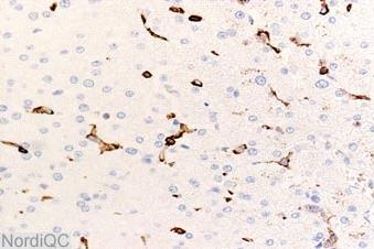
2 Tumours of unknown origin: Histology Brain tumour - biopsy


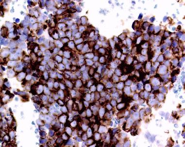
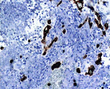
3 CD45 VIM Tumours of unknown origin: Immunohistochemistry Pan-CK S-100
4 IHC classification of the Unknown Primary Tumour UPT: A tumour appearing in metastatic setting without a histologically proven primary tumour. UPT pose an increasing challenge for the pathologist - due to the progress in surgical and oncological treatment possibilities.
5 IHC classification of the Unknown Primary Tumour New, relatively specific antibodies give the pathologist more and better diagnostic tools. But the diagnostic work also become more complex in terms of planning, optimization of protocols, interpretation of reaction patterns and error trapping.
6 IHC classification of the Unknown Primary Tumour 10-15% of cancers remain UPTs +??% uncertain if primary or metastatic - liver, lung, bone, lymph nodes, brain, peritoneum... Undifferentiated (5-10%) - carcinomas, sarcomas, melanomas, germ cell tumours - malignant lymphomas Adenocarcinomas (80-90%) - lung, breast, prostate, colorectum, ovary, pancreas Squamous cell carcinomas (5-10%) - lung, esophagus, uterine cervix
7 IHC classification of the Unknown Primary Tumour Differences in prognosis Differences in treatment regimes malignant lymphomas carcinomas (breast, prostate, ovary...) sarcomas (GIST, synovial sarcoma...) germ cell tumours Pathology tests cost effective Pathology tests save patient discomfort The patient s right to know The risk of hereditary cancer
8 IHC classification of the Unknown Primary Tumour Most likely diagnoses Relevant differential diagnoses Optimal selection of antibodies for a diagnostic algorithm Primary and secondary antibody panels Turn-around-time Laboratory expenses


9 IHC classification of the Unknown Primary Tumour Pathologist - knowledge, acceptance, skill Tumour material - diagnostic markers Antibodies available - applic. in diagnostic algorithms Methods - protocol: sensitivity, specificity, reliability - interpretation: cut-off level for positivity clinical relevance GIST: S-100B Protease GIST: S-100B MWO


10 IHC classification of the Unknown Primary Tumour Pathologist - knowledge, acceptance, skill Tumour material - diagnostic markers Antibodies available - applic. in diagnostic algorithms Methods - protocol: sensitivity, specificity, reliability - interpretation: cut-off level for positivity clinical relevance positive = any staining CK 20 negative = < 50% stained cells CK 20

11 March 2008
12
13

14 Planning diagnostic immunohistochemistry
15 Planning diagnostic immunohistochemistry
16 Planning diagnostic immunohistochemistry

17
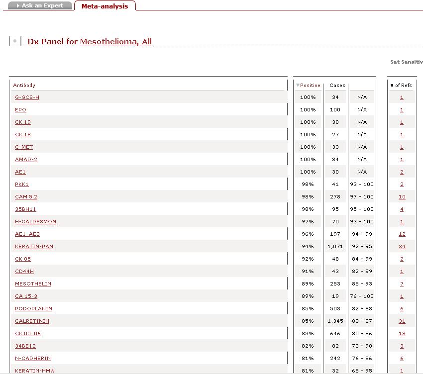
18 my.statdxpathiq.com
19
20
21
22
23
24

25 ?????
26
27
28


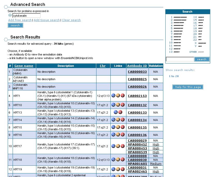
29 Human protein Atlas AE1/AE3
30

31
32
33 Primary panel for the unknown primary tumour Real CD45 CK S-100 VIM Haematolymphoid +/(-) -/(+) -/(+) +/(-) Epithelial - +/(-) -/+ -/+ Mesothelial Mesenchymal and neuronal Non-neuronal neuroepithelial - -/(+) -/ /(+) + + Germ cell - -/+ -/+ +

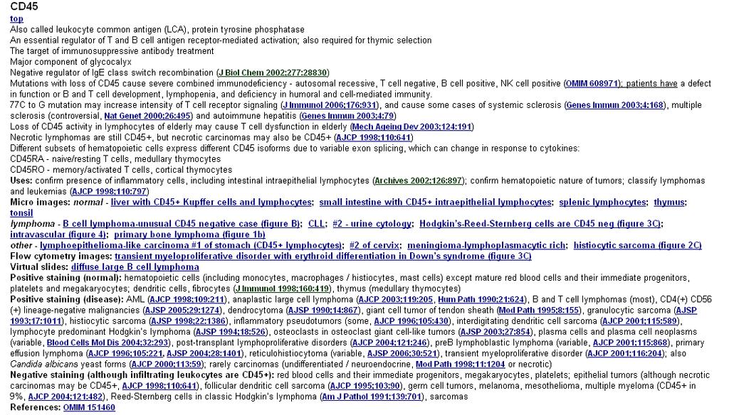
34 CD45 - Leucocyte common antigen (LCA) Transmembrane protein tyrosin phospatase essential for haematopoietic signal transduction and cell activation Membrane associated component: 5 isotypes Intracellular component: one common type

35 CD45 - Leucocyte common antigen (LCA) Transmembrane protein tyrosin phospatase essential for haematopoietic signal transduction and cell activation Membrane associated component: 5 isotypes Intracellular component: one common type Large majority of haematolymphoid cells Lost in maturing erythocytes, megakaryocytes and plasmacells Never found in non-haematolymphoid cells


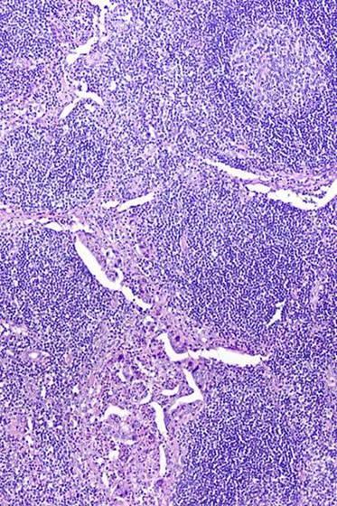
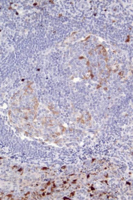
36 CD45 - Leucocyte common antigen (LCA) Normal lymph node Malignant lymphoma
37 CD45 - Leucocyte common antigen (LCA) More than 90% of lymphomas are positive Negative on: - some Acute Lymphoblastic Leukaemia/LBL - plasma cell malignancies - HR-S cells in classic Hodgkin Lymphoma - some Anaplastic Large Cell Lymphoma (ALCL) - ALK+ Large B-Cell Lymphoma Exceptionally positive in non haematol. tumours?
")

38 CD45 - Leucocyte common antigen (LCA) Liver Brain Critical assay performance control


39 CD45 negative HRS cells in classic Hodgkin Lymphoma CD45 positive LH cells in NLPHL CD45 weaker staining in B-CLL CD45 weak reactivity in ALCL 39
40 CD45 NordiQC run assessment-cd45.htm Which is best?
41 CD45 NordiQC run assessment-cd45.htm Optimal Insufficient


42 Optimal Insufficient CD45 NordiQC run assessment-cd45.htm B-CLL 56% of labs 18% of labs

 CD45 RO ~")

43 CD45 - Leucocyte common antigen (LCA) CD45 RO ~ T-cells Lymph node/tonsil CD45 RA ~ B-cells


44 CD45 - Leucocyte common antigen (LCA)


45 CD45 - Leucocyte common antigen (LCA)

46 CD45 - Leucocyte common antigen (LCA)

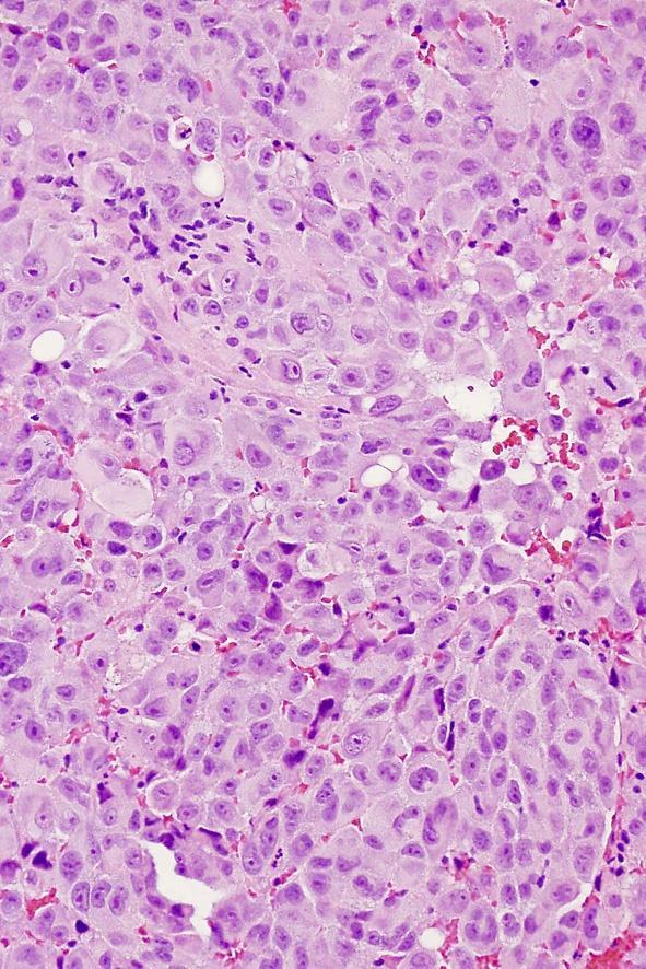
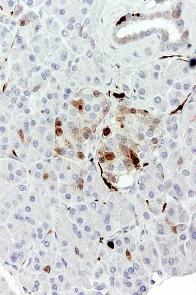
47 Pancreas tumour

48 Pancreas tumour CD45

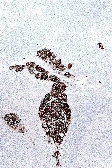
49 Pancreas tumour CD163

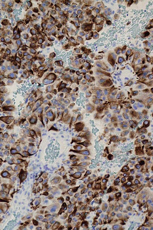
50 Pancreas tumour: undifferentiated carcinoma CK
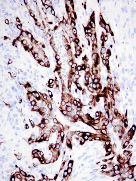
51 Primary panel for the unknown primary tumour Haematolymphoid CD45 CK S-100 VIM +/(-) -/(+) -/(+) +/(-) Epithelial - +/(-) -/+ -/+ Mesothelial Mesenchymal and neuronal Non-neuronal neuroepithelial - -/(+) -/ /(+) + + Germ cell - -/+ -/+ +
 # Microtubuli")
52 Cellular filaments # Microfilaments: (6 nm) # Intermediate filaments (7-11 nm) # Microtubuli (23 nm)
 and microtubules (23 nm) Important for mechanical strength and cellular")
53 Intermediate filaments Group of mainly cytoplasmic filaments 7 11 nm in diameter Part of the cytoskeleton in virtually all cells, creating as meshwork and connecting nuclear membrane with cell membrane Often associated with microfilaments (6 nm) and microtubules (23 nm) Important for mechanical strength and cellular functions

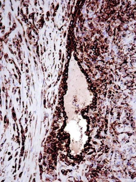
54 Intermediate filaments tetrameric units Central core formed by eight tetramers Nagle, AJSP 1988, 12:4

55 Intermediate filaments - 5 classes I acidic cytokeratins II basic-neutral cytokeratins III vimentin, desmin, glial fibrillary acidic protein, peripherin IV neurofilament protein, α-internexin, nestin V lamins

56 Cytokeratins as tonofilaments Cytokeratin intermediate filaments attached to desmosomes Drochmans et al. J Cell Biol. 1978, 79:427
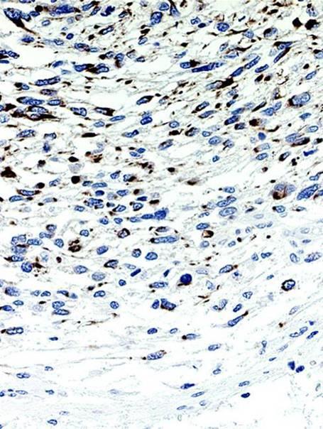
57 Cytokeratins in diagnostic pathology Cytokeratins (CKs) belong to the most fundamental markers of epithelial differentiation CKs comprise a large family of subtypes. Different cell types express different patterns of CK subtypes Cancers generally express CK patterns that at least in part represent the pattern of the putative cell of origin Metastases express CK patterns fairly concordant with those of the primary tumours
58 HE CK Micrometastases identified by cytokeratin


59 HE-frozen CK-frozen Carcinoma in frosen section identified by cytokeratin


60 Sarcomatoid carcinoma CK LMW Cytokeratins in carcinomas with aberrant growth patterns


61 Low molecular weight cytokeratins in carcinomas Carcinomas always LMW-CK-positive, except some cases of Renal cell carcinoma Adrenal cortical carcinoma Small cell carcinoma CK8: Adrenal cortical carcinoma CK8: Renal cell carcinoma CK: renal cell carcinoma
62 Primary panel for the unknown primary tumour Haematolymphoid CD45 CK S-100 VIM +/(-) -/(+) -/(+) +/(-) Epithelial - +/(-) -/+ -/+ Mesothelial Mesenchymal and neuronal Non-neuronal neuroepithelial - -/(+) -/ /(+) + + Germ cell - -/+ -/+ +


63 Cytokeratins in non-epithelial tumours CK8 CD45 42 y, tumour infiltrating retroperitoneum Malignant lymphoma! CD79a
64 Primary panel for the unknown primary tumour Haematolymphoid CD45 CK S-100 VIM +/(-) -/(+) -/(+) +/(-) Epithelial - +/(-) -/+ -/+ Mesothelial Mesenchymal and neuronal Non-neuronal neuroepithelial - -/(+) -/ /(+) + + Germ cell - -/+ -/+ +
65 Cytokeratins in malignant mesothelioma CK8 CK5
66 Primary panel for the unknown primary tumour Haematolymphoid CD45 CK S-100 VIM +/(-) -/(+) -/(+) +/(-) Epithelial - +/(-) -/+ -/+ Mesothelial Mesenchymal and neuronal Non-neuronal neuroepithelial - -/(+) -/ /(+) + + Germ cell - -/+ -/+ +


67 Cytokeratins in sarcomas Synovial sarcoma angiosarcoma

68 Cytokeratins in non-epithelial tumours Leiomyosarcoma
69 Primary panel for the unknown primary tumour Haematolymphoid CD45 CK S-100 VIM +/(-) -/(+) -/(+) +/(-) Epithelial - +/(-) -/+ -/+ Mesothelial Mesenchymal and neuronal Non-neuronal neuroepithelial - -/(+) -/ /(+) + + Germ cell - -/+ -/+ +
70 Cytokeratins in malignant melanoma CK
71 Primary panel for the unknown primary tumour Haematolymphoid CD45 CK S-100 VIM +/(-) -/(+) -/(+) +/(-) Epithelial - +/(-) -/+ -/+ Mesothelial Mesenchymal and neuronal Non-neuronal neuroepithelial - -/(+) -/ /(+) + + Germ cell - -/+ -/+ +


72 Cytokeratins in germ cell tumours CK8: Seminoma CK8: Embr. carcinoma

73 Cytokeratins: retrieval causing false negativity HIER Proteolysis AE1 detects CK8 after HIER only AE1 does not detect CK18 AE3 does not detect CK8/CK18 SCLC Proteolysis CK: renal cell carcinoma


74 Cytokeratins: retrieval causing false negativity TP FN HIER Proteolysis RCC TP CK: renal cell carcinoma FN
75 Primary panel for the unknown primary tumour Haematolymphoid CD45 CK S-100 VIM +/(-) -/(+) -/(+) +/(-) Epithelial - +/(-) -/+ -/+ Mesothelial Mesenchymal and neuronal Non-neuronal neuroepithelial - -/(+) -/ /(+) + + Germ cell - -/+ -/+ +
76 S-100 protein Family of acid calcium binding proteins 9/13 kda Located in nuclei, cytoplasm and cell membranes At least 10 α-chains and one β-chain creating homo- and heterodimers S-100 β-chain mainly found in Melanocytes Glial cells Langerhans cells / interdigitating reticulum cells Fat cells Myoepithelial cells Polyclonal antibodies primarily detects the β-chain

77 S-100 protein brain chondrocytes

78 S-100 protein Tonsil

79 S-100 protein pancreas


80 S-100 in malignant tumours mal. melanoma breast carcinoma


81 S-100 protein To HIER or not.. S100B Frozen IHC Proteolytic HIER
82 Primary panel for the unknown primary tumour Real CD45 CK S-100 VIM Haematolymphoid +/(-) -/(+) -/(+) +/(-) Epithelial - +/(-) -/+ -/+ Mesothelial Mesenchymal and neuronal Non-neuronal neuroepithelial - -/(+) -/ /(+) + + Germ cell - -/+ -/+ +

83 Vimentin Cytoplasmic intermediate filament, 57 kda Present in all mesenchymal cells Present in early stages of all cells, replaced by other intermediate filaments in most non-mesenchymal cells Coexpressed with cytokeratin in some epithelia Endometrium, renal tubules, thyroid gland Coexpressed with cytokeratin in some non-epithelial cells Mesothelium


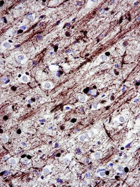
84 Vimentin in normal tissue HE Normal brain VIM


85 Vimentin in carcinomas renal cell carcinoma endometrioid carcinoma


86 Vimentin in non-epithelial tumours mal. melanoma mal. mesothelioma
87 Secondary panels for carcinoma identification/subclassification Cytokeratin subtypes Oncofetal proteins Transcription factors Neuroendocrine proteins Hormone receptors Secretory proteins Cell adhesion molecules... Breast markers Lung markers GI-markers Fem.gen.tract markers Urinary tract markers Prostate markers Squamous cell markers Mesothelial markers NE cell markers Liver markers Adrenal cortical markers Germinal cell markers
Classification of the unknown primary tumour: the primary IHC panel
 CIQC/CAP-ACP SEMINAR 2013: DIAGNOSTIC IHC AND MOLECULAR PATHOLOGY Classification of the unknown primary tumour: the primary IHC panel Aalborg University Hospital Denmark Tumours of unknown origin: Histology
The unkown primary tumour: IHC Classification, antibody selection, protocol optimization, controls and EQA (part I)
") The unkown primary tumour: IHC Classification, antibody selection, protocol optimization, Mogens Vyberg Professor of Clinical Pathology Director of NordiQC Aalborg University Hospital, Aalborg, Denmark
The unkown primary tumour: IHC Classification, antibody selection, protocol optimization, Mogens Vyberg Professor of Clinical Pathology Director of NordiQC Aalborg University Hospital, Aalborg, Denmark
The unknown primary tumour: IHC classification part I, the primary panel - Antibody selection, protocol optimization, controls and EQA
 The unknown primary tumour: IHC classification part I, Mogens Vyberg Professor of Clinical Pathology Director of NordiQC Aalborg University Hospital, Aalborg, Denmark the primary panel - Antibody selection,
Immunohistochemical classification of lung carcinomas and mesotheliomas. Prof. Mogens Vyberg NordiQC Institute of Pathology Aalborg, Denmark
 Immunohistochemical classification of lung carcinomas and mesotheliomas Prof. Mogens Vyberg NordiQC Institute of Pathology Aalborg, Denmark Endobronchial ultrasound guided transbronchial needle biopsy
Diagnostic IHC in lung and pleura pathology
 Diagnostic IHC in lung and pleura pathology Mogens Vyberg Professor of Clinical Pathology Director of NordiQC Aalborg University Hospital, Aalborg, Denmark WHO 2004 and Web Malignant mesothelioma Epithelioid
Nordic Immunohistochemical Quality Control
 Nordic Immunohistochemical Quality Control Immunohistochemistry in the classifiation of neoplasias of the alimentary tract & External Quality Assurance of Immunohistochemistry for GI cancer markers Mogens
External Quality Assessment of melanocytic marker analyses NordiQC experience
 External Quality Assessment of melanocytic marker analyses NordiQC experience Jan Klos MD, Department of Pathology Stavanger University Hospital Norway 1 Content 18 Runs = 2112 submissions between 2001-2014
Cancers of unknown primary : Knowing the unknown. Prof. Ahmed Hossain Professor of Medicine SSMC
 Cancers of unknown primary : Knowing the unknown Prof. Ahmed Hossain Professor of Medicine SSMC Definition Cancers of unknown primary site (CUPs) Represent a heterogeneous group of metastatic tumours,
The impact of proficiency testing on lab immunoassays
 The impact of proficiency testing on lab immunoassays Mogens Vyberg Professor of Clinical Pathology Director of NordiQC Aalborg University Hospital, Aalborg, Denmark Nordic Immunohistochemical Quality
The unkown primary tumour: IHC Classification, antibody selection, protocol optimization, controls and EQA (part II)
") The unkown primary tumour: IHC Classification, antibody selection, protocol optimization, Mogens Vyberg Professor of Clinical Pathology Director of NordiQC Aalborg University Hospital, Aalborg, Denmark
The unkown primary tumour: IHC Classification, antibody selection, protocol optimization, Mogens Vyberg Professor of Clinical Pathology Director of NordiQC Aalborg University Hospital, Aalborg, Denmark
Breast cancer: IHC classification. Mogens Vyberg Professor of Clinical Pathology Director of NordiQC Aalborg University Hospital, Aalborg, Denmark
 Breast cancer: IHC classification Mogens Vyberg Professor of Clinical Pathology Director of NordiQC Aalborg University Hospital, Aalborg, Denmark http://upload.wikimedia.org/wikipedia/commons/1/1a/breast.svg
Charles Halsey, DVM, PhD, DACVP Pfizer, Inc. IHC Resources
 Charles Halsey, DVM, PhD, DACVP Pfizer, Inc. IHC Resources 1 IHC Identification Targets Specimens Controls 2 Tissue controls Trouble Spots 3 The Key to Description IHC Description 4 Intermediate Filaments
NordiQC External Quality Assurance in Immunohistochemistry
 NordiQC External Quality Assurance in Immunohistochemistry Mogens Vyberg Professor of Clinical Pathology Director of NordiQC Aalborg University Hospital, Aalborg, Denmark AALBORG (~ 200.000 inhabitants)
C.L. Davis Foundation Descriptive Veterinary Pathology Course
 C.L. Davis Foundation 2015 Descriptive Veterinary Pathology Course IHC Resources IHC Identification Targets Antibodies Antibodies 1 Antibodies Specimens Antigen Retrieval Unmasks antigen epitopes Methods
Applications of IHC. Determination of the primary site in metastatic tumors of unknown origin
 Applications of IHC Determination of the primary site in metastatic tumors of unknown origin Classification of tumors that appear 'undifferentiated' by standard light microscopy Precise classification
CODING TUMOUR MORPHOLOGY. Otto Visser
 CODING TUMOUR MORPHOLOGY Otto Visser INTRODUCTION The morphology describes the tissue of the tumour closest to normal tissue Well differentiated tumours are closest to normal Undifferentiated tumours show
IHC Panels as an Aid in Diagnostic Decision Making
 IHC Antibody Test Selection Using a Panel Approach Steven Westra B.S. Reagent Product Specialist Leica Biosystems IHC Panels as an Aid in Diagnostic Decision Making Diagnostic Use of Tumors Using Algorithms
Mesothelioma: diagnostic challenges from a pathological perspective. Naseema Vorajee August 2016
 Mesothelioma: diagnostic challenges from a pathological perspective Naseema Vorajee August 2016 Naseema.vorajee@nhls.ac.za Pleural diseases (whether neoplastic, reactive or infective) may have similar
Histopathological diagnosis of CUP
 Histopathological diagnosis of CUP Dr Karin Oien karin.oien@glasgow.ac.uk Disclosure slide Dr Karin Oien has no financial interests in any company mentioned in this presentation. Dr Karin Oien is conducting
I. Diagnosis of the cancer type in CUP
 Latest Research: USA I. Diagnosis of the cancer type in CUP II. Outcomes of site-specific therapy of the cancer type in CUP a. Prospective clinical trial b. Retrospective clinical trials 1 Latest Research:
Quality Assurance in Immunohistochemistry: Experiences from NordiQC
 Nordic immunohistochemical Quality Control 2 Quality Assurance in Immunohistochemistry: Experiences from NordiQC Prof. Mogens Vyberg NordiQC Institute of Pathology Aalborg University Hospital Aalborg,
Reporting of carcinoma of unknown primary tumour (CUP)
") Reporting of carcinoma of unknown primary tumour (CUP) Prof John Schofield Kent Oncology Centre with grateful thanks to Dr Karin Oien University of Glasgow Royal College of Pathologists Cancer datasets
Reporting of carcinoma of unknown primary tumour (CUP) Prof John Schofield Kent Oncology Centre with grateful thanks to Dr Karin Oien University of Glasgow Royal College of Pathologists Cancer datasets
Differential diagnosis of HCC
 Hepatocellular Carcinoma Quest for an Ideal Immunohistochemical Panel Sanjay Kakar, MD UCSF Differential diagnosis of HCC Hepatocellular lesions Adenoma, FNH, HG dysplasia Adenocarcinoma CholangioCA, metastasis
MEDICAL POLICY Gene Expression Profiling for Cancers of Unknown Primary Site
 POLICY: PG0364 ORIGINAL EFFECTIVE: 04/22/16 LAST REVIEW: 07/26/18 MEDICAL POLICY Gene Expression Profiling for Cancers of Unknown Primary Site GUIDELINES This policy does not certify benefits or authorization
performed to help sway the clinician in what the appropriate diagnosis is, which can substantially alter the treatment of management.
 Hello, I am Maura Polansky at the University of Texas MD Anderson Cancer Center. I am a Physician Assistant in the Department of Gastrointestinal Medical Oncology and the Program Director for Physician
Corporate Medical Policy
 Corporate Medical Policy Microarray-based Gene Expression Testing for Cancers of Unknown File Name: Origination: Last CAP Review: Next CAP Review: Last Review: microarray-based_gene_expression_testing_for_cancers_of_unknown_primary
57th Annual HSCP Spring Symposium 4/16/2016
 An Unusual Malignant Spindle Cell Lesion to Involve the Breast Erinn Downs-Kelly, D.O. Associate Professor of Pathology University of Utah & ARUP Laboratories No disclosures Case 39 y/o female with no
Technology from Abcam
 CD2 (EP222) CD2 is one of the earliest T-cell lineage restricted antigens to appear during T-cell differentiation and only rare CD2+ cells can be found in the bone marrow. Anti-CD2 is a pan-t-cell antigen
Pathology Mystery and Surprise
 Pathology Mystery and Surprise Tim Smith, MD Director Anatomic Pathology Medical University of South Carolina Disclosures No conflicts to declare Some problem cases Kidney tumor Scalp tumor Bladder tumor
Assessment Run CK19
 Assessment Run 29 200 CK9 The slide to be stained for CK9 comprised:. Appendix, 2. Thyroid gland, 3. Pancreas, 4. Ductal breast carcinoma, 5. Esophagus, 6. Papillary thyroid carcinoma. All tissues were
Tumour Structure and Nomenclature. Paul Edwards. Department of Pathology and Cancer Research UK Cambridge Institute, University of Cambridge
 Tumour Structure and Nomenclature Paul Edwards Department of Pathology and Cancer Research UK Cambridge Institute, University of Cambridge Malignant Metastasis Core idea of cancer Normal Cell Slightly
Quality Control/Quality Assurance in Diagnostic Immunohistochemistry
 CIHRT Exhibit P- Page Quality Control/Quality Assurance in Diagnostic Immunohistochemistry Emina Torlakovic, MD, PhD College of Medicine University of Saskatchewan Emina Emilia Torlakovic, MD, PhD Associate
The role of immunohistochemistry in surgical pathology of the uterine corpus and cervix
 The role of immunohistochemistry in surgical pathology of the uterine corpus and cervix Prof. Ben Davidson, MD PhD Department of Pathology, Norwegian Radium Hospital, Oslo University Hospital, Oslo, Norway
The Panel Approach to Diagnostics. Lauren Hopson International Product Specialist Cell Marque Corporation
 The Panel Approach to Diagnostics Lauren Hopson International Product Specialist Cell Marque Corporation Cell Marque Rocklin, California About Cell Marque: IVD primary antibody manufacturer Distributors
INTRODUCTION TO PATHOLOGICAL TECHNIQUES. 1. Types of routine biopsy procedures 2. Special exams (IHC, FISH)
") INTRODUCTION TO PATHOLOGICAL TECHNIQUES 1. Types of routine biopsy procedures 2. Special exams (IHC, FISH) Biopsy-Indications Diffuse/multifocal lesions (neoplastic, inflammatory, etc) Etiology of the
INTRODUCTION TO PATHOLOGICAL TECHNIQUES 1. Types of routine biopsy procedures 2. Special exams (IHC, FISH) Biopsy-Indications Diffuse/multifocal lesions (neoplastic, inflammatory, etc) Etiology of the
Cytokeratin 19 (CK19)
") Assessment Run 34 202 Cytokeratin 9 (CK9) Material The slide to be stained for CK9 comprised:. Thyroid gland, 2. Appendix, 3. Esophagus, 4. Papillary thyroid carcinoma, 5 & 6. Pancreatic neuroendocrine
Assessment Run 34 202 Cytokeratin 9 (CK9) Material The slide to be stained for CK9 comprised:. Thyroid gland, 2. Appendix, 3. Esophagus, 4. Papillary thyroid carcinoma, 5 & 6. Pancreatic neuroendocrine
10 years of NordiQC Why are 30% of labs still getting it wrong?
 Mogens Vyberg & Søren Nielsen NordiQC Institute of Pathology Aalborg University Hospital Aalborg, Denmark May 29th 2015 10 years of NordiQC Why are 30% of labs still getting it wrong? Nothing to declare
Expression of Cytokeratin 5/6 in Epithelial Neoplasms: An Immunohistochemical Study of 509 Cases
 Expression of Cytokeratin 5/6 in Epithelial Neoplasms: An Immunohistochemical Study of 509 Peiguo G. Chu, M.D., Ph.D., Lawrence M. Weiss, M.D. Department of Pathology, City of Hope National Medical Center,
Schedule of Accreditation issued by United Kingdom Accreditation Service 2 Pine Trees, Chertsey Lane, Staines-upon-Thames, TW18 3HR, UK
 Schedule of ccreditation United Kingdom ccreditation Service 2 Pine Trees, Chertsey Lane, Staines-upon-Thames, TW18 3HR, UK External Quality ssessment Services for Cancer Diagnostics CIC Issue No: 005
Section 14 Other Cancers. Cancer of Unknown 113 Primary Site INTRODUCTION PATHOLOGIC EVALUATION
 Section 14 Other Cancers Cancer of Unknown 113 Primary Site F. Anthony Greco and John D. Hainsworth INTRODUCTION Cancer of unknown primary (CUP) site is a clinical syndrome that includes many types of
Insulinoma-associated protein (INSM1) is a sensitive and specific marker for lung neuroendocrine tumors in cytologic and surgical specimens
 is a sensitive and specific marker for lung neuroendocrine tumors in cytologic and surgical specimens") Insulinoma-associated protein (INSM1) is a sensitive and specific marker for lung neuroendocrine tumors in cytologic and surgical specimens Kartik Viswanathan, M.D., Ph.D New York Presbyterian - Weill
Insulinoma-associated protein (INSM1) is a sensitive and specific marker for lung neuroendocrine tumors in cytologic and surgical specimens Kartik Viswanathan, M.D., Ph.D New York Presbyterian - Weill
Lung Tumor Cases: Common Problems and Helpful Hints
 Lung Tumor Cases: Common Problems and Helpful Hints Brandon T. Larsen, MD, PhD Senior Associate Consultant Department of Laboratory Medicine and Pathology Mayo Clinic Arizona Arizona Society of Pathologists
Assessment Run GATA3
 Assessment Run 44 2015 GATA3 Material The slide to be stained for GATA3 comprised: 1. Tonsil 2. Kidney, 3. Urothelial carcinoma, 4. Breast ductal carcinoma, 5. Colon adenocarcinoma All tissues were fixed
Immunohistochemical Evaluation of Necrotic Malignant Melanomas
 Anatomic Pathology / EVALUATION OF NECROTIC MALIGNANT MELANOMAS Immunohistochemical Evaluation of Necrotic Malignant Melanomas Daisuke Nonaka, MD, Jordan Laser, MD, Rachel Tucker, HTL(ASCP), and Jonathan
Immunohistochemistry in Bone and Soft Tissue Tumors. Sahar Rassi Zankoul, MD
 Immunohistochemistry in Bone and Soft Tissue Tumors Sahar Rassi Zankoul, MD Introduction Bone tumors represent a wide variety of tumors of various origins and malignant potentials. These different tumor
NEW IHC A n t i b o d i e s
 NEW IHC Antibodies TABLE OF CONTENTS NEW IHC ANTIBODIES from Cell Marque CITED1 (5H6).... 1 Claudin 7 (5D10F3).... 1 GATA1 (4F5).... 1 Transgelin (2A10C2).... 1 NEW IHC ANTIBODIES using RabMAb Technology
Prof. Dr. med. Beata BODE-LESNIEWSKA Institute of Pathology and Molecular Pathology University Hospital; Zurich
 Prof. Dr. med. Beata BODE-LESNIEWSKA Institute of Pathology and Molecular Pathology University Hospital; Zurich 32 year old man 2 months history of growing left supraclavicular lymph nodes Antibiotic treatment
Neoplasia 2018 lecture 11. Dr H Awad FRCPath
 Neoplasia 2018 lecture 11 Dr H Awad FRCPath Clinical aspects of neoplasia Tumors affect patients by: 1. their location 2. hormonal secretions 3. paraneoplastic syndromes 4. cachexia Tumor location Even
SMH (Myosin, smooth muscle heavy chain)
") Material The slide to be stained for SMH comprised: Assessment Run 50 2017 SMH (Myosin, smooth muscle heavy chain) 1.Tonsil, 2. Esophagus, 3. Breast hyperplasia, 4. Breast ductal carcinoma in situ (DCIS),
Material The slide to be stained for SMH comprised: Assessment Run 50 2017 SMH (Myosin, smooth muscle heavy chain) 1.Tonsil, 2. Esophagus, 3. Breast hyperplasia, 4. Breast ductal carcinoma in situ (DCIS),
Enterprise Interest Nothing to declare
 Enterprise Interest Nothing to declare Diagnoses one would not like to miss in soft tissue pathology early in your career Marta Sbaraglia, MD Department of Pathology Hospital of Treviso University of Padua
MT09 - Normal Human Tissue Microarray, FDA
 Reveal Biosciences offers Histochemical Staining, Immunohistochemistry (IHC), In Situ Hybridization (ISH), Whole Slide Imaging, and Quantitative Image Analysis on any TMA MT09 - Normal Human Tissue Microarray,
The clinically challenging entity of liver metastasis from tumors of unknown primary
 The clinically challenging entity of liver metastasis from tumors of unknown primary Xuchen Zhang, MD, PhD Associate Professor of Pathology Department of Pathology Yale University School of Medicine Liver
What I Learned from 3 Cases and 3 Antibodies
 What I Learned from 3 Cases and 3 Antibodies Melinda Sanders, M.D Vanderbilt University Medical Center Professor of Pathology Consultant in Breast Pathology Disclosure of Relevant Financial Relationships
GUT-C 11/30/2017. Debasmita Das, M.D. PGY-1 Danbury Hospital
 GUT-C 11/30/2017 Debasmita Das, M.D. PGY-1 Danbury Hospital CLINICAL SUMMARY 8/2017 59 year old female Presented to the ED with 1 month history of general malaise, fever and weight loss PMH: Significant
Immunohistochemistry on Fluid Specimens: Technical Considerations
 Immunohistochemistry on Fluid Specimens: Technical Considerations Blake Gilks Dept of Pathology University of British Columbia, Vancouver, BC, Canada Disclosures None Learning Objectives At the end of
From Morphology to Molecular Pathology: A Practical Approach for Cytopathologists Part 1-Cytomorphology. Songlin Zhang, MD, PhD LSUHSC-Shreveport
 From Morphology to Molecular Pathology: A Practical Approach for Cytopathologists Part 1-Cytomorphology Songlin Zhang, MD, PhD LSUHSC-Shreveport I have no Conflict of Interest. FNA on Lymphoproliferative
4/12/2018. MUSC Pathology Symposium Kiawah Island April 18, Jesse K. McKenney, MD
 MUSC Pathology Symposium Kiawah Island April 18, 2018 Jesse K. McKenney, MD 1 Urothelial Carcinoma with Alternative Differentiation 2 Urothelial Carcinoma with Alternative Differentiation Recognition as
Objectives. Salivary Gland FNA: The Milan System. Role of Salivary Gland FNA 04/26/2018
 Salivary Gland FNA: The Milan System Dr. Jennifer Brainard Section Head Cytopathology Cleveland Clinic Objectives Introduce the Milan System for reporting salivary gland cytopathology Define cytologic
Cutaneous metastases. Thaddeus Mully. University of California, San Francisco Professor, Departments of Pathology and Dermatology
 Cutaneous metastases Thaddeus Mully University of California, San Francisco Professor, Departments of Pathology and Dermatology DISCLOSURE OF RELATIONSHIPS WITH INDUSTRY Thaddeus Mully Course C005 Essential
05/07/2018. Types of challenges. Challenging cases in uterine pathology. Case 1 ` 65 year old female Post menopausal bleeding Uterine Polyp
 Types of challenges Challenging cases in uterine pathology Nafisa Wilkinson Gynaecological Pathologist UCLH London Lack of complete history often, NO clinical history at all! Cases from other centres often
Role of immunohistochemistry in the differential diagnosis of malignant small round cell tumor: a study of 38 cases
 International Journal of Research in Medical Sciences Patel A et al. Int J Res Med Sci. 2015 Dec;3(12):3833-3839 www.msjonline.org pissn 2320-6071 eissn 2320-6012 Research Article DOI: http://dx.doi.org/10.18203/2320-6012.ijrms20151452
Immunopathology of Lymphoma
 Immunopathology of Lymphoma Noraidah Masir MBBCh, M.Med (Pathology), D.Phil. Department of Pathology Faculty of Medicine Universiti Kebangsaan Malaysia Lymphoma classification has been challenging to pathologists.
Neoplasia part I. Dr. Mohsen Dashti. Clinical Medicine & Pathology nd Lecture
 Neoplasia part I By Dr. Mohsen Dashti Clinical Medicine & Pathology 316 2 nd Lecture Lecture outline Review of structure & function. Basic definitions. Classification of neoplasms. Morphologic features.
Assessment Run
 Assessment Run 50 2017 S100 Material The slide to be stained for S100 comprised: 1. Appendix, 2. Tonsil, 3. Schwannoma, 4-5. Malignant melanoma, 6. Colon adenocarcinoma. All tissues were fixed in 10% neutral
Cerebral Parenchymal Lesions: I. Metastatic Neoplasms
 Chapter 4 Cerebral Parenchymal Lesions: I. Metastatic Neoplasms After one has reasonably ruled out the possibility of a nonneoplastic diagnosis (see Chap. 3), one is left with considering a diagnosis of
ACCME/Disclosures. Case History 4/13/2016. USCAP GU Specialty Conference Case 3. Ann Arbor, MI
 USCAP GU Specialty Conference Case 3 March 2016 L. Priya Kunju, M.D. University of Michigan Health System Ann Arbor, MI University of Michigan Health System ACCME/Disclosures The USCAP requires that anyone
Cutaneous Mesenchymal Neoplasms with EWSR1 Rearrangement
 Cutaneous Mesenchymal Neoplasms with EWSR1 Rearrangement By Konstantinos Linos MD, FCAP, FASDP Bone, Soft Tissue and Dermatopathology Assistant Professor of Pathology Dartmouth-Hitchcock Medical Center
Presentation material is for education purposes only. All rights reserved URMC Radiology Page 1 of 98
 Presentation material is for education purposes only. All rights reserved. 2011 URMC Radiology Page 1 of 98 Radiology / Pathology Conference February 2011 Brooke Koltz, Cytopathology Resident Presentation
Financial disclosures
 Cutaneous Mesenchymal Neoplasms with EWSR1 Rearrangement By Konstantinos Linos MD, FCAP, FASDP Bone, Soft Tissue and Dermatopathology Assistant Professor of Pathology Dartmouth-Hitchc Geisel School of
3/24/2017 DENDRITIC CELL NEOPLASMS: HISTOLOGY, IMMUNOHISTOCHEMISTRY, AND MOLECULAR GENETICS. Disclosure of Relevant Financial Relationships
 DENDRITIC CELL NEOPLASMS: HISTOLOGY, IMMUNOHISTOCHEMISTRY, AND MOLECULAR GENETICS Jason L. Hornick, M.D., Ph.D. Director of Surgical Pathology and Immunohistochemistry Brigham and Women s Hospital Professor
Recent advances in breast cancers
 Recent advances in breast cancers Breast cancer is a hetrogenous disease due to distinct genetic alterations. Similar morphological subtypes show variation in clinical behaviour especially in response
Serous Effusions. Spasenija Savic Prince, MD Pathology, University Hospital Basel, Switzerland
 Serous Effusions Spasenija Savic Prince, MD Pathology, University Hospital Basel, Switzerland Serous membrane Body cavities: Pleural Pericardial Peritoneal Effusion = Excess of fluid 80% Benign 20% Malignant
Differential diagnosis of hematolymphoid tumors composed of medium-sized cells. Brian Skinnider B.C. Cancer Agency, Vancouver General Hospital
 Differential diagnosis of hematolymphoid tumors composed of medium-sized cells Brian Skinnider B.C. Cancer Agency, Vancouver General Hospital Lymphoma classification Lymphoma diagnosis starts with morphologic
Oncology 101. Cancer Basics
 Oncology 101 Cancer Basics What Will You Learn? What is Cancer and How Does It Develop? Cancer Diagnosis and Staging Cancer Treatment What is Cancer? Cancer is a group of more than 100 different diseases
Optimization of antibodies, selection, protocols and controls Breast tumours
 Optimization of antibodies, selection, protocols and controls Breast tumours Søren Nielsen Project coordinator & Scheme Manager NordiQC Aalborg University Hospital, Denmark Breast panel: GCDFP-15 Mammaglobin
Enterprise Interest Nothing to declare
 Enterprise Interest Nothing to declare Biopsy diagnosis of renal tumors. Current applications Ondřej Hes Department of Pathology Charles University and University Hospital Plzeň Czech Republic Dealing
Kyle L. Ziegler, CTR. California Cancer Registry U.C. Davis Health System
 Kyle L. Ziegler, CTR California Cancer Registry U.C. Davis Health System Overview New Data Items Reportability Clarifications New Coding Rules Grade ICD-O-3 Changes Collaborative Stage v0205 2 New Data
LN04 - Lymphoma Tissue Microarray
 Reveal Biosciences offers Histochemical Staining, Immunohistochemistry (IHC), In Situ Hybridization (ISH), Whole Slide Imaging, and Quantitative Image Analysis on any TMA LN04 - Lymphoma Tissue Microarray
ONCOLOGY. Csaba Bödör. Department of Pathology and Experimental Cancer Research november 19., ÁOK, III.
 ONCOLOGY Csaba Bödör Department of Pathology and Experimental Cancer Research 2018. november 19., ÁOK, III. bodor.csaba1@med.semmelweis-univ.hu ONCOLOGY Characteristics of Benign and Malignant Neoplasms
ACCME/Disclosures ALK FUSION-POSITIVE MESENCHYMAL TUMORS. Tumor types with ALK rearrangements. Anaplastic Lymphoma Kinase. Jason L.
 Companion Meeting of the International Society of Bone and Soft Tissue Pathology The Evolving Concept of Mesenchymal Tumors ALK FUSION-POSITIVE MESENCHYMAL TUMORS Jason L. Hornick, MD, PhD March 13, 2016
New Developments in Immunohistochemistry for Gynecologic Pathology
 New Developments in Immunohistochemistry for Gynecologic Pathology Michael T. Deavers, M.D. Professor, Departments of Pathology and Gynecologic Oncology Immunohistochemistry in Gynecologic Pathology Majority
Chapter 2 Cell. Zhou Li Prof. Dept. of Histology and Embryology
 Chapter 2 Cell Zhou Li Prof. Dept. of Histology and Embryology The inner life of the cell Ⅰ. Plasma membrane (Plasmalemma) 1.1 The structure Unit membrane: inner layer 3-layered structure outer layer mediat
Tumores de células pequeñas, redondas y azules: diagnóstico diferencial cuando el tiempo apremia
 Tumores de células pequeñas, redondas y azules: diagnóstico diferencial cuando el tiempo apremia Sílvia Bagué Servei de Patologia Hospital de Sant Pau Barcelona Soft tissue sarcomas Heterogeneous group
Rare Cancers. Andrew J. Wagner, MD, PhD Center for Sarcoma and Bone Oncology Dana-Farber Cancer Institute Sarcoma Patient Symposium October 15, 2017
 Rare Cancers Andrew J. Wagner, MD, PhD Center for Sarcoma and Bone Oncology Dana-Farber Cancer Institute Sarcoma Patient Symposium October 15, 2017 Why should we care about Rare Cancers? Raise your hand
Lung Cytology: Lessons Learned from Errors in Practice
 Lung Cytology: Lessons Learned from Errors in Practice Stephen S. Raab, M.D. Department of Laboratory Medicine Eastern Health and Memorial University of Newfoundland, St. John s, NL and University of Washington,
SELECTED DILEMMAS IN RESPIRATORY CYTOPATHOLOGY (2 CASES)
") SELECTED DILEMMAS IN RESPIRATORY CYTOPATHOLOGY (2 CASES) Dr. Mariamma Joseph Professor of Pathology Division Head Cytopathology Department of Pathology and Laboratory Medicine LHSC and Western University
SELECTED DILEMMAS IN RESPIRATORY CYTOPATHOLOGY (2 CASES) Dr. Mariamma Joseph Professor of Pathology Division Head Cytopathology Department of Pathology and Laboratory Medicine LHSC and Western University
Impact of immunostaining of pulmonary and mediastinal cytology
 Impact of immunostaining of pulmonary and mediastinal cytology Harman Sekhon MD, PhD Director of Cytopathology Head of Ottawa-site Ontario Tumour Bank June 20, 2014 Disclaimer Pfizer: Honorarium-Advisory
List of Available TMAs in the PRN
 TMA RPCI_BrainCa01 RPCI_BrCa03 RPCI_BrCa04 RPCI_BrCa05 RPCI_BrCa0 RPCI_BrCa07 RPCI_BrCa08 RPCI_BrCa15 RPCI_BrCa1 RPCI_BrCa17 RPCI_BrCa18 RPCI_BrCa19 RPCI_BrCa20 RPCI_BrCa21 RPCI_BrCa24 RPCI_BrCa25 RPCI_BrCa2
RENAL EPITHELIAL NEOPLASMS: IS THERE A ROLE OF IMMUNOSTAINS IN DIAGNOSIS?
 RENAL EPITHELIAL NEOPLASMS: IS THERE A ROLE OF IMMUNOSTAINS IN DIAGNOSIS? John C. Cheville, M.D. Mayo Clinic and Mayo Foundation Rochester, MN The majority of renal epithelial neoplasms are diagnosed on
Lecture 13 - Intermediate filaments
 02.12.10 Lecture 13 - Intermediate filaments Intermediate filaments Present in nearly all animals, but absent from plants and fungi Rope-like network of filaments in the cell Principle function is maintenance
Update on Cutaneous Mesenchymal Tumors. Thomas Brenn
 Update on Cutaneous Mesenchymal Tumors Thomas Brenn Cutaneous Mesenchymal Tumours Wide morphological and biological spectrum Myofibroblastic, smooth muscle, neural, vascular, apidocytic, undifferentiated;
The role of appropriateness criteria in the planning of health services: the case of FDG-PET in oncology
 V Annual Meeting of Health Tecnology Assessment International (HTAi) MONTREAL 8 july 2008 The role of appropriateness criteria in the planning of health services: the case of FDG-PET in oncology L. BALLINI
ACCURACY OF IMMUNOHISTOCHEMISTRY IN EVALUATION
 POL J PATHOL 2011; 2: 95-100 ACCURACY OF IMMUNOHISTOCHEMISTRY IN EVALUATION OF MALIGNANT PLEURAL AND PERITONEAL EFFUSIONS FERESHTEH ENSANI, FARNAZ NEMATIZADEH, GITI IRVANLOU Department of Cytology, Cancer
Epithelial cell-cell adhesion molecule (Ep-CAM)
") Assessment Run 3 011 Epithelial cell-cell adhesion molecule (Ep-CAM) Material The slide to be stained for Ep-CAM comprised: 1. Appendix,. Kidney, 3. Adrenal gland, 4. Lung carcinoid, 5 & 6. Renal clear
Assessment Run 3 011 Epithelial cell-cell adhesion molecule (Ep-CAM) Material The slide to be stained for Ep-CAM comprised: 1. Appendix,. Kidney, 3. Adrenal gland, 4. Lung carcinoid, 5 & 6. Renal clear
Index. Cytoplasm, nonepithelial malignant tumor features 70
 Accurette device 23 Adenosarcoma, differential diagnosis 80, 81 Arias-Stella reaction 65 Atypical endocervical cells 8 Atypical endometrial cells 8 Atypical glandular cells (AGC) 8, 9 Atypical glandular
Estrogen receptor (ER)
") Assessment Run B7 204 Estrogen receptor (ER) Material The slide to be stained for ER comprised: No. Tissue ER-positivity* ER-intensity*. Uterine cervix 80-90% Moderate to strong 2. Breast carcinoma 0%
Assessment Run B7 204 Estrogen receptor (ER) Material The slide to be stained for ER comprised: No. Tissue ER-positivity* ER-intensity*. Uterine cervix 80-90% Moderate to strong 2. Breast carcinoma 0%
Radiology Pathology Conference
 Radiology Pathology Conference Nadia F. Yusaf, M.D. PGY-3 1/29/2010 Presentation material is for education purposes only. All rights reserved. 2010 URMC Radiology Page 1 of 90 Case 1 60 year- old man presents
Aggressive Malignant Mesothelioma In A Patient Without Previous Asbestos Exposure
 ISPUB.COM The Internet Journal of Pathology Volume 19 Number 1 Aggressive Malignant Mesothelioma In A Patient Without Previous Asbestos Exposure A Julai, G Wathuge Citation A Julai, G Wathuge. Aggressive
Schedule of Accreditation issued by United Kingdom Accreditation Service 2 Pine Trees, Chertsey Lane, Staines-upon-Thames, TW18 3HR, UK
 2 Pine Trees, Chertsey Lane, Staines-upon-Thames, TW18 3HR, UK Lister Hospital Contact: Heather Taylor Coreys Mill Lane Tel: +44 (0) 01438 285086 Stevenage E-Mail: Heathertaylor3@nhs.net SG1 4AB Website: