I. Membrane Proteins II. Intracellular Compartments III. Protein Translocation
|
|
|
- Louisa White
- 6 years ago
- Views:
Transcription
1 Lecture 3 I. Membrane Proteins II. Intracellular Compartments III. Protein Translocation Ref: MBoC (5th Edition), Alberts Johnson Lewis Raff Roberts Walter Chapter 10 Membrane Structure Chapter 12 Intracellular Compartments and Protein Sorting Hualin Zhong 2/04/2013 1

2 Many Membrane Proteins Diffuse in the Plane of the Membrane An experiment demonstrating the diffusion of proteins in the plasma membrane of mouse human hybrid cells. The mouse and human proteins are initially confined to their own halves of the newly formed heterocaryon plasma membrane, but they intermix over time. The two antibodies used to visualize the proteins can be distinguished in a fluorescence microscope because fluorescein is green and rhodamine is red. Figure

3 Fluorescence Recovery After Photobleaching (FRAP) Figure 10-36a A specific protein of interest can be expressed as a fusion protein with green fluorescent protein (GFP). In the FRAP technique, fluorescent molecules are bleached in a small area using a laser beam. The fluorescence intensity recovers as the bleached molecules diffuse away and unbleached molecules diffuse into the irradiated area. The diffusion coefficient is calculated from a graph of the rate of recovery: the greater the diffusion coefficient (mobility) of the membrane protein, the faster the recovery. 3

4 Fluorescence Loss In Photobleaching (FLIP) Figure 10-36b In the FLIP technique, a small area of membrane is irradiated continuously, and fluorescence is measured in a separate area. Fluorescence in the second area progressively decreases as fluorescent proteins diffuse out and bleached molecules diffuse in; eventually, all of the fluorescent protein molecules are bleached, as long as they are mobile and not permanently anchored to the cytoskeleton or extracellular matrix. 4


5 Cells Can Confine Proteins and Lipids to Specific Domains Within a Membrane Figure Figure Four ways of restricting the lateral mobility of specific plasma membrane proteins: (A) The proteins can self-assemble into large aggregates (e.g. bacteriorhodopsin in the purple membrane of Halobacterium); (B, C) they can be tethered by interactions with assemblies of macromolecules (B) outside or (C) inside the cell; (D) they can interact with proteins on the surface of another cell. In an epithelial cell, protein A and protein B can diffuse laterally in their own domains but are prevented from entering the other domain, at least partly by the specialized cell junction called a tight junction. Lipid molecules in the outer (noncytosolic) monolayer of the plasma membrane are likewise unable to diffuse between the two domains; lipids in the inner (cytosolic) monolayer, however, are able to do so (not shown). The basal lamina is a thin mat of extracellular matrix that separates epithelial sheets from other tissues 5
 (B) Spectrin dimers are linked together into a netlike meshwork by junctional complexes.")
.")
6 The Cortical Cytoskeleton Gives Membrane Mechanical Strength and Restricts Membrane Protein Figure 10-41a Figure 10-41b The Spectrin-based cytoskeleton on the cytosolic side of the human red blood cell plasma membrane (A) (B) Spectrin dimers are linked together into a netlike meshwork by junctional complexes. The junctional complexes are composed of short actin filaments, band 4.1, adducin, and a tropomyosin molecule. The cytoskeleton is linked to the membrane through two transmembrane proteins (band 3 & glycophorin). The EM shows the cytoskeleton on the cytosolic side of a red blood cell membrane after fixation and negative staining. 6

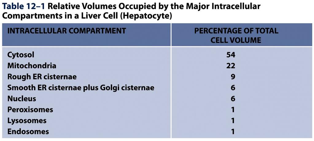
7 The Major Intracellular Compartments of an Animal Cell Figure 12-1 The cytoplasm consists of the cytosol and the cytoplasmic organelles. 7
8 8

9 Table

 Mitochondria (and plastids) are thought to have originated when a bacterium was engulfed by a larger preeucaryotic cell.")
10 Evolutionary Origins Explain the Topological Relationships of Organelles Hypothetical schemes for the evolutionary origins of some organelles. The origins of mitochondria, chloroplasts, ER, and the cell nucleus might explain the topological relationships of these compartments in eucaryotic cells. (A) (B) Figure 12-4 (A) A possible pathway for the evolution of the cell nucleus and the ER. (B) Mitochondria (and plastids) are thought to have originated when a bacterium was engulfed by a larger preeucaryotic cell. This could explain why they contain their own genomes and why the lumens of these organelles remain isolated from the membrane traffic that interconnects the lumens of many other intracellular compartments. 10

11 Topological Relationships Between Compartments of the Secretory and Endocytic Pathways in a Eucaryotic Cell Figure 12-5 Topologically equivalent spaces are shown in red. In principle, cycles of membrane budding and fusion permit the lumen of any of these organelles to communicate with any other and with the cell exterior by means of transport vesicles. Blue arrows indicate the extensive outbound and inbound vesicular traffic. *Some organelles, most notably mitochondria and (in plant cells) plastids, do not take part in this communication and are isolated from the traffic between organelles shown here. 11
12 A Simplified Roadmap of Protein Traffic Proteins can move from one compartment to another by gated transport (red), transmembrane transport (blue), or vesicular transport (green). The sorting signals that direct a given protein s movement through the system, and thereby determine its eventual location in the cell, are contained in each protein s amino acid sequence. The journey begins with the synthesis of a protein on a ribosome in the cytosol and terminates when the protein reaches its final destination. At each intermediate station (boxes), a decision is made as to whether the protein is to be retained in that compartment or transported further. In principle, a sorting signal could be required for either retention in or exit from a compartment. Figure


13 Signal sequence mediated intracellular protein translocation From Günter Blobel 13
14 Signal Sequences Direct Proteins to the Correct Cell Addresses Most protein sorting signals reside in a stretch of amino acid sequence, typically residues long. These signal sequences are often found at the N-terminus; specialized signal peptidases remove the signal sequence from the finished protein once the sorting process is complete. Signal sequences can also be internal stretches of amino acids, which remain part of the protein. In some cases, sorting signals are composed of multiple internal amino acid sequences that form a specific three-dimensional arrangement of atoms on the protein s surface, called a signal patch. * Each signal sequence specifies a particular destination in the cell. 14
15 Page 723 Molecular Biology of the Cell ( Garland Science 2008) 15

 (B) Figure 12-34 (A) (B) Part of the ER network in a cultured mammalian cell, stained with an antibody that binds to a protein retained")
16 The Endoplasmic Reticulum (ER) All eucaryotic cells have an ER. Its membrane typically constitutes more than half of the total membrane of an average animal cell. The ER is organized into a netlike labyrinth of branching tubules and flattened sacs that extends throughout the cytosol. (A) (B) Figure (A) (B) Part of the ER network in a cultured mammalian cell, stained with an antibody that binds to a protein retained in the ER. Part of an ER network in a living plant cell that was genetically engineered to express a fluorescent protein in the ER. 16

17 Co-translational and Post-translational Protein Translocation Figure Mammalian cells import most proteins into the ER co-translationally, whereas import proteins into mitochondria, chloroplasts, nuclei, and peroxisomes post-translationally. 17


 A 3-D reconstruction of a region of")
18 The ER Is Structurally and Functionally Diverse (B) Abundant smooth ER in a steroidhormone-secreting cell. (A) An EM of the rough ER in a pancreatic exocrine cell that makes and secretes large amounts of digestive enzymes every day. Figure (C) A 3-D reconstruction of a region of smooth ER and rough ER in a liver cell. 18


19 The Isolation of Purified Rough and Smooth Microsomes from the ER (A) A thin section electron micrograph of the purified rough ER fraction shows an abundance of ribosome-studded vesicles. (A) (B) When sedimented to equilibrium through a gradient of sucrose, the two types of microsomes separate from each other on the basis of their different densities. (B) Figure
.")
20 Signal Sequences Were First Discovered in Proteins Imported into the Rough ER The signal hypothesis. A simplified view of protein translocation across the ER membrane, as originally proposed. Figure When the ER signal sequence emerges from the ribosome, it directs the ribosome to a translocator on the ER membrane (a pore in the membrane through which the polypeptide is translocated). A signal peptidase is closely associated with the translocator and clips off the signal sequence during translocation, and the mature protein is released into the lumen of the ER immediately after synthesis. The translocator is closed until the ribosome has bound, so that the permeability barrier of the ER membrane is maintained at all times. For the history of the Signal Hypothesis, please listen to the Blobel s Nobel lecture: 20

21 A Signal-Recognition Particle (SRP) Directs ER Signal Sequences to a Specific Receptor in the Rough ER Membrane Figure The SRP binds to both the exposed ER signal sequence and the ribosome, thereby inducing a pause in translation. The SRP receptor in the ER membrane, which is composed of two different polypeptide chains, binds the SRP ribosome complex and directs it to the translocator. Because one of the SRP proteins and both chains of the SRP receptor contain GTP-binding domains, it is thought that conformational changes that occur during cycles of GTP binding and hydrolysis ensure that SRP release occurs only after the ribosome has become properly engaged with the translocator in the ER membrane. 21


22 The Signal-Recognition Particle (SRP) (A) A mammalian SRP is a rodlike complex containing six protein subunits and one RNA molecule. The SRP RNA forms the backbone that links the domain of the SRP containing the signal sequence binding pocket to the domain responsible for pausing translation. The 3-D outline of the SRP (shown in gray) was determined by cryo-electron microscopy. Where known, crystal structures of individual SRP pieces are fitted into the envelope and shown as ribbon diagrams. A bound signal sequence is shown as a green helix. (B) SRP bound to the ribsosome visualized by cryo-electron microscopy. SRP binds to the large ribosomal subunit so that its signal sequence binding pocket is positioned near the nascent chain exit site and its translational pause domain is positioned at the interface between the ribosomal subunits, where it interferes with elongation factor binding. Figure
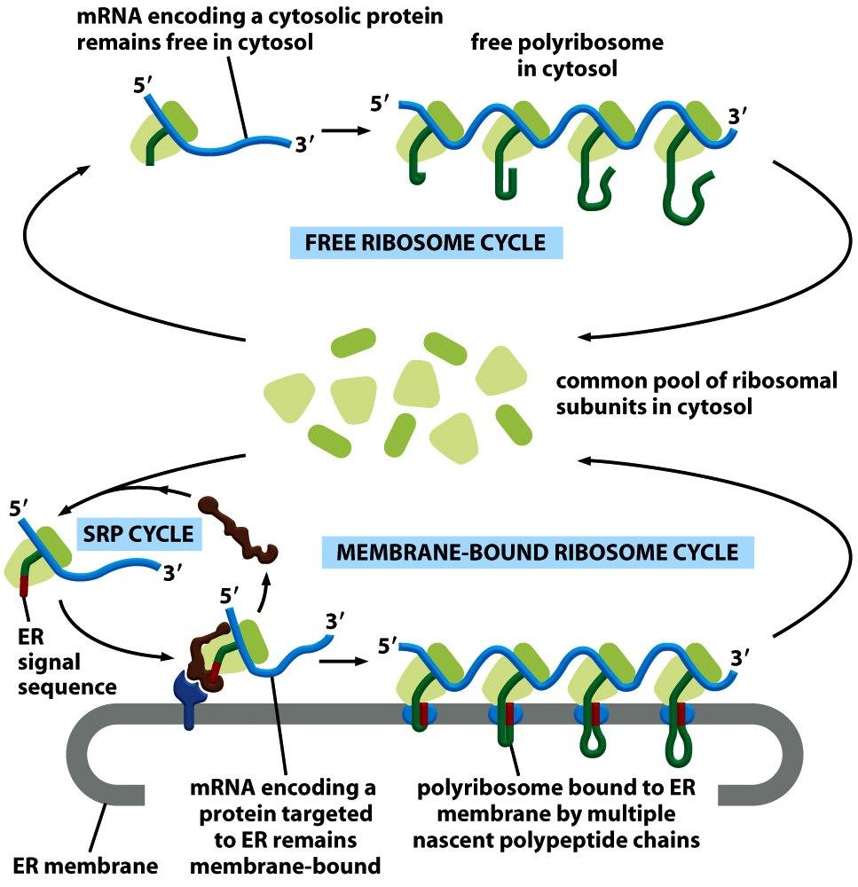

23 Free and Membrane-Bound Ribosomes Figure

24 Protein Translocation Can Be Driven Through Structurally Similar Translocators Figure No additional energy is needed An additional complex is attached to the Sec61 translocator and deposits BiP molecules onto the translocating chain as it emerges into the ER lumen. ATP-driven cycles of BiP binding and release pull the protein into the lumen ATP-hydrolysis-driven conformational changes drive a pistonlike motion in SecA ATPase, each cycle pushing about 20 aa of the protein chain through the pore of the translocator. 24
, the translocator opens its pore, allowing the transfer of the polypeptide chain across the lipid bilayer as a")
25 A Model for How a Soluble Protein Is Translocated Across the ER Membrane! Figure On binding an ER signal sequence (which acts as a starttransfer signal), the translocator opens its pore, allowing the transfer of the polypeptide chain across the lipid bilayer as a loop. After the protein has been completely translocated, the pore closes, but the translocator now opens laterally within the lipid bilayer, allowing the hydrophobic signal sequence to diffuse into the bilayer, where it is rapidly degraded. * In this figure, the ribosomes have been omitted for clarity. 25
 that functions as a start-transfer signal, as in Figure 12 45.")
26 How a Single-Pass Transmembrane Protein with a Cleaved ER Signal Sequence Is Integrated into the ER Membrane Figure In this protein, the co-translational translocation process is initiated by an N-terminal ER signal sequence (red) that functions as a start-transfer signal, as in Figure In addition to this start-transfer sequence, however, the protein also contains a stop-transfer sequence (orange).when the stop-transfer sequence enters the translocator and interacts with a binding site, the translocator changes its conformation and discharges the protein laterally into the lipid bilayer. * In this figure, the ribosomes have been omitted for clarity. 26
 or its N-terminus (pathway B) in the ER lumen.")
27 How a Single-Pass Transmembrane Protein with an Internal ER Signal Sequence Is Integrated into the ER Membrane An internal ER signal sequence (also binds to the SRP) that functions as start transfer signal can bind to the translocator in one of two different ways, leading to a membrane protein that has either its C-terminus (pathway A) or its N-terminus (pathway B) in the ER lumen. Proteins are directed into either pathway by features in the polypeptide chain flanking the internal start transfer sequence. * In this figure, the ribosomes have been omitted for clarity. Figure
 and initiates the transfer of the C-terminal part of the protein.")
28 Integration of a Double-Pass Membrane Protein with an Internal Signal Sequence into the ER Membrane Figure In this protein, an internal ER signal sequence acts as a start-transfer signal (as in Figure 12 47) and initiates the transfer of the C-terminal part of the protein. At some point after a stoptransfer sequence has entered the translocator, the translocator discharges the sequence laterally into the membrane. * In this figure, the ribosomes have been omitted for clarity. 28
 The hydrophobic region nearest the N-terminus serves as a start-transfer sequence that causes the preceding N-terminal portion of the protein to pass across the ER membrane.")
29 The Insertion of the Multipass Membrane Protein Rhodopsin into the ER Membrane Figure (A) A hydrophobicity (also called hydropathy) plot identifies seven short hydrophobic regions in rhodopsin. (B) The hydrophobic region nearest the N-terminus serves as a start-transfer sequence that causes the preceding N-terminal portion of the protein to pass across the ER membrane. Subsequent hydrophobic sequences function in alternation as start-transfer and stop-transfer sequences. (C) The final integrated rhodopsin has its N-terminus located in the ER lumen and its C- terminus located in the cytosol. The blue hexagons represent covalently attached oligosaccharides. Arrows indicate the paired start and stop signals inserted into the 29 translocator.
 The EmrE protein has two identical subunits with completely inverted topology. (C) The topology of aquaporin 1 changes before and after insertion into the membrane.")
30 Complex Membrane Protein Folding J U Bowie Science 2013;339: (A) The ClC chloride channel, which consists of two subunits, has a complex arrangement of helices. (B) The EmrE protein has two identical subunits with completely inverted topology. (C) The topology of aquaporin 1 changes before and after insertion into the membrane. The red arrows highlight the direction of helix rearrangements. Several helices must either insert into the membrane or flip orientations. (D) Topology rearrangements in lactose permease with varying lipid composition. The helices that undergo topology rearrangements in the distinct lipid environments are circled in the dashed red box.
Lecture 2 I. Membrane Proteins II. Intracellular Compartments
 Lecture 2 I. Membrane Proteins II. Intracellular Compartments Ref: MBoC (5th Edition), Alberts Johnson Lewis Raff Roberts Walter Chapter 10 Membrane Structure Chapter 12 Intracellular Compartments and
Intracellular Compartments and Protein Sorting
 Intracellular Compartments and Protein Sorting Intracellular Compartments A eukaryotic cell is elaborately subdivided into functionally distinct, membrane-enclosed compartments. Each compartment, or organelle,
Molecular Cell Biology Problem Drill 16: Intracellular Compartment and Protein Sorting
 Molecular Cell Biology Problem Drill 16: Intracellular Compartment and Protein Sorting Question No. 1 of 10 Question 1. Which of the following statements about the nucleus is correct? Question #01 A. The
Protein Trafficking in the Secretory and Endocytic Pathways
 Protein Trafficking in the Secretory and Endocytic Pathways The compartmentalization of eukaryotic cells has considerable functional advantages for the cell, but requires elaborate mechanisms to ensure
Lecture 4. Protein Translocation & Nucleocytoplasmic Transport
 Lecture 4 Protein Translocation & Nucleocytoplasmic Transport Chapter 12 MBoC (5th Edition) Alberts et al. Reference paper: Tran and Wente, Cell 125, 1041-1053, 2006 2/6/2013 1 A Model for How a Soluble
Practice Exam 2 MCBII
 1. Which feature is true for signal sequences and for stop transfer transmembrane domains (4 pts)? A. They are both 20 hydrophobic amino acids long. B. They are both found at the N-terminus of the protein.
Intracellular vesicular traffic. B. Balen
 Intracellular vesicular traffic B. Balen Three types of transport in eukaryotic cells Figure 12-6 Molecular Biology of the Cell ( Garland Science 2008) Endoplasmic reticulum in all eucaryotic cells Endoplasmic
Lecture 6 - Intracellular compartments and transport I
 01.25.10 Lecture 6 - Intracellular compartments and transport I Intracellular transport and compartments 1. Protein sorting: How proteins get to their appropriate destinations within the cell 2. Vesicular
Zool 3200: Cell Biology Exam 4 Part I 2/3/15
 Name: Key Trask Zool 3200: Cell Biology Exam 4 Part I 2/3/15 Answer each of the following questions in the space provided, explaining your answers when asked to do so; circle the correct answer or answers
Protein sorting (endoplasmic reticulum) Dr. Diala Abu-Hsasan School of Medicine
 Dr. Diala Abu-Hsasan School of Medicine") Protein sorting (endoplasmic reticulum) Dr. Diala Abu-Hsasan School of Medicine dr.abuhassand@gmail.com An overview of cellular components Endoplasmic reticulum (ER) It is a network of membrane-enclosed
Protein sorting (endoplasmic reticulum) Dr. Diala Abu-Hsasan School of Medicine dr.abuhassand@gmail.com An overview of cellular components Endoplasmic reticulum (ER) It is a network of membrane-enclosed
TRANSPORT PROCESSES. 1b. moving proteins into membranes and organelles
 1b. moving proteins into membranes and organelles SLIDE 1 A typical mammalian cell contains up to 10,000 different kinds of proteins. The vast majority of these proteins are synthesized by cytosolic ribosomes,
Essential Cell Biology
 Alberts Bray Hopkin Johnson Lewis Raff Roberts Walter Essential Cell Biology FOURTH EDITION Chapter 15 Intracellular Compartments and Protein Transport Copyright Garland Science 2014 CHAPTER CONTENTS MEMBRANE-ENCLOSED
2013 John Wiley & Sons, Inc. All rights reserved. PROTEIN SORTING. Lecture 10 BIOL 266/ Biology Department Concordia University. Dr. S.
 PROTEIN SORTING Lecture 10 BIOL 266/4 2014-15 Dr. S. Azam Biology Department Concordia University Introduction Membranes divide the cytoplasm of eukaryotic cells into distinct compartments. The endomembrane
Molecular Trafficking
 SCBM 251 Molecular Trafficking Assoc. Prof. Rutaiwan Tohtong Department of Biochemistry Faculty of Science rutaiwan.toh@mahidol.ac.th Lecture outline 1. What is molecular trafficking? Why is it important?
Chapter 4: Cell Structure and Function
 Chapter 4: Cell Structure and Function Robert Hooke Fig. 4-2, p.51 The Cell Smallest unit of life Can survive on its own or has potential to do so Is highly organized for metabolism Senses and responds
Summary of Endomembrane-system
 Summary of Endomembrane-system 1. Endomembrane System: The structural and functional relationship organelles including ER,Golgi complex, lysosome, endosomes, secretory vesicles. 2. Membrane-bound structures
10/13/11. Cell Theory. Cell Structure
 Cell Structure Grade 12 Biology Cell Theory All organisms are composed of one or more cells. Cells are the smallest living units of all living organisms. Cells arise only by division of a previously existing
The Cell Organelles. Eukaryotic cell. The plasma membrane separates the cell from the environment. Plasma membrane: a cell s boundary
 Eukaryotic cell The Cell Organelles Enclosed by plasma membrane Subdivided into membrane bound compartments - organelles One of the organelles is membrane bound nucleus Cytoplasm contains supporting matrix
Zool 3200: Cell Biology Exam 4 Part I 2/3/15
 Name: Trask Zool 3200: Cell Biology Exam 4 Part I 2/3/15 Answer each of the following questions in the space provided, explaining your answers when asked to do so; circle the correct answer or answers
Plasma Membrane. comprised of a phospholipid bilayer and embedded proteins separates the cells s contents from its surroundings
 Cell Organelles Plasma Membrane comprised of a phospholipid bilayer and embedded proteins separates the cells s contents from its surroundings Cytosol the fluid Cytoplasm cell interior, everything outside
Cytosol the fluid Cytoplasm cell interior, everything outside the nucleus but within the cell membrane, includes the organelles, cytosol, and
 Cell Organelles Plasma Membrane comprised of a phospholipid bilayer and embedded proteins Outer surface has oligosaccharides separates the cells s contents from its surroundings Cytosol the fluid Cytoplasm
1. to understand how proteins find their destination in prokaryotic and eukaryotic cells 2. to know how proteins are bio-recycled
 Protein Targeting Objectives 1. to understand how proteins find their destination in prokaryotic and eukaryotic cells 2. to know how proteins are bio-recycled As a protein is being synthesized, decisions
Don t Freak Out. Test on cell organelle on Friday!
 Cell Structure 1 Don t Freak Out Test on cell organelle on Friday! This test should be a buffer test and help raise your overall test score. All information will come from this week! 2 Cells Provide Compartments
A. Major parts 1. Nucleus 2. Cytoplasm a. Contain organelles (see below) 3. Plasma membrane (To be discussed in Cellular Transport Lecture)
 3. Plasma membrane (To be discussed in Cellular Transport Lecture)") Lecture 5: Cellular Biology I. Cell Theory Concepts: 1. Cells are the functional and structural units of living organisms 2. The activity of an organism is dependent on both the individual and collective
Lecture 5: Cellular Biology I. Cell Theory Concepts: 1. Cells are the functional and structural units of living organisms 2. The activity of an organism is dependent on both the individual and collective
A Tour of the Cell. Chapter 6. Biology Eighth Edition Neil Campbell and Jane Reece. PowerPoint Lecture Presentations for
 Chapter 6 A Tour of the Cell PowerPoint Lecture Presentations for Biology Eighth Edition Neil Campbell and Jane Reece Lectures by Chris Romero, updated by Erin Barley with contributions from Joan Sharp
The Cell. Biology 105 Lecture 4 Reading: Chapter 3 (pages 47 62)
") The Cell Biology 105 Lecture 4 Reading: Chapter 3 (pages 47 62) Outline I. Prokaryotic vs. Eukaryotic II. Eukaryotic A. Plasma membrane transport across B. Main features of animal cells and their functions
The Cell Biology 105 Lecture 4 Reading: Chapter 3 (pages 47 62) Outline I. Prokaryotic vs. Eukaryotic II. Eukaryotic A. Plasma membrane transport across B. Main features of animal cells and their functions
Human height. Length of some nerve and muscle cells. Chicken egg. Frog egg. Most plant and animal cells Nucleus Most bacteria Mitochondrion
 10 m 1 m 0.1 m 1 cm Human height Length of some nerve and muscle cells Chicken egg Unaided eye 1 mm Frog egg 100 µm 10 µm 1 µm 100 nm 10 nm Most plant and animal cells Nucleus Most bacteria Mitochondrion
Chapter 1 Plasma membranes
 1 of 5 TEXTBOOK ANSWERS Chapter 1 Plasma membranes Recap 1.1 1 The plasma membrane: keeps internal contents of the cell confined to one area keeps out foreign molecules that damage or destroy the cell
BIO 5099: Molecular Biology for Computer Scientists (et al)
") BIO 5099: Molecular Biology for Computer Scientists (et al) Lecture 15: Being a Eukaryote: From DNA to Protein, A Tour of the Eukaryotic Cell. Christiaan van Woudenberg Being A Eukaryote Basic eukaryotes
BIO 5099: Molecular Biology for Computer Scientists (et al) Lecture 15: Being a Eukaryote: From DNA to Protein, A Tour of the Eukaryotic Cell. Christiaan van Woudenberg Being A Eukaryote Basic eukaryotes
4 A Tour of the Cell CAMPBELL BIOLOGY IN FOCUS. Urry Cain Wasserman Minorsky Jackson Reece
 CAMPBELL BIOLOGY IN FOCUS Urry Cain Wasserman Minorsky Jackson Reece 4 A Tour of the Cell Lecture Presentations by Kathleen Fitzpatrick and Nicole Tunbridge Overview: The Fundamental Units of Life All
Lecture Series 4 Cellular Membranes. Reading Assignments. Selective and Semi-permeable Barriers
 Lecture Series 4 Cellular Membranes Reading Assignments Read Chapter 11 Membrane Structure Review Chapter 12 Membrane Transport Review Chapter 15 regarding Endocytosis and Exocytosis Read Chapter 20 (Cell
I. Fluid Mosaic Model A. Biological membranes are lipid bilayers with associated proteins
 Lecture 6: Membranes and Cell Transport Biological Membranes I. Fluid Mosaic Model A. Biological membranes are lipid bilayers with associated proteins 1. Characteristics a. Phospholipids form bilayers
17/01/2017. Protein trafficking between cell compartments. Lecture 3: The cytosol. The mitochondrion - the power plant of the cell
 ell biology 2017 version 13/1 2017 ote endosome vs lysosome handout Lecture 3: Text book Alberts et al.: hapter 12-14 (Topics covered by the lecture) A lot of reading! Focus on principles ell Biology interactive
Nucleic acids. Nucleic acids are information-rich polymers of nucleotides
 Nucleic acids Nucleic acids are information-rich polymers of nucleotides DNA and RNA Serve as the blueprints for proteins and thus control the life of a cell RNA and DNA are made up of very similar nucleotides.
Early scientists who observed cells made detailed sketches of what they saw.
 Early scientists who observed cells made detailed sketches of what they saw. Early scientists who observed cells made detailed sketches of what they saw. CORK Early scientists who observed cells made detailed
Chapter 3: Cell Structure and Function Assignment
 Chapter 3: Cell Structure and Function Assignment Provide full detail and be specific for full marks. 1. How do these organelles work together? 2 marks each = 10 marks a. lysosomes and vacuoles When material
Lecture 6 9/17 Dr. Hirsh Organization of Cells, continued
 Cell structure of Eukaryotic cells Lecture 6 9/17 Dr. Hirsh Organization of Cells, continued Lots of double-membraned organelles Existence of an Endo-membrane system separation of areas of cell, transport
CELL PARTS TYPICAL ANIMAL CELL
 AP BIOLOGY CText Reference, Campbell v.8, Chapter 6 ACTIVITY1.12 NAME DATE HOUR CELL PARTS TYPICAL ANIMAL CELL ENDOMEMBRANE SYSTEM TYPICAL PLANT CELL QUESTIONS: 1. Write the name of the cell part in the
AP Biology
 Tour of the Cell (1) 2007-2008 Types of cells Prokaryote bacteria cells - no organelles - organelles Eukaryote animal cells Eukaryote plant cells Cell Size Why organelles? Specialized structures - specialized
Cytoskeleton. Provide shape and support for the cell. Other functions of the cytoskeleton. Nucleolus. Nucleus
 Chapter 4: Cell Structure and Function Cytoskeleton The cytoskeleton is a network of fibers that organizes structures and activities in the cell. Microtubules (the largest) Intermediate fibers Microfilaments
Cellular compartments
 Cellular compartments 1. Cellular compartments and their function 2. Evolution of cellular compartments 3. How to make a 3D model of cellular compartment 4. Cell organelles in the fluorescent microscope
Thursday, October 16 th
 Thursday, October 16 th Good morning. Those of you needing to take the Enzymes and Energy Quiz will start very soon. Students who took the quiz Wednesday: Please QUIETLY work on the chapter 6 reading guide.
Moving Proteins into Membranes and Organelles
 13 Moving Proteins into Membranes and Organelles Review the Concepts 1. In eukaryotes, protein translocation across the endoplasmic reticulum (ER) membrane is most commonly cotranslational; it can also
Bio10 Cell Structure SRJC
 3.) Cell Structure and Function Structure of Cell Membranes Fluid mosaic model Mixed composition: Phospholipid bilayer Glycolipids Sterols Proteins Fluid Mosaic Model Phospholipids are not packed tightly
Structures in Cells. Cytoplasm. Lecture 5, EH1008: Biology for Public Health, Biomolecules
 Structures in Cells Lecture 5, EH1008: Biology for Public Health, Biomolecules Limian.zheng@ucc.ie 1 Cytoplasm Nucleus Centrioles Cytoskeleton Cilia Microvilli 2 Cytoplasm Cellular material outside nucleus
4/12/17. Cells. Cell Structure. Ch. 2 Cell Structure and Func.on. Range of Cell Sizes BIOL 100
 Ch. 2 Cell Structure and Func.on BIOL 100 Cells Fundamental units of life Cell theory All living things are composed of one or more cells. The cell is the most basic unit of life. All cells come from pre-existing
BIO 5099: Molecular Biology for Computer Scientists (et al) Lecture 15: Being A Eukaryote. Eukaryotic Cells. Basic eukaryotes have:
 Lecture 15: Being A Eukaryote. Eukaryotic Cells. Basic eukaryotes have:") BIO 5099: Molecular Biology for Computer Scientists (et al) Lecture 15: Being a Eukaryote: From DNA to Protein, A Tour of the Eukaryotic Cell. Christiaan van Woudenberg Being A Eukaryote Basic eukaryotes
BIO 5099: Molecular Biology for Computer Scientists (et al) Lecture 15: Being a Eukaryote: From DNA to Protein, A Tour of the Eukaryotic Cell. Christiaan van Woudenberg Being A Eukaryote Basic eukaryotes
A Tour of the Cell. Chapter 6. Biology. Edited by Shawn Lester. Inner Life of Cell. Eighth Edition Neil Campbell and Jane Reece
 Chapter 6 A Tour of the Cell Inner Life of Cell Edited by Shawn Lester PowerPoint Lecture Presentations for Biology Eighth Edition Neil Campbell and Jane Reece Lectures by Chris Romero, updated by Erin
The Cell. BIOLOGY OF HUMANS Concepts, Applications, and Issues. Judith Goodenough Betty McGuire
 BIOLOGY OF HUMANS Concepts, Applications, and Issues Fifth Edition Judith Goodenough Betty McGuire 3 The Cell Lecture Presentation Anne Gasc Hawaii Pacific University and University of Hawaii Honolulu
Structures in Cells. Lecture 5, EH1008: Biology for Public Health, Biomolecules.
 Structures in Cells Lecture 5, EH1008: Biology for Public Health, Biomolecules Limian.zheng@ucc.ie 1 Cytoplasm Nucleus Centrioles Cytoskeleton Cilia Microvilli 2 Cytoplasm Cellular material outside nucleus
Molecular Cell Biology 5068 In Class Exam 1 October 3, 2013
 Molecular Cell Biology 5068 In Class Exam 1 October 3, 2013 Exam Number: Please print your name: Instructions: Please write only on these pages, in the spaces allotted and not on the back. Write your number
(a) TEM of a plasma. Fimbriae. Nucleoid. Ribosomes. Plasma membrane. Cell wall Capsule. Bacterial chromosome
 TEM of a plasma. Fimbriae. Nucleoid. Ribosomes. Plasma membrane. Cell wall Capsule. Bacterial chromosome") 0 m m 0. m cm mm 00 µm 0 µm 00 nm 0 nm Human height Length of some nerve and muscle cells Chicken egg Frog egg Most plant and animal cells Most bacteria Smallest bacteria Viruses Proteins Unaided eye Light
0 m m 0. m cm mm 00 µm 0 µm 00 nm 0 nm Human height Length of some nerve and muscle cells Chicken egg Frog egg Most plant and animal cells Most bacteria Smallest bacteria Viruses Proteins Unaided eye Light
Chapter 7. (7-1 and 7-2) A Tour of the Cell
 A Tour of the Cell") Chapter 7 (7-1 and 7-2) A Tour of the Cell Microscopes as Windows to the World of Cells Cells were first described in 1665 by Robert Hooke. By the mid-1800s, the accumulation of scientific evidence led
Chapter 7 (7-1 and 7-2) A Tour of the Cell Microscopes as Windows to the World of Cells Cells were first described in 1665 by Robert Hooke. By the mid-1800s, the accumulation of scientific evidence led
Cell Structure and Function. Biology 12 Unit 1 Cell Structure and Function Inquiry into Life pages and 68-69
 Cell Structure and Function Biology 12 Unit 1 Cell Structure and Function Inquiry into Life pages 45 59 and 68-69 Assignments for this Unit Pick up the notes/worksheet for this unit and the project There
LIFE IS CELLULAR. Cell Theory. Cells Are Small. Prokaryotic Cell 10/4/15. Chapter 7 Cell Structure and Function
 Chapter 7 Cell Structure and Function The cell basic unit of life, all living things are made of a cell (unicellular) or more than one cell (multicellular). LIFE IS CELLULAR The invention of the microscope
BIOSC 041. v Today s lecture. v Today s lab. v Note- Monday is a holiday good time to do some reading!
 BIOSC 041 v Today s lecture Review questions Chapter 6, Cells More review questions v Today s lab Quick review of lab safety The Scientific Method start thinking about which environments you might want
endomembrane system internal membranes origins transport of proteins chapter 15 endomembrane system
 endo system chapter 15 internal s endo system functions as a coordinated unit divide cytoplasm into distinct compartments controls exocytosis and endocytosis movement of molecules which cannot pass through
Cell Structure & Function. Source:
 Cell Structure & Function Source: http://koning.ecsu.ctstateu.edu/cell/cell.html Definition of Cell A cell is the smallest unit that is capable of performing life functions. http://web.jjay.cuny.edu/~acarpi/nsc/images/cell.gif
Renáta Schipp Gergely Berta Department of Medical Biology
 The cell III. Renáta Schipp Gergely Berta Department of Medical Biology Size and Biology Biology is a visually rich subject many of the biological events and structures are smaller than the unaided human
A Tour of the Cell. reference: Chapter 6. Reference: Chapter 2
 A Tour of the Cell reference: Chapter 6 Reference: Chapter 2 Monkey Fibroblast Cells stained with fluorescent dyes to show the nucleus (blue) and cytoskeleton (yellow and red fibers), image courtesy of
Endomembrane system 11/1/2018. Endomembrane System. Direct physical continuity. Transfer of membrane segments as vesicles. Outer Nuclear envelope
 Endomembrane system Endomembrane System Outer Nuclear envelope Direct physical continuity Transfer of membrane segments as vesicles Endoplasmic reticulum BUT membranes are not identical in structure and
5/12/2015. Cell Size. Relative Rate of Reaction
 Cell Makeup Chapter 4 The Cell: The Fundamental Unit of Life We previously talked about the cell membrane The cytoplasm is everything inside the membrane, except the nucleus Includes Cytosol = liquid portion
(d) are made mainly of lipids and of proteins that lie like thin sheets on the membrane surface
 are made mainly of lipids and of proteins that lie like thin sheets on the membrane surface") Which of the following statements is no true? Biological membranes (a) are composed partly of amphipathic lipids (b) have hydrophobic and hydrophilic regions (c) are typically in a fluid state (d) are
Which of the following statements is no true? Biological membranes (a) are composed partly of amphipathic lipids (b) have hydrophobic and hydrophilic regions (c) are typically in a fluid state (d) are
Lecture Series 4 Cellular Membranes
 Lecture Series 4 Cellular Membranes Reading Assignments Read Chapter 11 Membrane Structure Review Chapter 21 pages 709-717 717 (Animal( Cell Adhesion) Review Chapter 12 Membrane Transport Review Chapter
Lecture Series 4 Cellular Membranes
 Lecture Series 4 Cellular Membranes Reading Assignments Read Chapter 11 Membrane Structure Review Chapter 12 Membrane Transport Review Chapter 15 regarding Endocytosis and Exocytosis Read Chapter 20 (Cell
PROTEIN TRAFFICKING. Dr. SARRAY Sameh, Ph.D
 PROTEIN TRAFFICKING Dr. SARRAY Sameh, Ph.D Overview Proteins are synthesized either on free ribosomes or on ribosomes bound to endoplasmic reticulum (RER). The synthesis of nuclear, mitochondrial and peroxisomal
Cells and Tissues 3PART A. PowerPoint Lecture Slide Presentation by Patty Bostwick-Taylor, Florence-Darlington Technical College
 PowerPoint Lecture Slide Presentation by Patty Bostwick-Taylor, Florence-Darlington Technical College Cells and Tissues 3PART A Cells and Tissues Carry out all chemical activities needed to sustain life
Types of cells. Cell size comparison. The Jobs of Cells 10/5/2015. Cells & Cell Organelles. Doing Life s Work
 Types of cells Prokaryote Cells & Cell Organelles bacteria cells Doing Life s Work Eukaryotes 2009-2010 animal cells plant cells Cell size comparison Animal cell Bacterial cell most bacteria (prokaryotic)
CHAPTER 4 - CELLS. All living things are made up of one or more cells. A cell is the smallest unit that can carry on all of the processes of life.
 CHAPTER 4 - CELLS Objectives Name the scientists who first observed living and nonliving cells. Summarize the research that led to the development of the cell theory. State the three principles of the
Lecture Series 5 Cellular Membranes
 Lecture Series 5 Cellular Membranes Cellular Membranes A. Membrane Composition and Structure B. Animal Cell Adhesion C. Passive Processes of Membrane Transport D. Active Transport E. Endocytosis and Exocytosis
A. Membrane Composition and Structure. B. Animal Cell Adhesion. C. Passive Processes of Membrane Transport. D. Active Transport
 Cellular Membranes A. Membrane Composition and Structure Lecture Series 5 Cellular Membranes B. Animal Cell Adhesion E. Endocytosis and Exocytosis A. Membrane Composition and Structure The Fluid Mosaic
A Tour of the Cell Lecture 2, Part 1 Fall 2008
 Cell Theory 1 A Tour of the Cell Lecture 2, Part 1 Fall 2008 Cells are the basic unit of structure and function The lowest level of structure that can perform all activities required for life Reproduction
Plant Cells. Chapter 3
 Plant Cells Chapter 3 Major Learning Objectives Contrast prokaryotic and eukaryotic cells Describe the functions of 10 parts of a plant cell Summarize the similarities and differences between plant cells
Renata Schipp Medical Biology Department
 Renata Schipp Medical Biology Department Deffinition of cell The cell is the smallest structural and functional unit of all known living organisms The cell was discovered by Robert Hooke in 1665 and also
Cells. Variation and Function of Cells
 Cells Variation and Function of Cells Cell Theory states that: 1. All living things are made of cells 2. Cells are the basic unit of structure and function in living things 3. New cells are produced from
MCB130 Midterm. GSI s Name:
 1. Peroxisomes are small, membrane-enclosed organelles that function in the degradation of fatty acids and in the degradation of H 2 O 2. Peroxisomes are not part of the secretory pathway and peroxisomal
CELL PART OF THE DAY. Chapter 7: Cell Structure and Function
 CELL PART OF THE DAY Chapter 7: Cell Structure and Function Cell Membrane Cell membranes are composed of two phospholipid layers. Cell membrane is flexible, not rigid The cell membrane has two major functions.
Study Guide for Biology Chapter 5
 Class: Date: Study Guide for Biology Chapter 5 Multiple Choice Identify the choice that best completes the statement or answers the question. 1. Which of the following led to the discovery of cells? a.
A Tour of the Cell Chapter 4. Outline. Early contributors to Understanding Cells. Cell Theory. Cell Size s Matt Schleiden & Ted Schann
 A Tour of the Cell Chapter 4 Outline History of the science behind cells Cell theory & its importance Why are cells small? Microscopes Cell structure and function Prokaryotic cells Eukaryotic cells Early
10 m Human height 1 m Length of some nerve and muscle cells eye 100 mm (10 cm) Chicken egg aid n 10 mm
 Chicken egg aid n 10 mm") Biology 112 Unit Three Chapter Four 1 Cell Sizes Smallest Bacteria Largest Bird egg Longest Giraffe s Nerve Cell Most Cells Diameter of 0.7µm to 105 µm 2 10 m 1 m 100 mm (10 cm) 10 mm (1 cm) Human height
Biology 112 Unit Three Chapter Four 1 Cell Sizes Smallest Bacteria Largest Bird egg Longest Giraffe s Nerve Cell Most Cells Diameter of 0.7µm to 105 µm 2 10 m 1 m 100 mm (10 cm) 10 mm (1 cm) Human height
Name: Multiple choice questions. Pick the BEST answer (2 pts ea)
") Exam 1 202 Oct. 5, 1999 Multiple choice questions. Pick the BEST answer (2 pts ea) 1. The lipids of a red blood cell membrane are all a. phospholipids b. amphipathic c. glycolipids d. unsaturated 2. The
Exam 1 202 Oct. 5, 1999 Multiple choice questions. Pick the BEST answer (2 pts ea) 1. The lipids of a red blood cell membrane are all a. phospholipids b. amphipathic c. glycolipids d. unsaturated 2. The
Organelles of the Cell & How They Work Together. Packet #7
 Organelles of the Cell & How They Work Together Packet #7 Introduction Introduction Organization of cells is basically similar in all cells. Additionally, most cells are tiny Ranging from 1 1000 cubic
1. endoplasmic reticulum This is the location where N-linked oligosaccharide is initially synthesized and attached to glycoproteins.
 Biology 4410 Name Spring 2006 Exam 2 A. Multiple Choice, 2 pt each Pick the best choice from the list of choices, and write it in the space provided. Some choices may be used more than once, and other
Structure of a Generalized Cell
 A Quick Tour Through A Cell BIO130 Lab 2 Exercise 4 The Cell: Anatomy Structure of a Generalized Cell -plasma membrane -cytoplasm: cytosol organelles -nucleus Play TourOfAnimalCell.mpg Plasma membrane
10/5/2015. Cell Size. Relative Rate of Reaction
 The Cell Biology 102 Fundamental unit of life Smallest unit that displays all the basic elements of life Lecture 5: Cells Cell Theory 1. All living things are made of one or more cells Cell Theory 2. The
Lecture Readings. Vesicular Trafficking, Secretory Pathway, HIV Assembly and Exit from Cell
 October 26, 2006 1 Vesicular Trafficking, Secretory Pathway, HIV Assembly and Exit from Cell 1. Secretory pathway a. Formation of coated vesicles b. SNAREs and vesicle targeting 2. Membrane fusion a. SNAREs
Zool 3200: Cell Biology Exam 4 Part II 2/3/15
 Name:Key Trask Zool 3200: Cell Biology Exam 4 Part II 2/3/15 Answer each of the following questions in the space provided, explaining your answers when asked to do so; circle the correct answer or answers
Organelles of the Cell & How They Work Together. Packet #5
 Organelles of the Cell & How They Work Together Packet #5 Developed by Mr. Barrow 2018 1 Introduction Organization of cells is basically similar in all cells. Additionally, most cells are tiny Ranging
Lecture 15. Membrane Proteins I
 Lecture 15 Membrane Proteins I Introduction What are membrane proteins and where do they exist? Proteins consist of three main classes which are classified as globular, fibrous and membrane proteins. A
Cell Overview. Hanan Jafar BDS.MSc.PhD
 Cell Overview Hanan Jafar BDS.MSc.PhD THE CELL is made of: 1- Nucleus 2- Cell Membrane 3- Cytoplasm THE CELL Formed of: 1. Nuclear envelope 2. Chromatin 3. Nucleolus 4. Nucleoplasm (nuclear matrix) NUCLEUS
Posttranslational Modification and Targeting of Proteins
 Posttranslational Modification and Targeting of Proteins Graduate Biochemistry Term 2/2016 Assist. Prof. Dr. Panida Khunkaewla School of Chemistry, Institute of Science Suranaree University of Technology
A Tour of the Cell. Chapter 4. Most cells are microscopic. Cells vary in size and shape
 Chapter 4 A Tour of the Cell Most cells are microscopic Cells vary in size and shape 10 m Human height 1 m Length of some nerve and muscle cells 100 mm (10 cm) 10 mm (1 cm) Chicken egg Unaided eye 1 mm
Biology 4410 First Examination Version B
 Biology 4410 Spring 2006 Name First Examination Version B This examination consists of two parts, a multiple-choice section and an essay section. Be sure to put your name on both the mark-sense sheet and
Endomembrane system, *Chloroplasts, *Mitochondria. *Learn these from text/connect1. Fertilization of a human cell
 Key Concepts: - Cells are the Basic Unit of Life Cell Theory, Surface to Volume - 2 Cell Types Prokaryotic, Eukaryotic - Cell Membrane Membrane Structure - Cell Organelles Endomembrane system, *Chloroplasts,
MULTIPLE CHOICE. Choose the one alternative that best completes the statement or answers the question.
 Exam Name MULTIPLE CHOICE. Choose the one alternative that best completes the statement or answers the question. 1) All of the following are synthesized along various sites of the endoplasmic reticulum
BIOLOGICAL CHEMISTRY Prof. J.H.P. Bayley, Dr. R.M. Adlington and Dr. L. Smith Trinity Term First Year. Lecture 2 Hagan Bayley
 BIOLOGICAL CHEMISTRY Prof. J.H.P. Bayley, Dr. R.M. Adlington and Dr. L. Smith Trinity Term 2007 - First Year Lecture 2 Hagan Bayley Introduction to the macromolecules of life and cell structures. Introduction
Anatomy Chapter 2 - Cells
 Cells Cells are the basic living structural, functional unit of the body Cytology is the branch of science that studies cells The human body has 100 trillion cells 200 different cell types with a variety
A TOUR OF THE CELL 10/1/2012
 A TOUR OF THE CELL Chapter 6 KEY CONCEPTS: Eukaryotic cells have internal membranes that compartmentalize their functions The eukaryotic cell s genetic instructions are housed in the nucleus and carried
MOLECULAR CELL BIOLOGY
 1 Lodish Berk Kaiser Krieger scott Bretscher Ploegh Matsudaira MOLECULAR CELL BIOLOGY SEVENTH EDITION CHAPTER 13 Moving Proteins into Membranes and Organelles Copyright 2013 by W. H. Freeman and Company
ORGANELLES OF THE ENDOMEMBRANE SYSTEM
 Membranes compartmentalize the interior of the cell and facilitate a variety of metabolic activities. Chloroplasts and a rigid cell wall are what distinguish a plant cell from an animal cell. A typical