The Neck. BY: Lina Abdullah & Rahaf Jreisat
|
|
|
- Toby Hampton
- 5 years ago
- Views:
Transcription

1 The Neck BY: Lina Abdullah & Rahaf Jreisat


2 Boundaries of the Neck: generally from base of the skull to root of the neck Superior margin :From superior nuchal line of occipital bone up to mastoid process down to margin of the mandible inferior margin: inlet of thoracic cavity, the clavicle and root of the neck cervical vertebral muscles subocciptal(part of deep muscles posteriorly and they form Remember that superior nuchal line lines posterior of the occipital bone triangular-like space ) and deep neck muscles mainly extends head and neck -rectus capitis posterior major m -rectus capitis posterior minor m -oblique capitis inferior m -oblique capitis superior m -Action of these muscles is extension the head -Recti muscles insert in inferior nuchal line of the occipital bones Anterior vertebral muscles flexes head and neck.longus coli m.longus capitis m :Capitis mean occipital bone so it attach from occipital to transverse processes of cervical vertebra Rectus capitis anterior m :anteriorly to cranium and vertebra of cervical


3 from transverse.anterior scalene m :processes o middle cerviacal vertebrae to first rib and anteriorly to groove for subclavian artery Lateral vertebral muscles: and neck laterally flex head.rectus capitis lateralis m :anterolateral to occipital condyle.splenius capitis m.levator scapulae m :Lateral flexion of the head on one side of scapula up to cervical Middle scalene.m : sup. attachment of transverse processes to first rib and posteriorly to groove for subclavian artery.posterior scalene m : also sup. Attachment of transverse processes of cervical vertebra to second rib So anterior to groove for subclavicn artery is anterior scalene But posteriorly to this groove is middle scalene and between middle and anterior scalene is brachial plexus all of lateral and anterior vertebra muscles are innervated by cervical nerves directly except levator scapulae,which is innervated by cervical nerves and brachial plexus
4 Superficial fascia: (externally) is layer of connective tissue that lies deeper to skin and part of hypodermis, it contains the platysma Deep fascia 3layers *Investing layer :The most superifical layer envelopes the trapezius and sternocleidomastoid muscles **Pretracheal layer :The middle layer has 2 parts: 1- Visceral part: it surrounds the thyroid, trachea,parathyroid,pharynx,larynx and esophagus the fascia surrounds the pharynx and down to esophagus ( in some references) known as buccopharyngeal fascia 2- muscular part :it surrounds the all anterior muscles of the neck (infrahyoid and suprahyoid muscles).***prevertebral layer: the deepest one,it surrounds all vertebral muscles and vertebrae Posterior to pharynx(posterior to pretracheal layer) Continue as axillary sheath Carotid sheath Thickening of the other layers,it is surrounded by all layers of deep fascia contents : Common & internal carotid.aa.intrnal jugular v.vagus n Deep cervical lymph nodes Cervical Fascia extensions Alar fascia: -Division from prevertebral fascia -From skull to T2 that means in superior mediastinum region (merge with )buccopharyngeal fascia anteriorly) Buccopharyngeal fascia : Superior & posterior continuation of the pretracheal fascia That surrounds the pharynx and esophagus The Merging between alar fascia and buccopharyngeal fascia forms space in sup.mediastinum known as real retropharyngeal space
5 Cervical Fascia: Spaces Retropharyngeal space : Between buccopharyngeal fascia and Prevertebral fascia Spread of infections (Real) Retropharyngeal space: Between the alar fascia and buccopharyngeal -Allow movement of pharynx, larynx, and trachea during swallowing -Continuous with superior mediastinum tot2 the spread of infection in real space stops at t2 where fusion of alar and buccopharyngeal posteriorly to alar Between the alar fascia and the Prevertebral fascia Continuous with mediastinum The risk that an infection in this space can spread directly to the thorax,because it continuous with mediastinum
 seperates the neck into anterior and posterior triangles.")
6 Slide 12: (Bear with me) Neck Triangles: Boundaries Anterior triangle: Anterior to SCM Carotid triangle Digastric triangle Submental triangle Muscular triangle Posterior triangle: Posterior to SCM Anterior to trapezius Occipital triangle Supraclavicular triangle *Sternocleidomastoid (SCM) seperates the neck into anterior and posterior triangles. *Anterior triangle: - Digastric (submandibular) triangle: between the anterior and posterior bellies of digastrics muscle, floor of digastrics: mylohyoid m. The rest of the triangles are separated by hyoid bone and sup&inf bellies of omohyoid m. carotid triangle : anterior to SCM, posteriosuperior to sup belly of omohyoid, and inferior to posterior belly of digastrics. - Submental: superior to hyoid bone, between anterior bellies of digastrics. Floor of submental triangle is made of mylohyoid muscles. - Muscular triangle: inferior to hyoid bone and posterior to superior belly of omohyoid.
7 *Posterior triangle: Inferior belly of omohyoid divides it into two subtriangles - Occipital triangle: superficial, superior to inf. belly of omohyoid, its floor is made of muscles (scalenius medius, levator scapulae, splenius capitis) - Supraclavicular triangle: superior to clavicle, inferior to inf. belly of omohyoid and lateral to SCM. Neck Triangles: Content Anterior Triangle: *Muscular triangle: -Infrahyoid muscles (sternohyoid, sternothyoid, thyrohyoid, omohyoid) -Viscera (thyroid cartilage, cricoid cartilage, trachea, thyroid gland isthmus and connections btw these structures) * Submental triangle: -Lymph nodes in submental groove that can be felt is they swell. * Digastric triangle: - Submandibular lymph nodes: drain nasal&oral cavity, pharynx - Submandibular salivary gland (superficial part) - Facial artery: pass deep to submandibular gland then transverse the lower edge of mandible (become superficial), you can feel it pulsing just inferior to masseteric muscle. Importance of this triangle: 1.injuries damage facial artery 2. Swelling of lymph nodes in common cold, sinusitis, *Carotid triangle: -Carotid sheath and its contents - Hypoglossal nerve before it goes deep to mylohyoid
 then passes deep towards SCM, it exits through posterior edge of SCM and transverses occipital triangle where it becomes superficial (separated from skin by investing deep fascia only) which")
8 - Cervical part of sympathetic trunk (posterior) Posterior Triangle: *Occipital triangle: - accessory nerve which innervates SCM and trapezius accessory nerve exits through jugular foramen(which is deep) then passes deep towards SCM, it exits through posterior edge of SCM and transverses occipital triangle where it becomes superficial (separated from skin by investing deep fascia only) which makes it more susceptible to injury. Any injury in this triangle can affect accessory nerve and leads to paralysis in trapezius muscle one can t lift his shoulder. *Supraclavicular triangle: -External jugular vein

9 معلش Sensory Innervations of the Neck *cervical plexus: -motor and sensory branches sensory branches exit posterior to SCM muscle and spread all over cervical region. -Deep: ansa cervicalis (part of carotid triangle) = hypoglossal nerve& C2& C3
: Veins (Right and left brachiocephalic), vagus")
10 innervate infrahyoid muscles. *Well be taken in another lecture Root of the Neck *Boundries: Ant: suprasternal angle Lateral: first rib Pos: body of T1 *Contents (from ant to pos): Veins (Right and left brachiocephalic), vagus and phrenic nerves, arteries, trachea, esophagus, thoracic duct & left lymphatic duct. Arteries of the Head and Neck Branches of external carorid artery: Superior thyroid artery
 Facial artery occipital artery (hypoglossal nerve exit just inferior to it) Posterior auricular artery Superficial temporal artery (pass superficial to zygomatic arch anterior to ear, you can feel")
11 Ascending pharyngeal artery Lingual artery (deep to mylohyoid m.) Facial artery occipital artery (hypoglossal nerve exit just inferior to it) Posterior auricular artery Superficial temporal artery (pass superficial to zygomatic arch anterior to ear, you can feel its pulse) Maxillary artery (Enter pteygopalatine fossa, pass with maxillary nerve) Mneumonic: Some American Ladies Find Our Pyramids Most Satisfactory Veins of the Head and Neck

12 *Internal Jugular vein drain all cranial cavity and a lot of superficial veins (facial, retromandiular, external jugular) *Retromandibular vein = union of maxillary vein and superficial temporal veins * External jugular can be drained in internal jugular or subclavian vein. Principal groups of lymph nodes Lymph drainage of Head and neck Regional lymph nodes: Occipital
 and submandibular lymph")
13 Retroauricular Parotid Buccal Submandibular Submental Anterior cervical Laryngeal Tracheal Superficial cervical Deep cervical: Jugular trunk *occipitaland & retroauricular are posterior. *The fate of all lymph all over head and neck: Deep Cervical *Deep cervical drain all superficial and ant group (pretracheal, paratracheal, retrotracheal, retropharyngeal) and submandibular lymph nodes. *Importance: knowing the sequence for tracing metastasis.
: -Junction of the superior & middle thirds of the posterior border of SCM junction between middle & lower")
 triangle -")
14 Occipital Triangle Above the inferior belly of omohyoid m. Spinal accessory nerve (XI): -Junction of the superior & middle thirds of the posterior border of SCM junction between middle & lower thirds of the anterior border of trapezius -Injury Anterior Triangle Carotid triangle: -Carotid sheath: Between sternocalvicular joint and the mid point between mastoid and angle of mandible - Hypoglossal nerve - Cervical sympathetic trunk Submandibular (digastric) triangle - Submandibular gland - Submandibular lymph nodes Surface Anatomy of the Neck Hyoid bone C3:
: - Vocal cords at the middle Cricoid cartilage C6: -")

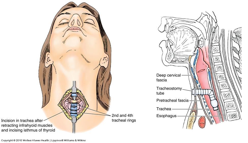
15 - Posterior to the mandible Laryngeal prominence (Adma s apple) tip (C4): - Vocal cords at the middle Cricoid cartilage C6: - Cricothyroid ligament - Cricothyrotomy First tracheal cartilage: - Tracheostomy Thyroid gland: - Isthmus 2nd 4th tracheal Rings Tracheostomy
16
Neck-2. Dr. Heba Kalbouneh Associate Professor of Anatomy and Histology
 Neck-2 ` Dr. Heba Kalbouneh Associate Professor of Anatomy and Histology Triangles of the neck Side of the neck Midline Lower border of mandible Line between angle of mandible and mastoid Superior nuchal
The Neck the lower margin of the mandible above the suprasternal notch and the upper border of the clavicle
 The Neck is the region of the body that lies between the lower margin of the mandible above and the suprasternal notch and the upper border of the clavicle below Nerves of the neck Cervical Plexus Is formed
Anatomy: head and Neck (6 questions) 1. Prevertebral Flexor Musculature (lying in front of the vertebrae) include all, EXCEPT: Longus Colli.
 1. Prevertebral Flexor Musculature (lying in front of the vertebrae) include all, EXCEPT: Longus Colli.") Anatomy: head and Neck (6 questions) 1. Prevertebral Flexor Musculature (lying in front of the vertebrae) include all, EXCEPT: Longus Colli. Rectus Capitis Anterior. Rectus Capitis Lateralis. Rectus Capitis
Anatomy: head and Neck (6 questions) 1. Prevertebral Flexor Musculature (lying in front of the vertebrae) include all, EXCEPT: Longus Colli. Rectus Capitis Anterior. Rectus Capitis Lateralis. Rectus Capitis
Veins of the Face and the Neck
 Veins of the Face and the Neck Facial Vein The facial vein is formed at the medial angle of the eye by the union of the supraorbital and supratrochlear veins. connected through the ophthalmic veins with
Anterior triangle of neck
 Anterior triangle of neck Dept. of Anatomy Zhou Hong Ying Outline boundary and subdivisions of ant. triangle contents of the triangle Muscles: suprahyoid muscles, infrahyoid muscles Nerves: CNⅩ, CNⅪ, CNⅫ,
Posterior Triangle of the Neck By Prof. Dr. Muhammad Imran Qureshi
 Posterior Triangle of the Neck By Prof. Dr. Muhammad Imran Qureshi For the purpose of anatomical description the neck is sub divided into two major triangles, the Anterior and the Posterior by muscle bellies
Prevertebral Region, Pharynx and Soft Palate
 Unit 20: Prevertebral Region, Pharynx and Soft Palate Dissection Instructions: Step1 Step 2 Step 1: Insert your fingers posterior to the sternocleidomastoid muscle, vagus nerve, internal jugular vein,
SCHOOL OF ANATOMICAL SCIENCES Mock Run Questions. 4 May 2012
 SCHOOL OF ANATOMICAL SCIENCES Mock Run Questions 4 May 2012 1. With regard to the muscles of the neck: a. the platysma muscle is supplied by the accessory nerve. b. the stylohyoid muscle is supplied by
Alexander C Vlantis. Selective Neck Dissection 33
 05 Modified Radical Neck Dissection Type II Alexander C Vlantis Selective Neck Dissection 33 Modified Radical Neck Dissection Type II INCISION Various incisions can be used for a neck dissection. The incision
Spinal nerves and cervical plexus Prof. Abdulameer Al Nuaimi. E mail: a.al E. mail:
 Spinal nerves and cervical plexus Prof. Abdulameer Al Nuaimi E mail: a.al nuaimi@sheffield.ac.uk E. mail: abdulameerh@yahoo.com Branches of ophthalmic artery Muscles of face A spinal nerve Spinal
Thyroid and Parathyroid Glands
 Thyroid and Parathyroid Glands Please view our Editing File before studying this lecture to check for any changes. Color Code Important Doctors Notes Notes/ explanation Objectives: By the end of the lecture,
OBJECTIVE: To obtain a fundamental knowledge of the root of the neck with respect to structure and function
 The root of the neck Jeff Dupree, Ph.D. e mail: jldupree@vcu.edu OBJECTIVE: To obtain a fundamental knowledge of the root of the neck with respect to structure and function READING ASSIGNMENT: Moore and
Tympanic Bulla Temporal Bone. Digastric Muscle. Masseter Muscle
 Superior view Hyoid Bone The hyoid bone does not articulate with any other bones. It is held in place by ligaments to the styloid process of the temporal bone and the thyroid cartilage of the larynx. It
THYROID & PARATHYROID. By Prof. Saeed Abuel Makarem & Dr. Sanaa Al-Sharawy
 THYROID & PARATHYROID By Prof. Saeed Abuel Makarem & Dr. Sanaa Al-Sharawy 1 OBJECTIVES By the end of the lecture, the student should be able to: Describe the shape, position, relations and structure of
Chapter 28: The neck. Fascia of the neck
 Chapter 28: The neck Fascia of the neck The superficial fascia is a fatty areolar layer between the skin and the more obvious deep fascia. It contains the platysma muscles and the external jugular veins
Surgical Anatomy of the Neck. M. J. Jurkiewicz, John Bostwick. Surgical Clinics of North America, Vol 54, No 6, December 1974.
 Surgical Anatomy of the Neck M. J. Jurkiewicz, John Bostwick Surgical Clinics of North America, Vol 54, No 6, December 1974. The radical neck dissection is a safe, effective therapeutic procedure for eradication
Lecture 07. Lymphatic's of Head & Neck. By: Dr Farooq Amanullah Khan PMC
 Lecture 07 Lymphatic's of Head & Neck By: Dr Farooq Amanullah Khan PMC Dated: 28.11.2017 Lymphatic Vessels Of the 800 lymph nodes in the human body, 300 are in the Head & neck region. The lymphatic vessels
The Pharynx. Dr. Nabil Khouri MD. MSc, Ph.D
 The Pharynx Dr. Nabil Khouri MD. MSc, Ph.D Introduction The pharynx is the Musculo-fascial halfcylinder that links the oral and nasal cavities in the head to the larynx and esophagus in the neck Common
REVIEW/PREVIEW OF HEAD AND NECK ANATOMY FOR ENT EXAM
 REVIEW/PREVIEW OF HEAD AND NECK ANATOMY FOR ENT EXAM - 2017 PALPATE CAROTID ARTERY: AT LEVEL OF CAROTID BIFURCATION VERTEBRAL LEVEL C4 Sternocleidomastoid Muscle INTERNAL CAROTID EXTERNAL CAROTID COMMON
For the following questions, indicate the letter that corresponds to the SINGLE MOST APPROPRIATE ANSWER
 GROSS ANATOMY EXAMINATION May 15, 2000 For the following questions, indicate the letter that corresponds to the SINGLE MOST APPROPRIATE ANSWER 1. Pain associated with an infection limited to the middle
CERVICAL LYMPH NODES
 CERVICAL LYMPH NODES (ANATOMY & EXAMINATION) Hemant (DTCD 1 st YEAR) 1. Lymphatic Tissues: A Type of connective tissue that contains large numbers of lymphocytes. 2. Lymphatic Vessels: Are Tubes that assist
General Anatomic Layout
 General Anatomic Layout 2 Core Messages At the start of the dissection exercise, we must take a panoramic look for orientation. We then establish the limits of the area of operation and the main landmarks.
Lecture 01. The Thyroid & Parathyroid Glands. By: Dr Farooq Khan PMC Date: 12 th March. 2018
 Lecture 01 The Thyroid & Parathyroid Glands By: Dr Farooq Khan PMC Date: 12 th March. 2018 INTRODUCTION LAYERS OF THE NECK The neck has four major compartments or layer which are enclosed by an outer musculofascial
Thyroid gland. importance. relations and connections. external laryngeal nerves. malformations.
 Thyroid gland 1. Recognize and understand the coverings of the thyroid gland and their clinical importance. 2. Recognize and understand the main parts of the thyroid gland and their locations, relations
Head & Neck Contouring
 Head & Neck Contouring Presented by James Wheeler, MD Center for Cancer Care Goshen, IN 46526 September 12, 2014 Special Thanks to: Spencer Boulter, Director of Operations (AAMD) Adam Moore, RT(T), CMD
Chapter 84: Surgical Anatomy. Raleigh E. Lingeman
 Chapter 84: Surgical Anatomy Raleigh E. Lingeman Surgeons doing head and neck surgery must first master the surgical anatomy and technique of doing neck dissection. Neck dissection is either the classic
HEAD & NECK ANATOMY - MCQ HEAD & NECK ANATOMY
 . ' HEAD & NECK ANATOMY I. Deep investing layer of cervical fascia splits to enclose: A. Sternocleidomastoid B. Trapezius C. Parotid gland D. Omohyoid 2. Regarding the prevertebral fascia, the following
3 Mohammad Al-Mohtasib Areej Mosleh
 3 Mohammad Al-Mohtasib Areej Mosleh ***Muscles Connecting the Upper Limb to the Vertebral Column 1.Trapezius Muscle ***The first muscle on the back is trapezius muscle, it s called so according
Larynx. Rudimentary. Behind the posterior surface : -stylopharyngeus - salpingopharyngeus -platopharyngeus
 Larynx The larynx is an organ that provides a protective sphincter at the inlet of the air passages and is responsible for voice production. It extends from C3-C6: *Posterior: the pharynx *Lateral: the
University of Palestine. Midterm Exam 2013/2014 Total Grade:
 Course No: DNTS2208 Course Title: Head and Neck Anatomy Date: 09/11/2013 No. of Questions: (50) Time: 1hour Using Calculator (No) University of Palestine Midterm Exam 2013/2014 Total Grade: Instructor
CHAPTER 9. The Neck THE "CERVICAL CAVITY" TRACHEA ESOPHAGUS THYROID GLAND PARATHYROID GLANDS
 281 CHAPTER 9 The Neck CERVICAL VERTEBRAE POSTERIOR TRIANGLE OF NECK Trapezius and Sternocleidomastoid Trapezius CLINICAL CONSIDERATIONS REGARDING TRAPEZIUS Sternocleidomastoid CLINICAL CONSIDERATIONS
Tikrit University collage of dentistry Dr.Ban I.S. head & neck anatomy 2 nd y. Lec [5] / Temporal fossa :
![Tikrit University collage of dentistry Dr.Ban I.S. head & neck anatomy 2 nd y. Lec [5] / Temporal fossa :](/thumbs/88/115294566.jpg "Tikrit University collage of dentistry Dr.Ban I.S. head & neck anatomy 2 nd y. Lec [5] / Temporal fossa :") Lec [5] / Temporal fossa : Borders of the Temporal Fossa: Superior: Superior temporal line. Inferior: gap between zygomatic arch and infratemporal crest of sphenoid bone. Anterior: Frontal process of the
Lec [5] / Temporal fossa : Borders of the Temporal Fossa: Superior: Superior temporal line. Inferior: gap between zygomatic arch and infratemporal crest of sphenoid bone. Anterior: Frontal process of the
Lies in front and sides of the neck. Consists of two lobe connected anterior to the trachea by an isthmus.
 THYROID GLAND 1 Lies in front and sides of the neck. Consists of two lobe connected anterior to the trachea by an isthmus. A small pyramidal lobe projects upwards from the left lobe in 40% of cases. The
THE NECK PART I BONES AND JOINTS OF THE NECK MUSCLES OF THE NECK SCM CAROTID TRIANGLE ENDOCRINE LAYER OF THE CERVICAL VISCERA
 THE NECK PART I BONES AND JOINTS OF THE NECK MUSCLES OF THE NECK SCM CAROTID TRIANGLE ENDOCRINE LAYER OF THE CERVICAL VISCERA THE NECK THE NECK The neck has four major compartments, which are enclosed
Anatomy of the Thyroid Gland
 Anatomy of the Thyroid Gland Introduction Nomenclature G, thyreos= shield, eidos= like Location Root of the neck ventrally (C5-T1) Function endocrine gland that secretes: Thyroxine (T4) T3 Calcitonin LWW,
Anatomy and Physiology II. Spine
 Anatomy and Physiology II Spine Bones and Other Structures Vertibrae Contains Cervical, Thoracic, Lumbar, Sacral and Coccygeal regions We use Capital letters to refer to these (C, T, L, S, and Co) and
ANTERIOR CERVICAL TRIANGLE (Fig. 2.1 )
") 2 Neck Anatomy ANTERIOR CERVICAL TRIANGLE (Fig. 2.1 ) The boundaries are: Lateral: sternocleidomastoid muscle Superior: inferior border of the mandible Medial: anterior midline of the neck This large triangle
2 Neck Anatomy ANTERIOR CERVICAL TRIANGLE (Fig. 2.1 ) The boundaries are: Lateral: sternocleidomastoid muscle Superior: inferior border of the mandible Medial: anterior midline of the neck This large triangle
PCM1 Physical Exam Skills Session: Head and Neck FACILITATOR & STUDENT COPY
 PATIENT CENTERED MEDICINE - 1 GOALS & OUTCOMES: PCM1 Physical Exam Skills Session: Head and Neck FACILITATOR & STUDENT COPY 1. To introduce the applied anatomy relevant for the examination of the head
1. Thyroxine (inactive form) also called T4 (90% of the secretion). 2. Triiodothyronine (active form) also called T3 (10% of the secretion).
 also called T4 (90% of the secretion). 2. Triiodothyronine (active form) also called T3 (10% of the secretion).") A Introduction The nomenclature of the thyroid gland comes from its close relation to the thyroid cartilage (the thyroid cartilage was named like this because thyroid means shield and it is shielding the
A Introduction The nomenclature of the thyroid gland comes from its close relation to the thyroid cartilage (the thyroid cartilage was named like this because thyroid means shield and it is shielding the
Candidate s instructions Look at this cross-section taken at the level of C5. Answer the following questions.
 Section 1 Anatomy Chapter 1. Trachea 1 Candidate s instructions Look at this cross-section taken at the level of C5. Answer the following questions. Pretracheal fascia 1 2 5 3 4 Questions 1. Label the
Surgical anatomy of thyroid and parathyroid glands
 Head & Neck Surgery Course Surgical anatomy of thyroid and parathyroid glands Dr Pierfrancesco PELLICCIA Pr Benjamin LALLEMANT Service ORL et CMF CHU de Nîmes CH de Arles Thyroid glands Dr Pierfrancesco
University of Palestine. Midterm Exam 2013/2014 Total Grade:
 [ Course No: DNTS2208 Course Title: Head and Neck Anatomy Date: 17/11/1024 No. of Questions: (52) Time: 2hours Using Calculator (No) University of Palestine Midterm Exam 2013/2014 Total Grade: Instructor
Anatomical Complications in General Surgery. John E. Skandalakis, Stephen W. Gray, Joseph S. Rowe. Chapter 1: The Neck
 Anatomical Complications in General Surgery John E. Skandalakis, Stephen W. Gray, Joseph S. Rowe Chapter 1: The Neck The human neck is so designed that the swelling of a normal structure or the presence
DESCRIPTION: This is the part of the trunk, which is located between the root of the neck and the superior border of the abdominal region.
 1 THE THORACIC REGION DESCRIPTION: This is the part of the trunk, which is located between the root of the neck and the superior border of the abdominal region. SHAPE : T It has the shape of a truncated
PTERYGOPALATINE FOSSA
 PTERYGOPALATINE FOSSA Outline Anatomical Structure and Boundaries Foramina and Communications with other spaces and cavities Contents Pterygopalatine Ganglion Especial emphasis on certain arteries and
Lecturer: Ms DS Pillay ROOM 2P24 25 February 2013
 Lecturer: Ms DS Pillay ROOM 2P24 25 February 2013 Thoracic Wall Consists of thoracic cage Muscle Fascia Thoracic Cavity 3 Compartments of the Thorax (Great Vessels) (Heart) Superior thoracic aperture
Right lung. -fissures:
 -Right lung is shorter and wider because it is compressed by the right copula of the diaphragm by the live.. 2 fissure, 3 lobes.. hilum : 2 bronchi ( ep-arterial, hyp-arterial ), one artery mediastinal
Lab Activity 11: Group I
 Lab Activity 11: Group I Muscles Martini Chapter 11 Portland Community College BI 231 Origin and Insertion Origin: The place where the fixed end attaches to a bone, cartilage, or connective tissue. Insertion:
NECK SUPERFICIAL STRUCTURES OF THE NECK Compartments: SCM, Posterior, Lateral, Anterior
 NECK SUPERFICIAL STRUCTURES OF THE NECK Compartments: SCM, Posterior, Lateral, Anterior BONES Cervical Vertebrae SCM Mastoid process of temp bone & Sup Nuchal line of occiput Manubrium & medial 1/3 1 clavicle
Parotid Gland, Temporomandibular Joint and Infratemporal Fossa
 M1 - Anatomy Parotid Gland, Temporomandibular Joint and Infratemporal Fossa Jeff Dupree Sanger 9-057 jldupree@vcu.edu Parotid gland: wraps around the mandible positioned between the mandible and the sphenoid
Suprahyoid and Infrahyoid Neck Overview
 10 Imaging Approaches & Indications Neither CT nor MR is a perfect modality for imaging the extracranial H&N. MR is most useful in the suprahyoid neck (SHN) because it is less affected by oral cavity dental
Gateway to the upper limb. An area of transition between the neck and the arm.
 Gateway to the upper limb An area of transition between the neck and the arm. Pyramidal space inferior to shoulder @ junction of arm & thorax Distribution center for the neurovascular structures that serve
The Thoracic wall including the diaphragm. Prof Oluwadiya KS
 The Thoracic wall including the diaphragm Prof Oluwadiya KS www.oluwadiya.com Components of the thoracic wall Skin Superficial fascia Chest wall muscles (see upper limb slides) Skeletal framework Intercostal
Dr.Ban I.S. head & neck anatomy 2 nd y. جامعة تكريت كلية طب االسنان املرحلة الثانية أ.م.د. بان امساعيل صديق 6102/6102
 جامعة تكريت كلية طب االسنان التشريح مادة املرحلة الثانية أ.م.د. بان امساعيل صديق 6102/6102 Parotid region The part of the face in front of the ear and below the zygomatic arch is the parotid region. The
Neck of Condylar. Process. Anterior Border of Ramus. Mandibular. Foramen. Posterior Border of Ramus Incisive Fossa.
 Learning Outcomes The Mandible Surface Anatomy Muscle Attachments The (FOM) Muscles of the FOM The Tongue Muscles of the Tongue The Submandibular Region Submandibular Gland Sublingual Gland Lingual The
Dr. Weyrich G07: Superior and Posterior Mediastina. Reading: 1. Gray s Anatomy for Students, chapter 3
 Dr. Weyrich G07: Superior and Posterior Mediastina Reading: 1. Gray s Anatomy for Students, chapter 3 Objectives: 1. Subdivisions of mediastinum 2. Structures in Superior mediastinum 3. Structures in Posterior
Mediastinum and pericardium
 Mediastinum and pericardium Prof. Abdulameer Al-Nuaimi E-mail: a.al-nuaimi@sheffield.ac.uk E. mail: abdulameerh@yahoo.com The mediastinum: is the central compartment of the thoracic cavity surrounded by
Clinical Anatomy of the Thyroid and Adrenal Glands
 Clinical Anatomy of the Thyroid and Adrenal Glands Handout download: http://www.oucom.ohiou.edu/dbms-witmer/gs-rpac.htm 28 October 2003 Lawrence M. Witmer, PhD Department of Biomedical Sciences College
Tikrit University College of Dentistry Dr.Ban I.S. head & neck anatomy 2 nd y.
 Lec [3]/The scalp The scalp extends from the supraorbital margins anteriorly to the nuchal lines at the back of the skull and down to the temporal lines at the sides. The forehead, from eyebrows to hairline,
Larynx - cartilaginous structure holding the vocal folds which protrude into airstream
 1! Larynx - cartilaginous structure holding the vocal folds which protrude into airstream 2! Flow increase - like thumb over garden hose Pressure drop - narrower space forces pressure drop due to speed
Face. Definition: The area between the two ears and from the chin to the eye brows. The muscles of the face
 Face Definition: The area between the two ears and from the chin to the eye brows. The muscles of the face The muscle of facial expression (include the muscle of the face and the scalp). All are derived
slide 23 The lobes in the right and left lungs are divided into segments,which called bronchopulmonary segments
 Done By : Rahmeh Alsukkar Date : 26 /10/2017 slide 23 The lobes in the right and left lungs are divided into segments,which called bronchopulmonary segments Each segmental bronchus passes to a structurally
APRIL
 APRIL - 2003 OCTOBER - 2003 February 2009 [KU 652] Sub. Code : 4131 FIRST B.D.S DEGREE EXAMINATION (Modified Regulations III) Paper I HUMAN ANATOMY, HISTOLOGY AND EMBRYOLOGY Time : Three hours
Identify the lines used in anatomical surface descriptions of the thorax. median line mid-axillary line mid-clavicular line
 L 14 A B O R A T O R Y Thorax THORACIC WALL Identify the lines used in anatomical surface descriptions of the thorax. median line mid-axillary line mid-clavicular line Identify the surface landmarks of
General Anatomy p. 1 Organization of the Human Body p. 1 Skeleton of the Human Body p. 4 Ossification of the Bones p. 6 Bone Structure p. 8 Joints p.
 General Anatomy p. 1 Organization of the Human Body p. 1 Skeleton of the Human Body p. 4 Ossification of the Bones p. 6 Bone Structure p. 8 Joints p. 10 Principal Joints (Immovable) p. 12 Synovial Joints
CHAPTER 7. The Neck RETROMANDIBULAR REGION
 205 CHAPTER 7 The Neck BODY WALL OF THE NECK Skeletal Components Cervical Vertebrae The Hyoid Bone and the Styloid Process of the Skull Thyroid Cartilage Cricoid Cartilage Muscular Components Cervical
Temporal fossa Infratemporal fossa Pterygopalatine fossa Terminal branches of external carotid artery Pterygoid venous plexus
 Outline of content Temporal fossa Infratemporal fossa Pterygopalatine fossa Terminal branches of external carotid artery Pterygoid venous plexus Boundary Content Communication Mandibular division of trigeminal
ANTERIOR CERVICAL TRIANGLE (FIG. 2.1 )
") 2 Neck Anatomy ANTERIOR CERVICAL TRIANGLE (FIG. 2.1 ) The boundaries are: Lateral: sternocleidomastoid muscle Superior: inferior border of the mandible Medial: anterior midline of the neck This large triangle
2 Neck Anatomy ANTERIOR CERVICAL TRIANGLE (FIG. 2.1 ) The boundaries are: Lateral: sternocleidomastoid muscle Superior: inferior border of the mandible Medial: anterior midline of the neck This large triangle
Mediastinum It is a thick movable partition between the two pleural sacs & lungs. It contains all the structures which lie
 Dr Jamila EL medany OBJECTIVES At the end of the lecture, students should be able to: Define the Mediastinum. Differentiate between the divisions of the mediastinum. List the boundaries and contents of
Head and Neck Image 頭頸部放射影像學
 Head and Neck Image 頭頸部放射影像學 陳家媛 台北醫學大學 - 市立萬芳醫院 cychen@wanfang.gov.tw Normal Suprahyoid neck: the old way Nasopharynx Oropharynx Oral cavity Staging of SCC Spaces of Suprahyoid Neck: a New Way Deep
Anatomy and Physiology II. Review Spine and Neck
 Anatomy and Physiology II Review Spine and Neck Spine regions How many cervical vertibrae are there? 7 The curvature is the cervical region posterior? Concave posterior How many thoracic? And curvature?
Learning Outcomes. The Carotid 20/02/2013. Scalp, Face, Parotid. Layers of the Scalp. The Parotid Gland. The Scalp. The Carotid The Facial Artery
 Learning Outcomes The Scalp Layers of the Scalp Bleeding from the Scalp The Carotid The Facial Artery Major Muscles of the Face and Jaw(s) Muscles of Mastication Muscles of Facial Expression The Parotid
Clarification of Terms
 Clarification of Terms The Spine, Spinal Column, and Vertebral Column are synonymous terms referring to the bony components housing the spinal cord Spinal Cord = made of nervous tissue Facet = a small,
Clarification of Terms
 Clarification of Terms The Spine, Spinal Column, and Vertebral Column are synonymous terms referring to the bony components housing the spinal cord Spinal Cord = made of nervous tissue Facet = a small,
Acknowledgments Figure Credits List of Clinical Blue Boxes Introduction to Clinically Oriented Anatomy Approaches to Studying Anatomy p.
 Preface p. ix Acknowledgments p. xi Figure Credits p. xv List of Clinical Blue Boxes p. xix Introduction to Clinically Oriented Anatomy Approaches to Studying Anatomy p. 2 Regional Anatomy p. 2 Systemic
function - sensory & postganglionic sympathetic [communication from the internal carotid plexus in the cavernous sinus] innervation of the mucosa of
![function - sensory & postganglionic sympathetic [communication from the internal carotid plexus in the cavernous sinus] innervation of the mucosa of](/thumbs/74/71276096.jpg "function - sensory & postganglionic sympathetic [communication from the internal carotid plexus in the cavernous sinus] innervation of the mucosa of") Nerves I. Cranial nerves A. Olfactory (CN I) 1. Olfactory bulb 2. Olfactory tract B. Optic n. (CNII) function - carries visual sensory information from the neural retina to the diencephalon & midbrain
Nerves I. Cranial nerves A. Olfactory (CN I) 1. Olfactory bulb 2. Olfactory tract B. Optic n. (CNII) function - carries visual sensory information from the neural retina to the diencephalon & midbrain
THE DESCENDING THORACIC AORTA
 Intercostal Arteries and Veins Each intercostal space contains a large single posterior intercostal artery and two small anterior intercostal arteries. The anterior intercostal arteries of the lower spaces
SURGERY. This article is one of a. of the Head and Neck
 SURGERY of the Head and Neck Mary Sutton, CST, CFA This article is one of a series that will discuss head and neck surgeries from an otolaryngology perspective. Most of these surgeries involve cancer,
Anatomy 2. Parotid bed (V.imp): meaning that gland is sleeping on structures and they are:
: meaning that gland is sleeping on structures and they are:") Anatomy 2 Parotid Gland: "refer to previous sheet for extra details." Its pyramidal in shape, apex is toward pharynx. Its Medial surface is divided into Anterio-medial and posterio-medial and its posterio-medial
Anatomy 2 Parotid Gland: "refer to previous sheet for extra details." Its pyramidal in shape, apex is toward pharynx. Its Medial surface is divided into Anterio-medial and posterio-medial and its posterio-medial
Objectives. Thoracic Inlet. Thoracic Inlet Boundaries. Thoracic Inlet Sagittal View ANTERIOR SCALENE ANTERIOR SCALENE
 Objectives Thoracic Inlet Deborah L. Reede M.D. SUNY Downstate Medical Center Learn the anatomy of the thoracic inlet (TI) Review the clinical and radiographic findings of common lesions encountered in
Temporal region. temporal & infratemporal fossae. Zhou Hong Ying Dept. of Anatomy
 Temporal region temporal & infratemporal fossae Zhou Hong Ying Dept. of Anatomy Temporal region is divided by zygomatic arch into temporal & infratemporal fossae. Temporal Fossa Infratemporal fossa Temporal
The Anatomy Coloring Book Wynn Kapit Lawrence M. Elson Fourth Edition
 The Anatomy Coloring Book Wynn Kapit Lawrence M. Elson Fourth Edition Pearson Education Limited Edinburgh Gate Harlow Essex CM20 2JE England and Associated Companies throughout the world Visit us on the
10/14/2018 Dr. Shatarat
 2018 Objectives To discuss mediastina and its boundaries To discuss and explain the contents of the superior mediastinum To describe the great veins of the superior mediastinum To describe the Arch of
Lec [8]: Mandibular nerve:
![Lec [8]: Mandibular nerve:](/thumbs/94/121295776.jpg "Lec [8]: Mandibular nerve:") Lec [8]: Mandibular nerve: The mandibular branch from the trigeminal ganglion lies in the middle cranial fossa lateral to the cavernous sinus. With the motor root of the trigeminal nerve [motor roots lies
Lec [8]: Mandibular nerve: The mandibular branch from the trigeminal ganglion lies in the middle cranial fossa lateral to the cavernous sinus. With the motor root of the trigeminal nerve [motor roots lies
Oral cavity : consist of two parts: the oral vestibule and the oral cavity proper. Oral vestibule : is slit like space between.
 Oral cavity Oral cavity : consist of two parts: the oral vestibule and the oral cavity proper Oral vestibule : is slit like space between the teeth, buccal gingiva, lips, and cheeks 1 Oral cavity Oral
HEAD & NECK. FACE Muscles, nerve supply - sensory and motor, blood supply, Danger area Level 2: Applied anatomy
 HEAD & COURSE CONTENT COMPETENCIES The first year medical student should be able to understand and describe the gross anatomy of muscles, fascia, vessels, nerves, bones, joints and viscera of the head
Structure and Nerve Supply of The Larynx
 Kingdom of Bahrain Arabian Gulf University College of Medicine and Medical sciences Structure and Nerve Supply of The Larynx This presentation was originally prepared by: Dr. Kumar Notes were added by:
Chapter 5: Other mediastinal structures. The Large Arteries. The Aorta. Ascending aorta
 Chapter 5: Other mediastinal structures The Large Arteries The Aorta The aorta is the main arterial trunk of the systemic circulation and in the healthy state its wall contain a large amount of yellow
thoracic cage inlet and outlet landmarks of the anterior chest wall muscles of the thoracic wall sternum joints ribs intercostal spaces diaphragm
 Thoracic Wall Lecture Objectives Describe the shape and outline of the thoracic cage including inlet and outlet. Describe the anatomical landmarks of the anterior chest wall. List various structures making
Structure Location Function
 Frontal Bone Cranium forms the forehead and roof of the orbits Occipital Bone Cranium forms posterior and inferior portions of the cranium Temporal Bone Cranium inferior to the parietal bone forms the
Upper Respiratory Tract
 Upper Respiratory Tract Lectures Objectives Describe the structure of nasal cavity including nasal septum. Describe the structure of lateral wall of nasal cavity including conchae and meatuses. Locate
"The Space Between Us:" A Radiographic Review of Common and Uncommon Pathologic Findings within the Deep Spaces of the Neck
 "The Space Between Us:" A Radiographic Review of Common and Uncommon Pathologic Findings within the Deep Spaces of the Neck Poster No.: C-2457 Congress: ECR 2015 Type: Educational Exhibit Authors: A. K.
Note : I put the sheet's info within the slides to easily understand this lecture Done by : Zaid Al-Ghnaneem
 Note : I put the sheet's info within the slides to easily understand this lecture Done by : Zaid Al-Ghnaneem Thoracic Wall Lecture Objectives Describe the shape and outline of the thoracic cage including
3-Deep fascia: is absent (except over the parotid gland & buccopharngeal fascia covering the buccinator muscle)
") The Face 1-Skin of the Face The skin of the face is: Elastic Vascular (bleed profusely however heal rapidly) Rich in sweat and sebaceous glands (can cause acne in adults) It is connected to the underlying
The Face 1-Skin of the Face The skin of the face is: Elastic Vascular (bleed profusely however heal rapidly) Rich in sweat and sebaceous glands (can cause acne in adults) It is connected to the underlying
Lung & Pleura. The Topics :
 Lung & Pleura The Topics : The Trachea. The Bronchi. The Brochopulmonary Segments. The Lungs. The Hilum. The Pleura. The Surface Anatomy Of The Lung & Pleura. The Root & Hilum. - first of all, the lung
Cranial nerves.
 Cranial nerves eaglezhyxzy@163.com Key Points of Learning Name Components Passing through Peripheral distribution Central connection Function Cranial nerves Ⅰ olfactory Ⅱ optic Ⅲ occulomotor Ⅳ trochlear