Utilitarian Shoulder Approach for Malignant. tumor resection.
|
|
|
- Tamsin Hart
- 6 years ago
- Views:
Transcription

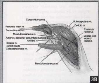
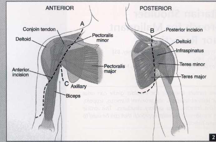
1 Utilitarian Shoulder Approach for Malignant Tumor Resection James C. Wittig, MD* Jacob Bickels, MDt Felasfa Wodajo, MDT Kristen L. Kellar-Graney, BS$ Isaac Meller, MDt Martin M. Malawer, MDT Malignant tumors involving the shoulder girdle can arise from four distinct locations: the proximal humerus, scapula, periscapular muscles, and axillary structures. This article describes a utilitarian shoulder approach that can be used to racprt thvcp tilmnrc The utilitarian shoulder approach can be used to resect shoulder girdle tumors. It accomplishes wide exposure of all shoulder girdle tumors and the adjacent neurovascular structures, facilitating mobilization and protection of all key structures and enabling adequate tumor resection. Malignant tumors involving the shoulder girdle can arise from four distinct locations: the proximal humerus, scapula, periscapular muscles, and axillary structures (eg, axillary lymph nodes, nerves, and vessels). Surgical resection often is technically challenging as most tumors in these locations From the *Department of Orthopedic Surger, New York University Medical Center and th Hospital for Joint Diseases, New York, NY; th tnational Unit of Orthopedic Oncology, Tel-Avi Souraskv Medical Center, Tel-Aviv, Israel; and th *Division of Orthopedic Oncology, Washingto Cancer Institute, Washington Hospital Cente Washington, DC. Reprint requests: Martin M. Malawer, ML Division of Orthopedic Oncology, Ste C Irving St NW, Washington, DC ple muscles, and often require resection via anterior and posterior exposure (Figure 1). An adequate surgical margin and a safe resection are best facilitated by complete exposure of the tumor and axillary vessels/brachial plexus. To reduce the surgical complexity and complications associated with limbsparing surgery for tumors in this region, the senior author (M.M.M.) has devel- oped an approach for resection, termed the utilitarian shoulder approach, which accomplishes safe exposure of the neu- rovascular structures; permits a reliable, wide excision of all tumors; and reduces morbidity associated with resections in this d cult anatomic area.' The utilitarian shoulder approach is based on an incision with three arms: anterior ( A), posterior (B), and anteroinferior (C) (Figure 2). The anterior approach (extended deltopectoral groove incision A) combined with detachment and mobilization of the pectoralis major and strap muscles (eg, coracobrachialis, short head of the biceps, pectoralis major), permits complete visualization of neurovascular structures (eg, the infraclavicular Reprinted with permission; SLACK Incorporated, O All rights reserved.
2
 is a modification of the anterior approach that extends medially from the deltopectoral groove and curves into the axilla.")

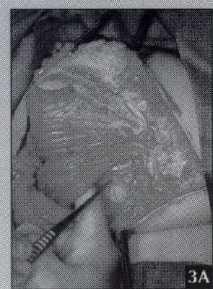
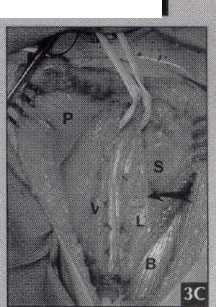
3 brachial plexus and axillary vessels). The posterior approach (B incision) accomplishes wide exposure of the scapula, deltoid, trapezius, rhomboids, and latissimus dorsi. The anteroinferior axillary incision (incision C) is a modification of the anterior approach that extends medially from the deltopectoral groove and curves into the axilla. It provides wide exposure of the axillary contents and is useful for large tumors in this area. The success of the utilitarian shoulder approach is based on wide exposure and exploration of the tumor, axillary vessels, and brachial plexus. This facilitates mobilization and protection of all key structures amenable to preservation. Anterior and posterior approaches can be combined, which is particularly useful for large scapular and proximal humeral tumors. For smaller or lowgrade tumors, any portion of any arm can be used or extended, depending on the anatomic location and extent of the tumor. In instances where the tumor is deemed unresectable after anterior exploration, the approach can be converted to a forequarter amputation without unnecessary contamination of skin and muscle flaps. SURGICAL TECHNIQUES Utilitarian Incision The utilitarian incision consists of three arms (A, B, and C) (Figure 2). Each arm of the incision results in the formation of a skin flap in which the base is at least as wide as the length of the flap. The anterior arm (A) is used primarily for proximal humerus resections and is modified for axillary tumors (C). The incision is extended posterolaterally over the shoulder (portion of B incision) for an extra-articular proximal humerus resection. A portion of the anterior incision (A) and the entire posterior incision (B) are used for extra-articular scapula resections and total scapulectomies. Any combination of incisions can be used for resecting soft-tissue sarcomas; this decision is based on the muscle of origin. Each arm is used to varying aegrees aepenamg on me anawunc location and extent of the tumor. We recommend an anterior approach be performed first so immediate exploration and mobilization of the brachial plexus can occur. At this point, if the tumor is deemed unresectable, the procedure can be converted to a forequarter amputation. Following the incision, skin flaps (fasciocutaneous, if possible) are raised to gain adequate exposure. Incision A: Anterior Approach The anterior incision (extended deltopectoral incision) extends from the middle third of the clavicle and passes 1 cm medial to the coracoid process, across the axillary fold, and distally along the anteromedial aspect of the arm, following the course of the neurovascular bundle. Medial and lateral fasciocutaneous skin flaps are raised. The key step for exposing the neurovascular bundle is releasing the pectoralis major from its humeral insertion and releasing the strap muscles (eg, pectoralis minor, short head of the biceps, and coracobrachialis) from their insertions on the coracoid (Figure 3). After the pectoralis major is released, the musculocutaneous nerve is dissected at the point where it enters the coracobrachialis and short head of the biceps (2-7 cm inferior to the coracoid). The 5lloll llcau ul Lllc ulecps allu l.olal.o - brachialis are then released from their coracoid insertions while protecting the underlying plexus and musculocutaneous nerve. The pectoralis minor is released from the coracoid (Figure 3). The entire neurovascular bundle from the clavicle to the proximal humerus can now be visualized. Next, the axillary nerve, at the lower border of the subscapularis muscle, and the radial nerve, at the lower border of the latissimus dorsi muscle, are identified and surrounded with vessel loops. Anterior and posterior humeral circumflex vessels are isolated and doubly lig

4 ated with #0 silk ties and metallic clips. This step is crucial for mobilization of the neurovascular bundle. The axillary nerve is ligated for proximal humerus tumors that require an extra-articular resection. Scapular and large axillary tumors may require ligation of any of the following structures: upper and lower subscapular nerves, subscapular artery, thoracodorsal nerve, lateral thoracic artery, and long thoracic nerve. Incision B: Posterior Incision The posterior incision begins superiorly over the shoulder, extending from the clavicular origin of the extended deltopectoral (anterior) incision. It follows the axillary border of the scapula to its tip and curves medially approximately 2-3 cm to permit construction of a large, medially-based flap. A fasciocutaneous (not subcutaneous) flap is used if deemed oncologically safe. Traditional scapular incisions have been constructed obliquely across the body of the scapula and have not allowed for adequate visualization and mobilization of the periscapular muscles.', ' This medially-based flap not only fully exposes the scapula, rhomboids, levator scapulae, and trapezius muscles, but also permits entrance to the posterior axillary space. A lateral flap also is developed that exposes the deltoid, proximal triceps, and teres minor and teres major muscles. Thus, all appropriate muscles can be resected or preserved and later transferred during the reconstruction phase of the procedure. For scapular tumors, early exploration of the chest wall once the rhomboid muscles are detached is performed initially to determine chest wall involvement and hence operability. Incision C: Axillary Approach The axillary incision is useful for resecting large axillary tumors that fill the axillary space (Figure 2). The axillary incision is a 6-cm extension of the anterior incision. It courses medial and inferior to the pectoralis major toward the base of the axillary space. It permits complete visualization of the inferior aspect of the axilla and lateral chest wall. The anterior component permits mobilization of the neurovascular structures whereas the axillary extension allows access to the axillary contents. TUMOR RESECTION Proximal Humerus Tumors When an extra-articular resection of the proximal humerus is performed, anterior exposure of the neurovascular structure and ligation of the axillary nerve is performed first. Then the incision is extended posterolaterally (portion of B), over the top of the shoulder (Figure 4). A medially-based fasciocutaneous skin flap is developed. The trapezius is released from the scapula and lateral clavicle. An incision is then made through the rotator cuff medial to the coracoid, extending from the subscapularis and passing sequentially through the supraspinatus, infraspinatus, and teres minor muscles. The latissimus dorsi and teres major are then released from the proximal humerus. Osteotomies are made through the humeral shaft at least 2 cm distal to the anatomic extent of the tumor, through the lateral one-third of the clavicle, and through the scapula medial to the corticoid. An extra-articular resection is completed (type VB resection, Malawer classification). Care must be taken at this point to avoid unnecessary traction on the axillary artery and the brachial plexus. The resected specimen consists of the proximal humerus removed en bloc with the lateral portion of the scapula, intact glenohumeral capsule, overlying rotator cuff, proximal portion of the long head of the biceps tendon, and the deltoid muscle. For an intra-articular resection (type IA resection, Malawer classification), only the anterior arm of the approach is used. The deltoid, rotator cuff, and capsule are released from the humerus. The axillary nerve is preserved. Humeral osteotomy is performed. Scapular Tumors In an extra-articular scapula resection, following neurovascular explowww.orthobluejournal.com

.")
5 I ration (A incision), the incision is extended posterolaterally to create a wide, medially-based skin flap (B incision) (Figure 5). All periscapular muscles, specifically the trapezius, rhomboids, levator scapulae, and latissimus dorsi, are sequentially released and tagged with a #0 nonabsorbable suture. An osteotomy is made through the proximal humerus at the inferior border of the subscapularis muscle (extraarticular osteotomy). A clavicular osteotomy is made through the lateral one-third of the clavicle. The scapula and tumor are removed en bloc with rotator cuff, capsule, and proximal humerus (Malawer type IVA resection). Axillary Tumors Incision C is used for axillary tumors. Release and mobilization of the pectoralis major muscle toward the chest wall provides excellent exposure of the entire axilla. Following dissection and mobilization of the brachial plexus and neurovascular structures from the tumor and ligation of the tethering neurovascular structures, the tumor can be removed en bloc (Figure 6). Soft-Tissue Tumors Soft-tissue sarcomas that arise in proximity to the neurovascular bundle are approached in a similar manner to that of axillary tumors. The utilitarian incision that is used depends on the muscle origin and the extent of the tumor. In general, at least the entire muscle of origin is resected for highgrade sarcomas. Soft-tissue sarcomas rarely invade adjacent bony structures and rarely require en bloc resection with a portion of the shoulder girdle. If bony invasion occurs, resection and reconstruction follow the principles outlined for scapular or proximal humerus tumors. tion of the proximal humerus or scapula, although the utilitarian approach for resection described herein may be used in conjunction with any reconstruction method. Following bony reconstruction, softtissue reconstruction commences. The short head of the biceps is tenodesed proximally to the coracoid (intra-articular proximal humerus reconstruction) or to the clavicle (extra-articular proximal humerus reconstruction) or pectoralis major (total scapula reconstruction). The pectoralis minor also is tenodesed back to its origin, when possible, or to the scapula to protect the neurovascular structures. The pectoralis major is repaired to its humeral insertion, or in cases requiring extra-articular proximal humerus reconstruction, transferred to cover the prosthesis with soft tissues. The latissimus dorsi may be transferred laterally to function as an external rotator following extraarticular proximal humerus resection. In total scapula reconstruction, the periscapular muscles are tenodesed to the prosthesis with heavy nonabsorbable sutures or tapes. In total scapula reconstruction, the periscapular muscles are tenodesed to the prosthesis in a manner that covers the entire prosthesis with muscle. Following isolated axillary tumor resection, the distal (numeral) cut edge of the latissimus dorsi is rotated into the defect and sutured to the superficial surface of the subscapularis muscle to fill the dead space. Largebore closed suction drains are routinely placed prior to skin closure (Figure 7). DISCUSSION Traditionally, shoulder girdle tumors often have been resected through exposures that provide poor visualization of neurovascular structures. 6 Proximal RECONSTRUCTION AND CLOSURE Following resection, bony reconstruction is performed for tumors that required a major bony resection. We recommend endoprosthetic reconstrucwww.orthobluejournal.com a
6 humeral tumors have been resected through a limited anterior deltopectoral approach; scapular and periscapular tumors through a limited oblique surgical incision based over the posterior scapula.'-' Axillary tumors have been resected through a limited inferiorlybased axillary approach." In all cases, poor exposure of the brachial plexus and axillary vessels may result in unnecessary complications and the inability to resect the tumor with adequate surgical margins. 8,9 We describe a utilitarian approach that has been used for surgical resection of proximal humerus, scapula, and axillary tumors in 155 patients. This approach permits wide exposure of the infraclavicular brachial plexus, axillary vessels, and major nerves, (axillary, musculocutaneous, median, ulnar, and radial). The incision consists of three components: anterior, posterior, and axillary. Any portion, or all three elements, can be used for resection of bone and soft-tissue tumors of the proximal humerus, scapula, periscapular muscles, and axilla. Releasing the pectoralis major from the proximal humerus and the coracoid insertions of the strap muscles is the key to exposure of the brachial plexus and axillary vessels. This ensures safe mobilization of major neurovascular structures. Optimal surgical margins are facilitated, and unnecessary complications (eg, inadvertent neurovascular injury) and local recurrence are minimized. Pectoralis major, triceps, and biceps strength return with rehabilitation. This approach is recommended for major resection of bone and soft-tissue tumors of the shoulder girdle. REFERENCES 1. Malawer MM, Meller I. A new surgical classification system for shoulder girdle resection: analysis of 38 patients. Clin Orthop. 1981; 267: Malawer MM, Sugarbaker PH, Lampert M, Baker AR, Geber NL. The Tikhoff-Linberg procedure: report often patients and presentation of a modified technique for tumors of the proximal humerus. Surgery ; 97: Malawer MM. Tumors of the shoulder girdle: technique of resection and description of the surgical classification system. Orthop Clin North Am. 1991; 22: Malawer MM. Surgical techniques and results of limb-sparing surgery for high-grade bone sarcomas of the knee and shoulder. Orthopedics ; 8: Enneking WE. Shoulder girdle. In: Musculoskeletal Tumor Surgery. New York, NY: Churchill Livingstone; 1983: Papioannou AN, Francis KC. Scapulectomy for the treatment of primary malignant tumors of the scapula. Clin Orthop. 1965; 41: Asavamongkolkul A, Eckardt JJ, Either FR, Dorey FJ, Ward WG, Kelly CM. Endoprosthetic reconstruction for malignant upper extremity tumors. Clin Orthop. 1999; 360: Ivens D, Hoe AL, Podd TJ, Hamilton CR, Taylor I, Roye GT. Assessment of morbidity from complete axillary dissection. Br J Cancer 1992; 66: Roses DF, Brooks AD, Harris MN, Shapiro RL, Mitnick J. Complications of level I and II axill ary dissection in the treatment of carcinoma of the breast. Ann Surg. 1999; 230: Section Editor: Steven F. Harwin, MD 484
MUSCLES. Anconeus Muscle
 LAB 7 UPPER LIMBS MUSCLES Anconeus Muscle anconeus origin: distal end of dorsal surface of humerus insertion: lateral surface of ulna from distal margin of the semilunar notch to proximal end of the olecranon
Region of upper limb attachment to the trunk Proximal segment of limb overlaps parts of the trunk (thorax and back) and lower lateral neck.
 and lower lateral neck.") Region of upper limb attachment to the trunk Proximal segment of limb overlaps parts of the trunk (thorax and back) and lower lateral neck. includes Pectoral Scapular Deltoid regions of the upper limb
Region of upper limb attachment to the trunk Proximal segment of limb overlaps parts of the trunk (thorax and back) and lower lateral neck. includes Pectoral Scapular Deltoid regions of the upper limb
Upper limb Pectoral region & Axilla
 Upper limb Pectoral region & Axilla 黃敏銓 mchuang@ntu.edu.tw 1 Pectoral region Intercostal nerve Anterior branch of lateral cutaneous branch Lateral cutaneous branch Anterior cutaneous branch Anterior cutaneous
Scapular and Deltoid Regions
 M1 Gross and Developmental Anatomy Scapular and Deltoid Regions Dr. Peters 1 Outline I. Skeleton of the Shoulder and Attachment of the Upper Extremity to Trunk II. Positions and Movements of the Scapula
Gateway to the upper limb. An area of transition between the neck and the arm.
 Gateway to the upper limb An area of transition between the neck and the arm. Pyramidal space inferior to shoulder @ junction of arm & thorax Distribution center for the neurovascular structures that serve
Key Relationships in the Upper Limb
 Key Relationships in the Upper Limb This list contains some of the key relationships that will help you identify structures in the lab. They are organized by dissection assignment as defined in the syllabus.
FUNCTIONAL ANATOMY OF SHOULDER JOINT
 FUNCTIONAL ANATOMY OF SHOULDER JOINT ARTICULATION Articulation is between: The rounded head of the Glenoid cavity humerus and The shallow, pear-shaped glenoid cavity of the scapula. 2 The articular surfaces
Upper limb Arm & Cubital region 黃敏銓
 Upper limb Arm & Cubital region 黃敏銓 1 Arm Lateral intermuscular septum Anterior (flexor) compartment: stronger Medial intermuscular septum Posterior (extensor) compartment 2 Coracobrachialis Origin: coracoid
Anatomy of the Shoulder Girdle. Prof Oluwadiya Kehinde FMCS (Orthop)
") Anatomy of the Shoulder Girdle Prof Oluwadiya Kehinde FMCS (Orthop) www.oluwadiya.com Bony Anatomy Shoulder Complex: Sternum(manubrium) Clavicle Scapula Proximal humerus Manubrium Sterni Upper part of
Anatomy of the Shoulder Girdle Prof Oluwadiya Kehinde FMCS (Orthop) www.oluwadiya.com Bony Anatomy Shoulder Complex: Sternum(manubrium) Clavicle Scapula Proximal humerus Manubrium Sterni Upper part of
3 Mohammad Al-Mohtasib Areej Mosleh
 3 Mohammad Al-Mohtasib Areej Mosleh ***Muscles Connecting the Upper Limb to the Vertebral Column 1.Trapezius Muscle ***The first muscle on the back is trapezius muscle, it s called so according
Joint G*H. Joint S*C. Joint A*C. Labrum. Humerus. Sternum. Scapula. Clavicle. Thorax. Articulation. Scapulo- Thoracic
 A*C Joint Scapulo- Thoracic Articulation Thorax Sternum Clavicle Scapula Humerus S*C Joint G*H Joint Labrum AC Ligaments SC Ligaments SC JOINT AC Coracoacromial GH GH Ligament Complex Coracoclavicular
THE SHOULDER JOINT T H E G L E N O H U M E R A L ( G H ) J O I N T
 J O I N T") THE SHOULDER JOINT T H E G L E N O H U M E R A L ( G H ) J O I N T CLARIFICATION OF TERMS Shoulder girdle = scapula and clavicle Shoulder joint (glenohumeral joint) = scapula and humerus Lippert, p115
THE SHOULDER JOINT T H E G L E N O H U M E R A L ( G H ) J O I N T CLARIFICATION OF TERMS Shoulder girdle = scapula and clavicle Shoulder joint (glenohumeral joint) = scapula and humerus Lippert, p115
MUSCLES OF SHOULDER REGION
 Dr Jamila EL Medany OBJECTIVES At the end of the lecture, students should: List the name of muscles of the shoulder region. Describe the anatomy of muscles of shoulder region regarding: attachments of
Axilla and Brachial Region
 L 4 A B O R A T O R Y Axilla and Brachial Region BRACHIAL PLEXUS 5 Roots/Rami (ventral rami C5 T1) 3 Trunks Superior (C5, C6) Middle (C7) Inferior (C8, T1) 3 Cords Lateral Cord (Anterior Superior and Anterior
Muscles of the Upper Limb
 Muscles of the Upper Limb anterior surface of ribs 3 5 coracoid process Pectoralis minor pectoral nerves protracts / depresses scapula Serratus anterior Subclavius ribs 1-8 long thoracic nerve rib 1 ----------------
G24: Shoulder and Axilla
 G24: Shoulder and Axilla Syllabus - Pg. 2 ANAT 6010- Medical Gross Anatomy David A. Morton, Ph.D. Objectives Upper limb Systemically: Bones (joints) Muscles Nerves Vessels (arteries/veins) Fascial compartments
Osteosarcoma of the Proximal Humerus: Long-Term Results With Limb-Sparing Surgery
 CLINICAL ORTHOPAEDICS AND RELATED RESEARCH Number397,pp.156-176 02002 Uppincott Williams & Wilkins. Inc., Osteosarcoma of the Proximal Humerus: Long-Term Results With Limb-Sparing Surgery James C. Wittig,
The Arm and Cubital Fossa
 The Arm and Cubital Fossa Dr. Andrew Gallagher School of Anatomical Sciences University of the Witwatersrand Introduction The ARM (BRACHIUM) is the most proximal segment of the upper limb musculoskeletal
7/31/2012 THE SHOULDER JOINT CLARIFICATION OF TERMS OSTEOLOGY OF THE GH JOINT(BONES)
") THE SHOULDER JOINT T H E G L E N O H U M E R AL ( G H ) J O I N T CLARIFICATION OF TERMS Shoulder girdle = scapula and clavicle Shoulder joint (glenohumerual joint) = scapula and Lippert, p115 OSTEOLOGY
THE SHOULDER JOINT T H E G L E N O H U M E R AL ( G H ) J O I N T CLARIFICATION OF TERMS Shoulder girdle = scapula and clavicle Shoulder joint (glenohumerual joint) = scapula and Lippert, p115 OSTEOLOGY
Upper Limb Muscles Muscles of Axilla & Arm
 Done By : Saleh Salahat Upper Limb Muscles Muscles of Axilla & Arm 1) Muscles around the axilla A- Muscles connecting the upper to thoracic wall (4) 1- pectoralis major Origin:- from the medial half of
Human Anatomy Biology 351
 1 Human Anatomy Biology 351 Upper Limb Exam Please place your name on the back of the last page of this exam. You must answer all questions on this exam. Because statistics demonstrate that, on average,
VENOUS DRAINAGE O US F UPPER UPPER LIM B BY dr.fahad Ullah
 VENOUS DRAINAGE OF UPPER LIMB BY dr.fahad Ullah Venous drainage of the supper limb The venous system of the upper limb drains deoxygenated blood from the arm, forearm and hand It can anatomically be divided
SHOULDER JOINT ANATOMY AND KINESIOLOGY
 SHOULDER JOINT ANATOMY AND KINESIOLOGY SHOULDER JOINT ANATOMY AND KINESIOLOGY The shoulder joint, also called the glenohumeral joint, consists of the scapula and humerus. The motions of the shoulder joint
Pectoral region. Lecture 2
 Pectoral region Lecture 2 Muscle Action Each muscle has: Origin Beginning. Insertion End. Body (belly). Law: When a muscle performs its action, its insertion, moves towards its origin. Spring 2016 Dr.
BRACHIAL PLEXUS. DORSAL SCAPULAR NERVE (C5) supraclavicular branch innervates rhomboids (major and minor) and levator scapulae
 supraclavicular branch innervates rhomboids (major and minor) and levator scapulae") THE BRACHIAL PLEXUS DORSAL SCAPULAR NERVE (C5) supraclavicular branch innervates rhomboids (major and minor) and levator scapulae SCHEMA OF THE BRACHIAL PLEXUS THE BRACHIAL PLEXUS PHRENIC NERVE supraclavicular
THE BRACHIAL PLEXUS DORSAL SCAPULAR NERVE (C5) supraclavicular branch innervates rhomboids (major and minor) and levator scapulae SCHEMA OF THE BRACHIAL PLEXUS THE BRACHIAL PLEXUS PHRENIC NERVE supraclavicular
Al-Balqa Applied University
 Al-Balqa Applied University Faculty Of Medicine *You can use this checklist as a guide to you for the lab. the items on this checklist represent the main features of the models that you have to know for
Pectoral region. Lecture 2
 Pectoral region Lecture 2 Muscle Action Each muscle has: Origin Beginning. Insertion End. Body (belly). Law: When a muscle performs its action, its insertion, moves towards its origin. Spring 2016 Dr.
Netter's Anatomy Flash Cards Section 6 List 4 th Edition
 Netter's Anatomy Flash Cards Section 6 List 4 th Edition https://www.memrise.com/course/1577581/ Section 6 Upper Limb (66 cards) Plate 6-1 Humerus and Scapula: Anterior View 1.1 Acromion 1.2 Greater tubercle
This figure (of humerus) is from Dr. Maher's newest slides. -Its added here just for consideration-
 is from Dr. Maher's newest slides. -Its added here just for consideration-") This figure (of humerus) is from Dr. Maher's newest slides. -Its added here just for consideration- Slides of Anatomy Please note : These slides are Dr. Maher Hadidi s slides of spring 2016 and were edited
This figure (of humerus) is from Dr. Maher's newest slides. -Its added here just for consideration- Slides of Anatomy Please note : These slides are Dr. Maher Hadidi s slides of spring 2016 and were edited
Brachial plexuses and axillary lymph nodes
 Brachial plexuses and axillary lymph nodes Introduction about nervous system nervous system central nervous system periphral nervous system brain spinal cord 31 pairs of spinal nerves 12 paris of cranial
The shoulder girdle consists of the glenohumeral, acromioclavicular, sternoclavicular and scapulothoracic joints
 Anatomy of Shoulder Girdle The shoulder girdle consists of the glenohumeral, acromioclavicular, sternoclavicular and scapulothoracic joints Glenohumeral Joint A ball and socket synoval joint with a large
The Clavicle Right clavicle Deltoid tubercle: Conoid tubercle, conoid ligamen Impression for the
 The Clavicle Muscle Attachment Sites in the Upper Limb Pectoralis major Right clavicle Smooth superior surface of the shaft, under the platysma muscle tubercle: attachment of the deltoid Acromial facet
region of the upper limb between the shoulder and the elbow Superiorly communicates with the axilla.
 1 region of the upper limb between the shoulder and the elbow Superiorly communicates with the axilla. Inferiorly, a number of important structures pass between arm & forearm through cubital fossa. 2 medial
Practical 2 Worksheet
 Practical 2 Worksheet Upper Extremity BONES 1. Which end of the clavicle is on the lateral side (acromial or sternal)? 2. Describe the difference in the appearance of the acromial and sternal ends of the
Breast conservation surgery and sentinal node biopsy: Dr R Botha Moderator: Dr E Osman
 Breast conservation surgery and sentinal node biopsy: Dr R Botha Moderator: Dr E Osman Breast anatomy: Breast conserving surgery: The aim of wide local excision is to remove all invasive and in situ
The arm: *For images refer back to the slides
 The arm: *For images refer back to the slides Muscles of the arm: deltoid, triceps (which is located at the back of the arm), biceps and brachialis (it lies under the biceps), brachioradialis (it lies
Sports Medicine Part II : ANATOMY OF THE SPINE, ABDOMEN AND SHOULDER COMPLEX
 Sports Medicine 25 1.1 Part II : ANATOMY OF THE SPINE, ABDOMEN AND SHOULDER COMPLEX c.w.p. Wagner High School, Sports Medicine, A. Morgan, T. Morgan & A. Eastlake, 2008 Muscles of the Upper Limbs In this
Muscle Action Origin Insertion Nerve Innervation Chapter Page. Deltoid. Trapezius. Latissimus Dorsi
 Muscle Action Origin Insertion Nerve Innervation Chapter Page All Fibers Abduct the shoulder (glenohumeral joint) Deltoid Anterior Fibers Flex the shoulder (G/H joint) Horizontally adduct the shoulder
Muscular Nomenclature and Kinesiology - One
 Chapter 16 Muscular Nomenclature and Kinesiology - One Lessons 1-3 (with lesson 4) 1 Introduction 122 major muscles covered in this chapter Chapter divided into nine lessons Kinesiology study of human
Peripheral Nervous Sytem: Upper Body
 Peripheral Nervous Sytem: Upper Body MSTN121 - Neurophysiology Session 10 Department of Myotherapy Cervical Plexus Accessory nerve (CN11 + C1-5) Motor: trapezius and sternocleidomastoid Greater auricular
Returning the Shoulder Back to Optimal Function. Scapula. Clavicle. Humerus. Bones of the Shoulder (Osteology) Joints of the Shoulder (Arthrology)
 Joints of the Shoulder (Arthrology)") Returning the Shoulder Back to Optimal Function Sternum Clavicle Ribs Scapula Humerus Bones of the Shoulder (Osteology) By Rick Kaselj Clavicle Scapula Medial Left Anterior Clavicle Inferior View 20 degree
Returning the Shoulder Back to Optimal Function Sternum Clavicle Ribs Scapula Humerus Bones of the Shoulder (Osteology) By Rick Kaselj Clavicle Scapula Medial Left Anterior Clavicle Inferior View 20 degree
URSA HEMI-SHOULDER ARTHROPLASTY B I O T E K
 URSA HEMI-SHOULDER ARTHROPLASTY SURGICAL TECHNIQUE B I O T E K 2 Surgical Position Once general anesthesia has been satisfactorily induced, or a supraclavicular nerve block has been given, the patient
Fascial Compartments of the Upper Arm
 Fascial Compartments of the Upper Arm The upper arm is enclosed in a sheath of deep fascia and has two fascial septa: 1- Medial fascial septum (medial intermuscular septum): attached to the medial supracondylar
Shoulder Biomechanics
 Shoulder Biomechanics Lecture originally developed by Bryan Morrison, Ph.D. candidate Arizona State University Fall 2000 1 Outline Anatomy Biomechanics Problems 2 Shoulder Complex Greatest Greatest Predisposition
26/9/2016. Anatomy. 1 Nour Erekat Wejdan Amer
 26/9/2016 Anatomy st 1 Nour Erekat Wejdan Amer Notes before we start the lecture. Bring any colored Atlas with you to the lab. The main reference is clinical anatomy by regions by Richard snell the 9 th
Muscles in the Shoulder, Chest, Arm, Stomach, and Back
 Muscles in the Shoulder, Chest, Arm, Stomach, and Back Shoulder Muscles Deltoid Supraspinatus Infraspinatus Teres Major Teres Minor Subscapularis Deltoid (Delts) Function: Raises the upper arm Origin:
Glenohumeral Joint. Glenohumeral Joint. Glenohumeral Joint. Glenohumeral Joint. Glenohumeral Joint. Glenohumeral Joint
 The Shoulder Joint Chapter 5 The Shoulder Joint Manual of Structural Kinesiology R.T. Floyd, EdD, ATC, CSCS McGraw-Hill Higher Education. All rights reserved. 5-1 Shoulder joint is attached to axial skeleton
Introduction to anatomy
 Introduction to anatomy Dr. Maher Hadidi Fareed Halteh 3 7/2/2013 Subscapularis: It is located on the anterior side of the scapula. It has a triangular shape. It is like Pectoralis major and Teres major
The Upper Limb II. Anatomy RHS 241 Lecture 11 Dr. Einas Al-Eisa
 The Upper Limb II Anatomy RHS 241 Lecture 11 Dr. Einas Al-Eisa Sternoclavicular joint Double joint.? Each side separated by intercalating articular disc Grasp the mid-portion of your clavicle on one side
Supplied in part by the musculocutaneous nerve. Forms the axis of rotation in movements of pronation and supination
 Anatomy: Upper limb (15 questions) 1. Latissimus Dorsi: Is innervated by the dorsal scapular nerve Lies above feres major muscle Medially rotates the humerus All of the above 2. Supinator muscle is: Deep
Forelimb Amputation. Indications. Pre operatively 11/12/2008. Technique Preparing for surgery. Bone neoplasia. Severe trauma/paralysis
 Forelimb Amputation January 10, 2007 Amy Bringardner Becky Pentecost Indications Bone neoplasia Osteosarcoma Fibrosarcoma Chondrosarcoma Severe trauma/paralysis Brachial plexus avulsion Infection Congenital
Shoulder Arthroscopy Portals
 Shoulder Arthroscopy Portals Alper Deveci and Metin Dogan 7 7.1 Bony Landmarks Before starting shoulder arthroscopy, the patient must be positioned and draping applied. Then the bony landmarks are identified
Continuing Education: Shoulder Stability
 Continuing Education: Shoulder Stability Anatomy & Kinesiology: The GHJ consists of the articulation of three bones: the scapula, clavicle and humerus. The scapula has three protrusions: the coracoid,
Anatomy and Physiology II. Review Shoulder Girdle New Material Upper Extremities - Bones
 Anatomy and Physiology II Review Shoulder Girdle New Material Upper Extremities - Bones Anatomy and Physiology II Shoulder Girdle Review Questions From Last Lecture Can you identify the following muscles?
Shoulder: Clinical Anatomy, Kinematics & Biomechanics
 Shoulder: Clinical Anatomy, Kinematics & Biomechanics Dr. Alex K C Poon Department of Orthopaedics & Traumatology Pamela Youde Nethersole Eastern Hospital Clinical Anatomy the application of anatomy to
Anatomage Table Instructors Guide- Upper Limb
 The Upper Limb Anatomage Table Instructors Guide- Upper Limb Table of Contents Upper Limb 1- The Skeletal System...3 1: Clavicle...3 2: Scapula...5 3: Shoulder (Glenohumeral) and Proximal Humerus...7 4:
The Free Upper Limb. Bone of the Arm. aus: Platzer, Locomotor System (ISBN ), 2009 Georg Thieme Verlag KG
, 2009 Georg Thieme Verlag KG") : ones, Ligaments, Joints The Free The bones of the free upper limb are The humerus The radius and ulna The carpal bones The metacarpal bones The phalanges one of the Arm Humerus (A H) The humerus articulates
: ones, Ligaments, Joints The Free The bones of the free upper limb are The humerus The radius and ulna The carpal bones The metacarpal bones The phalanges one of the Arm Humerus (A H) The humerus articulates
*the Arm* -the arm extends from the shoulder joint (proximal), to the elbow joint (distal) - it has one bone ; the humerus which is a long bone
, to the elbow joint (distal) - it has one bone ; the humerus which is a long bone") *the Arm* -the arm extends from the shoulder joint (proximal), to the elbow joint (distal) - it has one bone ; the humerus which is a long bone - muscles in the arm : *brachialis muscle *Biceps brachii
*the Arm* -the arm extends from the shoulder joint (proximal), to the elbow joint (distal) - it has one bone ; the humerus which is a long bone - muscles in the arm : *brachialis muscle *Biceps brachii
David G. Simpson, Ph.D.
 David G. Simpson, Ph.D. ARM & CUBITAL FOSSA Revised 7/08 Text References Moores 3 rd ed., p402 408, 436 439, 439 443, 478, 481 LEARNING OBJECTIVES: 1. Describe the humerus, indicating the sites of muscle
STEP #1: Anatomy STEP #2: Awareness STEP #3: Action
 Stabilize the Scapula in Three Easy Steps by NFPT Idea World 2016: Session 611 Saturday July 16th 7:30-9:20am Beverly Hosford, MA STEP #1: Anatomy STEP #2: Awareness STEP #3: Action 1. Anatomy *Memorize
Trapezius transfer in brachial plexus palsy
 Upper limb Trapezius transfer in brachial plexus palsy CORRELATION OF THE OUTCOME WITH MUSCLE POWER AND OPERATIVE TECHNIQUE O. Rühmann, S. Schmolke, M. Bohnsack, J. Carls, C. J. Wirth From Hannover Medical
Scapular & Parascapular flap FLAP TERRITORY ANATOMY. is normally accompanied by two venae comitantes.
 Scapular & Parascapular flap FLAP TERRITORY This is a composite flap that is situated over the scapula with various incisional arrangements. It can be harvested as a skin and subcutaneous tissue flap,
Anatomy Workshop Upper Extremity David Ebaugh, PT, PhD Workshop Leader. Lab Leaders: STATION I BRACHIAL PLEXUS
 Anatomy Workshop Upper Extremity David Ebaugh, PT, PhD Workshop Leader Lab Leaders: STATION I BRACHIAL PLEXUS A. Posterior cervical triangle and axilla B. Formation of plexus 1. Ventral rami C5-T1 2. Trunks
Immediate post surgical findings of soft tissue swelling, subcutaneous emphysema, and skin staples for reverse total shoulder arthroplasty.
 Immediate post surgical findings of soft tissue swelling, subcutaneous emphysema, and skin staples for reverse total shoulder arthroplasty. REVERSE TOTAL SHOULDER ARTHROPLASTY WITH FRACTURED ACROMION Above:
SHOULDER ANATOMY Karl Wieser, MD Department of Orthopedics, University of Zurich, Balgrist, Switzerland
 20th Course in Shoulder Surgery Balgrist SHOULDER ANATOMY Karl Wieser, MD Department of Orthopedics, University of Zurich, Balgrist, Switzerland www.balgrist.ch ANATOMY OVERVIEW courtesy of Georg Lajtai
REFERENCE DIAGRAMS OF UPPER LIMB MUSCLES: NAMES, LOCATIONS, ATTACHMENTS, FUNCTIONS MUSCLES CONNECTING THE UPPER LIMB TO THE AXIAL SKELETON
 REFERENCE DIAGRAMS OF UPPER LIMB MUSCLES: NAMES, LOCATIONS, ATTACHMENTS, FUNCTIONS MUSCLES CONNECTING THE UPPER LIMB TO THE AXIAL SKELETON A25LAB EXERCISES: UPPER LIMB MUSCLES Page 1 MUSCLES CONNECTING
WEEKEND THREE HOMEWORK
 WEEKEND THREE HOMEWORK READING ASSIGNMENTS Salvo Massage Therapy Principles and Practice 4 th Edition Muscolino The Muscular System Manual Muscolino The Muscle and Bone Palpation Manual Ch. 19 Skeletal
Anatomy of the Musculoskeletal System
 Anatomy of the Musculoskeletal System Kyle E. Rarey, Ph.D. Department of Anatomy & Cell Biology and Otolaryngology University of Florida College of Medicine Outline of Presentation Vertebral Column Upper
SHOULDER PROBLEMS & ARTHROSCOPIC MANAGEMENT
 SHOULDER PROBLEMS & ARTHROSCOPIC MANAGEMENT DR.SHEKHAR SRIVASTAV Sr. Consultant-KNEE & SHOULDER Arthroscopy Sant Parmanand Hospital,Delhi Peculiarities of Shoulder Elegant piece of machinery It has the
Research Article A Novel Surgical Approach to Lipomatous Tumours of the Deltoid Region
 Sarcoma Volume 2010, Article ID 495834, 4 pages doi:10.1155/2010/495834 Research Article A Novel Surgical Approach to Lipomatous Tumours of the Deltoid Region Emad Al Absi, Tamanna Karim, Nigel Colterjohn,
213: HUMAN FUNCTIONAL ANATOMY: PRACTICAL CLASS 1: Proximal bones, plexuses and patterns
 213: HUMAN FUNCTIONAL ANATOMY: PRACTICAL CLASS 1: Proximal bones, plexuses and patterns CLAVICLE Examine an isolated clavicle and compare it with a clavicle on an articulated skeleton. Viewed from above,
The posterolateral thoracotomy is still probably the
 Posterolateral Thoracotomy Jean Deslauriers and Reza John Mehran The posterolateral thoracotomy is still probably the most commonly used incision in general thoracic surgery. It provides not only excellent
Pectoral girdle, SUPERIEUR ARM AND HAND. Danil Hammoudi.MD
 Pectoral girdle, SUPERIEUR ARM AND HAND Danil Hammoudi.MD The pectoral girdle is the set of bones which connect the upper limb to the axial skeleton on each side. It consists of the clavicle scapula in
Connects arm to thorax 3 joints. Glenohumeral joint Acromioclavicular joint Sternoclavicular joint
 Connects arm to thorax 3 joints Glenohumeral joint Acromioclavicular joint Sternoclavicular joint Scapula Elevation Depression Protraction (abduction) Retraction (adduction) Downward Rotation Upward Rotation
SHOULDER PAIN. A Real Pain in the Neck. Michael Wolk, MD Northeastern Rehabilitation Associates October 31, 2017
 SHOULDER PAIN A Real Pain in the Neck Michael Wolk, MD Northeastern Rehabilitation Associates October 31, 2017 THE SHOULDER JOINT (S) 1. glenohumeral 2. suprahumeral 3. acromioclavicular 4. scapulocostal
JMSCR Volume 03 Issue 01 Page January 2015
 www.jmscr.igmpublication.org Impact Factor 3.79 ISSN (e)-2347-176x Is Surgical Exposure of Scapula Through Direct Lateral Approach Preferable? Authors Dr. Dibakar Ray 1, Dr. Ashoke Kumar Chanda 2, Dr.
RECURRENT SHOULDER DISLOCATIONS WITH ABSENT LABRUM
 RECURRENT SHOULDER DISLOCATIONS WITH ABSENT LABRUM D R. A M R I S H K R. J H A M S ( O R T H O ) A S S I S T A N T P R O F E S S O R M E D I C A L C O L L E G E, K O L K A T A LABRUM Function as a chock-block,
Dr. Mahir Alhadidi Anatomy Lecture #9 Feb,28 th 2012
 Quick Revision: Upper arm is divided into two compartments: 1. Anterior Compartment: Contains three muscles (Biceps brachii, Coracobrachialis, Brachialis). Innervated by Musculocutaneous nerve. 2. Posterior
G25: Brachium. ANAT Medical Gross Anatomy. David A. Morton, Ph.D.
 G25: Brachium ANAT 6010- Medical Gross Anatomy David A. Morton, Ph.D. Brachial Plexus Randy Travis Drinks Cold Beer What muscle(s) enable her to do the following exercise? What muscle(s) enable her to
The suction cup mechanism is enhanced by the slightly negative intra articular pressure within the joint.
 SHOULDER INSTABILITY Stability A. The stability of the shoulder is improved by depth of the glenoid. This is determined by: 1. Osseous glenoid, 2. Articular cartilage of the glenoid, which is thicker at
Osteopathic Considerations in Shoulder Pain. Kristen Brusky DO February 22, 2018
 Osteopathic Considerations in Shoulder Pain Kristen Brusky DO February 22, 2018 Overview Importance of pectoral girdle Ligaments, ligaments tensegrity Bones, joints, muscles Neurovasculature Innervation
Shoulder Joint Examination. Shoulder Joint Examination. Inspection. Inspection Palpation Movement. Look Feel Move
 Shoulder Joint Examination History Cuff Examination Instability Examination AC Joint Examination Biceps Tendon Examination Superior Labrum Examination Shoulder Joint Examination Inspection Palpation Movement
The Muscular System. Chapter 10 Part C. PowerPoint Lecture Slides prepared by Karen Dunbar Kareiva Ivy Tech Community College
 Chapter 10 Part C The Muscular System Annie Leibovitz/Contact Press Images PowerPoint Lecture Slides prepared by Karen Dunbar Kareiva Ivy Tech Community College Table 10.9: Muscles Crossing the Shoulder
Gross Anatomy Questions That Should be Answerable After October 27, 2017
 Gross Anatomy Questions That Should be Answerable After October 27, 2017 1. The inferior angle of the scapula of a woman who was recently in an automobile accident seems to protrude making a ridge beneath
The Shoulder. Anatomy and Injuries PSK 4U Unit 3, Day 4
 The Shoulder Anatomy and Injuries PSK 4U Unit 3, Day 4 Shoulder Girdle Shoulder Complex is the most mobile joint in the body. Scapula Clavicle Sternum Humerus Rib cage/thorax Shoulder Girdle It also includes
DK7215-Levine-ch12_R2_211106
 12 Arthroscopic Rotator Interval Closure Andreas H. Gomoll Department of Orthopedic Surgery, Brigham and Women s Hospital, Harvard Medical School, Boston, Massachusetts, U.S.A. Brian J. Cole Departments
Upper extremity. Part I
 Part I Upper extremity 1 Fractures of the clavicle Peter V. Giannoudis 1.1 OPEN REDUCTION AND INTERNAL FIXATION (ORIF) OF MIDSHAFT FRACTURES Indications (a) Open fractures. (b) Painful non-union. (c) Associated
BLUE SKY SCHOOL OF PROFESSIONAL MASSAGE AND THERAPEUTIC BODYWORK. Musculoskeletal Anatomy & Kinesiology II REVIEW
 BLUE SKY SCHOOL OF PROFESSIONAL MASSAGE AND THERAPEUTIC BODYWORK Musculoskeletal Anatomy & Kinesiology II REVIEW MSAK101-II Session 4 LEARNING OBJECTIVES: By the end of this session, the student will be
SUPERIEUR ARM AND HAND
 Pectoral girdle, SUPERIEUR ARM AND HAND Danil Hammoudi.MD The pectoral girdle is the set of bones which connect the upper limb to the axial skeleton on each side. It consists of the clavicle scapula in
BRACHIAL PLEXUS 11/12/2014 كيف تتكون الضفيرة FORMATION ENLARGEMENT (INTUMESCENCE) OF THE SPINAL CORD. Grey matter. Cervical intumescence - C 6 - T 2
 OF THE SPINAL CORD. Grey matter. Cervical intumescence - C 6 - T 2") BRACHIAL PLEXUS Prof. Fawzy Elnady ENLARGEMENT (INTUMESCENCE) OF THE SPINAL CORD Grey matter Cervical intumescence - C 6 - T 2 Lumbar intumescence - L 4 S 2 كيف تتكون الضفيرة FORMATION The ventral rami
BRACHIAL PLEXUS Prof. Fawzy Elnady ENLARGEMENT (INTUMESCENCE) OF THE SPINAL CORD Grey matter Cervical intumescence - C 6 - T 2 Lumbar intumescence - L 4 S 2 كيف تتكون الضفيرة FORMATION The ventral rami
The Elbow and the cubital fossa. Prof Oluwadiya Kehinde
 The Elbow and the cubital fossa Prof Oluwadiya Kehinde www.oluwadiya.com Elbow and Forearm Anatomy The elbow joint is formed by the humerus, radius, and the ulna Bony anatomy of the elbow Distal Humerus
STRUCTURAL BASIS OF MEDICAL PRACTICE EXAMINATION 5. September 30, 2011
 STRUCTURAL BASIS OF MEDICAL PRACTICE EXAMINATION 5 September 30, 2011 PART l. Answer in the space provided. (12 pts) 1. Identify the structures. (2 pts) EXAM NUMBER A. Suprascapular nerve B. Axillary nerve
Modified Radical Mastectomy
 Modified Radical Mastectomy Valerie L. Staradub, MD, and Monica Morrow, MD S urgical management options for breast cancer include modified radical mastectomy (MRM), MRM with immediate reconstruction, and
Motion of Left Upper Extremity During A Right- Handed Golf Swing
 Motion of Left Upper Extremity During A Right- Handed Golf Swing Description of Movement While the movement required for a golf swing requires many muscles, joints, & ligaments throughout the body, the
medial half of clavicle; Sternum; upper six costal cartilages External surfaces of ribs 3-5
 MUSCLE ORIGIN INSERTION ACTION NERVE Pectoralis Major medial half of clavicle; Sternum; upper six costal cartilages Lateral lip of intertubercular groove of horizontal adduction Medial and lateral pectoral
Due in Lab weeks because of Thanksgiving Prelab #10. Homework #8. Both sides! Both sides!
 Lab 8 MUSCLES Due in Lab 10 2 weeks because of Thanksgiving Prelab #10 Both sides! Homework #8 Both sides! Refer to Muscles 22-23 Naming of muscles Origin Site of muscle attachment that doesn t move during
The pectoral region. University of Babylon College of Medicine Dr.HaythemAli Alsayigh M.B.CH.B.-F.I.M.B.S. Surgical Clinical Anatomy
 The pectoral region University of Babylon College of Medicine Dr.HaythemAli Alsayigh M.B.CH.B.-F.I.M.B.S. Surgical Clinical Anatomy Objective Study the Bones and Joints A. Clavicle (collarbone) B. Scapula
BICEPTOR Tenodesis System
 BICEPTOR Tenodesis System Sub-Pectoral Biceps Tenodesis A Shoulder Series Technique Guide As described by: Nikhil N. Verma, MD As described by: Nikhil N. Verma, MD Midwest Orthopedics at Rush Chicago,
SHOULDER INSTABILITY
 SHOULDER INSTABILITY Dr.KN Subramanian M.Ch Orth., FRCS (Tr & Orth), CCT Orth(UK) Consultant Orthopaedic Surgeon, Special interest: Orthopaedic Sports Injury, Shoulder and Knee Surgery, SPARSH Hospital
Acromion splitting approach for extensive exposure of shoulder and proximal humerus
 Original Article Acromion splitting approach for extensive exposure of shoulder and proximal humerus Dominic Puthoor 1, Prabhakaran Jayaprakashan 2 www.kjoonline.org Amala Institute of Medical Sciences