Muscular System. Dr. Gary Mumaugh
|
|
|
- Edgar Hugo Nicholson
- 6 years ago
- Views:
Transcription


1 Muscular System Dr. Gary Mumaugh

2 The Muscular System. Superficial Deep Structural and functional organization of muscles Platysma Frontalis Orbicularis oculi Zygomaticus major Masseter Orbicularis oris Sternocleidomastoid Trapezius Muscles of the head and neck Muscles of the trunk Deltoid Pectoralis major Biceps brachii Brachioradialis Flexor carpi radialis External abdominal oblique Tensor fasciae latae Pectoralis minor Coracobrachialis Serratus anterior Brachialis Rectus abdominis Supinator Flexor digitorum profundus Flexor pollicis longus Transverse abdominal Internal abdominal oblique Pronator quadratus Muscles acting on the shoulder and upper limb Adductor longus Sartorius Rectus femoris Vastus lateralis Vastus medialis Adductors Vastus lateralis Vastus intermedius Gracilis Muscles acting on the hip and lower limb Fibularis longus Tibialis anterior Extensor digitorum longus Gastrocnemius Soleus Extensor digitorum longus (a) Anterior view

3 Anterior View The 40 superficial muscles here are divided into 10 regional areas of the body

4 Posterior View The 27 superficial muscles here are divided into seven regional areas of the body
5 Organization of Muscles about 600 human skeletal muscles constitute about half of our body weight three kinds of muscle tissue skeletal, cardiac, smooth specialized for one major purpose converting the chemical energy in ATP into the mechanical energy of motion myology the study of the muscular system
6 The Functions of Muscles Movement move from place to place, movement of body parts and body contents in breathing, circulation, feeding and digestion, defecation, urination, and childbirth role in communication speech, writing, and nonverbal communications Stability maintain posture by preventing unwanted movements antigravity muscles resist the pull of gravity and prevent us from falling or slumping over stabilize joints
7 Control of openings and passageways sphincters internal muscular rings that control the movement of food, bile, blood, and other materials Heat production by skeletal muscles as much as 85% of our body heat

8 Connective Tissues of a Muscle Tendon Fascia Skeletal muscle Muscle fascicle Nerve Blood vessels Epimysium Perimysium Endomysium Muscle fiber Muscle fascicle Perimysium Muscle fiber (a)
9 Connective Tissues of a Muscle endomysium thin sleeve of loose connective tissue surrounding each muscle fiber allows room for capillaries and nerve fibers to reach each muscle fiber perimysium slightly thicker layer of connective tissue fascicles bundles of muscle fibers wrapped in perimysium carry larger nerves and blood vessels, and stretch receptors
10 epimysium fibrous sheath surrounding the entire muscle outer surface grades into the fascia inner surface sends projections between fascicles to form perimysium fascia sheet of connective tissue that separates neighboring muscles or muscle groups from each other and the subcutaneous tissue

11 Fascicle Orientation of Muscles Unipennate T riangular Bipennate Parallel Multipennate Fusiform Tendon Circular Belly Pectoralis major Tendon Rectus abdominis Biceps brachii Palmar interosseous Rectus femoris Deltoid Orbicularis oculi strength of a muscle and the direction of its pull are determined partly by the orientation of its fascicles.
12 Muscle Attachments indirect attachment to bone tendons bridge the gap between muscle ends and bony attachment the collagen fibers of the endo-, peri-, and epimysium continue into the tendon from there into the periosteum and the matrix of bone very strong structural continuity from muscle to bone aponeurosis tendon is a broad, flat sheet (palmar aponeurosis direct (fleshy) attachment to bone little separation between muscle and bone muscle seems to immerge directly from bone margins of brachialis, lateral head of triceps brachii some skeletal muscles do not insert on bone, but in dermis of the skin muscles of facial expression
13 Origin bony attachment at stationary end of muscle Belly thicker, middle region of muscle between origin and insertion Insertion bony attachment to mobile end of muscle Origins Scapula Extensors: Triceps brachii Long head Lateral head Insertion Origins Humerus Bellies Flexors: Biceps brachii Brachialis Insertion Radius Ulna
14 Functional Groups of Muscles action the effects produced by a muscle to produce or prevent movement prime mover (agonist) - muscle that produces most of force during a joint action synergist - muscle that aids the prime mover stabilizes the nearby joint modifies the direction of movement antagonist - opposes the prime mover relaxes to give prime mover control over an action preventing excessive movement and injury

15 Functional Muscle Groups. prime mover - brachialis Origins Scapula Origins Humerus synergist - biceps brachii Extensors: Triceps brachii Long head Lateral head Insertion Bellies Flexors: Biceps brachii Brachialis Insertion Radius Ulna antagonist - triceps brachii fixator - muscle that holds scapula firmly in place rhomboids


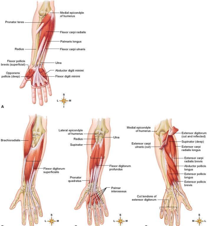
16 Intrinsic and Extrinsic Muscles (b) Intermediate flexor Flexor digitorum superficialis tendons Flexor digitorum profundus tendons Common flexor tendon Flexor digitorum superficialis Flexor pollicis longus intrinsic muscles entirely contained within a region, such as the hand both its origin and insertion there extrinsic muscles act on a designated region, but has its origin elsewhere fingers extrinsic muscles in the forearm Tendon sheath Tendon of flexor digitorum profundus Tendon of flexor digitorum superficialis Lumbricals Opponens digiti minimi Flexor digiti Abductor digiti minimi Flexor retinaculum Tendons of: Flexor carpi ulnaris Flexor digitorum superficialis Palmaris longus. (a) Palmar aspect, superficial First dorsal interosseous Adductor pollicis Tendon of flexor pollicis longus Flexor pollicis brevis Abductor pollicis brevis Opponens pollicis T endons of: Abductor pollicis longus Flexor carpi radialis Flexor pollicis longus
17 Muscle Innervation innervation of a muscle refers to the identity of the nerve that stimulates it enables the diagnosis of nerve, spinal cord, and brainstem injuries from their effects on muscle function spinal nerves arise from the spinal cord emerge through intervertebral foramina immediately branch into a posterior and anterior ramus innervate muscles below the neck cranial nerves arise from the base of the brain emerge through skull foramina innervate the muscles of the head and neck numbered I to XII
18 Muscles of Facial Expression muscles that insert in the dermis and subcutaneous tissues tense the skin and produce facial expressions innervated by facial nerve (CN VII) paralysis causes face to sag found in scalp, forehead, around the eyes, nose and mouth, and in the neck

19 Muscles in Facial Expression 19

20 20
 crucial importance to speech intrinsic muscles of tongue vertical, transverse, and longitudinal fascicles Palatoglossus Styloglossus Inferior longitudinal muscle of tongue Genioglossus")
21 Muscles of Chewing and Swallowing extrinsic muscles of the tongue tongue is very agile organ pushes food between molars for chewing (mastication) forces food into the pharynx for swallowing (deglutition) crucial importance to speech intrinsic muscles of tongue vertical, transverse, and longitudinal fascicles Palatoglossus Styloglossus Inferior longitudinal muscle of tongue Genioglossus Mylohyoid (cut) Geniohyoid Styloid process Mastoid process Posterior belly of digastric (cut) Superior pharyngeal constrictor (cut) Stylohyoid Middle pharyngeal constrictor Hyoglossus Hyoid bone Larynx Inferior pharyngeal constrictor Trachea Esophagus

 (a) Lateral view Lateral pterygoid plate Medial pterygoid plate Lateral pterygoid muscle")
22 Muscles of Chewing four pairs of muscles produce the biting and chewing movements of the mandible depression to open mouth elevation biting and grinding protraction incisors can cut retraction make rear teeth meet lateral and medial excursion grind food temporalis, masseter, medial pterygoid, lateral pterygoid innervated by mandibular nerve which is a branch of the trigeminal (V). Temporalis Orbicularis oris Buccinator Masseter (cut) (a) Lateral view Lateral pterygoid plate Medial pterygoid plate Lateral pterygoid muscle Medial pterygoid muscle Interior of oral cavity (b) Posterior view

23 23
24 Muscles Acting on the Head originate on the vertebral column, thoracic cage, and pectoral girdle insert on the cranial bones actions flexion (tipping head forward) sternocleidomastoid scalenes extension (holding the head erect) trapezius splenius capitis semispinalis capitis
25 Actions lateral flexion (tipping head to one side) rotation (turning the head to the left and right) may cause contralateral movement movement of the head toward the opposite side may cause ipsilateral movement movement of the head toward the same side

26 Muscles of the Face

27 Muscles of the Neck: Head Movements

28

29 Triangles of the Neck Anterior triangles A1. Muscular A2. Carotid A3. Submandibular A4. Suprahyoid Posterior triangles P1. Occipital P2. Omoclavicular A4 A3 A2 A1 P2 P1 Sternocleidomastoid
30 Muscles of the Trunk three functional groups muscles of respiration muscles that support abdominal wall and pelvic floor movement of vertebral column
31 Muscles of Respiration breathing requires the use of muscles enclosing thoracic cavity diaphragm, external and internal intercostal, inspiration air intake expiration expelling air other muscles of chest and abdomen that contribute to breathing sternocleidomastoid, scalenes of neck pectoralis major and serratus anterior of chest latissimus dorsi of back abdominal muscles internal and external obliques, and transverse abdominis some anal muscles
 in relaxation of diaphragm it rises shrinks the thoracic cavity (expiration) (b) Inferior view of diaphragm Xiphoid process of sternum Inferior vena cava Ribs Esophagus Central tendon")
32 Muscles of Respiration - Diaphragm muscular dome between thoracic and abdominal cavities muscle fascicles extend to a fibrous central tendon contraction flattens diaphragm enlarges thoracic cavity (inspiration) in relaxation of diaphragm it rises shrinks the thoracic cavity (expiration) (b) Inferior view of diaphragm Xiphoid process of sternum Inferior vena cava Ribs Esophagus Central tendon of diaphragm Aorta Vertebral column
 Lateral view of")
33 Muscles of Respiration - Intercostals external intercostals elevates ribs expand thoracic cavity create partial vacuum causing inflow of air. External intercostals internal intercostals depresses and retracts ribs compresses thoracic cavity expelling air Internal intercostals (a) Lateral view of intercostal muscles

34 Muscles of the Anterior Abdominal Wall four pairs of sheetlike muscles external abdominal oblique internal abdominal oblique transverse abdominal rectus abdominis strengthen abdominal wall. Muscles: External abdominal oblique Internal abdominal oblique Transverse abdominal Rectus abdominis Rectus sheath Peritoneum Posterior Anterior Subcutaneous fat Linea alba Aponeurosis of Transverse abdominal Internal abdominal oblique External abdominal oblique Skin 10-34

35 Muscles of the Abdominal Wall Figure 10.11a
36 external abdominal oblique most superficial of lateral abdominal muscles supports abdominal viscera against pull of gravity stabilizes vertebral column during heavy lifting maintains posture compresses abdominal organs aids in forced expiration rotation at waist Pectoralis major Latissimus dorsi Serratus anterior Tendinous intersections Rectus sheath (cut edges) Rectus sheath Umbilicus Linea semilunaris Linea alba Aponeurosis of external abdominal oblique Transverse abdominal Internal abdominal oblique (cut) External abdominal oblique (cut) Rectus abdominis Inguinal ligament (a) Superficial
37 internal abdominal oblique intermediate layer of lateral abdominal muscles unilateral contraction causes ipsilateral rotation of waist aponeurosis tendons of oblique and transverse muscles broad, fibrous sheets Subclavius Pectoralis minor (cut) Pectoralis minor Internal intercostals Serratus anterior External intercostals Rectus abdominis (cut) Rectus sheath External abdominal oblique (cut) Internal abdominal oblique Inguinal ligament Internal abdominal oblique (cut) Posterior wall of rectus sheath (rectus abdominis removed) Transverse abdominal (cut) (b) Deep
 Rectus sheath External abdominal oblique (cut) Internal abdominal oblique Inguinal ligament Internal")
38 transverse abdominal deepest of lateral abdominal muscles horizontal fibers compresses abdominal contents contributes to movements of vertebral column Subclavius Pectoralis minor (cut) Pectoralis minor Internal intercostals Serratus anterior External intercostals Rectus abdominis (cut) Rectus sheath External abdominal oblique (cut) Internal abdominal oblique Inguinal ligament Internal abdominal oblique (cut) Posterior wall of rectus sheath (rectus abdominis removed) Transverse abdominal (cut) (b) Deep
39 rectus abdominis flexes lumbar region of vertebral column produces forward bending at the waist extends from sternum to pubis rectus sheath encloses muscle three transverse tendinous intersections divide rectus abdominis into segments six pack Copyright The McGraw-Hill Companies, Inc. Permission required for reproduction or display. Pectoralis major Latissimus dorsi Serratus anterior Tendinous intersections Rectus sheath (cut edges) Rectus sheath Umbilicus Linea semilunaris Linea alba Aponeurosis of external abdominal oblique Transverse abdominal Internal abdominal oblique (cut) External abdominal oblique (cut) Rectus abdominis Inguinal ligament (a) Superficial

40 Superficial Muscles of Back. Superficial Deep Sternocleidomastoid Trapezius Deltoid Erector spinae Latissimus dorsi External abdominal oblique Thoracolumbar fascia Gluteus medius Semispinalis capitis Splenius capitis Levator scapulae Rhomboideus minor Rhomboideus major Supraspinatus Infraspinatus Teres minor Teres major Serratus anterior Serratus posterior inferior External abdominal oblique Internal abdominal oblique Gluteus minimus extend, rotate, and laterally flex vertebral column upper limb movement Gluteus maximus Lateral rotators

41

42 Deep Muscles of the Back. erector spinae iliocostalis, longissimus, spinalis from cranium to sacrum extension and lateral flexion of vertebral column semispinalis thoracis extension and contralateral rotation of vertebral column quadratus lumborum aids respiration ipsilateral flexion of lumbar vertebral column multifidus stabilizes adjacent vertebrae maintains posture Superior nuchal line Longissimus capitis Splenius capitis Serratus posterior superior Splenius cervicis Erector spinae: Iliocostalis Longissimus Spinalis Serratus posterior inferior Internal abdominal oblique External abdominal oblique (cut) Semispinalis capitis Semispinalis cervicis Semispinalis thoracis Multifidus Quadratus lumborum

43 Deep Back Muscles
44 Muscles of the Pelvic Floor three layers of muscles and fasciae that span pelvic outlet penetrated by anal canal, urethra, and vagina perineum diamond-shaped region between the thighs bordered by four bony landmarks urogenital triangle anterior half of perineum anal triangle posterior half of perineum three layers or compartments of the perineum superficial perineal space three muscles ischiocavernosus, bulbospongiosus, superficial transverse peritoneal middle compartment - spanned by urogenital diaphragm composed of a fibrous membrane and two or three muscles deep transverse perineal muscle, external urethral and anal sphincters compressor urethrae in females only pelvic diaphragm deepest layer consists of two muscle pairs levator ani and coccygeus
 Superficial perineal space, inferior")
45 Superficial Perineal Space. Male Ischiocavernosus Perineal raphe Bulbospongiosus Superficial transverse perineal muscle Levator ani Gluteus maximus (a) Superficial perineal space, inferior view muscles found just deep to the skin ischiocavernosus maintains erection bulbospongiosus aids in erection, expels remaining urine

46 Muscles of Pelvic Diaphragm Pubic symphysis Pubic ramus External urethral sphincter Deep transverse perineal muscle Perineal body External anal sphincter (b) Urogenital diaphragm, inferior view deepest compartment of the perineum pelvic diaphragm two muscle pairs levator ani - supports viscera and defecation coccygeus - supports and elevates pelvic floor
47 Hernias hernia any condition in which the viscera protrudes through a weak point in the muscular wall of the abdominopelvic cavity inguinal hernia most common type of hernia (rare in women) viscera enter inguinal canal or even the scrotum hiatal hernia stomach protrudes through diaphragm into thorax overweight people over 40 umbilical hernia viscera protrude through the navel
48 Muscles Acting on Shoulder and Upper Limb compartments spaces in which muscles are organized and are separated by fibrous connective tissue sheets (fasciae) each compartment contains one or more functionally related muscles along with their nerve and blood supplies muscles of upper limbs divided into anterior and posterior compartments muscles of lower limbs divided into anterior, posterior, medial, and lateral compartments compartment syndrome one of the muscles or blood vessels in a compartment is injured
49 Compartment Syndrome fasciae of arms and legs enclose muscle compartments very snugly if a blood vessel in a compartment is damaged, blood and tissue fluid accumulate in the compartment fasciae prevent compartment from expanding with increasing pressure compartment syndrome mounting pressure on the muscles, nerves and blood vessel triggers a sequence of degenerative events blood flow to compartment is obstructed by pressure if ischemia (poor blood flow) persists for more than 2 4 hours, nerves begin to die after 6 hours, muscles begin to die nerves can regenerate after pressure relieved, but muscle damage is permanent myoglobin in urine indicates compartment syndrome treatment immobilization of limb and fasciotomy incision to relieve compartment pressure
50 Muscles Acting on the Shoulder originate on the axial skeleton insert on clavicle and scapula scapula loosely attached to thoracic cage capable of great movement rotation, elevation, depression, protraction, retraction clavicle braces the shoulder and moderates movements
51 Anterior Muscles of Pectoral Girdle pectoralis minor ribs 3-5 to coracoid process of scapula draws scapula laterally serratus anterior ribs 1-9 to medial border of scapula abducts and rotates or depresses scapula Subclavius Pectoralis minor (cut) Pectoralis minor Internal intercostals Serratus anterior External intercostals Rectus abdominis (cut) Rectus sheath External abdominal oblique (cut) Internal abdominal oblique Inguinal ligament Internal abdominal oblique (cut) Posterior wall of rectus sheath (rectus abdominis removed) Transverse abdominal (cut) (b) Deep
 Serratus anterior Elevation Levator scapulae Trapezius (superior part) Rhomboideus major Rhomboideus minor Medial rotation Levator scapulae Rhomboideus")
52 Muscles Acting on Scapula. Lateral rotation Trapezius (superior part) Serratus anterior Elevation Levator scapulae Trapezius (superior part) Rhomboideus major Rhomboideus minor Medial rotation Levator scapulae Rhomboideus major Rhomboideus minor Depression Trapezius (inferior part) Serratus anterior Retraction Rhomboideus major Rhomboideus minor Trapezius Protraction Pectoralis minor Serratus anterior
53 Posterior Muscles of Pectoral Girdle four muscles of posterior group trapezius - superficial levator scapulae, rhomboideus minor, and rhomboideus major - deep trapezius stabilizes scapula and shoulder elevates and depresses shoulder apex levator scapulae elevates scapula flexes neck laterally rhomboideus minor retracts scapula and braces shoulder rhomboideus major same as rhomboideus minor Sternocleidomastoid Trapezius Deltoid Erector spinae Latissimus dorsi External abdominal oblique Thoracolumbar fascia Gluteus medius Gluteus maximus Superficial. Deep Semispinalis capitis Splenius capitis Levator scapulae Rhomboideus minor Rhomboideus major Supraspinatus Infraspinatus Teres minor Teres major Serratus anterior Serratus posterior inferior External abdominal oblique Internal abdominal oblique Gluteus minimus Lateral rotators 10-53

54

55

56 56

57 Muscles Acting on Arm nine muscles cross the shoulder joint and insert on humerus two are axial muscles because they originate on axial skeleton pectoralis major flexes, adducts, and medially rotates humerus latissimus dorsi adducts and medially rotated humerus.. Deltoid Clavicle Sternum Pectoralis major Supraspinatus Spine of scapula Greater tubercle of humerus Infraspinatus Humerus Teres minor Teres major Triceps brachii: Lateral head Long head Medial head Biceps brachii Brachialis Brachioradialis Coracobrachialis Triceps brachii: Lateral head Long head Latissimus dorsi (a) Anterior view (b) Posterior view

58

59 Muscles Acting on Arm seven scapular muscles originate on scapula deltoid rotates and abducts arm intramuscular injection site teres major extension and medial rotation of humerus coracobrachialis flexes and medially rotates arm remaining four form the rotator cuff that reinforce the shoulder joint Copyright The McGraw-Hill Companies, Inc. Permission required for reproduction or display. Deltoid Triceps brachii: Lateral head Long head Medial head Biceps brachii Brachialis Brachioradialis (a) Anterior view Clavicle Sternum Pectoralis major Coracobrachialis Supraspinatus Spine of scapula Greater tubercle of humerus Infraspinatus Humerus Teres minor Teres major Triceps brachii: Lateral head Long head Latissimus dorsi (b) Posterior view
60 Rotator Cuff Muscles tendons of the remaining four scapular muscles form the rotator cuff SITS muscles for the first letter of their names supraspinatus infraspinatus teres minor subscapularis tendons of these muscles merge with the joint capsule of the shoulder as they cross it in route to the humerus holds head of humerus into glenoid cavity supraspinatus tendon most easily damaged

61 Rotator Cuff Muscles 61 Anterior Posterior Rotator cuff (SITS) muscles: Supraspinatus Infraspinatus Teres minor Subscapularis Clavicle Acromion Coracoid process Glenoid cavity Inferior angle

62
63 Muscles Acting on Forearm elbow and forearm capable of flexion, extension, pronation, and supination carried out by muscles in both brachium (arm) and antebrachium (forearm) muscles with bellies in the arm (brachium) principal elbow flexors anterior compartment brachialis and biceps brachii brachialis produces 50% more power than biceps brachii brachialis is prime mover of elbow flexion principal elbow extensor posterior compartment triceps brachii prime mover of elbow extension
64 Muscles Acting on Forearm muscles with bellies in the forearm (antebrachium) most forearm muscles act on the hand and wrist brachioradialis flexes elbow anconeus extends elbow pronator quadratus prime mover in forearm pronation pronator teres assists pronator quadratus in pronation supinator supinates the forearm
65 Muscles Acting on Forearm principal flexor brachialis Copyright The McGraw-Hill Companies, Inc. Permission required for reproduction or display. Biceps brachii: Long head Short head synergistic flexors biceps brachii brachioradialis principal extensor triceps brachii Deltoid Triceps brachii: Lateral head Long head Medial head Biceps brachii Brachialis Brachioradialis Copyright The McGraw-Hill Companies, Inc. Permission required for reproduction or display. (a) Anterior view Clavicle Sternum Pectoralis major Coracobrachialis (c) Anterior view Copyright The McGraw-Hill Companies, Inc. Permission required for reproduction or display. Supraspinatus Spine of scapula Greater tubercle of humerus Infraspinatus Humerus Teres minor Teres major Triceps brachii: Lateral head Long head Latissimus dorsi (b) Posterior view 10-65

66
67

 Muscle actions in supination (b) Muscle actions in supination (a) Supination")
68 Supination and Pronation Copyright The McGraw-Hill Companies, Inc. Permission required for reproduction or display. Copyright The McGraw-Hill Companies, Inc. Permission required for reproduction or display. Lateral epicondyle Medial epicondyle Supinator Pronator teres Lateral epicondyle Medial epicondyle Supinator Pronator teres Ulna Radius Pronator quadratus Biceps brachii Ulna Radius Pronator quadratus Biceps brachii Radius Supinator Bursa Ulna Radius Supinator Bursa Ulna (b) Muscle actions in supination (b) Muscle actions in supination (a) Supination (c) Pronation supination supinator muscle palm facing anteriorly or superiorly pronation pronator quadratus and pronator teres palm faces posteriorly or inferiorly

69 Anterior Muscles on Wrist and Hand extrinsic muscles of the forearm intrinsic muscles in the hand itself extrinsic muscle actions flexion and extension of wrist and digits radial and ulnar flexion finger abduction and adduction thumb opposition. Biceps brachii Triceps brachii Anterior view Brachialis Common flexor tendon Pronator teres Aponeurosis of biceps brachii Brachioradialis Flexor carpi radialis Palmaris longus Flexor carpi ulnaris Flexor digitorum superficialis Common flexor tendon Supinator Interosseous membrane Flexor digitorum profundus Extensor carpi radialis longus and brevis Flexor digitorum superficialis Flexor pollicis longus Flexor pollicis longus Flexor retinaculum Palmar aponeurosis Flexor digitorum superficialis tendons Flexor digitorum superficialis tendons Flexor digitorum profundus tendons Flexor digitorum profundus tendons (a) Superficial flexors (b) Intermediate flexor (c) Deep flexors
70 Anterior Muscles on Wrist and Hand Anterior (Flexor) Compartment superficial layer flexor carpi radialis flexor carpi ulnaris flexor digitorum superficialis palmaris longus Anterior (Flexor) Compartment deep layer flexor digitorum profundus flexor pollicis longus

71 71

72 Posterior Muscles on Wrist & Hand extension of wrist and fingers, adduct / abduct wrist extension and abduction of thumb (pollicis) brevis - short, ulnaris - on ulna side of forearm Triceps brachii Posterior view Anconeus Flexor carpi ulnaris Extensor carpi ulnaris Extensor digiti minimi Brachioradialis Extensor carpi radialis longus Extensor carpi radialis brevis Extensor digitorum Abductor pollicis longus Extensor pollicis brevis Olecranon Extensor pollicis longus Extensor indicis Anconeus Supinator Abductor pollicis longus Extensor pollicis brevis Tendon of extensor indicis Tendons of extensor digitorum Extensor pollicis longus Tendons of extensor carpi radialis longus and brevis (a) Superficial extensors (b) Deep extensors
73 Posterior Muscles on Wrist and Hand Posterior (Extensor) Compartment superficial layer extensor carpi radialis longus extensor carpi radialis brevis extensor digitorum extensor digiti minimi extensor carpi ulnaris Posterior (Extensor) Compartment deep layer abductor pollicis longus extensor pollicis brevis extensor pollicis longus extensor indicis
74 Carpal Tunnel Syndrome flexor retinaculum bracelet-like fibrous sheet that the flexor tendons of the extrinsic muscles that flex the wrist pass on their way to their insertions carpal tunnel tight space between the flexor retinaculum and the carpal bones flexor tendons passing through the tunnel are enclosed in tendon sheaths enable tendons to slide back and forth quite easily carpal tunnel syndrome - prolonged, repetitive motions of wrist and fingers can cause tissues in the carpal tunnel to become inflamed, swollen, or fibrotic puts pressure on the median nerve of the wrist that passes through the carpal tunnel along with the flexor tendons tingling and muscular weakness in the palm and medial side of the hand pain may radiate to arm and shoulder treatment anti-inflammatory drugs, immobilization of the wrist, and

75 Carpal Tunnel Syndrome Tendon of flexor digitorum superficialis Lumbrical Opponens digiti minimi repetitive motions cause inflammation and pressure on median nerve Flexor digiti minimi brevis Abductor digiti minimi Pisiform bone Flexor digitorum superficialis Adductor pollicis Flexor pollicis brevis Abductor pollicis brevis Tendon of extensor pollicis brevis Tendon of flexor carpi radialis Tendon sheath Tendon of flexor digitorum profundus Tendon of flexor digitorum superficialis Lumbricals Opponens digiti minimi Flexor digiti minimi brevis Abductor digiti minimi First dorsal interosseous Adductor pollicis Tendon of flexor pollicis longus Flexor pollicis brevis Abductor pollicis brevis Opponens pollicis (b) Palmar dissection, superficial Flexor retinaculum Tendons of: Flexor carpi ulnaris Flexor digitorum superficialis Palmaris longus Tendons of: Abductor pollicis longus Flexor carpi radialis Flexor pollicis longus (a) Palmar aspect, superficial 10-75

76 76
77 Intrinsic Hand Muscles thenar group form thick, fleshy mass at base of thumb adductor pollicis abductor pollicis brevis flexor pollicis brevis opponens pollicis Hypothenar group - fleshy base of the little finger abductor digiti minimi flexor digiti minimi brevis opponens digiti minimi Midpalmar group hollow of palm dorsal interosseous muscles (4) palmar interosseous muscles (3) lumbricals (4 muscles) Tendon sheath Tendon of flexor digitorum profundus Tendon of flexor digitorum superficialis Lumbricals Opponens digiti minimi Flexor digiti minimi brevis Abductor digiti minimi Flexor retinaculum Tendons of: Flexor carpi ulnaris Flexor digitorum superficialis Palmaris longus First dorsal interosseous Adductor pollicis Tendon of flexor pollicis longus Flexor pollicis brevis Abductor pollicis brevis Opponens pollicis Tendons of: Abductor pollicis longus Flexor carpi radialis Flexor pollicis longus (a) Palmar aspect, superficial 10-77
78 Muscles on the Hip and Lower Limb largest muscles found in lower limb less for precision, more for strength needed to stand, maintain balance, walk, and run several cross and act on two or more joints leg the part of the limb between the knee and ankle foot includes tarsal region (ankle), metatarsal region, and the toes
79 Muscles Acting on the Hip and Femur. anterior muscles of the hip iliacus flexes thigh at hip iliacus portion arises from iliac crest and fossa psoas major flexes thigh at hip arises from lumbar vertebrae they share a common tendon on the femur Iliopsoas: Iliacus Psoas major Pectineus Adductor magnus Adductor brevis Adductor longus Gracilis Insertion of gracilis on tibia Piriformis Obturator externus

80 80

81 81
82 Posterior Muscles on Hip and Femur lateral and posterior muscles of the hip tensor fasciae latae extends knee, laterally rotates knee gluteus maximus forms mass of the buttock prime hip extensor provides most of lift when you climb stairs gluteus medius and minimus abduct and medially rotate thigh Iliac crest Gluteus medius Sacrum Gluteus maximus Coccyx Ischial tuberosity Superficial Deep Gluteus minimus Lateral rotators: Piriformis Gemellus superior Obturator internus Obturator externus Gemellus inferior Quadratus femoris

83 83

84 Posterior Muscles on Hip and Femur lateral rotators - six muscles inferior to gluteus minimus deep to the two other gluteal muscles gemellus superior gemellus inferior obturator externus obturator internus piriformis quadratus femoris Iliac crest Gluteus medius Sacrum Gluteus maximus Coccyx Ischial tuberosity Superficial Deep Gluteus minimus Lateral rotators: Piriformis Gemellus superior Obturator internus Obturator externus Gemellus inferior Quadratus femoris
 compartment of thigh five muscles act as primary adductors of the thigh adductor brevis adductor longus adductor magnus gracilis pectineus Iliopsoas:")
85 Muscles Acting on Hip and Femur. medial (adductor) compartment of thigh five muscles act as primary adductors of the thigh adductor brevis adductor longus adductor magnus gracilis pectineus Iliopsoas: Iliacus Psoas major Pectineus Adductor magnus Adductor brevis Adductor longus Gracilis Insertion of gracilis on tibia Piriformis Obturator externus
86 Muscles on the Knee and Leg anterior (extensor) compartment of the thigh contains large quadriceps femoris muscle prime mover of knee extension most powerful muscle in the body has four heads rectus femoris, vastus lateralis, vastus medialis, and vastus intermedius all converge on single quadriceps (patellar) tendon extends to patella then continues as patellar ligament inserts on tibial tuberosity sartorius longest muscle in the body tailor s muscle

87 Muscles Acting on the Knee and Leg Iliac crest. Iliopsoas: Iliacus Psoas major L5 Anterior superior iliac spine Tensor fasciae latae Iliotibial band Medial compartment: Adductor magnus Pectineus Adductor brevis Adductor longus Gracilis Anterior compartment: Sartorius Quadriceps femoris: Vastus intermedius Rectus femoris Vastus lateralis Vastus medialis Quadriceps femoris tendont Patella Patellar ligament (a) Superficial (b) Deep

88 88

89 Muscles Acting on the Knee and Leg posterior (flexor) compartment of the thigh contains hamstring muscles from lateral to medial; biceps femoris semitendinosus semimembranosus Gluteus medius Gluteus maximus Gracilis Adductor magnus Iliotibial band Vastus lateralis Hamstring group: Biceps femoris Long head Short head Semitendinosus Semimembranosus

90 Anterior Compartment of Leg. Patella Patellar ligament Fibularis longus Fibularis brevis Extensor digitorum longus Extensor retinacula Tibia Gastrocnemius Soleus Tibialis anterior Tibialis anterior Extensor hallucis brevis Extensor digitorum brevis Extensor hallucis longus Fibularis tertius (a) (b) (c) (d) Extensor digitorum longus anterior (extensor) compartment of the leg dorsiflex the ankle prevent toes from scuffing when walking fibularis (peroneus) tertius extensor digitorum longus extensor hallucis longus tibialis anterior

91 91
 Fibularis longus Soleus Gastrocnemius")
92 Posterior Compartment of Leg Superficial Group. Gastrocnemius: Medial head Lateral head Tendon of gastrocnemius Plantaris Popliteus Tendon of plantaris Flexor digitorum longus Heads of gastrocnemius (cut) Fibularis longus Soleus Gastrocnemius (cut) Fibularis longus Fibularis brevis Flexor hallucis longus Calcaneal tendon Calcaneus three muscles of the superficial group gastrocnemius - plantar flexes foot, flexes knee soleus plantar flexes foot plantaris - weak synergist of triceps (a) (b)
 Calcaneus Plantar surface of the foot (a) four muscles in the deep group")
93 Posterior Compartment of Leg Deep Group Plantaris (cut) Tibialis posterior Flexor digitorum longus Popliteus Gastrocne -mius (cut) Soleus (cut) Fibula (b) (c) Flexor digitorum longus Tibialis posterior Fibularis longus Flexor hallucis longus Fibularis brevis Popliteus Flexor hallucis longus Calcaneal tendon (cut) Calcaneus Plantar surface of the foot (a) four muscles in the deep group flexor digitorum longus flexes phalanges flexor hallucis longus flexes great toe tibialis posterior inverts foot popliteus acts on knee (d)
 Calcaneus (a)")
94 Lateral (Fibular) Compartment of the Leg Popliteus Soleus (cut) Flexor digitorum longus Plantaris (cut) Gastrocnemius (cut) Fibula Tibialis posterior Fibularis longus Flexor hallucis longus Fibularis brevis two muscles in this compartment fibularis longus fibularis brevis both plantar flex and evert the foot provides lift and forward thrust Calcaneal tendon (cut) Calcaneus (a)
 Layer 1, plantar view Abductor hallucis Flexor digitorum brevis Plantar aponeurosis")
95 Intrinsic Muscles of Foot. support for arches abduct and adduct the toes flex the toes Flexor digiti minimi brevis Abductor digiti minimi (a) Layer 1, plantar view Abductor hallucis Flexor digitorum brevis Plantar aponeurosis (cut) Calcaneus Quadratus plantae Flexor digitorum brevis (cut) (b) Layer 2, plantar view Lumbricals Flexor hallucis longus tendon Flexor digitorum longus tendon Abductor hallucis (cut) one dorsal muscle extensor digitorum brevis extends toes Flexor digiti minimi brevis Quadratus plantae (cut) Adductor hallucis Flexor hallucis brevis Flexor hallucis longus tendon (cut) Abductor hallucis (cut) Flexor digitorum longus tendon (cut) Plantar interosseous Dorsal interosseous dorsal view (c) Layer 3, plantar view (d) Layer 4, plantar view (e) Layer 4, dorsal view
96 Athletic Injuries muscles and tendons are vulnerable to sudden and intense stress proper conditioning and warm-up needed common injuries; compartment syndrome shinsplints pulled hamstrings tennis elbow pulled groin rotator cuff injury treat with rest, ice, compression and elevation no pain, no gain is a dangerous misconception
Due in Lab weeks because of Thanksgiving Prelab #10. Homework #8. Both sides! Both sides!
 Lab 8 MUSCLES Due in Lab 10 2 weeks because of Thanksgiving Prelab #10 Both sides! Homework #8 Both sides! Refer to Muscles 22-23 Naming of muscles Origin Site of muscle attachment that doesn t move during
11/15/2018. Temporalis Elevates & retracts mandible. Masseter = Prime mover of jaw closure. Levator scapulae Supraspinatus Clavicle.
 Due in Lab 10 Lab 8 MUSCLES 2 weeks because of Thanksgiving Prelab #10 Both sides! Homework #8 Both sides! Refer to Muscles 22-23 Examples of Origin & Insertion Naming of muscles Origin Site of muscle
Human Anatomy and Physiology I Laboratory
 Human Anatomy and Physiology I Laboratory Gross Anatomy of the Muscular System (Two weeks) 1 This lab involves study of the laboratory exercise Gross Anatomy of the Muscular System. Complete the Review
Epicranius (frontal belly) Zygomaticus minor. Zygomaticus major Buccinator
 Zygomaticus minor. Zygomaticus major Buccinator") Epicranius (frontal belly) Zygomaticus minor Zygomaticus major Buccinator Masseter Digastric (posterior belly) Stylohyoid Sternocleidomastoid Trapezius Scalenus Omohyoid (inferior belly) Orbicularis oris
Epicranius (frontal belly) Zygomaticus minor Zygomaticus major Buccinator Masseter Digastric (posterior belly) Stylohyoid Sternocleidomastoid Trapezius Scalenus Omohyoid (inferior belly) Orbicularis oris
Muscle Anatomy Review Chart
 Muscle Anatomy Review Chart BACK Superficial (5) Trapezius Transverse cervical a. Latissimus dorsi Thoracodorsal a. Rhomboideus major Dorsal scapular a. Rhomboideus minor Levator scapulae Intermediate
Muscle fiber (cell) Blood vessel. Perimysium. Epimysium. Fascicle (wrapped by perimysium) Endomysium (between fibers) Tendon. Bone
 Blood vessel. Perimysium. Epimysium. Fascicle (wrapped by perimysium) Endomysium (between fibers) Tendon. Bone") Figure 6.1 Connective tissue wrappings of skeletal muscle. Blood vessel Muscle fiber (cell) Perimysium Epimysium Fascicle (wrapped by perimysium) Tendon Endomysium (between fibers) Bone Figure 6.15 Superficial
Figure 6.1 Connective tissue wrappings of skeletal muscle. Blood vessel Muscle fiber (cell) Perimysium Epimysium Fascicle (wrapped by perimysium) Tendon Endomysium (between fibers) Bone Figure 6.15 Superficial
Biology 2401 Muscles List for CPC models
 Biology 2401 List for CPC models Italicized muscles are dissect and similar in the cat = Dissect and note the differences in human and cat Major of the Human Head Facial Expression Epicranius frontalis
Muscular System Dr. Gary Mumaugh
 Muscular System Dr. Gary Mumaugh 1 Organization of Muscles about 600 human skeletal muscles constitute about half of our body weight three kinds of muscle tissue o skeletal, cardiac, smooth specialized
Temporalis Elevates & retracts mandible. Masseter Elevates mandible. Sternocleidomastoid Neck flexion. Trapezius Elevates & depresses shoulders
 Anterior Posterior Temporalis Elevates & retracts mandible Masseter Elevates mandible Sternocleidomastoid Neck flexion Trapezius Elevates & depresses shoulders Masseter Elevates mandible Temporalis Elevates
Synergist Muscles. Shoulder (glenohumeral joint) Flexion Deltoid (anterior fibers) Pectoralis major (upper fibers) Biceps Brachii Coracobrachialis
 Flexion Deltoid (anterior fibers) Pectoralis major (upper fibers) Biceps Brachii Coracobrachialis") Synergist Muscles Dr Gene Desepoli DrGeneLMT@gmail.com Shoulder (glenohumeral joint) Deltoid (anterior fibers) Pectoralis major (upper fibers) Biceps Brachii Coracobrachialis Deltoid (posterior fibers)
Synergist Muscles Dr Gene Desepoli DrGeneLMT@gmail.com Shoulder (glenohumeral joint) Deltoid (anterior fibers) Pectoralis major (upper fibers) Biceps Brachii Coracobrachialis Deltoid (posterior fibers)
A. All movements require muscle which are organs using chemical energy to contract.
 Ch 8 Muscles Introduction: A. All movements require muscle which are organs using chemical energy to contract. B. The three types of muscle in the body are skeletal, smooth, and cardiac muscle. C. This
Chapter 10. An Overview of the Muscle System
 Chapter 10 An Overview of the Muscle System The Muscular System Superficial Deep Deep Superficial Frontalis Platysma Deltoid Pectoralis major Biceps brachii Brachioradialis Flexor carpi radialis External
5/21/2013. Muscle Anatomy. Thursday January, 24 th, Skeletal Muscle. Smooth Muscle. Cardiac Muscle
 Muscle Anatomy Thursday January, 24 th, 2013 Skeletal Muscle Cardiac Muscle Smooth Muscle 1 Smooth Muscle 1. Found in the walls of the digestive system, bladder, uterus and blood vessels 2. Involuntary
3/27/2012. Muscle Classification: Functional Groups. Interactions of Skeletal Muscles. Naming Skeletal Muscles. Naming Skeletal Muscles
 Interactions of Skeletal Muscles Skeletal muscles work together or in opposition Muscles only pull (never push) As muscles shorten, the insertion generally moves toward the origin Whatever a muscle (or
The Muscular System. Chapter 10 Part D. PowerPoint Lecture Slides prepared by Karen Dunbar Kareiva Ivy Tech Community College
 Chapter 10 Part D The Muscular System Annie Leibovitz/Contact Press Images PowerPoint Lecture Slides prepared by Karen Dunbar Kareiva Ivy Tech Community College Table 10.14: Muscles Crossing the Hip and
Prime movers provide the major force for producing a specific movement Antagonists oppose or reverse a particular movement Synergists
 Dr. Gary Mumaugh Prime movers provide the major force for producing a specific movement Antagonists oppose or reverse a particular movement Synergists Add force to a movement Reduce undesirable or unnecessary
Chapter 11: The Muscular System. Copyright 2009, John Wiley & Sons, Inc.
 Chapter 11: The Muscular System Muscle Attachment Sites: Origin & Insertion n Skeletal muscles cause movements by exerting force on tendons, which pulls on bones or other structures. n Articulating bones
A&P 1 Muscle In-Lab Guide
 A&P 1 Muscle In-Lab Guide This lab guide includes a table with all the muscles you need to ID, along with their origins, insertions and actions Dashed lines means ignore. If several actions are listed,
Lectures Muscular System 10-1
 Lectures 12-14 Muscular System 10-1 Properties of Muscle Ability of a muscle to shorten with force Capacity of muscle to respond to a stimulus Muscle can be stretched to its normal resting length and beyond
The Human Muscular System Required reading before beginning this lab: Saladin, KS: Human Anatomy 5th ed (2017) Chapters 10, 11, 12 INTRODUCTION
 Chapters 10, 11, 12 INTRODUCTION") Biology 322: Human Anatomy The Human Muscular System Required reading before beginning this lab: Saladin, KS: Human Anatomy 5 th ed (2017) Chapters 10, 11, 12 INTRODUCTION We will use a number of lab periods
Biology 322: Human Anatomy The Human Muscular System Required reading before beginning this lab: Saladin, KS: Human Anatomy 5 th ed (2017) Chapters 10, 11, 12 INTRODUCTION We will use a number of lab periods
SKELETAL MUSCLE ANATOMY
 SKELETAL MUSCLE ANATOMY OUTLINE I. Anatomical Terms of Motion II. Head, Face & Neck Muscles III. Anterior Torso Muscles IV. Posterior Torso Muscles V. Arm & Shoulder Muscles VI. Leg & Hip Muscles 2 ANATOMICAL
Scapula Spine Lateral edge of clavicle. Medial border Scapula. Medial border of Scapula, between superior angle and root of spine. Scapula.
 Muscle attachments and actions answer sheet Muscle Origins insertions Movements Joints crossed Trapezius Base of skull Spinous process of C7 Thoracic Spine Lateral edge of clavicle Elevation Retraction
Lab Exercise #5 The Muscular System Student Performance Objectives
 Student Performance Objectives The material that you are required to learn in this exercise can be found in either the lecture text or the supplemental materials provided in lab. Prior to coming to class,
Contents. Preface xv. SECTION 1: Introduction to the Bodynamic System 1. SECTION 2: The Bodynamic Psycho-Motor Anatomy 29
 Contents Preface xv SECTION 1: Introduction to the Bodynamic System 1 Definitions in the Bodynamic System 3 Ego Formation through the Coding Elements 9 Examples of Formation of Coding 17 Using This Book
The Muscular System. Chapter 10 Part C. PowerPoint Lecture Slides prepared by Karen Dunbar Kareiva Ivy Tech Community College
 Chapter 10 Part C The Muscular System Annie Leibovitz/Contact Press Images PowerPoint Lecture Slides prepared by Karen Dunbar Kareiva Ivy Tech Community College Table 10.9: Muscles Crossing the Shoulder
ACTIVITIES 5 & 6: APPENDICULAR AND AXIAL MUSCLES
 ACTIVITIES 5 & 6: APPENDICULAR AND AXIAL MUSCLES Objectives: 1) How to get ready: Read Chapter 11 & 12, McKinley et al., Human Anatomy, 4e. All text references are for this textbook. Begin identifying
Chapter 10: Muscular System: Gross Anatomy
 Chapter 10: Muscular System: Gross Anatomy I. General Principles A. General Terminology 1. Tendons attach 2. What is an aponeurosis? 3. The points of muscle attachment are called & 4. How is the "origin"
The Muscular System Lab Power Point
 The Muscular System Lab Power Point Myoneural Junction Sarcoplasm Nucleus Myofibrils Sarcomere (black line to black line) Sarcolemma Myoneural space Nucleus Endomysium Motor Neuron Muscles of Facial Expression
The Muscular System PART C. PowerPoint Lecture Slide Presentation by Patty Bostwick-Taylor, Florence-Darlington Technical College
 PowerPoint Lecture Slide Presentation by Patty Bostwick-Taylor, Florence-Darlington Technical College The Muscular System 6 PART C Five Golden Rules of Skeletal Muscle Activity Table 6.2 Muscles and Body
Chapter 9. The Muscular System
 1 Chapter 9 The Muscular System 2 Introduction Skeletal muscles: movement in environment Smooth muscles: intestines, ureters, veins and arteries Cardiac muscle: pumps blood through heart and blood vessels
Head & Neck The muscle names are followed by the chapter number
 Head & Neck The muscle names are followed by the chapter number. Splenius capitis (9) 2. Occipitalis (2) Temporalis () 3. Temporalis () 4. Semispinalis capitis (9) Facial / Scalp (2) 5. Temporalis () Facial
The Muscular System. PowerPoint Lecture Presentations prepared by Jason LaPres. Lone Star College North Harris Pearson Education, Inc.
 11 The Muscular System PowerPoint Lecture Presentations prepared by Jason LaPres Lone Star College North Harris An Introduction to the Muscular System The Muscular System Consists only of skeletal muscles
In-Depth Foundations: Anatomy Terms to Know
 Be familiar with / able to identify and define all the following parts. The Spine Cranium Vertebrae Cervical, Thoracic, Lumbar Sacrum Coccyx Bones of Upper Body Cranium Mastoid process; Occipital condyle,
BIOH111. o Cell Module o Tissue Module o Skeletal system o Integumentary system o Muscle system o Nervous system o Endocrine system
 BIOH111 o Cell Module o Tissue Module o Skeletal system o Integumentary system o Muscle system o Nervous system o Endocrine system TEXTBOOK AND REQUIRED/RECOMMENDED READINGS o Principles of anatomy and
Cadaver Muscular System Practice Practical
 Cadaver Muscular System Practice Practical Station 1 Station 1 1. Specific structure 1. Rectus sheath 2. Red line 2. Linea alba Station 2 Station 2 3. Red muscle 1. Rectus abdominis 4. Red muscle actions
Muscles of the Cat. N Deltoid MUSCLES OF THE CHEST. Pectoralis major. (This muscle is superior to Pectoralis minor) MUSCLES OF THE CHEST
 MUSCLES OF THE CHEST") MUSCLES OF THE CHEST Pectoralis major (This muscle is superior to Pectoralis minor) 1. MUSCLES OF THE CHEST Pectoralis minor (This muscle is inferior to Pectoralis major) 2. MUSCLES OF THE ARM Deltoid
MUSCLES OF THE CHEST Pectoralis major (This muscle is superior to Pectoralis minor) 1. MUSCLES OF THE CHEST Pectoralis minor (This muscle is inferior to Pectoralis major) 2. MUSCLES OF THE ARM Deltoid
Human Anatomy Lab #7: Muscles of the Cadaver
 Human Anatomy Lab #7: Muscles of the Cadaver Table of Contents: Expected Learning Outcomes.... 1 Introduction...... 1 Identifying Muscles on Yourself.... 2 Muscles of the Anterior Trunk and Arm.. 2 Muscles
The Muscular System Outline 10.1 For any movement, muscles can act in one of three ways (pp ) A. Muscles only pull; they never push, and as a
 A. Muscles only pull; they never push, and as a") The Muscular System Outline 10.1 For any movement, muscles can act in one of three ways (pp. 321 322) A. Muscles only pull; they never push, and as a muscle shortens, the insertion is pulled toward the
The Muscular System Outline 10.1 For any movement, muscles can act in one of three ways (pp. 321 322) A. Muscles only pull; they never push, and as a muscle shortens, the insertion is pulled toward the
Chapter 10. The Muscular System. AP1-Lecture Goodwin College. Introduction. In this chapter we will cover: 4/27/2015
 Chapter 10 The Muscular System AP1-Lecture Goodwin College Introduction In this chapter we will cover: Structural and functional organization of muscles Muscles of the head and neck Muscles of the trunk
BIOH111. o Cell Module o Tissue Module o Skeletal system o Integumentary system o Muscle system o Nervous system o Endocrine system
 BIOH111 o Cell Module o Tissue Module o Skeletal system o Integumentary system o Muscle system o Nervous system o Endocrine system TEXTBOOK AND REQUIRED/RECOMMENDED READINGS o Principles of anatomy and
Bio 113 Anatomy and Physiology The Muscles. Muscles of the Head and Neck. Masseter. Orbicularis occuli. Orbicularis oris. Sternocleidomastoid
 Bio 113 Anatomy and Physiology The Muscles Muscles of the Head and Neck Masseter Orbicularis occuli Orbicularis oris Sternocleidomastoid Temporalis BIO 113 Fall 2011 Muscles Page 1 of 5 Muscles of the
List of Muscles and Function. Region View Muscle Function Facial Anterior/Oblique Occipitofrontalis front belly Raises eyebrows
 List of Muscles and Function Region View Muscle Function Facial Anterior/Oblique Occipitofrontalis front belly Raises eyebrows Orbicularis oculi Closes eye Orbicularis oris Purses lips Zygomaticus minor/major
Muscles of the lower extremities. Dr. Nabil khouri MD, MSc, Ph.D
 Muscles of the lower extremities Dr. Nabil khouri MD, MSc, Ph.D Posterior leg Popliteal fossa Boundaries Biceps femoris (superior-lateral) Semitendinosis and semimembranosis (superior-medial) Gastrocnemius
Biology 218 Human Anatomy. Adapted from Martini Human Anatomy 7th ed. Chapter 12 Surface Anatomy and Cross-Sectional Anatomy
 Adapted from Martini Human Anatomy 7th ed. Chapter 12 Surface Anatomy and Introduction Surface anatomy is the study of anatomical landmarks on the exterior of the human body Knowledge of surface anatomy
TABLES OF MUSCLE ACTIONS, INNERVATIONS, AND ATTACHMENTS
 TABLES OF MUSCLE ACTIONS, INNERVATIONS, AND ATTACHMENTS Table 1-1 ERECTOR SPINAE MUSCLES Intrinsic muscles producing extension and/or lateral of the spine Muscle Joint and Action Innervation Inferior Attachment
Exercise Science Section 3: The Muscular System
 Exercise Science Section 3: The Muscular System An Introduction to Health and Physical Education Ted Temertzoglou Paul Challen ISBN 1-55077-132-9 Major Functions of Muscles Movement Includes: breathing,
The muscular system I Muscles of the head neck and trunk
 The muscular system I Muscles of the head neck and trunk Dr. Nabil Khouri Dr. Nabil Khouri MD MSc, PhD Interactions of Skeletal Muscles Skeletal muscles work together or in opposition Muscles only pull
lesser trochanter of femur lesser trochanter of femur iliotibial tract (connective tissue) medial surface of proximal tibia
 medial surface of proximal tibia") LOWER LIMB MUSCLES OF THE APPENDICULAR SKELETON The muscles that act on the lower limb fall into three groups: those that move the thigh, those that move the lower leg, and those that move the ankle, foot,
LOWER LIMB MUSCLES OF THE APPENDICULAR SKELETON The muscles that act on the lower limb fall into three groups: those that move the thigh, those that move the lower leg, and those that move the ankle, foot,
CHAPTER 11 LECTURE OUTLINE I. INTRODUCTION
 CHAPTER 11 LECTURE OUTLINE I. INTRODUCTION A. The muscular system specifically concerns skeletal muscles and associated connective tissue that make individual muscle organs. B. This chapter discusses how
Human Anatomy Biology 351
 1 Human Anatomy Biology 351 Upper Limb Exam Please place your name on the back of the last page of this exam. You must answer all questions on this exam. Because statistics demonstrate that, on average,
The Muscular System Part A
 10 The Muscular System Part A Lecture Presentation by Lori Garrett Section 1: Functional Organization of the Muscular System Learning Outcomes 10.1 Describe the general function of the body s axial and
Muscular Nomenclature and Kinesiology - One
 Chapter 16 Muscular Nomenclature and Kinesiology - One Lessons 1-3 (with lesson 4) 1 Introduction 122 major muscles covered in this chapter Chapter divided into nine lessons Kinesiology study of human
The Muscular System. Part A
 The Muscular System Part A 10 The Muscular System Part A 10 Hold onto your glutes, this is a big one. 10 Interactions of Skeletal Muscles Skeletal muscles work together or in opposition Muscles only pull
Muscles of the Upper Limb
 Muscles of the Upper Limb anterior surface of ribs 3 5 coracoid process Pectoralis minor pectoral nerves protracts / depresses scapula Serratus anterior Subclavius ribs 1-8 long thoracic nerve rib 1 ----------------
Lab 9: Learn origin and insertion for each of the listed muscles. For Exercise 15, do Activities 1-6 in 9 th edition, Activities 1-4 in 10 th edition
 The Muscular System Exercises 14, 15, and 16 (begins: page 187 in 9 th and 10 th editions) Exercises 12, 13, and 14 (begins: page 185 in 11 th edition, page 189 in 12 th edition) Lab 8 and 9 Objectives
Cat Muscles Flashcards Mt SAC
 1. MUSCLES OF THE CHEST Pectoralis major (This muscle is superior to Pectoralis minor) 2. MUSCLES OF THE CHEST Pectoralis minor (This muscle is inferior to Pectoralis major) 3. MUSCLES OF THE ARM AD CHEST
Biology 218 Human Anatomy. Adapted from Martini Human Anatomy 7th ed. Chapter 10 The Muscular System Axial Musculature
 Adapted from Martini Human Anatomy 7th ed. Chapter 10 The Muscular System Axial Musculature Introduction The skeletal muscle of the body can be subdivided into: Axial musculature Muscles that position
Human Anatomy Biology 351
 Human Anatomy Biology 351 Lower Limb Please place your name on the back of the last page of this exam. You must answer all questions on this exam. Because statistics demonstrate that, on average, between
Muscles of Lesson Five. Muscular Nomenclature and Kinesiology - Two. Muscles of Lesson Five, cont. Chapter 16
 Chapter 16 Muscular Nomenclature and Kinesiology - Two Lessons 5-6 Muscles of Lesson Five Iliopsoas (psoas major, iliacus) Hip outward rotators (piriformis, gemellus superior, gemellus inferior, obturator
Biol 353 Pre-Professional Human Anatomy Exam III Fall 2017 page 1 of 8
 Biol 353 Pre-Professional Human Anatomy Exam III Fall 2017 page 1 of 8 IMPORTANT INSTRUCTIONS: ANSWER ONLY 50 QUESTIONS. Do not answer more than 50 questions. If you answer more than 50 questions, then
Musculoskeletal Anatomy Coloring Book
 Musculoskeletal Anatomy Coloring Book Muscolino, Joseph E. ISBN-13: 9780323057219 Table of Contents Introduction â How to Use This Book 1. The Skeletal System Bones of the Head â Anterior View Bones of
Name this muscle. Name this muscle
 this muscle this muscle Pectoralis Major Pectoralis Minor Serratus anterior Pectoralis minor Serratus anterior this muscle Deltoid: The major abductor of the upper limb this muscle this muscle this muscle
Lower limb summary. Anterior compartment of the thigh. Done By: Laith Qashou. Doctor_2016
 Lower limb summary Done By: Laith Qashou Doctor_2016 Anterior compartment of the thigh Sartorius Anterior superior iliac spine Upper medial surface of shaft of tibia 1. Flexes, abducts, laterally rotates
Muscles of the Hip 1. Tensor Fasciae Latae O: iliac crest I: lateral femoral condyle Action: abducts the thigh Nerve: gluteal nerve
 Muscles of the Hip 1. Tensor Fasciae Latae O: iliac crest I: lateral femoral condyle Action: abducts the thigh Nerve: gluteal nerve 2. Gluteus Maximus O: ilium I: femur Action: abduct the thigh Nerve:
REFERENCE DIAGRAMS OF UPPER LIMB MUSCLES: NAMES, LOCATIONS, ATTACHMENTS, FUNCTIONS MUSCLES CONNECTING THE UPPER LIMB TO THE AXIAL SKELETON
 REFERENCE DIAGRAMS OF UPPER LIMB MUSCLES: NAMES, LOCATIONS, ATTACHMENTS, FUNCTIONS MUSCLES CONNECTING THE UPPER LIMB TO THE AXIAL SKELETON A25LAB EXERCISES: UPPER LIMB MUSCLES Page 1 MUSCLES CONNECTING
Lab Activity 11: Group I
 Lab Activity 11: Group I Muscles Martini Chapter 11 Portland Community College BI 231 Origin and Insertion Origin: The place where the fixed end attaches to a bone, cartilage, or connective tissue. Insertion:
Muscles Built on the Maniken
 Muscles Built on the Maniken Facial Muscle Group 1. Temporalis O temporal fossa I anterior border of the ramus of the mandible A elevates the mandible (bite muscle) and holds jaw while at rest 2. Procerus
Copy Right- Hongqi ZHANG-Department of Anatomy-Fudan University. Systematic Anatomy. Locomotor system - Part 6
 Systematic Anatomy Locomotor system - Part 6 Muscles of abdomen Muscles of the upper limb Dr.Hongqi Zhang ( 张红旗 ) Email: zhanghq58@126.com 1 Muscles of abdomen Muscles of the upper limb Muscles of abdomen
Masseter- in front of ear Temporalis Mandible
 Frontal Belly (Epicranius) Occipital Belly (Epicranius) Orbicularis Oculi Orbicularis Oris Zygomaticus minor Zygomaticus major Buccinator Facial Expression Origin- stays still Raises eyebrows Galea aponeurotica
Monday, November 13, 2017 A & P 2401
 Monday, November 13, 2017 A & P 2401 Today you will complete the following handouts. Study the last part of the handout for this will be on your quiz, which will be on Wednesday. It is titled steps of
In which arm muscle are intramuscular injections most often given? (not in text)
") AP1 Lab 9 - Muscles of the Arms and Legs Locate the following muscles on the models and on yourself. Recall anatomical position. Directional terms such as anterior, posterior, lateral, etc. all assume
AP1 Lab 9 - Muscles of the Arms and Legs Locate the following muscles on the models and on yourself. Recall anatomical position. Directional terms such as anterior, posterior, lateral, etc. all assume
Skeleton. The left forearm is in the position of supination, the right in pronation.
 S ystemic review A Skeleton A from the front B B from behind The left forearm is in the position of supination, the right in pronation. Skull Mandible Hyoid bone Cervical vertebrae Clavicle Sternum Costal
Chiropractic Technician Class
 Chiropractic Technician Class Presentation By: Dr. Kay Miller. The Role of Exercise as it Relates to Our Musculoskeletal System Introduction to the topic and Preliminary Physical exam Musculoskeletal anatomy:
Exercise Science Section 3: The Muscular System
 Exercise Science Section 3: The Muscular System An Introduction to Health and Physical Education Ted Temertzoglou Paul Challen ISBN 1-55077-132-9 Major Functions of Muscles Movement v Includes: breathing,
medial half of clavicle; Sternum; upper six costal cartilages External surfaces of ribs 3-5
 MUSCLE ORIGIN INSERTION ACTION NERVE Pectoralis Major medial half of clavicle; Sternum; upper six costal cartilages Lateral lip of intertubercular groove of horizontal adduction Medial and lateral pectoral
The Clavicle Right clavicle Deltoid tubercle: Conoid tubercle, conoid ligamen Impression for the
 The Clavicle Muscle Attachment Sites in the Upper Limb Pectoralis major Right clavicle Smooth superior surface of the shaft, under the platysma muscle tubercle: attachment of the deltoid Acromial facet
Muscles in the Shoulder, Chest, Arm, Stomach, and Back
 Muscles in the Shoulder, Chest, Arm, Stomach, and Back Shoulder Muscles Deltoid Supraspinatus Infraspinatus Teres Major Teres Minor Subscapularis Deltoid (Delts) Function: Raises the upper arm Origin:
LEARN - INSPIRE - SUCCEED
 Anatomy and Physiology Workbook LEARN - INSPIRE - SUCCEED Label The Skeletal System Fibula Lumbar vertebrae Patella Sternum Ilium Femur Scapula Phalanges Sacrum Ischium Tarsals Cranium Clavicle Pubis Ribs
Match the types of muscle tissues with the words and phrases. 1) Skeletal 2) Smooth 3) Cardiac 2 Walls of blood vessels. 2 Walls of digestive tract
 Skeletal 2) Smooth 3) Cardiac 2 Walls of blood vessels. 2 Walls of digestive tract") S T U D Y G U I D E. Types of Muscle Tissues Match the types of muscle tissues with the words and phrases. ) Skeletal ) Smooth ) Cardiac, Striated Walls of blood vessels, Single nucleus Heart muscle, Involuntary
S T U D Y G U I D E. Types of Muscle Tissues Match the types of muscle tissues with the words and phrases. ) Skeletal ) Smooth ) Cardiac, Striated Walls of blood vessels, Single nucleus Heart muscle, Involuntary
Biology 353 Pre-Professional Human Anatomy Exam III Fall 2016 page 1 of 8
 Biology 353 Pre-Professional Human Anatomy Exam III Fall 2016 page 1 of 8 IMPORTANT INSTRUCTIONS: ANSWER ONLY 50 QUESTIONS. Do not answer more than 50 questions. If you answer more than 50 questions, then
Muscles of the Gluteal Region
 Muscles of the Gluteal Region 1 Some of the most powerful in the body Extend the thigh during forceful extension Stabilize the iliotibial band and thoracolumbar fascia Related to shoulders and arms because
BIO130 Lab Practice Exam 2 Questions
 BIO130 Lab Practice Exam 2 Questions 1. Refer to Figure 1 and answer the following: Name the covering labeled Name the tubular portion labeled Name the hollow part labeled Name the material labeled Name
Bone Practical. Labs Muscle Labs. Final Practical. Divisions of the Muscular System. Quiz format
 Bone Practical Labs 17 + 18 Muscles Wed 7/11 @ 8am 40 50 stations About half axial, half appendicular bones Disarticulated bones: Skulls, partial skulls, vertebrae, ribs, skeletons, arm bones, leg bones,
Appendix. Useful Anatomical Data of Clinical Significance
 Appendix Useful Anatomical Data of Clinical Significance Appendix Outline Respiratory System 426 Table I. Important Airway Distances (Adult) 426 Table II. Important Data Concerning the Trachea 426 Musculoskeletal
Human Muscles (Anterior View) Model 3-44
 Model 3-44") Human Muscles (Anterior View) Model 3-44 Temporalis Frontalis Orbicularis Occuli Orbicularis Oris Masseter Sternocleidomastoid Orbicularis Occuli Human Muscles (Anterior View) Model 3-65 Temporalis Masseter
Human Muscles (Anterior View) Model 3-44 Temporalis Frontalis Orbicularis Occuli Orbicularis Oris Masseter Sternocleidomastoid Orbicularis Occuli Human Muscles (Anterior View) Model 3-65 Temporalis Masseter
Module 7 - The Muscular System Muscles of the Arm and Trunk
 Module 7 - The Muscular System Muscles of the Arm and Trunk This Module will cover the muscle anatomy of the arms and trunk. We have already seen the muscles that move the humerus, so this module will
Human Anatomy Biology 351
 Human Anatomy Biology 351 Lower Limb Please place your name on the back of the last page of this exam. You must answer all questions on this exam. Because statistics demonstrate that, on average, between
Anatomy & Physiology. Muscles of the Lower Limbs.
 Anatomy & Physiology Muscles of the Lower Limbs http://www.ishapeup.com/musclecharts.html Muscles of the Lower Limbs Among the strongest muscles in the body. Because pelvic girdle is composed of heavy,
Naming Skeletal Muscles
 Naming Skeletal Muscles Direction of Muscle Fibers Action Location Origin & Insertion Skeletal Muscle Size Shape Number Of Origins Direction of Muscle Fibers Relative to the Midline RECTUS = parallel to
DISSECTION 1: SKELETAL MUSCLES
 8546d_c01_1-42 6/21/02 1:34 PM Page 4 mac62 mac62:1253_ge: 4 Cat Dissection DISSECTION 1: SKELETAL MUSCLES Many skeletal muscles of the cat are similar to human muscles. This dissection will reinforce
BLUE SKY SCHOOL OF PROFESSIONAL MASSAGE AND THERAPEUTIC BODYWORK. Musculoskeletal Anatomy & Kinesiology II REVIEW
 BLUE SKY SCHOOL OF PROFESSIONAL MASSAGE AND THERAPEUTIC BODYWORK Musculoskeletal Anatomy & Kinesiology II REVIEW MSAK101-II Session 4 LEARNING OBJECTIVES: By the end of this session, the student will be
BIOL 4260 Human Evolu3onary Anatomy Lecture 12: Limb Development. Lecture 2: Fossil Record
 BIOL 4260 Human Evolu3onary Anatomy Lecture 12: Limb Development Lecture 2: Fossil Record Outline Limb Evolution Limb Development Limb Function A Few Definitions Appendicular skeleton girdles & limbs Pectoral
Lumbar Plexus. Ventral rami L1 L4 Supplies: Major nerves.. Abdominal wall External genitalia Anteromedial thigh
 Lower Limb Nerves Lectures Objectives Describe the structure and relationships of the plexuses of the lower limb. Describe the course, relationships and structures supplied for the major nerves of the
Test Bank for The Human Body in Health and Illness 4th Edition by Herlihy
 Test Bank for The Human Body in Health and Illness 4th Edition by Herlihy Chapter 9: Muscular System Test Bank MULTIPLE CHOICE 1. Which of the following muscles is described as striated and involuntary?
Sports Medicine Part II : ANATOMY OF THE SPINE, ABDOMEN AND SHOULDER COMPLEX
 Sports Medicine 25 1.1 Part II : ANATOMY OF THE SPINE, ABDOMEN AND SHOULDER COMPLEX c.w.p. Wagner High School, Sports Medicine, A. Morgan, T. Morgan & A. Eastlake, 2008 Muscles of the Upper Limbs In this
ARM Brachium Musculature
 ARM Brachium Musculature Coracobrachialis coracoid process of the scapula medial shaft of the humerus at about its middle 1. flexes the humerus 2. assists to adduct the humerus Blood: muscular branches
The Muscular System. - composed of mostly skeletal muscle tissue, nervous tissue, blood and connective tissue
 The Muscular System Every action the body takes utilizes a muscular activity. Some of the muscles of the body are under voluntary control (skeletal muscles), and by using these muscle, you are able to
Lab Activity 11: Group II
 Lab Activity 11: Group II Muscles Martini Chapter 11 Portland Community College BI 231 Origin and Insertion Origin: The place where the fixed end attaches to a bone, cartilage, or connective tissue. Insertion:
MUSCLES OF THE LOWER LIMBS
 MUSCLES OF THE LOWER LIMBS Naming, location and general function Dr. Nabil khouri ROLES THAT SHOULD NOT BE FORGOTTEN Most anterior compartment muscles of the hip and thigh Flexor of the femur at the hip
Location Terms. Anterior and posterior. Proximal and Distal The term proximal (Latin proximus; nearest) describes where the appendage joins the body.
 describes where the appendage joins the body.") HUMAN ANAT OMY Location Terms Anterior and posterior In human anatomical usage, anterior refers to the front of the individual. Similarly, posterior refers to the back of the subject. In standard anatomical
HUMAN ANAT OMY Location Terms Anterior and posterior In human anatomical usage, anterior refers to the front of the individual. Similarly, posterior refers to the back of the subject. In standard anatomical