Inflammation Laboratory 2. Shannon Martinson: VPM 152: March 2012
|
|
|
- Barbara Short
- 5 years ago
- Views:
Transcription

1 Inflammation Laboratory 2 Shannon Martinson: VPM 152: March 2012


2 Reminder - Creating a Morphologic Diagnosis for Inflammatory Lesions Organ and Process Exudate Distribution Duration Severity Generally speaking = Organ + itis Egs: Hepatitis, Pleuritis, Nephritis, Gastritis.. Serous, Fibrinous, Suppurative, Necrotizing, Fibrinonecrotizing, Hemorrhagic, Granulomatous Focal, multifocal, locally extensive, coalescing, diffuse Peracute, Acute, Subacute, Chronic, Chronic-active Mild, Moderate, Severe


3 A little practice. What is the term for pus in the thorax Pyothorax

4 A little practice. Morphologic Diagnosis? Peritonitis, serofibrinous, diffuse, acute, marked

5 A little practice. What form of necrosis does this represent? Liquefactive Is this acute or chronic? What is the exudate? CHRONIC suppurative inflammation Skin abscess!

6 A little practice. What is inflamed? Tonsils = Tonsillitis Pharynx = Pharyngitis Tongue = Glossitis

7 A little practice. Is this acute or chronic? ACUTE Fibrinous pleuritis

8 A little practice. What is the term for this pattern of inflammation in the lung Bronchopneumonia cranioventral distribution

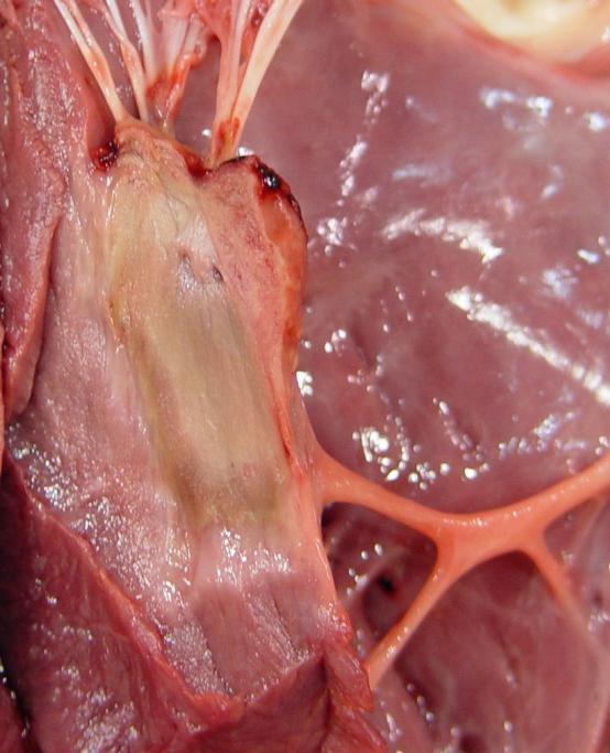
9 Inflammation Case 1 Heart and lungs from a pig History: Poor doing, cyanosis of the ears and tail
 irregular masses (1-2 cm) of friable to firm")
10 Inflammation Case 1 Description? The aortic valve is completely effaced by exophytic (vegetative) irregular masses (1-2 cm) of friable to firm tan material with a roughened surface. There is narrowing of the valve orifice.
 and the parietal")
11 Inflammation Case 1 Description? There is diffuse firm adhesion of the visceral pericardium (epicardium) and the parietal pericardium by a 0.5 to 1 cm thick layer of tough tan tissue.
12 Inflammation Case 1 Possible sequellae? Morphologic Diagnosis? 1. Valvular endocarditis, fibrinosuppurative, diffuse (aortic valve), subacute/chronic-active, severe 2. Fibrous pericardial adhesions, diffuse, chronic, moderate

13 Inflammation Case 2 Lungs and heart from a ewe (you may have seen this before!) History of respiratory disease (coughing, dyspnea)

14 Inflammation Case 2 Description? The right lung is firm, with diffuse thickening and white discolouration of the pleura. Similar changes are present in the left lung, but affecting only the cranioventral portion.
15 Inflammation Case 2 Description? On cut section, similar white firm tissue replaces the parenchyma and contains multiple round pockets of viscous yellow-tinged material surrounded by thick connective tissue capsules.
16 Inflammation Case 2 Morphologic Diagnosis? Diffuse pulmonary and pleural fibrosis with multifocal abscesses, chronic, severe (right lung In the left lung it s locally extensive)


17 Inflammation Case 2 Abscess Morphologic Diagnosis? Pneumonia, suppurative, multifocal, chronic, severe, with extensive pulmonary and pleural fibrosis
18 Inflammation Case 3 Urinary tract from a dog. History of hematuria, lethargy, anorexia
19 Inflammation Case 3 Description? The left kidney is moderately enlarged with roughening of the capsular surface, mild adhesion of the capsule and dilation of the pelvis. The renal papilla is tan and friable and the surrounding medullary tissue is red to dark brown with a small cleft separating the two. The urothelial lining is granular/roughened.


20 Inflammation Case 3 Morphologic Diagnosis? Pyelonephritis, necrohemorrhagic, diffuse (unilateral), acute/subacute, severe


21 Inflammation Case 4 Heart from a 3 month old calf History of not doing well murmur detected on PE
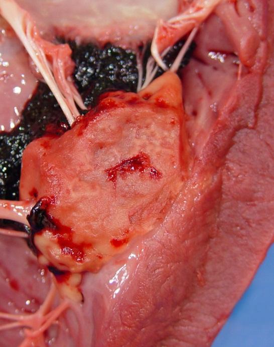
22 Description? Inflammation Case 4
23 Inflammation Case 4 Description? Within the papillary muscle of the LV is a 2.5 x 3 cm irregular region of pale tan discolouration with extension through the endocardium just beneath the left AV valve. The endocardial surface is raised and roughened in the affected area.


24 Inflammation Case 4 Morphologic Diagnosis? Myocarditis/mural endocarditis, necrotizing, locally extensive, subacute/acute, severe
25 Inflammation Case 5 Tongue, larynx, trachea from a lamb History of respiratory distress
26 Inflammation Case 5 Description? A 4 x 3 cm raised mass of slightly friable tan material is focally adhered to the mucosal surface of the larynx at the level of the right vocal fold. On the opposing surface is a similar smaller lesion.

27 Inflammation Case 5 Morphologic Additional info: This was caused by Fusobacterium necrophorum Disease name = Necobacillosis Diagnosis? Laryngitis, fibrinonecrotizing, locally extensive (multifocal), subacute, severe

28 Inflammation Case 6 Bladder and penis from a 2 year old male cat History of oliguria (not able to urinate)
29 Inflammation Case 6 Description? The bladder is distended with thickening of the wall and extensive bright red discolouration of the mucosa. Friable tan material is present in the lumen/ loosely adhered to the roughened mucosa.
30 Inflammation Case 6 Description? Within the penis, yellow-tan friable to pasty material occludes the urethral lumen and there is thickening of the wall of the urethra.


31 Inflammation Case 6 Note: Much of the exudate in the urethra (and bladder) is likely struvite uroliths, protein, and sloughed epithelial cells. This is case of FLUDT and urethral obstruction ( blocked cat ) Morphologic Diagnosis? Cystitis, fibrinohemorrhagic, diffuse, acute, severe Urethritis, fibrinous, locally extensive, acute, severe
32 Liver from a lamb Found dead Inflammation Case 7
33 Inflammation Case 7 Description? Numerous, multifocal to coalescing pale, tan, discrete, cm in greatest diameter foci are scattered randomly throughout the liver. These areas extend deep into the parenchyma on cut surface.


34 Inflammation Case 7 Morphologic Diagnosis? Additional info: This was caused by Fusobacterium necrophorum Disease name = Necobacillosis Hepatitis, necrotizing, multifocal to coalescing, acute, severe
35 Inflammation Case 8 Cranial mesenteric artery from 16 year old mare History of intermittent colic

36 Inflammation Case 8 Aorta Cranial mesenteric artery Cranial mesenteric artery from 16 year old mare History of intermittant colic
37 Inflammation Case 8 Description? The proximal 8 10 cm of the cranial mesenteric artery is irregularly dilated and a small amount of friable tan material is adhered to the thickened, roughened intimal surface.


38 Inflammation Case 8 Morphologic Diagnosis? Cranial mesenteric arteritis, necrotizing (eosinophilic?) segmental, chronic, severe
: Strongylus")
39 Inflammation Case 8 Additional info: Slender worms were embedded in the fibrin in the lumen (thrombus): Strongylus vulgaris (L4)

40 Liver from a caribou No history provided Inflammation Case 9
41 Inflammation Case 9 Description? Several round cyst-like cavities, each surrounded by a thick fibrous capsule, are present multifocally within the liver. Within these structures, there are coiled trematodes (dorsoventrally flattened, measuring ~ 5 8 cm x 3 4 cm with a ventral sucker). Fine black linear tracts are also present multifocally.
42 Inflammation Case 9 Morphologic Diagnosis? Hepatitis (cholangiohepatitis), necrotizing/eosinophilic (?), multifocal, moderate / severe, chronic These tend to be associated with bile ducts

.")
43 Inflammation Case 9 Extra info: Fascioloides magna Morphologic Diagnosis? This is a tough one : there really is no visible exudate parasites induce eosinophils so it s a valid choice. Also there must have been necrosis in order for the cysts to get large (and replace parenchyma). Hepatitis (cholangiohepatitis), necrotizing/eosinophilic (?), multifocal, moderate / severe, chronic
44 Questions?
Inflammation Laboratory 1
 Inflammation Laboratory 1 Lab1 Emphasis: The exudates of acute inflammation Descriptions Morphologic Diagnoses Shannon Martinson: http://people.upei.ca/smartinson VPM 152: March 2013 Describing Lesions
Inflammation Laboratory 1
 Inflammation Laboratory 1 Lab1 Emphasis: The exudates of acute inflammation Descriptions Morphologic Diagnoses Shannon Martinson: http://people.upei.ca/smartinson VPM 152: February 2012 Describing Lesions
Inflammation Laboratory 3 Emphasis: Chronic inflammation and healing. Shannon Martinson: VPM 152: April 2013
 Inflammation Laboratory 3 Emphasis: Chronic inflammation and healing Shannon Martinson: http://people.upei.ca/smartinson VPM 152: April 2013 Example A Reproductive tract and colon/rectum from a sheep Previous
Describing and interpreting gross lesions. Prepared for VPM 4600, May 2018; Shannon Martinson
 Describing and interpreting gross lesions Prepared for VPM 4600, May 2018; Shannon Martinson How to Describe (and Interpret) Lesions Step 1 Step 2 Step 3 Step 4 Look at the specimen: Is it normal or abnormal
Respiratory Pathology Lab 2: Lung. Shannon Martinson,
 Respiratory Pathology Lab 2: Lung Shannon Martinson, 2017 http://people.upei.ca/smartinson/ Case 1 Signalment: 9 month old DSH cat History: Poor doer with stunted growth One month of lethargy one day the
VPM Pigment and other tissue deposits. Shannon Martinson
 VPM 152 - Pigment and other tissue deposits Shannon Martinson http://people.upei.ca/smartinson/ Case 1 Signalment: 2 month old heifer beef calf Clinical History: Lateral recumbency for 4 days Tachycardia,
Cardiovascular Pathology Lab. Shannon Martinson,
 Cardiovascular Pathology Lab Shannon Martinson, 2017 http://people.upei.ca/smartinson/ Case 1 Signalment: 10 year old MC DSH Cat History Heart murmur detected on PE recommended cardiac US Blood work was
VPM Pigment and other tissue deposits. Shannon Martinson
 VPM 152 - Pigment and other tissue deposits Shannon Martinson http://people.upei.ca/smartinson/ Case 1: Signalment: 2 month old heifer beef calf Clinical History: Lateral recumbency for 4 days. Tachycardia,
Cellular Pathology Gross Pathology Laboratory 2 Cell Injury. VPM 152: General Pathology Instructor: Chelsea Martin Winter 2016
 Cellular Pathology Gross Pathology Laboratory 2 Cell Injury VPM 152: General Pathology Instructor: Chelsea Martin Winter 2016 Gross Specimens The following slides consist of images from the specimens presented
Respiratory Pathology Lab 1: Upper Respiratory Tract. Shannon Martinson,
 Respiratory Pathology Lab 1: Upper Respiratory Tract Shannon Martinson, 2017 http://people.upei.ca/smartinson/ Case 1 Signalment: 5 year old dog History: 2 month history of nasal discharge Decreased airflow
Pathology of the Alimentary Tract
 Pathology of the Alimentary Tract Lab 2: Lower alimentary tract SI, LI, cecum, and peritoneum GIST in the cecum of a dog Shannon Martinson: http://people.upei.ca/smartinson VPM 221: November, 2011 3 year
INFLAMMATION & REPAIR
 INFLAMMATION & REPAIR Histopath Laboratory 1 Winter 2013 Chelsea Martin Special thanks to Drs. Hanna and Forzan Goals: Examine Tissue and Identify the Organ Describe the lesion, grossly and histologically
Cellular Pathology. Histopathology Lab #2 (web) Paul Hanna Jan 2018
 Paul Hanna Jan 2018") Cellular Pathology Histopathology Lab #2 (web) Paul Hanna Jan 2018 Slide #91 Clinical History: a necropsy was performed on an aged cat the gross pathological changes included: widespread subcutaneous edema
Cellular Pathology Histopathology Lab #2 (web) Paul Hanna Jan 2018 Slide #91 Clinical History: a necropsy was performed on an aged cat the gross pathological changes included: widespread subcutaneous edema
URINARY SYSTEM. Lecturer Dr.Firdous M.Jaafar Department of anatomy/histology section Lecture 3
 URINARY SYSTEM Lecturer Dr.Firdous M.Jaafar Department of anatomy/histology section Lecture 3 Objectives 1- Describe the structure of the urinary bladder, 2- Describe the structure of the ureters, bladder,
Liver Lab #2. Bacterial Hepatitis
 Liver Lab #2 Bacterial Hepatitis Case: O12561-04. Adult ewe. Describe the lesion: Multifocal large nodules ranging in size from 1-3.5cm in greatest diameter are present within the liver and are filled
Pathology of the Hematopoietic System - Lab.
 Pathology of the Hematopoietic System - Lab http://people.upei.ca/smartinson/ Shannon Martinson, September 2015 Case #1 Signalment: 96 kg gilt History: Pig from minimal disease herd. Sudden death Case
Disturbances of Circulation, Lab 1: Edema and Congestion/Hyperemia. Shannon Martinson, Feb
 Disturbances of Circulation, Lab 1: Edema and Congestion/Hyperemia Shannon Martinson, Feb 2017 http://people.upei.ca/smartinson/ Case #1 Signalment and History: 6-month old feeder lamb found dead on pasture
Urinary Anatomy. Lab 40. Kidneys. Nephrons. Renal Corpuscle
 Urinary Anatomy Lab 40. Urinary Anatomy and Kidney Dissection Kidneys: filters blood, produces urine Ureters: convey urine to bladder Bladder: holding tank Urethra: carries urine to the outside for elimination
Abdominal ultrasound:
 Abdominal ultrasound: Non-traumatic acute abdomen Wittanee Na-ChiangMai, MD Department of Radiology ChiangMai University 26/04/2017 Contents Technique of examination Normal anatomy Emergency conditions
WSC , Conference 9, Case 1. Tissue from a nyala.
 WSC 2009-2010, Conference 9, Case 1. Tissue from a nyala. MICROSCOPIC DESCRIPTION: Heart, atrium (1 pt.): Approximately 40% of the atrial myocardium is replaced by areas of fibrous connective tissue (1
Disturbances of Circulation. Histopathology Lab #2 (Web)
") Disturbances of Circulation Histopathology Lab #2 (Web) Paul Hanna Winter 2015 Slide #96 History: pig was fine in the morning & found dead in the afternoon there was ~100 mls of clear fluid in the pericardial
Disturbances of Circulation Histopathology Lab #2 (Web) Paul Hanna Winter 2015 Slide #96 History: pig was fine in the morning & found dead in the afternoon there was ~100 mls of clear fluid in the pericardial
Firm Texture. (chronic) Cut surface: purulent exudate in bronchi Sequels: Abscesses,
 Cut surface: purulent exudate in bronchi Sequels: Abscesses,") 2008 Classification of Pneumonias in Domestic Animals There is no universal classification! Based on texture, distribution of lesions and type of exudate, pneumonias in domestic animals are currently classified
2008 Classification of Pneumonias in Domestic Animals There is no universal classification! Based on texture, distribution of lesions and type of exudate, pneumonias in domestic animals are currently classified
Post Mortal Approach to the Respiratory System Part 1
 Post Mortal Approach to the Respiratory System Part 1 System examination Before the carcass is opened examination of the nasal openings is carried out. Observe for any evidence of nasal discharge or nasal
Pathology of the Respiratory System 5: Lung and Thoracic Cavity
 Pathology of the Respiratory System 5: Lung and Thoracic Cavity Shannon Martinson, Jan 2017 http://people.upei.ca/smartinson/ VPM 222 Systemic Pathology DISORDERS OF THE LUNG Congenital Pigmentary deposition
Characteristic. Course of disease:short Days--one month Changes : Alteration, exudation Tissue destruction Inflammation cells: major neutrophils
 ACUTE INFLAMMATION Characteristic Course of disease:short Days--one month Changes : Alteration, exudation Tissue destruction Inflammation cells: major neutrophils TYPES Serous Inflammation Fibrinous Inflammation
Group B: Organ systems (digestive, respiratory, urinary, genital system, heart, glands and skin) green
 green") Group B: Organ systems (digestive, respiratory, urinary, genital system, heart, glands and skin) green Digestive system 1. Teeth Main points: external and internal structure of a tooth, fixation of a tooth
Group B: Organ systems (digestive, respiratory, urinary, genital system, heart, glands and skin) green Digestive system 1. Teeth Main points: external and internal structure of a tooth, fixation of a tooth
What s Your Diagnosis?
 What s Your Diagnosis? Signalment: 5 year old MC Belgian Malinois Presenting Complaint: Perineal hernia as well as not eating or defecating History: The patient presented to the KSU VHC on 7/28/2018 for
Proceedings of the 34th World Small Animal Veterinary Congress WSAVA 2009
 www.ivis.org Proceedings of the 34th World Small Animal Veterinary Congress WSAVA 2009 São Paulo, Brazil - 2009 Next WSAVA Congress : Reprinted in IVIS with the permission of the Congress Organizers IMAGING
HISTOPATHOLOGY. Shannon Martinson
 HISTOPATHOLOGY Shannon Martinson March 2013 Case #1 History: 8 year old beagle Neck pain for the past couple of weeks Paresis, followed by paralysis developed over the past few days Gross Description courtesy
Acute flank pain in children: Imaging considerations
 Acute flank pain in children: Imaging considerations Carlos J. Sivit MD Rainbow Babies and Children s Hospital Case Western Reserve School of Medicine Flank pain Results from distention of ureter or renal
Chylothorax Basics OVERVIEW GENETICS SIGNALMENT/DESCRIPTION OF PET
 Chylothorax Basics OVERVIEW Chylo- refers to chyle; thorax refers to the chest Chyle is a milky to slightly yellow fluid composed of lymph and fats (rich in triglycerides) taken up from the intestines
Development of Respiratory System. Dr. Sanaa Alshaarawy& Dr. Saeed Vohra
 Development of Respiratory System Dr. Sanaa Alshaarawy& Dr. Saeed Vohra OBJECTIVES At the end of the lecture the students should be able to: Identify the development of the laryngeotracheal (respiratory)
5 DISTURBANCES IN CIRCULATION. Congestion / Hyperemia Haemorrhage Thrombosis Embolism Ischemia Infarction Oedema Shock Sludged blood Model Questions
 5 DISTURBANCES IN CIRCULATION Congestion / Hyperemia Haemorrhage Thrombosis Embolism Ischemia Infarction Oedema Shock Sludged blood Model Questions CONGESTION/ HYPEREMIA Hyperemia is increased amount of
09-Mar-15 PNEUMONIA RESPIRATORY SYSTEM L-3
 RESPIRATORY SYSTEM L-3 Professor Department of Pathology, University of Agriculture, Faisalabad. Email: mtjaved@uaf.edu.pk Web: https://sites.geocities.ws/mtjaved PNEUMONIA The pulmonary inflammatory response
Anatomical Considerations for Lab Practical II
 Anatomical Considerations for Lab Practical II For each of the following please be prepared to provide: Identification System Organ(s) or ducts to Function(s) location which it is attached Use your lecture
US in non-traumatic acute abdomen. Lalita, M.D. Radiologist Department of radiology Faculty of Medicine ChiangMai university
 US in non-traumatic acute abdomen Lalita, M.D. Radiologist Department of radiology Faculty of Medicine ChiangMai university Sagittal Orientation Transverse (Axial) Orientation Coronal Orientation Intercostal
Connective Tissue. Consists of two basic elements: Cells and Extra-cellular matrix
 Connective Tissue Consists of two basic elements: Cells and Extra-cellular matrix True Connective Tissue Cells Fibroblasts: Secrete both fibers and ground substance of the matrix (wandering) Macrophages:
General Anatomy of Urinary System
 General Anatomy of Urinary System URINARY SYSTEM ORGANS Kidneys (2) Ureters (2) Urinary bladder Urethra KIDNEY FUNCTIONS Control blood volume and composition KIDNEY FUNCTIONS Filter blood plasma, eliminate
ATHEROSCLEROSIS. Secondary changes are found in other coats of the vessel wall.
 ATHEROSCLEROSIS Atherosclerosis Atherosclerosis is a disease process affecting the intima of the aorta and large and medium arteries, taking the form of focal thickening or plaques of fibrous tissue and
Bio& 241 Unit 1 / Lecture 4
 Bio& 241 Unit 1 / Lecture 4 Connective Tissue Consists of two basic elements: Cells and Extra-cellular matrix 1 True Connective Tissue Cells Fibroblasts: Secrete both fibers and ground substance of the
Liver Pathology Lab 1. Shannon Martinson, 2017
 Liver Pathology Lab 1 Shannon Martinson, 2017 http://people.upei.ca/smartinson/ Case 1 Signalment: 10 year old MC DSH cat History: Inappetence and weight loss Fluid in the abdomen noted on US Esophageal
Guidelines, Policies and Statements D5 Statement on Abdominal Scanning
 Guidelines, Policies and Statements D5 Statement on Abdominal Scanning Disclaimer and Copyright The ASUM Standards of Practice Board have made every effort to ensure that this Guideline/Policy/Statement
DIABETES MELLITUS: COMPLICATION. Benyamin Makes Dept. of Anatomic Pathology FMUI - Jakarta
 DIABETES MELLITUS: COMPLICATION Benyamin Makes Dept. of Anatomic Pathology FMUI - Jakarta COMPLICATION OF DIABETES Susceptibility to infections including tuberculosis, pneumonia, pyelonephritis, and mucocutaneous
Pathology of the Hematopoietic System. Case studies
 Pathology of the Hematopoietic System Case studies Shannon Martinson, September 2015 Signalment: 9 yr-old MC cat Case Study 1 History: Cat had been anorexic and developed bleeding in the eyes Physical
KIDNEY Locate the following structures on the sheep kidney and human kidney models: pelvis
 159 Lab 11: Urinary System Anatomy and Physiology, Reproductive System Anatomy Unit 15: Urinary System Unit 16: Reproductive Systems Cat Dissection: Photo Atlas, Chapter 19 Ex. 15-1: Urinary System Anatomy,
2014 Descriptive Vet Path Course. Gross Exam #2 KEY
 2014 Descriptive Vet Path Course Gross Exam #2 KEY 2014 DESCRIPTIVE VETERINARY PATHOLOGY COURSE GROSS EXAM #2 1. Tissue from a chicken. Morphologic Diagnosis: Multifocal proliferative and ulcerative facial
The Respiratory System. Dr. Ali Ebneshahidi
 The Respiratory System Dr. Ali Ebneshahidi Functions of The Respiratory System To allow gases from the environment to enter the bronchial tree through inspiration by expanding the thoracic volume. To allow
Endocrine Lab. Heather Fenton VPM 222 November
 Endocrine Lab Heather Fenton VPM 222 November 27 2012 Case 1: Nursery pig Case 1: Nursery pig Description: There are multifocal round (approximately 1cm diameter) firm lesions within the adrenal gland
Urinary system. Urinary system
 INTRODUCTION. Several organs system Produce urine and excrete it from the body Maintenance of homeostasis. Components. two kidneys, produce urine; two ureters, carry urine to single urinary bladder for
This is not a required assignment but it is recommended.
 SU 12 Name: This is not a required assignment but it is recommended. BIO 116 - Anatomy & Physiology II Practice Assignment 2 - The Respiratory and Cardiovascular Systems 1. The exchange of oxygen and carbon
A Frame of Reference for Anatomical Study. Anatomy and Physiology Mr. Knowles Chapter 1 Liberty Senior High School
 A Frame of Reference for Anatomical Study Anatomy and Physiology Mr. Knowles Chapter 1 Liberty Senior High School Anatomical Terms of Direction and Position Created for communicating the direction and
Module: Foundation Principles of Life Science for Midwifery Practice. WHH1008-N
 Module: Foundation Principles of Life Science for Midwifery Practice. WHH1008-N 2015 Welcome to the Anatomy Workbook. This directed learning has been developed to prepare you for lectures designed to study
Mediastinum and pericardium
 Mediastinum and pericardium Prof. Abdulameer Al-Nuaimi E-mail: a.al-nuaimi@sheffield.ac.uk E. mail: abdulameerh@yahoo.com The mediastinum: is the central compartment of the thoracic cavity surrounded by
Post Mortem Examination of the Urinary System
 Post Mortem Examination of the Urinary System System examination Figure 1 Figure 2 Place kidney on a flat surface and apply dorsal pressure with your hand (Figure 1). While applying this dorsal pressure
The Kidney Dissection (photos curtosy of Murray Jensen at UMN)
") CJ Shuster AP2 Lab Addenum Kidney Dissection 1 The Kidney Dissection (photos curtosy of Murray Jensen at UMN) BACKGROUND INFORMATION The human urinary system consists of two kidneys, two ureters, one urinary
CJ Shuster AP2 Lab Addenum Kidney Dissection 1 The Kidney Dissection (photos curtosy of Murray Jensen at UMN) BACKGROUND INFORMATION The human urinary system consists of two kidneys, two ureters, one urinary
Lab Activity 31. Anatomy of the Urinary System. Portland Community College BI 233
 Lab Activity 31 Anatomy of the Urinary System Portland Community College BI 233 Urinary System Organs Kidneys Urinary bladder: provides a temporary storage reservoir for urine Paired ureters: transport
LADIS Case of the Month
 November 2018 LADIS Case of the Month Drs Valentin Janvier and Brieuc Cossic Hospital for Animals and Animal Health Diagnostic Center Signalment and presenting complaint 13 year old Thoroughbred gelding
PATHOLOGY OF THE CARDIOVASCULAR SYSTEM
 PATHOLOGY OF THE CARDIOVASCULAR SYSTEM Lecture 3: Pericardium and Endocardium Shannon Martinson, 2018 VPM 2220 Systemic Pathology II http://people.upei.ca/smartinson/ PERICARDIUM AND EPICARDIUM Normal
Auscultation of the lung
 Auscultation of the lung Auscultation of the lung by the stethoscope. *Compositions of the stethoscope: 1-chest piece 2-Ear piece 3-Rubber tubs *Auscultation area of the lung(triangle of auscultation).
THE HEART OBJECTIVES: LOCATION OF THE HEART IN THE THORACIC CAVITY CARDIOVASCULAR SYSTEM
 BIOLOGY II CARDIOVASCULAR SYSTEM ACTIVITY #3 NAME DATE HOUR THE HEART OBJECTIVES: Describe the anatomy of the heart and identify and give the functions of all parts. (pp. 356 363) Trace the flow of blood
URINARY SYSTEM CHAPTER 28 I ANATOMY OF THE URINARY SYSTEM. Student Name
 Student Name CHAPTER 28 URINARY SYSTEM L iving produces wastes. Wherever people live or work or play, wastes accumulate. To keep these areas healthy, there must be a method of disposing of these wastes
Chapter 23. The Nephron. (functional unit of the kidney
 Chapter 23 The Nephron (functional unit of the kidney Renal capsule The Nephron Renal cortex Nephron Collecting duct Efferent arteriole Afferent arteriole (a) Renal corpuscle: Glomerular capsule Glomerulus
Canine Liver Eneku Wilfred Bovine Pathology
 2012-1-3 Canine Liver Eneku Wilfred Bovine Pathology Contributor: New Mexico Department of Agriculture Veterinary Diagnostic Services Signalment: 5 month old male Weimaraner dog (Canis familiaris) History:
CARCINOMA OF ESOPHAGUS PERFORATING THE AORTA*
 CARCINOMA OF ESOPHAGUS PERFORATING THE AORTA* HERBERT J. SCHATTENBERG AND JOSEPH ZISKIND From the Department of Pathology, Graduate School, Tulane University, and the Charity Hospital, New Orleans Perforation
Necrosis is death of cells and tissues in the living animal. Focal/ Multifocal necrosis- terms used for one
 Necrosis Necrosis Necrosis is death of cells and tissues in the living animal. Focal/ Multifocal necrosis- terms used for one or more, small, clearly defined areas of necrosis. Diffuse necrosis- term used
4. Describe the body cavities, what organs are found in each and be able to identify them on a diagram.
 Health Science I Final Exam Review 1. Define ANATOMY & PHYSIOLOGY 2. List and describe the characteristics of life 3. Know the levels of organization, from simplest to most complex 4. Describe the body
Figure 10.1A Transparency Master 79
 Brain Carotid arteries Jugular vein Right front leg Lungs (inflated) Cranial Right atrium To left front leg Left subclavian Bronchus capillaries Brachiocephalic vein Left atrium Dorsal aorta Right ventricle
CARDIOVASCULAR PATHOLOGY LABORATORY CASES AND SLIDES. VPM Lisa Miller CARDIOVASCULAR LAB
 CARDIOVASCULAR PATHOLOGY LABORATORY CASES AND SLIDES VPM 222 2009 Lisa Miller CARDIOVASCULAR LAB Case 1 Heart - Echoplane section from a 7-month-old Labrador retriever Give two morphologic diagnoses 1.
THE HEART. A. The Pericardium - a double sac of serous membrane surrounding the heart
 THE HEART I. Size and Location: A. Fist-size weighing less than a pound (250 to 350 grams). B. Located in the mediastinum between the 2 nd rib and the 5 th intercostal space. 1. Tipped to the left, resting
Respiratory System. Functional Anatomy of the Respiratory System
 Respiratory System Overview of the Respiratory System s Job Major Duty Respiration Other important aspects ph control Vocalization Processing incoming air Protection Metabolism (ACE) What structures allow
Histopathology: pulmonary pathology
 Histopathology: pulmonary pathology These presentations are to help you identify basic histopathological features. They do not contain the additional factual information that you need to learn about these
Most abundant and widely distributed tissues in the body Binds, support, and strengthen body tissues, protect and insulate internal organ, serve as
 Connective tissue Most abundant and widely distributed tissues in the body Binds, support, and strengthen body tissues, protect and insulate internal organ, serve as major transport system, compartmentalizes
H I S T O L O G Y O F T H E U R I N A R Y S Y S T E M
 SCPA 602- Anatomical Basis For Pathological Study H I S T O L O G Y O F T H E U R I N A R Y S Y S T E M S O M P H O N G N A R K P I N I T, M. D. D E P A R T M E N T O F P A T H O B I O L O G Y F A C U
Date Lab Pd. Lecture Notes (57)
") Name SECTION OBJECTIVES Describe the locations of the major body cavities List the organs located in each major body cavity Name the membranes associated with the thoracic and abdominopelvic cavities Name
Name SECTION OBJECTIVES Describe the locations of the major body cavities List the organs located in each major body cavity Name the membranes associated with the thoracic and abdominopelvic cavities Name
THE RESPIRATORY SYSTEM
 THE RESPIRATORY SYSTEM Functions of the Respiratory System Provides extensive gas exchange surface area between air and circulating blood Moves air to and from exchange surfaces of lungs Protects respiratory
Bladder Case 1 SURGICAL PATHOLOGY REPORT. Procedure: Cystoscopy, transurethral resection of bladder tumor (TURBT)
") Bladder Case 1 February 17, 2007 Specimen (s) received: Bladder Tumor Pre-operative Diagnosis: Bladder Cancer Post operative Diagnosis: Bladder Cancer Procedure: Cystoscopy, transurethral resection of
Bladder Case 1 February 17, 2007 Specimen (s) received: Bladder Tumor Pre-operative Diagnosis: Bladder Cancer Post operative Diagnosis: Bladder Cancer Procedure: Cystoscopy, transurethral resection of
Epithelium. Four primary tissue types:
 Epithelium Four primary tissue types: Epithelial (covering) Connective (support) Nervous (control) Muscular (movement) Smooth muscle Cardiac muscle Skeletal muscle 1 Epithelial Tissue Features Epithelial
GUIDE TO: Diagnosing Coccidiosis & Necrotic Enteritis
 GUIDE TO: Diagnosing Coccidiosis & Necrotic Enteritis Site of Infection Species E. acervulina E. brunetti E. maxima E. mivati E. tenella E. necatrix Oocyst Size 2µ{ 18.3 x 14.6 24.6 x 18.8 30.5 x 20.7
CELL AND TISSUE INJURY COURSE-II PATHOLOGY LABORATORY. PATHOLOGY of MASS LESIONS and TISSUE DEFECTS -MACROSCOPY Assoc. Professor Rengin Ahıskalı
 CELL AND TISSUE INJURY COURSE-II PATHOLOGY LABORATORY PATHOLOGY of MASS LESIONS and TISSUE DEFECTS -MACROSCOPY Assoc. Professor Rengin Ahıskalı M1 - RENAL TUBERCULOSIS cavitary areas caseous necrosis fibrous
Inflammation. First Lab.
 Inflammation First Lab. The cardinal signs of inflammation are rubor (redness), calor (heat), tumor (swelling), dolor (pain), and loss of function. Seen here is skin with erythema, compared to the more
CYSTIC DISEASES of THE KIDNEY. Dr. Nisreen Abu Shahin
 CYSTIC DISEASES of THE KIDNEY Dr. Nisreen Abu Shahin 1 Types of cysts 1-Simple Cysts 2-Dialysis-associated acquired cysts 3-Autosomal Dominant (Adult) Polycystic Kidney Disease 4-Autosomal Recessive (Childhood)
27-Apr-15 1 UAF ANOMALIES OF DEVELOPMENT RENAL SYSTEM - 1 DR. MUHAMMAD TARIQ JAVED UAF UAF
 RENAL SYSTEM - 1 DR. MUHAMMAD TARIQ JAVED Professor, Department of Pathology, Faculty of Veterinary Science, University of Agriculture, Faisalabad, Pakistan. Email: mtjaved@uaf.edu.pk RENAL AGENESIS Renal
Unit II Problem 2 Pathology: Pneumonia
 Unit II Problem 2 Pathology: Pneumonia - Definition: pneumonia is the infection of lung parenchyma which occurs especially when normal defenses are impaired such as: Cough reflex. Damage of cilia in respiratory
The Urinary System Pearson Education, Inc.
 26 The Urinary System Introduction The urinary system does more than just get rid of liquid waste. It also: Regulates plasma ion concentrations Regulates blood volume and blood pressure Stabilizes blood
Pathology of the Hematopoietic System GROSS/HISTO LAB
 Pathology of the Hematopoietic System GROSS/HISTO LAB Paul Hanna (thanks to Dr s Aburto, Martinson & Fenton) Fall 2014 Slide 1 Spleen from a Beaver Give a morphologic diagnosis and possible etiology &
My Patient Has Abdominal Pain PoCUS of the Biliary Tract and the Urinary Tract
 My Patient Has Abdominal Pain PoCUS of the Biliary Tract and the Urinary Tract Objectives PoCUS for Biliary Disease PoCUS for Renal Colic PoCUS for Urinary Retention Biliary Disease A patient presents
What s Your Diagnosis??? Renée Fahrenholz, Class of 2012
 Renée Fahrenholz, Class of 2012 What s Your Diagnosis??? Signalment Emma, a 9 year old, Female, Spayed, Domestic Short Haired Feline Presenting Complaint Weight loss, vomited the morning of her visit,
Chapter 14. Circulatory System Images. VT-122 Anatomy & Physiology II
 Chapter 14 Circulatory System Images VT-122 Anatomy & Physiology II The mediastinum Dog heart Dog heart Cat heart Dog heart ultrasound Can see pericardium as distinct bright line Pericardial effusion Fluid
Pathology of the Liver and Biliary Tract 5 Diseases of the Biliary Tract. Shannon Martinson, April 2016
 Pathology of the Liver and Biliary Tract 5 Diseases of the Biliary Tract Shannon Martinson, April 2016 http://people.upei.ca/smartinson/ OUTLINE Normal anatomy & function Hepatobiliary Injury and responses
Lecture 7. The Urinary System
 Lecture 7 The Urinary System Copyright 2006 Thomson Delmar Learning The Urinary System The urinary system removes wastes from the body The urinary system also maintains homeostasis or a constant internal
Urinary system. Urinary system
 Distal convoluted tubule (DCT) Highly coiled, ~ 5 mm in length Last part of the nephron. Wall; simple cuboidal epithelium Less metabolically active than the PCT no brush border light eosinophilic cytoplasm
Ischaemia It means local anemia, it is characterized by a decrease amount of blood in an organ or region. Causes of Ischemia: *1.
 المرحلة الثالثة م. هالة عباس ناجي Ischaemia It means local anemia, it is characterized by a decrease amount of blood in an organ or region. Causes of Ischemia: *1.External pressure upon an artery e.g:
Urinary System. Unit 6.12 (6 th Edition) Chapter 7.12 (7 th Edition)
 Chapter 7.12 (7 th Edition)") Urinary System Unit 6.12 (6 th Edition) Chapter 7.12 (7 th Edition) 1 Learning Objectives Identify the major organs of the urinary system and their functions. Explain the major functions of the kidneys.
Urinary System Unit 6.12 (6 th Edition) Chapter 7.12 (7 th Edition) 1 Learning Objectives Identify the major organs of the urinary system and their functions. Explain the major functions of the kidneys.
SESSION 2: THE MOUTH AND PHARYNX
 SESSION 2: THE MOUTH AND PHARYNX 9 In the pig s digestive tract, food flows in only one direction from mouth to anus.this allows for greatly specialized sections that can act independently of each other.
the Cardiovascular System I
 the Cardiovascular System I By: Dr. Nabil A Khouri MD, MsC, Ph.D MEDIASTINUM 1. Superior Mediastinum 2. inferior Mediastinum Anterior mediastinum. Middle mediastinum. Posterior mediastinum Anatomy of
Sampled: 06/05/2017 Paramus, NJ Received: 06/13/2017 (201) Finalized: 07/27/2017. Anatomic Pathology
 Finalized: 07/27/2017. Anatomic Pathology") Owner: 240 Farrier Road,, Ithaca, NY 14853 Ph: 607-253-3900 Fax: 607-253-3943 https://ahdc.vet.cornell.edu Page 1 of 7 Oradell Animal Hospital Inc - (3382) Dr John Lucy 580 Winters Ave Sampled: 06/05/2017
Owner: 240 Farrier Road,, Ithaca, NY 14853 Ph: 607-253-3900 Fax: 607-253-3943 https://ahdc.vet.cornell.edu Page 1 of 7 Oradell Animal Hospital Inc - (3382) Dr John Lucy 580 Winters Ave Sampled: 06/05/2017
Urinary System Laboratory
 Urinary System Laboratory 1 Adrenal gland Organs of The Urinary System Renal artery and vein Kidney Ureter Urinary bladder Figure 26.1 2 Urethra Functions of the urinary system organs: Urethra expels urine
PATHOLOGY Intracellular Degeneration LAB 1
 PATHOLOGY Intracellular Degeneration LAB 1 Cellular swelling Liver Organ :- Liver Lesion :- 1. Narrowing of hepatic sinusoids due to the swelling of hepatocyte. 2. The cytoplasm of affected hepatocyte
Avian Pathology. Bacterial diseases: histo slides. ECVP-ESVP Summer School 2012 Frédérique NGUYEN
 Avian Pathology Bacterial diseases: histo slides ECVP-ESVP Summer School 2012 Frédérique NGUYEN Bacterial diseases: histo slides B1. Turkey. Organs? Morphologic diagnosis? Special procedure? B2. Hen. Organ?
Organs Histology D. Sahar AL-Sharqi. Respiratory system
 Respiratory system The respiratory system provides for exchange of O2 and CO2 to and from the blood. Respiratory organs include the lungs and a branching system of bronchial tubes that link the sites of