Respiratory Pathology Lab 2: Lung. Shannon Martinson,
|
|
|
- Arthur Stone
- 5 years ago
- Views:
Transcription


1 Respiratory Pathology Lab 2: Lung Shannon Martinson,
2 Case 1 Signalment: 9 month old DSH cat History: Poor doer with stunted growth One month of lethargy one day the cat was barely able to walk, stumbling Fluid noted in chest on radiographs modified transudate based on cytology Fluid in abdomen and enlarged kidneys noted on US Hyperproteinemia characterized by hyperglobulinemia
3 Case 1
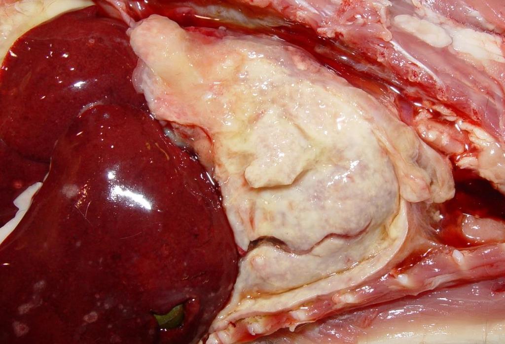
4 Case 1 Description On the right side, the visceral and parietal pleura are diffusely covered by a thick adherent layer of somewhat friable tan to yellow material There is possible extension into the pulmonary parenchyma
5 Case 1 Morph Diag Pleuritis, pyogranulomatous (fibrino-suppurative), diffuse, right thoracic cavity (+/- pneumonia) What are some possible etiologies for this lesion? Mutated feline coronavirus* Pasteurella multocida (implantation from a bite wound)


 How")


6 Case 1 Look at the kidneys, what is the most likely etiology? Mutated feline coronavirus What is the name of this disease? Feline Infectious Peritonitis (FIP) How would you confirm the diagnosis? Histology +/- IHC, PCR Antemortem: Clin Path + Serology? Many cats will have positive serology but not FIP


7 Case 2 Signalment: 50 day old Shorthorn MC calf Clinical History: Calf was weak, down, unable to move with its eyes rolled back Calf was doing well the previous evening The calf had scours 4 weeks prior
8 Case 2 Description There is cranioventral consolidation and dark discolouration of the left and right lungs involving ~ 50% of the total lung field On section, there are scattered deposits of pasty white-yellow exudate
9 Case 2 Morph Diagnosis Bronchopneumonia, suppurative, locally extensive (bilateral), subacute to chronic, severe (with abscesses or bronchiectasis) What is the likely portal of entry for the lesion? Aerogenous
, subacute")
 What is the likely portal of entry")
10 Case 2 Morph Diagnosis Bronchopneumonia, suppurative, locally extensive (bilateral), subacute to chronic, severe (with abscesses or bronchiectasis) What is the likely portal of entry for the lesion? Aerogenous
11 Case 2 Possible Etiologies Pasteurella multocida Histophilus somni Mycoplasma bovis Trueperella pyogenes +/- Mannheimia hemolytica Predisposing factors Viral infection BRSV, BPIV-3, BoHV-1 Environmental / Management stressors Temperature, Humidity, Transport, Crowding, ect


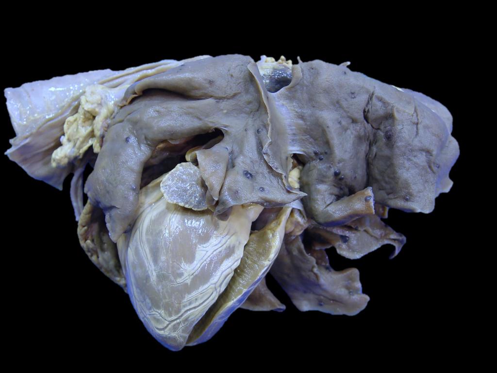
12 Case 3 Signalment: 4 5 week old pig Clinical History: Multiple animals have poor performance post-weaning, chronic coughing, dyspnea and weight loss
13 Case 3 Description The lungs are uncollapsed with a diffuse elastic texture and light brown colouration There is mild expansion of the interlobular septa The tracheobronchial LNs are enlarged homogenous and tan
14 Case 3 Poss. Morph Diagnoses Interstitial pneumonia, diffuse, subacute, severe Pulmonary edema, diffuse, severe How would you achieve a diagnosis? Histology!
15 Case 3 Possible etiologies for interstitial pneumonia Viral PPRS virus PCV-2 Swine infuenza virus Sepsis Salmonella Nematode migration Ascaris suum larval migration How would you determine the cause? Submit fresh lung for viral testing (PCR, FAT, etc) Submit fresh lung +/- other organs for bacterial culture Fixed lung for histology (+/- IHC)
16 Case 3 Possible etiologies for interstitial pneumonia Viral PPRS virus PCV-2 Swine infuenza virus Sepsis Salmonella Nematode migration Ascaris suum larval migration Possible port of entry? Hematogenous Aerogenous

17 Case 4 Signalment: ~ 3 week old female lamb Clinical History: History of scours and dehydration Found dead
18 Case 4 Description Scattered randomly throughout the left and right lungs, but often clustered at the periphery of the lobes, are multifocal to coalescing, variably discrete small (2 5 mm) yellow foci
19 Case 4 Morphologic Diagnosis Embolic pneumonia, suppurative, multifocal to coalescing, acute (subacute), moderate
20 Case 4 Possible port of entry? Hematogenous Possible underlying cause (source of agent)? Typically showering from a bacterial infection Valvular endocarditis (R heart) Omphalophlebitis Hepatic abscess Sepsis
21 Case 4 Possible port of entry? Hematogenous Can you relate the clinical signs to the lesions? Possible that diarrhea and dehydration impaired pulmonary defence mechanisms Damage to the intestinal wall (enteritis) could have allowed entry for bacteria to the enter the blood

22 Case 5 Signalment: 12 year old MC, DLH cat Clinical History: Dramatic weight loss over a month or two More recently dyspnea, tachypnea
23 Case 5

 What type of")
24 Case 5 Description Several, tan-grey, firm nodular masses are scattered randomly throughout the lung. They range in size from cm diameter and often have an umbilicated surface They extend deeply into the parenchyma on section and are often cavitated (cystic or necrotic) What type of disease process does this represent? Neoplasia

25 Case 5 What are some differentials for a neoplastic lesion in the lung? Morph Diag Epithelial** Pulmonary carcinoma (primary) Metastatic carcinoma Mesenchymal or round cell? Less likely due to umbilication of the masses!! Fibrosarcoma Chondrosarcoma Metastatic sarcoma Histiocytic sarcoma Lymphoma Malignant neoplasia, multifocal, lung How would you achieve a diagnosis? Histology!
 Metastasis to the regional LN +/- Metastasis to the other viscera Primary or secondary?")
26 Case 5 Morph Diag Carcinoma, lung Possible sequelae? Metastasis within the lung (as seen here) Metastasis to the regional LN +/- Metastasis to the other viscera Primary or secondary? This can be hard when there are multiple masses Is one much larger than the others? Check other organs for a primary source
27 Case 6 Signalment: 10 year old MC German Shepherd Clinical History: 5 days: lame on the right hind end Acutely weak and brought in on emergency PE: Pale mucous membranes On US, large amount of fluid in the abdomen abdominocentesis showed abundant erythrocytes and high protein Developed signs of shock - resuscitation was attempted unsuccessfully

28 Case 6
, soft to firm, brownred, nodular masses The right auricle is thickened, dark brown and irregular")
29 Case 6 Description Scattered randomly throughout the lungs are numerous, small (0.3 1 cm), soft to firm, brownred, nodular masses The right auricle is thickened, dark brown and irregular due to an ill-defined mass

30 Case 6 What type of disease process does this represent? Neoplasia Differentials Hemangiosarcoma Melanoma Hemangiosarcoma Metastatic melanoma
31 Case 6 What type of disease process does this represent? Neoplasia How would you achieve a diagnosis? Histology! Hemangiosarcoma Metastatic melanoma


32 Case 6 Probable Diagnosis Hemangiosarcoma Mass type lesion in the right auricle Metastases to the lung Other organs typically involved Spleen Liver Any other organ: kidney, muscle, brain, etc
33 Case 6 Cause of shock Blood loss (hemoabdomen) Cause of hemoabdomen? Rupture of an abdominal mass
")

34 Case 6 Cause of shock Blood loss (hemoabdomen) Cause of hemoabdomen? Rupture of an abdominal mass

35
Pathology of the Respiratory System 4: Pneumonia
 Pathology of the Respiratory System 4: Pneumonia Shannon Martinson, March 2016 http://people.upei.ca/smartinson/ VPM 222 Systemic Pathology LUNG PNEUMONIA Review Classification of Pneumonia Diffuse LUNG
Respiratory Pathology Lab 1: Upper Respiratory Tract. Shannon Martinson,
 Respiratory Pathology Lab 1: Upper Respiratory Tract Shannon Martinson, 2017 http://people.upei.ca/smartinson/ Case 1 Signalment: 5 year old dog History: 2 month history of nasal discharge Decreased airflow
Pathology of the Respiratory System 5: Lung and Thoracic Cavity
 Pathology of the Respiratory System 5: Lung and Thoracic Cavity Shannon Martinson, Jan 2017 http://people.upei.ca/smartinson/ VPM 222 Systemic Pathology DISORDERS OF THE LUNG Congenital Pigmentary deposition
Inflammation Laboratory 1
 Inflammation Laboratory 1 Lab1 Emphasis: The exudates of acute inflammation Descriptions Morphologic Diagnoses Shannon Martinson: http://people.upei.ca/smartinson VPM 152: February 2012 Describing Lesions
Inflammation Laboratory 1
 Inflammation Laboratory 1 Lab1 Emphasis: The exudates of acute inflammation Descriptions Morphologic Diagnoses Shannon Martinson: http://people.upei.ca/smartinson VPM 152: March 2013 Describing Lesions
Pathology of the Alimentary Tract
 Pathology of the Alimentary Tract Lab 2: Lower alimentary tract SI, LI, cecum, and peritoneum GIST in the cecum of a dog Shannon Martinson: http://people.upei.ca/smartinson VPM 221: November, 2011 3 year
Inflammation Laboratory 2. Shannon Martinson: VPM 152: March 2012
 Inflammation Laboratory 2 Shannon Martinson: http://people.upei.ca/smartinson VPM 152: March 2012 Reminder - Creating a Morphologic Diagnosis for Inflammatory Lesions Organ and Process Exudate Distribution
Liver Lab #2. Bacterial Hepatitis
 Liver Lab #2 Bacterial Hepatitis Case: O12561-04. Adult ewe. Describe the lesion: Multifocal large nodules ranging in size from 1-3.5cm in greatest diameter are present within the liver and are filled
Pathology of the Hematopoietic System. Case studies
 Pathology of the Hematopoietic System Case studies Shannon Martinson, September 2015 Signalment: 9 yr-old MC cat Case Study 1 History: Cat had been anorexic and developed bleeding in the eyes Physical
Describing and interpreting gross lesions. Prepared for VPM 4600, May 2018; Shannon Martinson
 Describing and interpreting gross lesions Prepared for VPM 4600, May 2018; Shannon Martinson How to Describe (and Interpret) Lesions Step 1 Step 2 Step 3 Step 4 Look at the specimen: Is it normal or abnormal
Firm Texture. (chronic) Cut surface: purulent exudate in bronchi Sequels: Abscesses,
 Cut surface: purulent exudate in bronchi Sequels: Abscesses,") 2008 Classification of Pneumonias in Domestic Animals There is no universal classification! Based on texture, distribution of lesions and type of exudate, pneumonias in domestic animals are currently classified
2008 Classification of Pneumonias in Domestic Animals There is no universal classification! Based on texture, distribution of lesions and type of exudate, pneumonias in domestic animals are currently classified
Inflammation Laboratory 3 Emphasis: Chronic inflammation and healing. Shannon Martinson: VPM 152: April 2013
 Inflammation Laboratory 3 Emphasis: Chronic inflammation and healing Shannon Martinson: http://people.upei.ca/smartinson VPM 152: April 2013 Example A Reproductive tract and colon/rectum from a sheep Previous
Pathology of the Hematopoietic System - Lab.
 Pathology of the Hematopoietic System - Lab http://people.upei.ca/smartinson/ Shannon Martinson, September 2015 Case #1 Signalment: 96 kg gilt History: Pig from minimal disease herd. Sudden death Case
HISTOPATHOLOGY. Shannon Martinson
 HISTOPATHOLOGY Shannon Martinson March 2013 Case #1 History: 8 year old beagle Neck pain for the past couple of weeks Paresis, followed by paralysis developed over the past few days Gross Description courtesy
VPM Pigment and other tissue deposits. Shannon Martinson
 VPM 152 - Pigment and other tissue deposits Shannon Martinson http://people.upei.ca/smartinson/ Case 1 Signalment: 2 month old heifer beef calf Clinical History: Lateral recumbency for 4 days Tachycardia,
Cellular Pathology Gross Pathology Laboratory 2 Cell Injury. VPM 152: General Pathology Instructor: Chelsea Martin Winter 2016
 Cellular Pathology Gross Pathology Laboratory 2 Cell Injury VPM 152: General Pathology Instructor: Chelsea Martin Winter 2016 Gross Specimens The following slides consist of images from the specimens presented
Pathology of the Liver and Biliary Tract 5 Diseases of the Biliary Tract. Shannon Martinson, March 2017
 Pathology of the Liver and Biliary Tract 5 Diseases of the Biliary Tract Shannon Martinson, March 2017 http://people.upei.ca/smartinson/ OUTLINE Normal anatomy & function Hepatobiliary injury and responses
like humans, have well-developed mediastinal separation between the left and right hemithorax, thus unilateral changes can occur. On the other hand,
 Tutorial Module 6 Thoracic Cavity and Tumors of Lung and Pleura Alfonso López Atlantic Veterinary College University of Prince Edward Island Canada 2009 Enero 3 Thoracic Cavity There are significant anatomical
09-Mar-15 PNEUMONIA RESPIRATORY SYSTEM L-3
 RESPIRATORY SYSTEM L-3 Professor Department of Pathology, University of Agriculture, Faisalabad. Email: mtjaved@uaf.edu.pk Web: https://sites.geocities.ws/mtjaved PNEUMONIA The pulmonary inflammatory response
VPM Pigment and other tissue deposits. Shannon Martinson
 VPM 152 - Pigment and other tissue deposits Shannon Martinson http://people.upei.ca/smartinson/ Case 1: Signalment: 2 month old heifer beef calf Clinical History: Lateral recumbency for 4 days. Tachycardia,
Proceedings of the World Small Animal Veterinary Association Sydney, Australia 2007
 Proceedings of the World Small Animal Sydney, Australia 2007 Hosted by: Next WSAVA Congress THE LAST GASP II: LUNGS AND THORAX David Holt, BVSc, Diplomate ACVS University of Pennsylvania School of Veterinary
Unit II Problem 2 Pathology: Pneumonia
 Unit II Problem 2 Pathology: Pneumonia - Definition: pneumonia is the infection of lung parenchyma which occurs especially when normal defenses are impaired such as: Cough reflex. Damage of cilia in respiratory
Cardiovascular Pathology Lab. Shannon Martinson,
 Cardiovascular Pathology Lab Shannon Martinson, 2017 http://people.upei.ca/smartinson/ Case 1 Signalment: 10 year old MC DSH Cat History Heart murmur detected on PE recommended cardiac US Blood work was
LADIS Case of the Month
 November 2018 LADIS Case of the Month Drs Valentin Janvier and Brieuc Cossic Hospital for Animals and Animal Health Diagnostic Center Signalment and presenting complaint 13 year old Thoroughbred gelding
Thoracic Cavity and Tumors of Lung and Pleura
 Tutorial Module 6 Thoracic Cavity and Tumors of Lung and Pleura Alfonso López Atlantic Veterinary College University of Prince Edward Island Canada Sept 28, 2014 Thoracic Cavity There are anatomical differences
Liver Pathology Lab 1. Shannon Martinson, 2017
 Liver Pathology Lab 1 Shannon Martinson, 2017 http://people.upei.ca/smartinson/ Case 1 Signalment: 10 year old MC DSH cat History: Inappetence and weight loss Fluid in the abdomen noted on US Esophageal
Disturbances of Circulation, Lab 1: Edema and Congestion/Hyperemia. Shannon Martinson, Feb
 Disturbances of Circulation, Lab 1: Edema and Congestion/Hyperemia Shannon Martinson, Feb 2017 http://people.upei.ca/smartinson/ Case #1 Signalment and History: 6-month old feeder lamb found dead on pasture
Neoplasms of the Canine, Feline and Lemur Liver:
 Neoplasms of the Canine, Feline and Lemur Liver: Classification and Prognosis Annual Seminar of the French Society of Veterinary Pathology John M. Cullen VMD PhD DACVP North Carolina State University Primary
Exploring BPHS lesions time trends: July March 2015
 Exploring BPHS lesions time trends: July 2005 - March 2015 Prepared for: Ouafa Doxon BPHS Steering Group Members BPEX Prepared by: Carla Gomes, Madeleine Henry ERU-SRUC Contact: Epidemiology Research Unit,
Pathology of the Liver and Biliary Tract 5 Diseases of the Biliary Tract. Shannon Martinson, April 2016
 Pathology of the Liver and Biliary Tract 5 Diseases of the Biliary Tract Shannon Martinson, April 2016 http://people.upei.ca/smartinson/ OUTLINE Normal anatomy & function Hepatobiliary Injury and responses
Liver. Liver. System examination
 Liver System examination Examine the liver in situ after opening the abdominal cavity examine the liver in context with other changes in the abdominal cavity. The image on the left is of a ruptured liver
What s Your Diagnosis? Signalment: Species: Canine Breed: Golden Retriever Sex: Female (spayed) Date of Birth: 04/01/99
 Date of Birth: 04/01/99") What s Your Diagnosis? Signalment: Species: Canine Breed: Golden Retriever Sex: Female (spayed) Date of Birth: 04/01/99 Presenting Complaint: Acute onset of lethargy Vomited twice (partially digested food)
What s Your Diagnosis? Signalment: Species: Canine Breed: Golden Retriever Sex: Female (spayed) Date of Birth: 04/01/99 Presenting Complaint: Acute onset of lethargy Vomited twice (partially digested food)
INFLAMMATION & REPAIR
 INFLAMMATION & REPAIR Histopath Laboratory 1 Winter 2013 Chelsea Martin Special thanks to Drs. Hanna and Forzan Goals: Examine Tissue and Identify the Organ Describe the lesion, grossly and histologically
TUMOR,NEOPLASM. Pathology Department, Zhejiang University School of Medicine,
 TUMOR,NEOPLASM Pathology Department, Zhejiang University School of Medicine, 马丽琴,maliqin198@zju.edu.cn The points in this chapter What is a neoplasm (conception) Morphology of neoplasm Macroscopy of Neoplasm
What s Your Diagnosis?
 What s Your Diagnosis? Signalment: 5 year old MC Belgian Malinois Presenting Complaint: Perineal hernia as well as not eating or defecating History: The patient presented to the KSU VHC on 7/28/2018 for
Table of Contents. Preface xi. Acknowledgments xiii. Part I Overview of the Diagnostic Process 1. 1 Overview of Grading and Staging 3
 Table of Contents Preface xi Acknowledgments xiii Part I Overview of the Diagnostic Process 1 1 Overview of Grading and Staging 3 Identification of the process 3 Identification of tumor types 5 Grading
Canine Histiocytic Disorders DR. MEREDITH GAUTHIER, DVM DACVIM (ONCOLOGY) OCTOBER 29, 2015
 OCTOBER 29, 2015") Canine Histiocytic Disorders DR. MEREDITH GAUTHIER, DVM DACVIM (ONCOLOGY) OCTOBER 29, 2015 Canine Histiocytes! Cells derived from CD34+ stem cells and blood monocytes! Macrophages! Dendritic cells (DC)!
Canine Histiocytic Disorders DR. MEREDITH GAUTHIER, DVM DACVIM (ONCOLOGY) OCTOBER 29, 2015 Canine Histiocytes! Cells derived from CD34+ stem cells and blood monocytes! Macrophages! Dendritic cells (DC)!
Cellular Pathology. Histopathology Lab #2 (web) Paul Hanna Jan 2018
 Paul Hanna Jan 2018") Cellular Pathology Histopathology Lab #2 (web) Paul Hanna Jan 2018 Slide #91 Clinical History: a necropsy was performed on an aged cat the gross pathological changes included: widespread subcutaneous edema
Cellular Pathology Histopathology Lab #2 (web) Paul Hanna Jan 2018 Slide #91 Clinical History: a necropsy was performed on an aged cat the gross pathological changes included: widespread subcutaneous edema
Pathology of the Hematopoietic System. Lecture 3: Spleen and thymus
 Pathology of the Hematopoietic System Lecture 3: Spleen and thymus Shannon Martinson, March 2012 Spleen Structure and Function Present in the left cranial part of the abdomen within the greater omentum
Resident Case Review CHEST. Daria Manos CAR 2016
 Resident Case Review CHEST CAR 2016 Daria Manos Disclosure Speakers bureau, Roche CAR 2016 Daria Manos 1. Recognize common and critical chest radiograph and computed tomography signs and use these clues
Endocrine Lab. Heather Fenton VPM 222 November
 Endocrine Lab Heather Fenton VPM 222 November 27 2012 Case 1: Nursery pig Case 1: Nursery pig Description: There are multifocal round (approximately 1cm diameter) firm lesions within the adrenal gland
Pathology of the Hematopoietic System GROSS/HISTO LAB
 Pathology of the Hematopoietic System GROSS/HISTO LAB Paul Hanna (thanks to Dr s Aburto, Martinson & Fenton) Fall 2014 Slide 1 Spleen from a Beaver Give a morphologic diagnosis and possible etiology &
Pathology of Pneumonia
 Pathology of Pneumonia Dr. Atif Ali Bashir Assistant Professor of Pathology College of Medicine Majma ah University Introduction: 5000 sq meters of area.! (olympic track) Filters >10,000 L of air / day!
Ocular Neoplasia What s Common? What s New? Richard R Dubielzig
 Ocular Neoplasia What s Common? What s New? Richard R Dubielzig Orbit 288 6% Tumors of the globe make up 3225 out of 6110 total neoplasms = 53%. Tumors of the conjunctiva make up 1192 out of 6110 total
WSC , Conference 9, Case 1. Tissue from a nyala.
 WSC 2009-2010, Conference 9, Case 1. Tissue from a nyala. MICROSCOPIC DESCRIPTION: Heart, atrium (1 pt.): Approximately 40% of the atrial myocardium is replaced by areas of fibrous connective tissue (1
PATHOLOGY OF LIVER & BILIARY TRACT. Lecture 5. Idiopathic & proliferative conditions; diseases of the biliary tract
 PATHOLOGY OF LIVER & BILIARY TRACT Lecture 5 Idiopathic & proliferative conditions; diseases of the biliary tract Enrique Aburto Winter 2015 IX. Diseases of uncertain origin Equine serum hepatitis Idiopathic
What s Your Diagnosis?
 What s Your Diagnosis? Courtney S. Wait Signalment: 11 year old FS Labrador Retriever Presenting Complaint/History: The patient presented to the referring DVM for inappetance, vomiting, lethargy, and anorexia.
Excavated pulmonary nodule: steps to diagnosis?
 Excavated pulmonary nodule: steps to diagnosis? Poster No.: C-1044 Congress: ECR 2014 Type: Authors: Keywords: DOI: Educational Exhibit W. Mnari, M. MAATOUK, A. Zrig, B. Hmida, M. GOLLI; Monastir/ TN Metastases,
Case # nd Annual SEVPAC May 17, Kathy-Anne Clarke
 Case # 10 42 nd Annual SEVPAC May 17, 2014 Kathy-Anne Clarke Google images Babu Babu is 10 year old spayed female French Bulldog Chronic weight loss over 4 months Febrile and lethargic at the referring
Key Difference - Pleural Effusion vs Pneumonia
 Difference Between Pleural Effusion and Pneumonia www.differencebetween.com Key Difference - Pleural Effusion vs Pneumonia Pleural effusion and pneumonia are two conditions that affect our respiratory
Post Mortal Approach to the Respiratory System Part 1
 Post Mortal Approach to the Respiratory System Part 1 System examination Before the carcass is opened examination of the nasal openings is carried out. Observe for any evidence of nasal discharge or nasal
Disorders of Cell Growth & Neoplasia. Histopathology Lab
 Disorders of Cell Growth & Neoplasia Histopathology Lab Paul Hanna April 2010 Case #84 Clinical History: 5 yr-old, West Highland White terrier. skin mass from axillary region. has been present for the
Principles of Surgical Oncology. Winnie Achilles Tierklinik Hollabrunn Lastenstrasse Hollabrunn
 Principles of Surgical Oncology Winnie Achilles Tierklinik Hollabrunn Lastenstrasse 2 2020 Hollabrunn boexi@gmx.de The first surgery provides the best chance for a cure in an animal with a tumor Clinical
How to Analyse Difficult Chest CT
 How to Analyse Difficult Chest CT Complex diseases are:- - Large lesion - Unusual or atypical pattern - Multiple discordant findings Diffuse diseases are:- - Numerous findings in both sides 3 basic steps
Amyloidosis (Disorder Caused by Deposition of Proteins [Amyloid] in Various Organs) Basics
![Amyloidosis (Disorder Caused by Deposition of Proteins [Amyloid] in Various Organs) Basics](/thumbs/75/72552398.jpg "Amyloidosis (Disorder Caused by Deposition of Proteins [Amyloid] in Various Organs) Basics") Amyloidosis (Disorder Caused by Deposition of Proteins [Amyloid] in Various Organs) Basics OVERVIEW A group of conditions of differing cause in which insoluble proteins (amyloid) are deposited outside
Amyloidosis (Disorder Caused by Deposition of Proteins [Amyloid] in Various Organs) Basics OVERVIEW A group of conditions of differing cause in which insoluble proteins (amyloid) are deposited outside
2015 Lab Animal 8/22/ Mouse. 2. Rat. 3. Mouse. Tissue from an Mouse: Morphologic Diagnosis: Rectal prolapse
 1. Mouse 2015 Lab Animal 2. Rat Tissue from an Mouse: Morphologic Diagnosis: Rectal prolapse Causes: 1. Syphacia obveluta 2. Citrobacter rodentum 3. Heicobacter sp 3. Mouse 2. Tissue from a Rat: Morphologic
RHODOCOCCUS EQUI. Post-mortem Environmental Persistence Specific Control Measures Release of Animals from Isolation
 RHODOCOCCUS EQUI Definition Clinical Signs Transmission Diagnostic Sampling, Testing and Handling Post-mortem Environmental Persistence Specific Control Measures Release of Animals from Isolation Biosecurity
Jin-An Huang VMRD Beijing
 Jin-An Huang VMRD Beijing Neonatal Scours BRD Septicemia Parasites Clostridia Anaplasma Babesia FMDV Different serotypes/subtypes, biosecurity, needs of marker vaccines with sterilizing immunity Johne
CARDIOVASCULAR PATHOLOGY LABORATORY CASES AND SLIDES. VPM Lisa Miller CARDIOVASCULAR LAB
 CARDIOVASCULAR PATHOLOGY LABORATORY CASES AND SLIDES VPM 222 2009 Lisa Miller CARDIOVASCULAR LAB Case 1 Heart - Echoplane section from a 7-month-old Labrador retriever Give two morphologic diagnoses 1.
Disorders of Cell Growth & Neoplasia
 General Pathology VPM 152 Disorders of Cell Growth & Neoplasia Lecture 3 Rate of growth, local invasion, and metastasis. Molecular basis of cancer (normal cell-cycle and cellular proliferation). Enrique
Complicated echinococcal cyst to Biopsy or not to biopsy. V. Rusanov MR Kramer Pulmonary Institute, Rabin medical center
 Complicated echinococcal cyst to Biopsy or not to biopsy V. Rusanov MR Kramer Pulmonary Institute, Rabin medical center Case 1 84 y.o. Male, Iraq descend, past smoker 40 PY Medical History- HTN, Rheumatoid
Histopathology: pulmonary pathology
 Histopathology: pulmonary pathology These presentations are to help you identify basic histopathological features. They do not contain the additional factual information that you need to learn about these
Pathological Investigations on Bovine Pheumonic Pasteurellosis by Use of Immunoperoxidase Technique
 JARQ 29, 13 1-136 (1995) Pathological Investigations on Bovine Pheumonic Pasteurellosis by Use of Immunoperoxidase Technique Makoto HARITANI Tohoku Branch Laboratory, National Institute of Animal Health
BOVINE RESPIRATORY DISEASE COMPLEX น.สพ.ฐปณ ฐ สงคส ภา สถาบ นส ขภาพส ตว แห งชาต
 BOVINE RESPIRATORY DISEASE COMPLEX น.สพ.ฐปณ ฐ สงคส ภา สถาบ นส ขภาพส ตว แห งชาต The Most Common Causes of Death Source: Department of Agriculture s Laboratories Quarterly Surveillance Report, 2015(IRELAND)
Clinical Manifestations of HIV
 HIV Symptoms Diane Havlir, MD Professor of Medicine and Chief, HIV/AIDS Division University of California, San Francisco (UCSF) WorldMedSchool; July 2, 2013 1 Clinical Manifestations of HIV! Result from
8/14/2017. Objective: correlate radiographic findings of common lung diseases to actual lung pathologic features
 What is that lung disease? Pulmonary Patterns & Correlated Pathology Dr. Russell Tucker, DACVR Objective: correlate radiographic findings of common lung diseases to actual lung pathologic features Improved
Chest Radiology Interpretation: Findings of Tuberculosis
 Chest Radiology Interpretation: Findings of Tuberculosis Get out your laptops, smart phones or other devices pollev.com/chestradiology Case #1 1 Plombage Pneumonia Cancer 2 Reading the TB CXR Be systematic!
Management of Pleural Effusion
 Management of Pleural Effusion Development of Pleural Effusion pulmonary capillary pressure (CHF) capillary permeability (Pneumonia) intrapleural pressure (atelectasis) plasma oncotic pressure (hypoalbuminemia)
Acute and Chronic Lung Disease
 KATHOLIEKE UNIVERSITEIT LEUVEN Faculty of Medicine Acute and Chronic Lung Disease W De Wever, JA Verschakelen Department of Radiology, University Hospitals Leuven, Belgium Clinical utility of HRCT To detect
CASE REPORTS. Inflammatory Polyp of the Bronchus. V. K. Saini, M.S., and P. L. Wahi, M.D.
 CASE REPORTS V. K. Saini, M.S., and P. L. Wahi, M.D. I n 1932 Jackson and Jackson [l] first reported a number of clinical cases under the title Benign Tumors of the Trachea and Bronchi with Especial Reference
Canine Cutaneous Melanoma
 Canine Cutaneous Melanoma By Elizabeth Downing Clinical Advisor: Dr. Angharad Waite, VMD Basic Science Advisor: Dr. Cheryl Balkman, DVM, DACVIM Senior Seminar Paper Cornell University College of Veterinary
(Iteceived for publication December 3, 1915)
") TRANSPLANTABLE SARCOMATA OF THE RAT LIVER ARISING IN THE WALLS OF PARASITIC CYSTS G. L. ROHDENBURG, M.D., AND F. D. BULLOCK, M.D. From Colurnbia University, George Crocker Special Re-search Fund, F. C.
TRANSPLANTABLE SARCOMATA OF THE RAT LIVER ARISING IN THE WALLS OF PARASITIC CYSTS G. L. ROHDENBURG, M.D., AND F. D. BULLOCK, M.D. From Colurnbia University, George Crocker Special Re-search Fund, F. C.
PULMONARY TUBERCULOSIS RADIOLOGY
 PULMONARY TUBERCULOSIS RADIOLOGY RADIOLOGICAL MODALITIES Medical radiophotography Radiography Fluoroscopy Linear (conventional) tomography Computed tomography Pulmonary angiography, bronchography Ultrasonography,
Acute pneumonia in a cat
 Acute pneumonia in a cat Elspeth Milne, Anita Schwartz, Alasdair Stuart, Danielle Gunn-Moore, Kerry Simpson and Sionagh Smith, Division of Veterinary Clinical Sciences, University of Edinburgh, United
PATHOGENS COMMONLY ASSOCIATED WITH BRONCHOPNEUMONIA:
 BOVINE RESPIRATORY DISEASE AN OVERVIEW OF A CONTINUING CHALLENGE Jeff Lakritz DVM, PhD, Dip ACVIM The Ohio State University Veterinary Medical Center Hospital for Farm Animals Bovine respiratory disease
1/13/2014. Proper Radiographs. Proper Radiographs. A Review of Pulmonary Patterns
 Live Webinar A Review of Pulmonary Patterns Sofija R. Liles, DVM, DACVR Proper Radiographs Which views? One lateral plus ventrodorsal (at least) Left lateral is best for thorax Three views for full metastatic
Bronchial syndrome. Atelectasis Draining bronchus Bronchiectasis
 Bronchial syndrome Atelectasis Draining bronchus Bronchiectasis Etienne Leroy Terquem Pierre L Her SPI / ISP Soutien Pneumologique International / International Support for Pulmonology Atelectasis Consequence
Systemic lupus erythematosus (SLE): Pleuropulmonary Manifestations
: Pleuropulmonary Manifestations") 08/30/10 09/26/10 Systemic lupus erythematosus (SLE): Pleuropulmonary Manifestations Camila Downey S. Universidad de Chile, School of Medicine, Year VII Harvard University, School of Medicine Sept 17,
08/30/10 09/26/10 Systemic lupus erythematosus (SLE): Pleuropulmonary Manifestations Camila Downey S. Universidad de Chile, School of Medicine, Year VII Harvard University, School of Medicine Sept 17,
Tuberculosis: The Essentials
 Tuberculosis: The Essentials Kendra L. Fisher, MD, PhD THORACIC TUBERCULOSIS: THE BARE ESSENTIALS Kendra Fisher MD, FRCP (C) Department of Radiology Loma Linda University Medical Center TUBERCULOSIS ()
HRCT in Diffuse Interstitial Lung Disease Steps in High Resolution CT Diagnosis. Where are the lymphatics? Anatomic distribution
 Steps in High Resolution CT Diagnosis Pattern of abnormality Distribution of disease Associated findings Clinical history Tomás Franquet MD What is the diagnosis? Hospital de Sant Pau. Barcelona Secondary
Index. B Biological factors, 2 Brain stem encephalitis, Burkitt s lymphoma, 83, 105
 Index A Acquired immunodeficiency syndrome (AIDS) abdomen gallbladder complications, 97, 107 109 gastrointestinal complications, 96, 105 106 liver complications, 97, 107 109 neoplasm, 99, 110 111 pancreas
Pathology of the Hematopoietic System. Lecture 3: Spleen and thymus
 Pathology of the Hematopoietic System Lecture 3: Spleen and thymus Shannon Martinson, March 2011 Spleen Structure and Function Present in the left cranial part of the abdomen within the greater omentum
Causes of pleural effusion and its imaging approach in pediatrics. M. Mearadji International Foundation for Pediatric Imaging Aid
 Causes of pleural effusion and its imaging approach in pediatrics M. Mearadji International Foundation for Pediatric Imaging Aid Pleural fluid A tiny amount of fluid in the pleural cavity is physiological.
EUROPEAN ASSOCIATION OF VETERINARY DIAGNOSTIC IMAGING EUROPEAN COLLEGE OF VETERINARY DIAGNOSTIC IMAGING
 EISAGOGIKO EUROPEAN ASSOCIATION OF VETERINARY DIAGNOSTIC IMAGING EUROPEAN COLLEGE OF VETERINARY DIAGNOSTIC IMAGING ARISTOTLE UNIVERSITY OF THESSALONIKI SCHOOL OF VETERINARY MEDICINE SECTION OF RADIOLOGY
Lecture Notes. Chapter 16: Bacterial Pneumonia
 Lecture Notes Chapter 16: Bacterial Pneumonia Objectives Explain the epidemiology Identify the common causes Explain the pathological changes in the lung Identify clinical features Explain the treatment
Tumors of the Spleen
 Tumors of the Spleen 803-808-7387 www.gracepets.com These notes are provided to help you understand the diagnosis or possible diagnosis of cancer in your pet. For general information on cancer in pets
What s your diagnosis? Malori Marotz. Squirt, an 8month old mix breed puppy. History:
 What s your diagnosis? Malori Marotz Squirt, an 8month old mix breed puppy History: The owner obtained squirt at 12 weeks of age. The owner reported that Squirt was passing soft stools lately and he is
August 2018 Imaging Case of the Month: Dyspnea in a 55-Year-Old Smoker. Michael B. Gotway, MD
 August 2018 Imaging Case of the Month: Dyspnea in a 55-Year-Old Smoker Michael B. Gotway, MD Department of Radiology Mayo Clinic Arizona Scottsdale, AZ USA Clinical History: A 55 year old woman presented
New lung lesion in a 55 year-old male treated with chemoradiation for non-small cell lung carcinoma
 July 2016 New lung lesion in a 55 year-old male treated with chemoradiation for non-small cell lung carcinoma Contributed by: Laurel Rose, MD, Resident Physician, Indiana University School of Medicine,
Recognizing African swine fever 13. Post mortem findings
 Recognizing African swine fever 13 Post mortem findings Carcasses of pigs that die in the acute stage of the disease are often in good condition. In white-skinned pigs, bluish-purple discolouration of
Sheet: Patho-Pulmonary infections Done by: Maen Faoury
 Sheet: Patho-Pulmonary infections Done by: Maen Faoury Pneumonitis : might be an infection or not. Chemical Pneumonitis : not an infection. Parenchyma : an infection.( تندرج تحت ال pneumonitis) Lung Parenchyma
Melanoma Case Scenario 1
 Melanoma Case Scenario 1 History and physical 11/5/16 Patient is a single, 48-year-old male in good health who presented to his primary physician for a yearly physical exam during which a 3.4 x 2.8 x 1.5
Cattle Farming. Our wealth Standby team
 1 Our wealth Standby team Dr. Soliman Mohammed Soliman Lecturer of Infectious Diseases, Faculty of Veterinary Medicine, Cairo University Cattle Farming Common Disease Problems Neonatal Calf Diarrhea (Scours)
(JPC ) Caprine lungs
 Caprine lungs") 2011-7-2 (JPC 3133973) Caprine lungs Bat Otgontugs Bovine Pathology Contributor: Natoinal Institute Animal Health, Tsukuba, Japan Signalment: 5-year 3-month old female Japanese native breed goat, (Capra
2011-7-2 (JPC 3133973) Caprine lungs Bat Otgontugs Bovine Pathology Contributor: Natoinal Institute Animal Health, Tsukuba, Japan Signalment: 5-year 3-month old female Japanese native breed goat, (Capra
Ocular Neoplasia CL Davis 9/08. Richard R Dubielzig
 Ocular Neoplasia CL Davis 9/08 Richard R Dubielzig 2135/5722 Canine Melanocytic Tumors Outside the Globe: 264 Conjunctival: 159 Eye Lid: 72 Skin: 33 Affecting the Globe: 1871 Anterior Uveal Melanocytoma:
Pathology of pulmonary tuberculosis. Dr: Salah Ahmed
 Pathology of pulmonary tuberculosis Dr: Salah Ahmed Is a chronic granulomatous disease, caused by Mycobacterium tuberculosis (hominis) Usually it involves lungs but may affect any organ or tissue Transmission:
SESSION 1: GENERAL (BASIC) PATHOLOGY CONCEPTS Thursday, October 16, :30am - 11:30am FACULTY COPY
 PATHOLOGY CONCEPTS Thursday, October 16, :30am - 11:30am FACULTY COPY") SESSION 1: GENERAL (BASIC) PATHOLOGY CONCEPTS Thursday, October 16, 2008 9:30am - 11:30am FACULTY COPY GOAL: Describe the basic morphologic (structural) changes which occur in various pathologic conditions.
SESSION 1: GENERAL (BASIC) PATHOLOGY CONCEPTS Thursday, October 16, 2008 9:30am - 11:30am FACULTY COPY GOAL: Describe the basic morphologic (structural) changes which occur in various pathologic conditions.
Chest X rays and Case Studies. No disclosures. Outline 5/31/2018. Carlo Manalo, M.D. Department of Radiology Loma Linda University Children s Hospital
 Chest X rays and Case Studies Carlo Manalo, M.D. Department of Radiology Loma Linda University Children s Hospital No disclosures. Outline Importance of history Densities delineated on radiography An approach
Interesting Cases. Pulmonary
 Interesting Cases Pulmonary 54M with prior history of COPD, hep B/C, and possible history of TB presented with acute on chronic dyspnea, and productive cough Hazy opacity overlying the left hemithorax