Imaging of Pediatric MSK Tumors
|
|
|
- Cornelia Jackson
- 5 years ago
- Views:
Transcription


1 Imaging of Pediatric MSK Tumors Kirsten Ecklund, M.D. Boston Children s Hospital Harvard Medical School kirsten.ecklund@childrens.harvard.edu
2 Tumor Imaging Goals Diagnosis Lesion characterization Benign vs malignant DDX Extent of disease Treatment Size, extent Treatment response Tissue characterization (necrosis vs growth) RECIST guidelines Surgical planning Relationship to neurovascular structures Measurements for custom reconstruction Surveillance Local recurrence Metastatic search
3 Current MR Imaging Goals Highest resolution even at small FOV Tissue characterization Functional imaging Metabolic imaging Decrease sedation Motion correction Increase acquisition speed

4 Normal RM Stress fx Diagnosis: EWS Leukemia


5 Tumor Mimics/Pitfalls Inflammatory lesions Osteoid osteoma Chondroblastoma Infection Myositis ossificans Histiocytosis CRMO (CNO) Trauma/stress fracture 19 y.o. right elbow mass


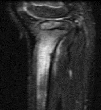
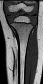
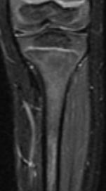
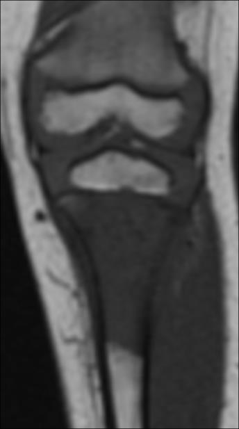
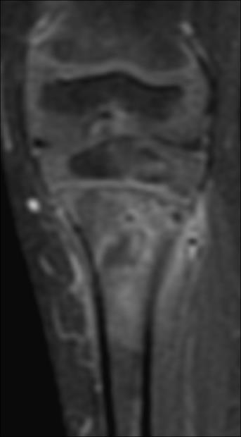
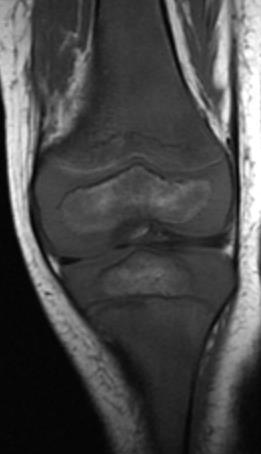
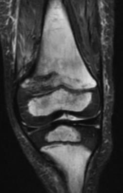
6 Two 15 year olds with rt knee pain D. Femur stress fx, p. tibia stress reaction Primary bone lymphoma


7 Primary Osseous Lymphoma 6% of 1 bone tumors, <10% of NHL Commonly involves epiphyses and equivalents MR - Infarct-like appearance, sequestra 10-30% multifocal 10-15% metastases at dx
8 90% of malignant pediatric bone tumors Osteosarcoma ~ 400 new cases/yr in U.S. #1 malignant bone tumor < 18 y.o. Peak age: y.o., boys > girls Sites: d. femur (75%), p. tibia, p. humerus Risk factors: prior xrt, genetic predisposition (retinoblastoma, Li- Fraumeni, Rothmund-Thomson) ES family of tumors ~ 200 new cases/yr Caucasian predominance Peak age: y.o Sites: axial (54%), appendicular (42%) No risk factors Translocation of chromosomes involving ESWR1 gene

")


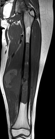
9 18 m.o. with Ewing Sarcoma Ewing family (EFT) Chromosome 22 EWS gene translocation Mesenchymal cell origin PET all pts (BS deemphasized

10 COG* imaging guidelines for Osteosarcoma Site Presentation / Pre-op Post-op, On ChemoRx Surveillance off Rx Primary tumor AP, lat XR MR + contrast XR q 16 wks MR (unless precluded by hardware) XR q3 mo x4, q6 mo x4, q12 mo x2 MR for sxs Bone Mets AP, lat XR MR + contrast MR if sxs XR q3 mo x4, q6 mo x4, q12 mo x2 MR for sxs Whole body MDP bone scan PET? MDP bone scan q16 wks PET? MDP bone scan q3 mo x4, q6 mo x4, q1 2 mo x2 PET to evaluate abnormal imaging Chest PA, lat XR CT XR q 2 mo. CT q 16 wks if abnormal in past XR q6 mo x 6 CT q3 mo x 4, q6 mo x 2 *Children s Oncology Group: Meyer, et al. Pediatric Blood Cancer 2008; 51:163

11 COG* imaging guidelines for Ewing Family of Tumors Site Presentation + prior to local control On ChemoRx, post local control Surveillance off Rx Primary tumor AP, lat XR MR + contrast XR ½ way thru and at end of rx MR/CT 3/4 months after local control XR q3 mo x8, q6 mo x6, q12 mo x5 MR for sxs Whole body MDP bone scan FDG-PET MDP bone scan at end of rx PET at end of rx MDP bone scan if sxs PET if sxs or abnormal imaging Chest PA, lat XR dx only CT CT ½ way thru and at end of rx XR q3 mo x 8, q6 mo x 6, q12 mo x 5 CT if abnormal CXR *Children s Oncology Group: Meyer, et al. Pediatric Blood Cancer 2008; 51:163
 Partial response = > 30% decrease in sum Complete response = disappearance of all lesions Unreliable for")
12 Response Evaluation Criteria in Solid Tumors (RECIST)* Applies to primary and mets together size-based assessment of tumor burden Sum of longest dimension of each lesion Requires measurable soft tissue Lesions must be > 10 mm Response: Progression = >20% increase in sum (or new lesions) Partial response = > 30% decrease in sum Complete response = disappearance of all lesions Unreliable for osteosarcoma > 20% show increased size despite rx response *Eisenhauer, et al. European J Cancer 2009:
13 Treatment Response MR Evaluation Diffusion weighted imaging (DWI) Dynamic contrast-enhanced MRI Whole body MR (FSE, DW) Spectroscopy Chemical shift (for marrow)

14 Comprehensive Bone Tumor Protocol Cor T1, FSEIR entire bone Sag T2 fat sat Ax PD, T2 fat sat DWI/ADC axial Spectroscopy- single voxel 3D GRE DCE pre + post C C+ T1 fs short and long axis

 Increased diffusion cell # (necrosis, cysts, fluid) cell membrane")
15 DWI - Principle Brownian motion of water in extracellular tissues Tissue structure/ cellularity Qualitative and Quantitative Restricted diffusion Decrease in extracellular space (edema) Increase in cell #, size (tumors, fibrosis) Increased diffusion cell # (necrosis, cysts, fluid) cell membrane permeability (ischemia)
16 DWI in MSK Tumors Diagnosis: general rules Malignant = restricted diffusion Benign tumors ~ less restricted diffusion Myxoid content = diffusion Response to therapy Necrosis diffusion Ischemia diffusion Recurrence/ metastases diffusion Post op change/hematoma diffusion


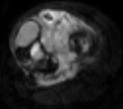
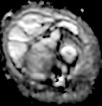
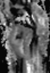
17 18 m.o. boy with left thigh pain, swelling Dx = Ewing Sarcoma


18 Same pt after 2 cycles chemo rx DWI = bright, suggests restricted diff ADC = bright increased diff, thus T2 effect Path: No viable tumor


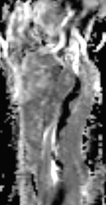
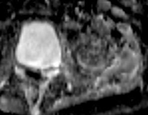
19 9 y.o. with left thigh pain Dx: telangiectatic osteosarcoma
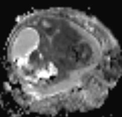
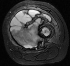
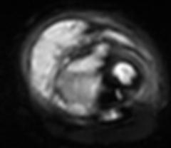
20 Telangiectatic Osteosarcoma: pre and post Dx s/p Rx FSE T2 fs DWI ADC
21 DWI Quantitative Assessment ADC value (apparent diffusion coefficient) Calculated from all b values in data set < 1.5 x 10⁻³ mm 2 /sec high cellularity Contaminated by capillary perfusion at low b PIDC (perfusion independent diff coef) Higher b value data only (slow diffusion) Eliminates perfusion effect < 1.1 x 10⁻³ mm 2 /sec high cellularity Correlation with % necrosis would be ideal


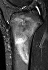
22 13 y.o. with proximal tibial osteosarcoma DX: ADC=2.5 FSEIR DWI ADC Map 3 cycles Rx: ADC=1.1


 ADC:")

23 12 y.o. with radiation induced, refractory OSA prox tibia T2 fs: DWI: (outside hospital exam, no DWI) dx s/p 2 cycles rx s/p change in rx


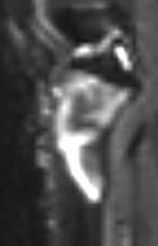
24 9 y.o. with limited ROM left hip: Synovial Dx s/p 3 cycles rx T2 fs T1 + gad DWI, b 500 ADC

25 Dynamic Contrast Enhanced MR Perfusion Diagnosis Response to therapy Pre-op angiography High temporal/ spatial resolution 3T T1 relax blood vs tissue CNR Parallel imaging + faster sequences TRICKS/ TWIST MRA 3D LAVA/ VIBE perfusion small vessel visualization

26 DCE: Qualitative Assessment Tumor margin delineation First pass - simplified Intense enhancement = residual/recurrent tumor Intermediate enhancement = post rx fibrosis Minimal enhancement = necrosis, edema, hemorrhage Guide dx or recurrence


27 DCE: Quantitative Assessment 7 y.o. boy with osteosarcoma s/p 2 cycles chemorx, pre-op eval 3D GRE VIBE/LAVA Time Intensity Curves Fem artery Intramedullary tumor Soft tissue mass
28 Surveillance Imaging Metastasis detection Problem-solving Local recurrence Hardware artifact mitigation MR techniques Dual energy CT FDG-PET and WB MR complimentary


29 12 y.o. with EWS rt humerus, staging studies


30 Ewing Sarcoma PET vs MRI in Response Assessment FSE-T2/FS Post-Gd T1/FS Post-ChemoRx Pre-ChemoRx 18 F-FDG-PET Courtesy of Stephan Voss, M.D.


31 Whole Body MR Metastatic survey Cancer predisposition surveillance Assessment of cortical bone, marrow, soft tissues 13 y.o. boy with NF1


32 19 y.o. with NF1, prior MPNST BCH: SUV > 4.5 g/ml, surgical sampling/rxn
 J.")
33 NF-1 and 18 F-FDG-PET: SUV and final histology: Benign plexiform -> atypical NF -> MPNST Courtesy of Stephan Voss, M.D. Tsai et al. (2012) J. NeuroOnc 108:469


34 NF-1 and MPNST SUV max = 5 Atyp NF SUV max = 4.2 Benign NF SUV max = 4.9 Atyp NF SUV max = 9.3 MPNST Courtesy of Stephan Voss, M.D.

35 Proton Spectroscopy Molecular tissue characterization Choline level (peak) Increased cell membrane turnover Correlates with malignancy Single voxel techniques most useful Susceptibility issues Costa FM, Canella C, Gasparetto E. Radiol Clin North Am, Nov 2011, 49(6)


36 Hardware Imaging: Artifact Reduction General: high BW, slice thick, matrix, ETL, TE SEMAC - optimized sequences STIR, TSE (WARP) 1.5 T preferred Conventional STIR Optimized STIR Optimized PD SPACE
")
37 Summary Conventional and functional imaging techniques are complimentary. Together they improve our confidence Dx Response to therapy Long term surveillance Size alone (RECIST) insufficient to assess rx response for OSA, EFT
Why Talk About Technique? MRI of the Knee:
 Why Talk About Technique? MRI of the Knee: Part 1 - Imaging Techniques Mark Anderson, M.D. University of Virginia Health Sciences Center Charlottesville, Virginia Always had an interest teach our fellows
High Field MR of the Spine
 Department of Radiology University of California San Diego 3T for MR Applications Advantages High Field MR of the Spine Increased signal-to-noise Better fat suppression Increased enhancement with gadolinium
Disclosures. Diffusion and Perfusion Imaging in the Head and Neck. Learning objectives ???
 Disclosures No relevant financial disclosures Diffusion and Perfusion Imaging in the Head and Neck Ashok Srinivasan, MD Associate Professor Director of Neuroradiology University of Michigan Health System
Functional aspects of anatomical imaging techniques
 Functional aspects of anatomical imaging techniques Nilendu Purandare Associate Professor & Consultant Radiologist Tata Memorial Centre Functional/metabolic/molecular imaging (radioisotope scanning) PET
Musculoskeletal Sarcomas
 Musculoskeletal Sarcomas Robert C. Orth, M.D., Ph.D. Edward B. Singleton Department of Pediatric Radiology Texas Children s Hospital Page 0 xxx00.#####.ppt 9/23/2012 9:01:18 AM No disclosures Page 1 xxx00.#####.ppt
Bone PET/MRI : Diagnostic yield in bone metastases and malignant primitive bone tumors
 Bone PET/MRI : Diagnostic yield in bone metastases and malignant primitive bone tumors Lars Stegger, Benjamin Noto Department of Nuclear Medicine University Hospital Münster, Germany Content From PET to
Pediatric Lymphoma Update from the Children s Oncology Group
 Pediatric Lymphoma Update from the Children s Oncology Group Stephan D. Voss, MD, PhD Department of Radiology Boston Children s Hospital Harvard Medical School Staging Assessment Disclosures None Acknowledgements:
Imaging: When to get MRI, CT or PET-CT?
 Imaging: When to get MRI, CT or PET-CT? Alina Uzelac, D.O. Assistant Clinical Professor Neuroradiology UCSF Department of Radiology and Biomedical Imaging San Francisco General Hospital Overview CT MRI
MRI of Pediatric Ankle and Foot. Mahesh Thapa, MD Associate Professor Seattle Children s University of Washington School of Medicine
 MRI of Pediatric Ankle and Foot Mahesh Thapa, MD Associate Professor Seattle Children s University of Washington School of Medicine Disclosures Under contract with Lippincott Williams and Wilkins (LWW)
PET-MRI in malignant bone tumours. Lars Stegger Department of Nuclear Medicine University Hospital Münster, Germany
 PET-MRI in malignant bone tumours Lars Stegger Department of Nuclear Medicine University Hospital Münster, Germany Content From PET to PET/MRI General considerations Bone metastases Primary bone tumours
Essentials of Clinical MR, 2 nd edition. 73. Urinary Bladder and Male Pelvis
 73. Urinary Bladder and Male Pelvis Urinary bladder carcinoma is best locally staged with MRI. It is important however to note that a thickened wall (> 5 mm) is a non-specific finding seen in an underfilled
ROUTINE MRI PROTOCOLS: I. Abdomen Plus Post Gadolinium Screening Pelvis
 ROUTINE MRI PROTOCOLS: I. Abdomen Plus Post Gadolinium Screening Pelvis Sequence Coverage Slice/Gap Notes COR T2 ssfse 32-40 6/-1 Coverage from all sequences is above the liver dome, through the kidneys.
Abdominal applications of DWI
 Postgraduate course, SPR San Antonio (Texas), May 14-15, 2013 Abdominal applications of DWI Rutger A.J. Nievelstein Wilhelmina Children s s Hospital, Utrecht (NL) Outline What is DWI? How to perform? Challenges
Effective local and systemic therapy is necessary for the cure of Ewing tumor Most chemotherapy regimens are a combination of cyclophosphamide,
 Ewing Tumor Perez Ewing tumor is the second most common primary tumor of bone in childhood, and also occurs in soft tissues Ewing tumor is uncommon before 8 years of age and after 25 years of age In the
MRI PEDIATRIC PROTOCOLS (Updated 6/19/2018)
") MRI PEDIATRIC PROTOCOLS (Updated 6/19/2018) *Please get or let us know where radiologist can review plain films. *For Texas Orthopedics and other Docs requesting only MSK section read for their pediatric
MRI PEDIATRIC PROTOCOLS (Updated 6/19/2018) *Please get or let us know where radiologist can review plain films. *For Texas Orthopedics and other Docs requesting only MSK section read for their pediatric
Orthopedic Hardware Imaging Part II: MRI v. Metal
 Orthopedic Hardware Imaging Trent Roth, MD And Lauren Ladd, MD Indiana University School of Medicine IU Health Physicians-Radiology Recap: Imaging Techniques Radiography Standard for initial and surveillance
T2, T2*, ute. Yeo Ju Kim. Radiology, Inha University Hospital, Incheon, Korea
 SY28-1 T2, T2*, ute Yeo Ju Kim Radiology, Inha University Hospital, Incheon, Korea T2 relaxation times relate to the rate of transverse magnetization decay, caused by the loss of phase coherence induced
MRI XR, CT, NM. Principal Modality (2): Case Report # 2. Date accepted: 15 March 2013
: Case Report # 2. Date accepted: 15 March 2013") Radiological Category: Musculoskeletal Principal Modality (1): Principal Modality (2): MRI XR, CT, NM Case Report # 2 Submitted by: Hannah Safia Elamir, D.O. Faculty reviewer: Naga R. Chinapuvvula, M.D.
Radiological Category: Musculoskeletal Principal Modality (1): Principal Modality (2): MRI XR, CT, NM Case Report # 2 Submitted by: Hannah Safia Elamir, D.O. Faculty reviewer: Naga R. Chinapuvvula, M.D.
Prostate MRI: Not So Difficult. Neil M. Rofsky, MD, FACR, FSCBTMR, FISMRM Dallas, TX
 Prostate MRI: Not So Difficult Neil M. Rofsky, MD, FACR, FSCBTMR, FISMRM Dallas, TX What is the biggest barrier to your practice incorporating prostate MRI? 1) I don t know how to read the cases 2) I don
Musculoskeletal MR Protocols
 Musculoskeletal MR Protocols Joint-based protocols MSK 1: Shoulder MRI MSK 1A: Shoulder MR arthrogram MSK 1AB: Shoulder MR arthrogram (instability protocol) MSK 2: Elbow MRI MSK 2A: Elbow MR arthrogram
MEASUREMENT OF EFFECT SOLID TUMOR EXAMPLES
 MEASUREMENT OF EFFECT SOLID TUMOR EXAMPLES Although response is not the primary endpoint of this trial, subjects with measurable disease will be assessed by standard criteria. For the purposes of this
Functional Chest MRI in Children Hyun Woo Goo
 Functional Chest MRI in Children Hyun Woo Goo Department of Radiology and Research Institute of Radiology Asan Medical Center, University of Ulsan College of Medicine, Seoul, Korea No ionizing radiation
Whole-Body Dynamic Contrast- Enhanced (DCE) MR Imaging in patients with myeloma
 MR Imaging in patients with myeloma") Whole-Body Dynamic Contrast- Enhanced (DCE) MR Imaging in patients with myeloma Alain Rahmouni, Department of Medical Imaging, Mondor Academic Hospital : Centre Hospitalo-Universitaire Henri Mondor-Assistance
Whole-Body Dynamic Contrast- Enhanced (DCE) MR Imaging in patients with myeloma Alain Rahmouni, Department of Medical Imaging, Mondor Academic Hospital : Centre Hospitalo-Universitaire Henri Mondor-Assistance
6/23/2009. Inversion Recovery (IR) Techniques and Applications. Variations of IR Technique. STIR, FLAIR, TI and TI Null. Applications of IR
 Techniques and Applications. Variations of IR Technique. STIR, FLAIR, TI and TI Null. Applications of IR") The Anatomy of Basic R Pulse Sequences Inversion Recovery () Techniques and Applications Chen Lin, PhD Indiana University School of edicine & Clarian Health Partners agnetization Preparation Section Chemical
The Anatomy of Basic R Pulse Sequences Inversion Recovery () Techniques and Applications Chen Lin, PhD Indiana University School of edicine & Clarian Health Partners agnetization Preparation Section Chemical
Publication for the Philips MRI Community
 FieldStrength Publication for the Philips MRI Community Issue 38 Summer 2009 Pediatric MSK imaging benefits from tailored scan protocols Vanderbilt University Children s Hospital builds dedicated scans
Update on RECIST and Staging of Common Pediatric Tumors Ethan A. Smith, MD
 Update on RECIST and Staging of Common Pediatric Tumors Ethan A. Smith, MD Section of Pediatric Radiology C.S. Mott Children s Hospital University of Michigan ethans@med.umich.edu Disclosures No relevant
Review Course «Musculoskeletal Oncology» October 6, 2011 UNIKLINIK BALGRIST. Imaging of Bone and Soft Tissue. Tumors
 Imaging of Bone and Soft Tissue Tumors Approach from a radiologist s point of view Florian Buck Radiology Radio- Radio- Oncologist Oncologist Orthopedist Orthopedist Patient Management Oncologist Oncologist
Radiographic Assessment of Response An Overview of RECIST v1.1
 Radiographic Assessment of Response An Overview of RECIST v1.1 Stephen Liu, MD Georgetown University May 15 th, 2015 Presentation Objectives To understand the purpose of RECIST guidelines To describe the
MARK D. MURPHEY MD, FACR. Physician-in-Chief, AIRP. Chief, Musculoskeletal Imaging
 ALPHABET SOUP AND CYSTIC LESIONS OF THE BONE MARK D. MURPHEY MD, FACR Physician-in-Chief, AIRP Chief, Musculoskeletal Imaging ALPHABET SOUP AND CYSTIC LESIONS OF THE BONE Giant cell tumor (GCT) Unicameral
MR Advance Techniques. Vascular Imaging. Class II
 MR Advance Techniques Vascular Imaging Class II 1 Vascular Imaging There are several methods that can be used to evaluate the cardiovascular systems with the use of MRI. MRI will aloud to evaluate morphology
Imaging of bone metastases
 Imaging of bone metastases Antoine Feydy Service de Radiologie B Hôpital Cochin APHP Université Paris Descartes antoine.feydy@aphp.fr MEXICO 2016 INTRODUCTION Diagnostic Imaging Imaging Modalities Strengths,
Advances in MRI for Radiation Therapy
 Advances in MRI for Radiation Therapy Jing Cai, PhD, DABR Associate Professor Department of Radiation Oncology Duke University Medical Center, Durham NC Advances in MRI Structural Imaging Fast Imaging
Personal use only. MRI Metal Artifact Reduction: Shoulder Implants and Arthroplasty. Reto Sutter, MD
 MRI Metal Artifact Reduction: Shoulder Implants and Arthroplasty Reto Sutter, MD University Hospital Balgrist Zurich University of Zurich Cor PD fat sat 56-year old male patient with positive lift-off
Prof. Dr. NAGUI M. ABDELWAHAB,M.D.; MARYSE Y. AWADALLAH, M.D. AYA M. BASSAM, Ms.C.
 Role of Whole-body Diffusion MR in Detection of Metastatic lesions Prof. Dr. NAGUI M. ABDELWAHAB,M.D.; MARYSE Y. AWADALLAH, M.D. AYA M. BASSAM, Ms.C. Cancer is a potentially life-threatening disease,
醫用磁振學 MRM 肌肉骨骼磁振造影簡介 肌肉骨骼磁振造影. 本週課程內容 General Technical Considerations 肌肉骨骼磁振造影簡介 盧家鋒助理教授國立陽明大學生物醫學影像暨放射科學系
 本週課程內容 http://www.ym.edu.tw/~cflu 肌肉骨骼磁振造影簡介 醫用磁振學 MRM 肌肉骨骼磁振造影 盧家鋒助理教授國立陽明大學生物醫學影像暨放射科學系 alvin4016@ym.edu.tw MRI of the musculoskeletal system (5th/6th edition) Editor: Thomas H. Berquist MD 2 General
Monitoring bony metastases response with diffusion MRI
 Monitoring bony metastases response with diffusion MRI Anwar Padhani MD Mount Vernon Hospital Cancer Centre London, UK Objectives To illustrate the potential of whole body DWI in the therapy response assessment
Screening for and Assessment of Osteonecrosis in Oncology Patients. Sue C. Kaste, DO SPR Postgraduate Course 2015
 Screening for and Assessment of Osteonecrosis in Oncology Patients Sue C. Kaste, DO SPR Postgraduate Course 2015 The author declares no potential conflicts of interest or financial disclosures Osteonecrosis
Topics. Musculoskeletal Infection Extremities. Detection of Infection. Role of Imaging in Extremity Infection. Detection of Infection
 Topics Musculoskeletal Infection Extremities Nuttaya Pattamapaspong M.D. Department of Radiology, Faculty of Medicine, Chiang Mai University, Chiang Mai, Thailand Role of imaging in extremity infection
Case Reports: Tumor Detection by Diffusion-Weighted MRI and ADC-Mapping with Correlation to PET/CT Results
 Case Reports: Tumor Detection by Diffusion-Weighted MRI and ADC-Mapping with Correlation to PET/CT Results Matthias Philipp Lichy, M.D.; Philip Aschoff, M.D.; Christina Pfannenberg, M.D.; Schlemmer Heinz-Peter,
Successful Breast MRI Program : The ingredients
 Successful Breast MRI Program : The ingredients Dr. Smriti Hari Associate Professor Deptt. Of Radiology All India Institute of Medical Sciences New Delhi How to perform Breast MRI Breast MRI descriptors
Whole Body MRI. Dr. Nina Tunariu. Prostate Cancer recurrence, progression and restaging
 Whole Body MRI Prostate Cancer recurrence, progression and restaging Dr. Nina Tunariu Consultant Radiology Drug Development Unit and Prostate Targeted Therapies Group 12-13 Janeiro 2018 Evolving Treatment
Emerging Techniques in Breast Imaging: Contrast-Enhanced Mammography and Fast MRI
 Emerging Techniques in Breast Imaging: Contrast-Enhanced Mammography and Fast MRI Lilian Wang, M.D. Breast Imaging Section Department of Radiology Northwestern Medicine Overview Rationale for new imaging
NEURO PROTOCOLS MRI NEURO PROTOCOLS (SIEMENS SCANNERS)
") Page 1 NEURO PROTOCOLS Brain Stroke Brain Brain with contrast Brain for seizures Brain for MS Brain for Pineal gland Sella FAST Scan for hydrocephalus MRA/MRV Brain MRA carotids 8 th nerve Cranial nerves
Page 1 NEURO PROTOCOLS Brain Stroke Brain Brain with contrast Brain for seizures Brain for MS Brain for Pineal gland Sella FAST Scan for hydrocephalus MRA/MRV Brain MRA carotids 8 th nerve Cranial nerves
Recommendations for cross-sectional imaging in cancer management, Second edition
 www.rcr.ac.uk Recommendations for cross-sectional imaging in cancer management, Second edition Musculoskeletal tumours Faculty of Clinical Radiology www.rcr.ac.uk Contents Primary bone tumours 3 Clinical
Introduction to Musculoskeletal Tumors. James C. Wittig, MD Orthopedic Oncologist Sarcoma Surgeon
 Introduction to Musculoskeletal Tumors James C. Wittig, MD Orthopedic Oncologist Sarcoma Surgeon www.tumorsurgery.org Definitions Primary Bone / Soft tissue tumors Mesenchymally derived tumors (Mesodermal)
Abdominal MRI Techniques in Pediatric Oncology
 Abdominal MRI Techniques in Pediatric Oncology Jonathan R. Dillman, M.D. Assistant Professor Departments of Radiology & Urology Section of Pediatric Radiology C.S. Mott Children s Hospital Disclosures
-Proton Beam Therapy in Paediatric Radiation Oncology -
 -Proton Beam Therapy in Paediatric Radiation Oncology - Beate Timmermann, M.D. West German Proton Therapy Centre Essen Germany Preview Survival Toxicity Why protons? (theoretically) Experiences so far
Bone Tumors Clues and Cues
 William Herring, M.D. 2002 Bone Tumors Clues and Cues In Slide Show mode, advance the slides by pressing the spacebar All Photos Retain the Copyright of their Authors Clues by Appearance of Lesion Patterns
Dr Claire Smith, Consultant Radiologist St James University Hospital Leeds
 Dr Claire Smith, Consultant Radiologist St James University Hospital Leeds Imaging in jaundice and 2ww pathway Image protocol Staging Limitations Pancreatic cancer 1.2.4 Refer people using a suspected
Emerging Referral Patterns for Whole-Body Diffusion Weighted Imaging (WB-DWI) in an Oncology Center
 in an Oncology Center") Emerging Referral Patterns for Whole-Body Diffusion Weighted Imaging (WB-DWI) in an Oncology Center Poster No.: C-1296 Congress: ECR 2014 Type: Scientific Exhibit Authors: G. Petralia 1, G. Conte 1, S.
Emerging Referral Patterns for Whole-Body Diffusion Weighted Imaging (WB-DWI) in an Oncology Center Poster No.: C-1296 Congress: ECR 2014 Type: Scientific Exhibit Authors: G. Petralia 1, G. Conte 1, S.
Thoracic Recurrences. Soft tissue recurrence
 Stereotactic body radiotherapy for thoracic and soft malignancies Alexander Gottschalk, M.D., Ph.D. Associate Professor Director of CyberKnife Radiosurgery Department of Radiation Oncology University of
Liver MRI in 30 minutes
 X Liver MRI in 30 minutes SCBT/MR Annual Meeting Salt Lake City September 18, 2016 Scott B. Reeder, MD, PhD Department of Radiology University of Wisconsin Madison, WI Disclosures University of Wisconsin-Madison
Outline. Neuroradiology. Diffusion Imaging in. Clinical Applications of. Basics of Diffusion Imaging. Basics of Diffusion Imaging
 Clinical Applications of Diffusion Imaging in Neuroradiology No disclosures Stephen F. Kralik Assistant Professor of Radiology Indiana University School of Medicine Department of Radiology and Imaging
Disseminated Primary Non-Hodgkin s Lymphoma of Bone : A Case Re p o r t 1
 Disseminated Primary Non-Hodgkin s Lymphoma of Bone : A Case Re p o r t 1 Hee-Jin Park, M.D., Sung-Moon Lee, M.D., Hee-Jung Lee, M.D., Jung-Sik Kim, M.D., Hong Kim, M.D. Primary lymphoma of bone is uncommon
Vertebral and Paravertebral Diseases
 Department of Radiology University of California San Diego Vertebral and Paravertebral Diseases John R. Hesselink, M.D. Vertebral / Paravertebral Disease (Extradural) Metastatic disease Primary bone tumors
Innovations in HCC Imaging: MDCT/MRI
 Innovations in HCC Imaging: MDCT/MRI Anthony E. Cheng, M.D. Cardinal MRI Center Cardinal Santos Medical Center, Wilson Street, San Juan Innovations in HCC Imaging: Goals/Objectives MDCT/MRI Learn the diagnostic
Laura Tormoehlen, M.D. Neurology and EM-Toxicology Indiana University
 Laura Tormoehlen, M.D. Neurology and EM-Toxicology Indiana University Disclosures! No conflicts of interest to disclose Neuroimaging 101! Plain films! Computed tomography " Angiography " Perfusion! Magnetic
Diffusion-Weighted Magnetic Resonance Imaging for the Evaluation of Musculoskeletal Tumors
 Diffusion-Weighted Magnetic Resonance Imaging for the Evaluation of Musculoskeletal Tumors Flávia Martins Costa, MD*, Elisa Carvalho Ferreira, MD, Evandro Miguelote Vianna, MD KEYWORDS Diffusion-weighted
Pediatric cancer. Kleebsabai Sanpakit, MD. Hemato/Oncology division, Department of Pediatrics Faculty of Medicine Siriraj hospital
 Pediatric cancer เทคน คการให รห สโรค Siriraj Cancer ICD-O Registry ฉบ บม ออาช พ Kleebsabai Sanpakit, MD. Hemato/Oncology division, Department of Pediatrics Faculty of Medicine Siriraj hospital Different
Neuroradiology MR Protocols
 Neuroradiology MR Protocols Brain protocols N 1: Brain MRI without contrast N 2: Pre- and post-contrast brain MRI N 3 is deleted N 4: Brain MRI without or pre-/post-contrast (seizure protocol) N 5: Pre-
CT & MRI of Benign Liver Neoplasms Srinivasa R Prasad
 CT & MRI of Benign Liver Neoplasms Srinivasa R Prasad No financial disclosures Acknowledgements Many thanks to Drs. Heiken, Narra & Menias (MIR) Dr. Sahani (MGH) for sharing images Benign Liver Tumors:
Dr Sneha Shah Tata Memorial Hospital, Mumbai.
 Dr Sneha Shah Tata Memorial Hospital, Mumbai. Topics covered Lymphomas including Burkitts Pediatric solid tumors (non CNS) Musculoskeletal Ewings & osteosarcoma. Neuroblastomas Nasopharyngeal carcinomas
Acknowledgements. Update of Focal Liver Lesions Goals. Focal Liver Lesions. Imaging Choices For Liver Lesions. Focal Liver Lesions
 Acknowledgements Update of Focal Liver Lesions 2012 Giles Boland Massachusetts General Hospital Harvard Medical School No disclosures Dushyant Sahani Mukesh Harisinghani Goals Focal liver lesions Imaging
diaphyseal femur; allograft-fibula
 54 yo 1st referral: Feb 6th, 2013 HPI: - Pain in left thigh for 1 year. Initially very low, on charge. - no night pain, no increased sweating. - no analgesics PMH: - Arterial hypertension 54 yo 1st referral:
LIVER IMAGING TIPS IN VARIOUS MODALITIES. M.Vlychou, MD, PhD Assoc. Professor of Radiology University of Thessaly
 LIVER IMAGING TIPS IN VARIOUS MODALITIES M.Vlychou, MD, PhD Assoc. Professor of Radiology University of Thessaly Hepatocellular carcinoma is a common malignancy for which prevention, screening, diagnosis,
Diffusion Weighted Imaging in Pediatric Body: Update
 Diffusion Weighted Imaging in Pediatric Body: Update Govind Chavhan, MD DABR Diagnostic Imaging Department The Hospital for Sick Children and University of Toronto, Canada 2 Outline Principles of diffusion
MR Tumor Staging for Treatment Decision in Case of Wilms Tumor
 MR Tumor Staging for Treatment Decision in Case of Wilms Tumor G. Schneider, M.D., Ph.D.; P. Fries, M.D. Dept. of Diagnostic and Interventional Radiology, Saarland University Hospital, Homburg/Saar, Germany
Tips and Tricks of State of the art MRA
 Tips and Tricks of State of the art MRA Mayil Krishnam, MD,MBA, MRCP,FRCR(UK) Professor of Radiology Director, Cardiovascular and Thoracic Imaging University of California, Irvine Objectives Technical
Perfusion MRI. Youngkyoo Jung, PhD Associate Professor Radiology, Biomedical Engineering, and Clinical & Translational Science Institute
 Perfusion MRI Youngkyoo Jung, PhD Associate Professor Radiology, Biomedical Engineering, and Clinical & Translational Science Institute Perfusion The delivery of blood to a capillary bed in tissue Perfusion
Diffusion Weighted Imaging in Prostate Cancer
 Diffusion Weighted Imaging in Prostate Cancer Disclosure Information Vikas Kundra, M.D, Ph.D. No financial relationships to disclose. Education Goals and Objectives To describe the utility of diffusion-weighted
MSK Interesting Cases. Dr Yap Sheau Huey
 MSK Interesting Cases Dr Yap Sheau Huey Case 1: History 41 y.o man, surf skier C/o pain over anterior left 5 th to 8 th ribs. Worse after sport activity. Chest Radiograph US Periostitis and early callus
Case Studies in the Skull Base
 Case Studies in the Skull Base Amy C Tsai, MD Neuroradiology Fellow Department of Radiology and Imaging Sciences University of Utah Health Sciences Center Salt Lake City, Utah, USA No disclosures related
Fluid-fluid levels in bone tumors: A pictorial review
 Fluid-fluid levels in bone tumors: A pictorial review Poster No.: C-578 Congress: ECR 2009 Type: Educational Exhibit Topic: Musculoskeletal Authors: L. Figueroa Nasra, C. Martín Hervás, M. Tapia-Viñé,
OASIS 1.2T: MULTIPARAMETRIC MRI OF PROSTATE CANCER
 OASIS 1.2T: MULTIPARAMETRIC MRI OF PROSTATE CANCER By Dr. John Feller, MD, Radiologist Desert Medical Imaging, Palm Springs, CA MRI is clinically accepted as the best imaging modality for displaying anatomical
MRI Abdomen Protocol Pancreas/MRCP with Contrast
 MRI Abdomen Protocol Pancreas/MRCP with Contrast Reviewed By: Brett Mollard, MD; Anna Ellermeier, MD Last Reviewed: July 2018 Contact: (866) 761-4200 Standard uses: 1. Characterization of cystic and solid
Role of Diffusion WIs and T 2 * GRE Pulse Sequences in Dubious Vertebral Marrow Pathological Lesions
 Journal of the Egyptian Nat. Cancer Inst., Vol. 19, No. 4, December: 254-262, 2007 Role of Diffusion WIs and T 2 * GRE Pulse Sequences in Dubious Vertebral Marrow Pathological Lesions OMAR M. OSMAN, M.D.*;
Non Contrast MRA. Mayil Krishnam. Director, Cardiovascular and Thoracic Imaging University of California, Irvine
 Non Contrast MRA Mayil Krishnam Director, Cardiovascular and Thoracic Imaging University of California, Irvine No disclosures Non contrast MRA-Why? Limitations of CTA Radiation exposure Iodinated contrast
PET Imaging in Langerhans Cell Histiocytosis
 PET Imaging in Langerhans Cell Histiocytosis Christiane Franzius Bremen, Germany Histiocytosis Histiocytosis Idiopathic proliferation of histiocytes Two types of histiocytes macrophages: antigen processing
Revised Dec Spine MR Protocols
 Spine MR Protocols Sp 1: Cervical spine MRI without contrast Sp 2: Pre- and post-contrast cervical spine MRI Sp 3: Pre- and post-contrast cervical spine MRI (multiple sclerosis protocol) Sp 4: Thoracic
Non-Contrast MRA. How and When 1996! Why Non-Contrast MRA? Angiography: What are our goals? Inflow Techniques Differences in excitation hx
 A major teaching hospital of Harvard Medical School Angiography: What are our goals? Non-Contrast MRA: How and When Neil M. Rofsky, M.D. Professor of Radiology, Harvard Medical School Director of MRI &
1) Diffusion weighted imaging DWI is a term used to describe moving molecules due to random thermal motion. This motion is restricted by boundaries
 Diffusion weighted imaging DWI is a term used to describe moving molecules due to random thermal motion. This motion is restricted by boundaries") 1) Diffusion weighted imaging DWI is a term used to describe moving molecules due to random thermal motion. This motion is restricted by boundaries such as ligaments, membranes and macro molecules. Diffusion
1) Diffusion weighted imaging DWI is a term used to describe moving molecules due to random thermal motion. This motion is restricted by boundaries such as ligaments, membranes and macro molecules. Diffusion
Imaging Tissue Response to Therapeutic Radiation
 1 Imaging Tissue Response to Therapeutic Radiation Sean P. Frigo, Ph.D. Assistant Professor Department of Human Oncology School of Medicine and Public Health University of Wisconsin Madison Slide 1 1 Based
F NaF PET/CT in the Evaluation of Skeletal Malignancy
 F NaF PET/CT in the Evaluation of Skeletal Malignancy Andrei Iagaru, MD September 26, 2013 School of of Medicine Ø Introduction Ø F NaF PET/CT in Primary Bone Cancers Ø F NaF PET/CT in Bone Metastases
objectives Pitfalls and Pearls in PET/CT imaging Kevin Robinson, DO Assistant Professor Department of Radiology Michigan State University
 objectives Pitfalls and Pearls in PET/CT imaging Kevin Robinson, DO Assistant Professor Department of Radiology Michigan State University To determine the regions of physiologic activity To understand
BODY MRI PROTOCOLS 1.5T
 BODY MRI PROTOCOLS 1.5T Table of Contents Non Specific...3-5 Renals.....6-8 Adrenals.....9-11 Pancreas..12-14 Liver 15-17 Liver No Gad.18-21 Liver Eovist..22-24 Liver Iron Quant 25-29 MRCP.30-34 ENTEROGRAPHY.35-38
Malignant Bone Tumours. PathoBasic, Daniel Baumhoer
 Malignant Bone Tumours PathoBasic, 20.03.18 Daniel Baumhoer FNCLCC Grading The differentiation score is defined as the extent to which a tumor resembles adult mesenchymal tissue (score 1), the extent to
PET/MR:Techniques, Indications and Applications
 PET/MR:Techniques, Indications and Applications Franz Wolfgang Hirsch Professor and Head of the Department of Pediatric Radiology University Hospital Leipzig / Germany Children s Hospital University Leipzig
MRI Assessment of the Right Ventricle and Pulmonary Blood Flow, Perfusion and Ventilation
 MRI Assessment of the Right Ventricle and Pulmonary Blood Flow, Perfusion and Ventilation Dr. Richard Thompson Department of Biomedical Engineering University of Alberta Heart and Lung Imaging Many Constantly
Armed Forces Institute of Pathology.
 Armed Forces Institute of Pathology www.radpath.com Armed Forces Institute of Pathology Breast Disease www.radpath.org Armed Forces Institute of Pathology Interpretation of Breast MRI Leonard M. Glassman
Current Clinical Practice. MR Imaging Evaluations. MRI Anatomic Review. Imaging to Address Clinical Challenges. Prostate MR
 BETH ISRAEL DEACONESS MEDICAL CENTER Prostate MR Neil M. Rofsky, MD Harvard Medical School Current Clinical Practice DIGITAL RECTAL EXAMINATION PSA ( ~ 20% False negative) BIOPSY (18-25% False negative)
RECENT ADVANCES IN CLINICAL MR OF ARTICULAR CARTILAGE
 In Practice RECENT ADVANCES IN CLINICAL MR OF ARTICULAR CARTILAGE By Atsuya Watanabe, MD, PhD, Director, Advanced Diagnostic Imaging Center and Associate Professor, Department of Orthopedic Surgery, Teikyo
RECIST 1.1 Criteria Handout. Medical Imaging. ICONplc.com/imaging
 RECIST 1.1 Criteria Handout Medical Imaging ICONplc.com/imaging 2 Contents Basic Paradigm 3 3 Image Acquisition 44 Measurable Lesions 55 Non-Measurable Lesions. 66 Special Lesion Types 77 Baseline Lesion
Oak foundation for donating the 3T Siemens Verio scanner. Board of directors BBH and Frh Hospitals for supporting the
 Knee pain and inflammation in the infrapatellar fat pad estimated by conventional and dynamic contrast-enhanced magnetic resonance imaging in obese patients with osteoarthritis: a crosssectional study
Magnetization Preparation Sequences
 Magnetization Preparation Sequences Acquisition method may not give desired contrast Prep block adds contrast (and/or encoding) MP-RAGE = Magnetization prepared rapid acquisition with gradient echo (Mugler,
Laura M. Fayad, MD. Associate Professor of Radiology, Orthopaedic Surgery & Oncology The Johns Hopkins University
 Society of Pediatric Radiology, May 2013 Laura M. Fayad, MD Associate Professor of Radiology, Orthopaedic Surgery & Oncology The Johns Hopkins University Describes surgical techniques that resect and reconstruct
Clinical Trials for Adult Brain Tumors - the Imaging Perspective
 Clinical Trials for Adult Brain Tumors - the Imaging Perspective Whitney B. Pope, M.D., Ph.D. Department of Radiology David Geffen School of Medicine at UCLA August 22, 2015 1 Disclosure of Financial Relationships
Imaging Decisions Start Here SM
 Owing to its high resolution and wide anatomic coverage, dynamic first-pass perfusion 320-detector-row CT outperforms PET/CT for distinguishing benign from malignant lung nodules, researchers from Japan
Perfusion Physics. ICMRI2018 March 29-31, 2018 Grand Hilton Hotel, Seoul, Korea. Asian Forum Ⅱ: Perfusion MRI SY24-1.
 SY24-1 Perfusion Physics Hiroyuki Kabasawa MR Collaborations and Development, GE Healthcare, Tokyo, Japan Perfusion is referred as the blood supply to micro capillary in tissue. Perfusion parameter such
Disclosures. Giant Cell Rich Tumors of Bone. Outline. The osteoclast. Giant cell rich tumors 5/21/11
 Disclosures Giant Cell Rich Tumors of Bone Andrew Horvai, MD, PhD Associate Clinical Professor, Pathology This lecture discusses "off label" uses of a number of pharmaceutical agents. The speaker is describing
Differentiation of osteoporosis from metastasis in the vertebral fracture using chemical shift and diffusion weighted imaging
 Differentiation of osteoporosis from metastasis in the vertebral fracture using chemical shift and diffusion weighted imaging Poster No.: C-0444 Congress: ECR 2012 Type: Educational Exhibit Authors: H.