Slide 1 Functional Imaging: A review of fmri, DTI and Non-invasive Perfusion Imaging. Slide 2. Slide 3. Overview
|
|
|
- Wilfred Briggs
- 6 years ago
- Views:
Transcription

1 Slide 1 Functional Imaging: A review of fmri, DTI and Non-invasive Perfusion Imaging Kristine Mosier DMD, Ph.D. Neuroradiology & Imaging Science Department of Radiology Clinical fmri, Chief Head & Neck Imaging Associate Professor of Radiology, Neuroscience and Biomedical Engineering Indiana University School of Medicine & IUPUI Slide 2 Overview Functional Brain Mapping Neurophysiology and hemodynamic basis of BOLD / CBF. fmri paradigms, data acquistion and processing. Clinical case examples. Diffusion Tensor Imaging (DTI) & Fibertracking. Non-invasive Perfusion Imaging (Arterial Spin Labeling). Cases Slide 3


2 Slide 4 Functional Imaging: fmri Brain activity can be mapped using either BOLD technique (Blood Oxygen Level Dependent) or rcbf. Both BOLD and CBF changes dependent on neurovascular coupling. BOLD signal most closely correlated with LFP (local field potentials). fmri performed in the neurosurgical setting to map eloquent cortex. Slide 5 Slide 6 fmri


3 Slide 7 BOLD mechanism: summary Neuronal activity focal net increase in blood flow and oxygenation. Increase in focal oxygenated blood decrease in deoxyhemoglobin less T2* effect increase in signal intensity. Slide 8 Slide 9
4 Slide 10 BOLD fmri: contrast mechanism Relative mismatch between O 2 delivery and O 2 extraction during activation period Blood flow is increased to activated regions of brain O 2 extraction also increased, but less than increase in O 2 delivery Slide 11 BOLD fmri: contrast mechanism Thus increased oxyhb at post-capillary level decreased deoxyhb DeoxyHb is paramagnetic decreases T2* (decreases signal) Decrease in local deoxyhb results in increased signal intensity on T2*-wtd images ( 1-5%) Slide 12 Basis of BOLD fmri From: Moseley ME & Glover GH. NeuroImaging Clinics of North America; Functional Neuroimaging 5(2): , 1995
:")

5 Slide 13 Basis of BOLD fmri From: Moseley ME & Glover GH. NeuroImaging Clinics of North America; Functional Neuroimaging 5(2): , 1995 Slide 14 Raw Image Time Series visual stim no stim visual stim no stim Slide 15 Difference Image Time Series visual stim no stim visual stim no stim


6 Slide 16 Peak BOLD signal arises at the level of the postcapillary venule. Problem: contribution to signal from draining veins spatial, temporal artifact. Animal expt. at high field (e.g. 7-9T) within 200 µm of LFP. Humans: several studies BOLD accurate to w/in 1cm of electrode. Slide 17 Slide 18 Echo Planar Imaging FFT k-space image space


7 Slide 19 fmri Data acquistion Acquire time-series of fast images while subject performs sensorimotor, language or cognitive task Process time-series data using statistical methods & compare signal change during task performance with signal during rest / baseline. Accurate processing: need to remove drift, motion, physiological noise. Slide 20 Patient Preparation Paradigm Design Data Acquisition fmri : Study Overview Data Transfer Image Reconstruction and Processing Workstation Statistical Maps Computation Visualization of Maps and Analysis Slide 21


 has real-time processing")
8 Slide 22 Slide 23 fmri Data: Statistical Analysis Most commercially available and custom algorithms use GLM (General Linear Model). Y = M*a+e; Y = data vector, M = model of amplitude response, a = response amplitude, e = noise. Current scanner software (GE, Siemens) has real-time processing using t-test: BEWARE; not all noise & artifact removed! Slide 24 fmri Post-Procesing: FT paradigm Raw EPI
 #frames=(144 144 144 144 144) ##datadir=(8-fmri-epi-ft 22-fMRI-EPI-RB 15-fMRI-EPI-LB 29-fMRI-EPI-WG 36-fMRI-EPI-RM 43-fMRI-EPI-NA) datadir=(10-ep2d_pace_stamp_ba")
9 #!/bin/bash # set HOST = `hostname` echo "subj=080821_hpa" read subj echo $subj stadir=/data6/${subj} refpath=/home/yang/fmriref #TR=3s #nslices=32 #frame=144 ##tasks=(ft RB LB VS RM WG NA) tasks=(bwchbd CLchbd) #frames=( ) ##datadir=(8-fmri-epi-ft 22-fMRI-EPI-RB 15-fMRI-EPI-LB 29-fMRI-EPI-WG 36-fMRI-EPI-RM 43-fMRI-EPI-NA) datadir=(10-ep2d_pace_stamp_ba 14-ep2d_pace_stamp_WG 16-ep2d_pace_stamp_NA 18-ep2d_pace_stamp_RM 20-ep2d_pace_stamp_PL 26-ep2d_pace_stamp_CS) cd ${stadir}/afni ## segmentation mprage in SPM5 and generate brain only mask on segemented brain ## coregister T2 or T1 to mprage in SPM5 and then coregister EPI-MC.nii to rt2.nii or rt1.nii ## coregister *MC.nii in SPM5 --- estimate ONLY ##first convert these spm_output_files back to afni #files=`ls r*mc*.nii` #for file in ${files} #do #echo run for ${file} # origfile=${file#r} # \rm tmp* # 3dresample -dxyz prefix tmp-rs -inset ${file} # 3dresample -master tmp-rs+orig -inset ${origfile} -prefix tmp-orig-rs # 3dWarpDrive -affine_general -base tmp-rs+orig -prefix ${origfile%.nii}-fix tmp-orig-rs+orig #done #files=`ls rmask*.nii` #for file in ${files} #do #echo run for ${file} # origfile=${file#r} # \rm tmp* # 3dresample -dxyz prefix tmp-rs -inset ${file} # 3dresample -master tmp-rs+orig -inset ${origfile} -prefix tmp-orig-rs # 3dWarpDrive -affine_general -base tmp-rs+orig -prefix ${origfile%.nii}-fix tmp-orig-rs+orig # ###3dresample -master ${origfile} -inset ${file} -prefix tmp-rs # ###3dWarpDrive -affine_general -base tmp-rs+orig -prefix ${origfile%.nii}-fix -twopass ${origfile} #done ## now analyze each task #echo all tasks are ${tasks[*]} n=0 for task in ${tasks[*]} do echo run for $task 3dvolreg -verbose -Fourier -prefix ${subj}-${task}-mc.nii -base 0 -zpad 4 -tshift 0-1Dfile ${subj}-${task}mc ${task}.nii 1dplot -ps -volreg -one -nopush -xlabel ${subj}-${task} ${subj}-${task}mc > ${subj}-${task}mc.ps 3dNotes -HH " " ${subj}-${task}-mc.nii.gz 3dAutomask -prefix Mask-${task} ${subj}-${task}-mc.nii.gz 3dDespike -prefix ${subj}-${task}-mcds ${subj}-${task}-mc.nii.gz 3dmerge -1blur_fwhm 5 -doall -prefix ${subj}-${task}-mcdssm ${subj}-${task}-mcds+orig file=${subj}-${task}-mcdssm+orig cp ${subj}-${task}mc mc 3dDeconvolve -input ${file} \ done -progress \ -mask Mask-${task}+orig \ -polort A -num_stimts 8 \ -stim_file 1 ${refpath}/test_on.wav.1d -stim_label 1 'On' \ -stim_file 2 ${refpath}/test_off.wav.1d -stim_label 2 'Off' \ -stim_file 3 'mc[0]' -stim_base 2 -stim_label 3 Roll \ -stim_file 4 'mc[1]' -stim_base 3 -stim_label 4 Pitch \ -stim_file 5 'mc[2]' -stim_base 4 -stim_label 5 Yaw \ -stim_file 6 'mc[3]' -stim_base 5 -stim_label 6 ds \ -stim_file 7 'mc[4]' -stim_base 6 -stim_label 7 dl \ -stim_file 8 'mc[5]' -stim_base 7 -stim_label 8 dp \ -num_glt 4 \ -glt_label 1 'On' -gltsym 'SYM: +On' \ -glt_label 2 'Off' -gltsym 'SYM: +Off' \ -glt_label 3 'On-Off' -gltsym 'SYM: +On -Off' \ -glt_label 4 'Off-On' -gltsym 'SYM: +Off -On' \ -bucket ${subj}-${task}+mlr -nocout -tout -fout -rout -vout \ -fitts ${subj}-${task}+fit -errts ${subj}-${task}+ert -xsave \ -cbucket ${subj}-${task}+cbk -x1d ${task} 3drefit -addfdr ${subj}-${task}+mlr+orig #3dFDR -input ${subj}-${task}+mlr+orig -mask_file Mask-${task}-fix+orig -prefix ${subj}-${task}+mlrfdr #3dNotes -HH " " ${subj}-${task}+mlrfdr+orig ((n=n+1)) Slide 25 fmri Post-Procesing: FT paradigm Bas_MoCo Slide 26 Post-Processing: AFNI Slide 27 fmri Post-Procesing: FT paradigm Motion Parameters


10 Slide 28 fmri Post-Procesing: FT paradigm <matrix # ni_type = "11*double" # ni_dimen = "144" # ColumnLabels = "Run#1Pol#0 ; Run#1Pol#1 ; Run#1Pol#2 ; On#0 ; Off#0 ; Roll#0 ; Pitch#0 ; Yaw#0 ; ds#0 ; dl#0 ; dp#0" # ColumnGroups = "3@-1,1,6@0,8" # RowTR = "2" # GoodList = "0..143" # NRowFull = "144" # RunStart = "0" # Nstim = "2" # StimBots = "3,10" # StimTops = "3,10" # StimLabels = "On ; dp" # Nglt = "4" # GltLabels = "On ; Off ; On-Off ; Off-On" # GltMatrix_ = "1,11,3@0,1,7@0" # GltMatrix_ = "1,11,4@0,1,6@0" # GltMatrix_ = "1,11,3@0,1,-1,6@0" # GltMatrix_ = "1,11,3@0,-1,1,6@0" # CommandLine = "3dDeconvolve -input OOS-FT-dsMCewsm+orig -progress automask -float - polort A -num_stimts 8 -stim_file 1 /home/yang/doc/fmriref/p4on5off_on_wav.1d -stim_label 1 On -stim_file 2 /home/yang/doc/fmriref/p4on5off_off_wav.1d -stim_label 2 Off -stim_file 3 'mc[0]' -stim_base 2 - stim_label 3 Roll -stim_file 4 'mc[1]' -stim_base 3 -stim_label 4 Pitch -stim_file 5 'mc[2]' -stim_base 4 -stim_label 5 Yaw -stim_file 6 'mc[3]' -stim_base 5 -stim_label 6 ds -stim_file 7 'mc[4]' -stim_base 6 -stim_label 7 dl -stim_file 8 'mc[5]' -stim_base 7 -stim_label 8 dp - num_glt 4 -glt_label 1 On -gltsym 'sym: +On' -glt_label 2 Off -gltsym 'sym: +Off' - glt_label 3 On-Off -gltsym 'sym: +On -Off' -glt_label 4 Off-On -gltsym 'sym: +Off -On' - bucket OOS-FT+mlr -nocout -tout -fout -rout -vout -fitts OOS-FT+fit -errts OOS-FT+ert - xsave -cbucket OOS-FT+cbk -x1d FT" # > Slide 29 fmri Post-Procesing: FT paradigm Intermediate T-map Slide 30 fmri Post-Procesing: FT paradigm Design
 Sensory: Visual field /")
 Not yet standardized:")
11 Slide 31 fmri Post-Procesing: FT paradigm Mean + T-map Slide 32 fmri Post-Procesing: FT paradigm GLM Slide 33 Sensorimotor fmri: Typical Tasks Gross motor: Finger-tapping, tongue tapping Fine motor: object manipulation (Mosier) Sensory: Visual field / retinotopic mapping Language: expressive & receptive speech Expressive speech: Word generation, Object naming, Rhyming Receptive speech: Passive Listening, Rhyming Memory: Working memory Other: Swallowing / articulated speech (Mosier) Not yet standardized: ASFNR working on that!


12 Slide 34 fmri: Choice of Tasks Location Gross Motor Fine Motor Tasks Language Frontal + + /- + Visual Field Working Memory Other Parietal Temporal + / /- Occipital + Object Naming Insular Slide 35 fmri: Patient Selection Intracranial lesion requiring eloquent cortex mapping. Patient able to undergo MR imaging at 1.5T or 3T (only 3T at IUPUI). AVM patients: specific clips not 3T. Stents; not all 3T Body habitus Claustrophobia Able to speak & understand English Able to 6 th grade level. Peds + /- Slide 36 Clinical fmri: Neurosurgical Mapping A fmri (Language rhyming task ) ESC IU Radiology fmri


13 Slide 37 Case 1: Oligodendroglioma; Bilateral Finger tapping Central sulcus Pre-central gyrus Slide 38 Case 1 Oligodendroglioma; Object manipulation right hand: fine motor, sensory tactile, proprioception Slide 39 Case 1: Oligodendroglioma Language: Word Generation: speech execution Activation


14 Slide 40 Case 1: Oligodendroglioma Language: Naming; semantic language: speech reception and execution Noise Activation Noise Slide 41 Case 1: Oligodendroglioma Language: Rhyming; semantic language Noise Slide 42 Case 2: Finger-tapping

15 Slide 43 Case 2: Object Manipulation Slide 44 Case 2: Working Memory Slide 45 fmri Brain Mapping Advantages: Non-invasive mapping of eloquent cortex w/ maps co-registered to anatomical images. In many institutions, this has completely replaced WADA testing. Disadvantages: Not all subjects are candidates: MRI safety, patient must be awake & cooperative, peds. Requires a team with expertise: neuroradiology, neurosurgery, neuropsych., MR physicists, image processing specialists, etc. Indirect measure of neuronal activity.


16 Slide 46 Case 3:WHO Grade II oligoastrocytoma Slide 47 Case 3:WHO Grade II oligoastrocytoma FT TT Naming Rhyming Slide 48 Case 3:WHO Grade II oligoastrocytoma


17 Slide 49 Case 4: Visual Cortex, Awake Slide 50 Case 4: Visual Cortex, Awake Note: patient has granted permission for his photos to be used for publication and teaching Slide 51 Case 4: Visual Cortex, Awake Note: patient has granted permission for his photos to be used for publication and teaching


18 Slide 52 Case 4:Oligoastrocytoma < Gr III Left Chkbd Right Chkbd Face Matching Slide 53 Slide 54


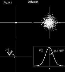
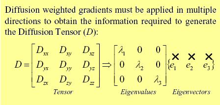
19 Slide 55 Slide 56 Fig. 9.3 Slide 57 MR Diffusion Tensor Imaging In liquids In tissues Anisotropy


20 Slide 58 Slide 59 Slide 60 DTI


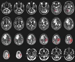
21 Slide 61 Slide 62 Slide 63 Case 5 - DTI/FA


22 Slide 64 Slide 65 Slide 66 Adapted from: Descoteaux et al DTI: Fiber Tracking


23 Slide 67 DTI at 3T Slide 68 Case 1 DTI Slide 69 Case 2: DTI


24 Slide 70 A C B D Slide 71 Slide 72


25 Slide 73 Case 6: 54 y.o. w/partial complex seizures & speech arrest Slide 74 Case 6: Bilateral Finger-tapping Slide 75 Case 6: Word Generation


26 Slide 76 Case 6: Naming Slide 77 Case 6: DTI Slide 78 Case 6: Perfusion (ASL)

27 Slide 79
functional MRI everything you always wanted to know, but never dared to MD PhD
 functional MRI everything you always wanted to know, but never dared to ask @MarionSmits, MD PhD Associate Professor of Neuroradiology Dept. of Radiology, Erasmus MC, Rotterdam (NL) Honorary Consultant
Define functional MRI. Briefly describe fmri image acquisition. Discuss relative functional neuroanatomy. Review clinical applications.
 Dr. Peter J. Fiester November 14, 2012 Define functional MRI. Briefly describe fmri image acquisition. Discuss relative functional neuroanatomy. Review clinical applications. Briefly discuss a few examples
Seamless pre-surgical fmri and DTI mapping
 Seamless pre-surgical fmri and DTI mapping Newest release Achieva 3.0T X-series and Eloquence enable efficient, real-time fmri for brain activity mapping in clinical practice at Nebraska Medical Center
Functional MRI and Diffusion Tensor Imaging
 Functional MRI and Diffusion Tensor Imaging Andrew Steven March 23, 2018 Ochsner Neuroscience Symposium None Disclosure 1 Objectives Review basic principles of BOLD fmri and DTI. Discuss indications and
Visualization strategies for major white matter tracts identified by diffusion tensor imaging for intraoperative use
 International Congress Series 1281 (2005) 793 797 www.ics-elsevier.com Visualization strategies for major white matter tracts identified by diffusion tensor imaging for intraoperative use Ch. Nimsky a,b,
Functional Magnetic Resonance Imaging with Arterial Spin Labeling: Techniques and Potential Clinical and Research Applications
 pissn 2384-1095 eissn 2384-1109 imri 2017;21:91-96 https://doi.org/10.13104/imri.2017.21.2.91 Functional Magnetic Resonance Imaging with Arterial Spin Labeling: Techniques and Potential Clinical and Research
Combining tdcs and fmri. OHMB Teaching Course, Hamburg June 8, Andrea Antal
 Andrea Antal Department of Clinical Neurophysiology Georg-August University Goettingen Combining tdcs and fmri OHMB Teaching Course, Hamburg June 8, 2014 Classical Biomarkers for measuring human neuroplasticity
The neurolinguistic toolbox Jonathan R. Brennan. Introduction to Neurolinguistics, LSA2017 1
 The neurolinguistic toolbox Jonathan R. Brennan Introduction to Neurolinguistics, LSA2017 1 Psycholinguistics / Neurolinguistics Happy Hour!!! Tuesdays 7/11, 7/18, 7/25 5:30-6:30 PM @ the Boone Center
Overview. Fundamentals of functional MRI. Task related versus resting state functional imaging for sensorimotor mapping
 Functional MRI and the Sensorimotor System in MS Nancy Sicotte, MD, FAAN Professor and Vice Chair Director, Multiple Sclerosis Program Director, Neurology Residency Program Cedars-Sinai Medical Center
P2 Visual - Perception
 P2 Visual - Perception 2014 SOSE Neuroimaging of high-level visual functions gyula.kovacs@uni-jena.de 11/09/06 Functional magnetic resonance imaging (fmri) The very basics What is fmri? What is MRI? The
HST.583 Functional Magnetic Resonance Imaging: Data Acquisition and Analysis Fall 2008
 MIT OpenCourseWare http://ocw.mit.edu HST.583 Functional Magnetic Resonance Imaging: Data Acquisition and Analysis Fall 2008 For information about citing these materials or our Terms of Use, visit: http://ocw.mit.edu/terms.
The Study of Brain Activity at Rest
 The Study of Brain Activity at Rest Activity Baseline in Brain Functional Studies Baseline Task Experimental Paradigm Block Design BOLD Resting State Brain Activity and the Default Mode Network A Definition
3/1/18. Overview of the Talk. Important Aspects of Neuroimaging Technology
 3/1/18 Considerations for the Use of Neuroimaging for Predicting Recovery of Speech and Language in Aphasia Linda I. Shuster, Ph.D., CCC-SLP Overview of the Talk Important aspects of neuroimaging technology
fmri: Interpretation, Limits and Potential Pitfalls
 fmri: Interpretation, Limits and Potential Pitfalls Seong-Gi Kim kimsg@pitt.edu www.kimlab.pitt.edu Mapping Brain Functions Stimulation/Task Functional Map (MRI) Pre-synaptic activity Post-synaptic activity
Biophysical and physiological bases of fmri signals: challenges of interpretation and methodological concerns
 Biophysical and physiological bases of fmri signals: challenges of interpretation and methodological concerns Antonio Ferretti aferretti@itab.unich.it Institute for Advanced Biomedical Technologies, University
BOLD signal dependence on blood flow and metabolism. Outline
 BOLD signal dependence on blood flow and metabolism R. Hoge, MGH NMR Center Outline physiological events accompanying neuronal activation factors affecting BOLD signal sensitivity BOLD response dynamics
Advances in Clinical Neuroimaging
 Advances in Clinical Neuroimaging Joseph I. Tracy 1, PhD, ABPP/CN; Gaelle Doucet 2, PhD; Xaiosong He 2, PhD; Dorian Pustina 2, PhD; Karol Osipowicz 2, PhD 1 Department of Radiology, Thomas Jefferson University,
Stuttering Research. Vincent Gracco, PhD Haskins Laboratories
 Stuttering Research Vincent Gracco, PhD Haskins Laboratories Stuttering Developmental disorder occurs in 5% of children Spontaneous remission in approximately 70% of cases Approximately 1% of adults with
Activated Fibers: Fiber-centered Activation Detection in Task-based FMRI
 Activated Fibers: Fiber-centered Activation Detection in Task-based FMRI Jinglei Lv 1, Lei Guo 1, Kaiming Li 1,2, Xintao Hu 1, Dajiang Zhu 2, Junwei Han 1, Tianming Liu 2 1 School of Automation, Northwestern
Daniel Bulte. Centre for Functional Magnetic Resonance Imaging of the Brain. University of Oxford
 Daniel Bulte Centre for Functional Magnetic Resonance Imaging of the Brain University of Oxford Overview Signal Sources BOLD Contrast Mechanism of MR signal change FMRI Modelling Scan design details Factors
fmri and Tractography in Preoperative Neurosurgical Planning Ronald L. Wolf, M.D., Ph.D. University of Pennsylvania Medical Center
 fmri and Tractography in Preoperative Neurosurgical Planning Ronald L. Wolf, M.D., Ph.D. University of Pennsylvania Medical Center Acknowledgements/Disclosures Radiology Ragini Verma Birkan Tunc Sumei
Epilepsy Surgery, Imaging, and Intraoperative Neuromonitoring: Surgical Perspective
 Epilepsy Surgery, Imaging, and Intraoperative Neuromonitoring: Surgical Perspective AC Duhaime, M.D. Director, Pediatric Neurosurgery, Massachusetts General Hospital Professor, Neurosurgery, Harvard Medical
Table 1. Summary of PET and fmri Methods. What is imaged PET fmri BOLD (T2*) Regional brain activation. Blood flow ( 15 O) Arterial spin tagging (AST)
 Regional brain activation. Blood flow ( 15 O) Arterial spin tagging (AST)") Table 1 Summary of PET and fmri Methods What is imaged PET fmri Brain structure Regional brain activation Anatomical connectivity Receptor binding and regional chemical distribution Blood flow ( 15 O)
Table 1 Summary of PET and fmri Methods What is imaged PET fmri Brain structure Regional brain activation Anatomical connectivity Receptor binding and regional chemical distribution Blood flow ( 15 O)
Use of Multimodal Neuroimaging Techniques to Examine Age, Sex, and Alcohol-Related Changes in Brain Structure Through Adolescence and Young Adulthood
 American Psychiatric Association San Diego, CA 24 May 2017 Use of Multimodal Neuroimaging Techniques to Examine Age, Sex, and Alcohol-Related Changes in Brain Structure Through Adolescence and Young Adulthood
POSITION TITLE: Sr. Research Imaging Specialist, Vanderbilt University Institute of Imaging Science
 NAME: Allen Timothy Newton OMB No. 0925-0001/0002 (Rev. 08/12 Approved Through 8/31/2015) BIOGRAPHICAL SKETCH Provide the following information for the Senior/key personnel and other significant contributors.
Functional MRI Mapping Cognition
 Outline Functional MRI Mapping Cognition Michael A. Yassa, B.A. Division of Psychiatric Neuro-imaging Psychiatry and Behavioral Sciences Johns Hopkins School of Medicine Why fmri? fmri - How it works Research
Invasive Evaluation for Epilepsy Surgery Lesional Cases NO DISCLOSURES. Mr. Johnson. Seizures at 29 Years of Age. Dileep Nair, MD Juan Bulacio, MD
 Invasive Evaluation for Epilepsy Surgery Lesional Cases NO DISCLOSURES Dileep Nair, MD Juan Bulacio, MD Mr. Johnson Seizures at 29 Years of Age Onset of seizures at 16 years of age bed wetting episodes
MULTIMODAL IMAGING IN TEMPORAL LOBE EPILEPSY JONATHAN WIRSICH
 Réunion Mensuelle de Neuroimagerie - 21 Mai 2015 MULTIMODAL IMAGING IN TEMPORAL LOBE EPILEPSY JONATHAN WIRSICH PhD Project Overview General Question: How are cognitive brain networks modified in epileptic
Neural Correlates of Human Cognitive Function:
 Neural Correlates of Human Cognitive Function: A Comparison of Electrophysiological and Other Neuroimaging Approaches Leun J. Otten Institute of Cognitive Neuroscience & Department of Psychology University
Pre-surgical planning for brain tumor resection using functional MRI
 June 2011 Divya S Bolar, HMSIV Pre-surgical planning for brain tumor resection using functional MRI Divya S. Bolar,, HMS IV 1 Our patient: clinical history 85-year year-old right-handed handed woman presents
Diffusion Tensor Imaging in brain tumours
 Diffusion Tensor Imaging in brain tumours @MarionSmits, MD PhD Associate Professor of Neuroradiology Dept. of Radiology, Erasmus MC, Rotterdam (NL) Honorary Consultant and Reader UCLH National Hospital
Neuroimaging. BIE601 Advanced Biological Engineering Dr. Boonserm Kaewkamnerdpong Biological Engineering Program, KMUTT. Human Brain Mapping
 11/8/2013 Neuroimaging N i i BIE601 Advanced Biological Engineering Dr. Boonserm Kaewkamnerdpong Biological Engineering Program, KMUTT 2 Human Brain Mapping H Human m n brain br in m mapping ppin can nb
BRAIN STATE CHANGE DETECTION VIA FIBER-CENTERED FUNCTIONAL CONNECTIVITY ANALYSIS
 BRAIN STATE CHANGE DETECTION VIA FIBER-CENTERED FUNCTIONAL CONNECTIVITY ANALYSIS Chulwoo Lim 1, Xiang Li 1, Kaiming Li 1, 2, Lei Guo 2, Tianming Liu 1 1 Department of Computer Science and Bioimaging Research
Introduction to Functional MRI
 Introduction to Functional MRI Douglas C. Noll Department of Biomedical Engineering Functional MRI Laboratory University of Michigan Outline Brief overview of physiology and physics of BOLD fmri Background
Combining microstimulation and fmri in an awake behaving monkey
 NEAAPM Peter Neurath Symposium Combining microstimulation and fmri in an awake behaving monkey Presented by: Leeland B. Ekstrom April 2006 Advisors: Wim Vanduffel & Bruce Rosen MGH / MIT / HMS Martinos
HST 583 fmri DATA ANALYSIS AND ACQUISITION
 HST 583 fmri DATA ANALYSIS AND ACQUISITION Neural Signal Processing for Functional Neuroimaging Neuroscience Statistics Research Laboratory Massachusetts General Hospital Harvard Medical School/MIT Division
BOLD signal compartmentalization based on the apparent diffusion coefficient
 Magnetic Resonance Imaging 20 (2002) 521 525 BOLD signal compartmentalization based on the apparent diffusion coefficient Allen W. Song a,b *, Harlan Fichtenholtz b, Marty Woldorff b a Brain Imaging and
An Unified Approach for fmri-measurements used by a New Real-Time fmri Analysis System
 An Unified Approach for fmri-measurements used by a New Real-Time fmri Analysis System Maurice Hollmann 1, Tobias Moench 1, Claus Tempelmann 2, Johannes Bernarding 1 1 Institute for Biometry and Medical
Resting-State functional Connectivity MRI (fcmri) NeuroImaging
 NeuroImaging") Resting-State functional Connectivity MRI (fcmri) NeuroImaging Randy L. Buckner et. at., The Brain s Default Network: Anatomy, Function, and Relevance to Disease, Ann. N. Y. Acad. Sci. 1124: 1-38 (2008)
Resting-State functional Connectivity MRI (fcmri) NeuroImaging Randy L. Buckner et. at., The Brain s Default Network: Anatomy, Function, and Relevance to Disease, Ann. N. Y. Acad. Sci. 1124: 1-38 (2008)
HST.583 Functional Magnetic Resonance Imaging: Data Acquisition and Analysis Fall 2008
 MIT OpenCourseWare http://ocw.mit.edu HST.583 Functional Magnetic Resonance Imaging: Data Acquisition and Analysis Fall 2008 For information about citing these materials or our Terms of Use, visit: http://ocw.mit.edu/terms.
FUNCTIONAL MRI IN EPILEPSY December 6 th 2013
 FUNCTIONAL MRI IN EPILEPSY December 6 th 2013 Matthias J Koepp, MD, PhD UCL Institute of Neurology National Hospital for Neurology and Neurosurgery London, UK American Epilepsy Society Annual Meeting Disclosure
Classification and Statistical Analysis of Auditory FMRI Data Using Linear Discriminative Analysis and Quadratic Discriminative Analysis
 International Journal of Innovative Research in Computer Science & Technology (IJIRCST) ISSN: 2347-5552, Volume-2, Issue-6, November-2014 Classification and Statistical Analysis of Auditory FMRI Data Using
PETER PAZMANY CATHOLIC UNIVERSITY Consortium members SEMMELWEIS UNIVERSITY, DIALOG CAMPUS PUBLISHER
 PETER PAZMANY CATHOLIC UNIVERSITY SEMMELWEIS UNIVERSITY Development of Complex Curricula for Molecular Bionics and Infobionics Programs within a consortial* framework** Consortium leader PETER PAZMANY
Supplementary Information Methods Subjects The study was comprised of 84 chronic pain patients with either chronic back pain (CBP) or osteoarthritis
 or osteoarthritis") Supplementary Information Methods Subjects The study was comprised of 84 chronic pain patients with either chronic back pain (CBP) or osteoarthritis (OA). All subjects provided informed consent to procedures
Supplementary Information Methods Subjects The study was comprised of 84 chronic pain patients with either chronic back pain (CBP) or osteoarthritis (OA). All subjects provided informed consent to procedures
Hemodynamics and fmri Signals
 Cerebral Blood Flow and Brain Activation UCLA NITP July 2011 Hemodynamics and fmri Signals Richard B. Buxton University of California, San Diego rbuxton@ucsd.edu... The subject to be observed lay on a
Supplementary Online Content
 Supplementary Online Content Gregg NM, Kim AE, Gurol ME, et al. Incidental cerebral microbleeds and cerebral blood flow in elderly individuals. JAMA Neurol. Published online July 13, 2015. doi:10.1001/jamaneurol.2015.1359.
Functional Magnetic Resonance Imaging of the Brain
 Page: 1 of 9 Last Review Status/Date: December 2016 Description Functional magnetic resonance imaging (fmri) is a noninvasive method for localizing areas of brain function and has been used for the presurgical
INTRO TO BOLD FMRI FRANZ JOSEPH GALL ( ) OUTLINE. MRI & Fast MRI Observations Models Statistical Detection
 OUTLINE. MRI & Fast MRI Observations Models Statistical Detection") INTRO TO BOLD FMRI 2014 M.S. Cohen all rights reserved mscohen@g.ucla.edu OUTLINE FRANZ JOSEPH GALL (1758-1828) MRI & Fast MRI Observations Models Statistical Detection PAUL BROCA (1824-1880) WILLIAM JAMES
INTRO TO BOLD FMRI 2014 M.S. Cohen all rights reserved mscohen@g.ucla.edu OUTLINE FRANZ JOSEPH GALL (1758-1828) MRI & Fast MRI Observations Models Statistical Detection PAUL BROCA (1824-1880) WILLIAM JAMES
Speed, Comfort and Quality with NeuroDrive
 Speed, Comfort and Quality with NeuroDrive Echelon Oval provides a broad range of capabilities supporting fast, accurate diagnosis of brain conditions and injuries. From anatomical depiction to vascular
DATA MANAGEMENT & TYPES OF ANALYSES OFTEN USED. Dennis L. Molfese University of Nebraska - Lincoln
 DATA MANAGEMENT & TYPES OF ANALYSES OFTEN USED Dennis L. Molfese University of Nebraska - Lincoln 1 DATA MANAGEMENT Backups Storage Identification Analyses 2 Data Analysis Pre-processing Statistical Analysis
APPLICATIONS OF ASL IN NEUROSCIENCE
 APPLICATIONS OF ASL IN NEUROSCIENCE Luis Hernandez-Garcia, Ph.D. Functional MRI laboratory University of Michigan 1 OVERVIEW Quick review of ASL The niche for ASL Examples of practical applications in
Diffusion Tensor Imaging 12/06/2013
 12/06/2013 Beate Diehl, MD PhD FRCP University College London National Hospital for Neurology and Neurosurgery Queen Square London, UK American Epilepsy Society Annual Meeting Disclosure None Learning
Cortical hypoperfusion in Parkinson's disease assessed with arterial spin labeling MRI
 Cortical hypoperfusion in Parkinson's disease assessed with arterial spin labeling MRI Poster No.: C-0609 Congress: ECR 2013 Type: Scientific Exhibit Authors: S. Aoki, K. Kamagata, Y. Motoi, K. Kamiya,
Concurrent near-infrared spectroscopy (NIRS) and functional magnetic resonance imaging (fmri) of the brain
 and functional magnetic resonance imaging (fmri) of the brain") Motor cortex activation fmri Near-infrared imaging Concurrent near-infrared spectroscopy (NIRS) and functional magnetic resonance imaging (fmri) of the brain Sergio Fantini s group, Department of Biomedical
Motor cortex activation fmri Near-infrared imaging Concurrent near-infrared spectroscopy (NIRS) and functional magnetic resonance imaging (fmri) of the brain Sergio Fantini s group, Department of Biomedical
Perfusion MRI. Youngkyoo Jung, PhD Associate Professor Radiology, Biomedical Engineering, and Clinical & Translational Science Institute
 Perfusion MRI Youngkyoo Jung, PhD Associate Professor Radiology, Biomedical Engineering, and Clinical & Translational Science Institute Perfusion The delivery of blood to a capillary bed in tissue Perfusion
Brain and Cognition. Cognitive Neuroscience. If the brain were simple enough to understand, we would be too stupid to understand it
 Brain and Cognition Cognitive Neuroscience If the brain were simple enough to understand, we would be too stupid to understand it 1 The Chemical Synapse 2 Chemical Neurotransmission At rest, the synapse
PHYSICS OF MRI ACQUISITION. Alternatives to BOLD for fmri
 PHYSICS OF MRI ACQUISITION Quick Review for fmri HST-583, Fall 2002 HST.583: Functional Magnetic Resonance Imaging: Data Acquisition and Analysis Harvard-MIT Division of Health Sciences and Technology
MEDICAL REVIEW Functional Magnetic Resonance Imaging: From Acquisition to Application
 Functional Magnetic Resonance Imaging: From Acquisition to Application Gail Yarmish * and Michael L. Lipton *, Departments of Radiology *, and Neuroscience Albert Einstein College of Medicine Bronx, New
Titelmaster The physics of functional magnetic resonance imaging (fmri)
") Titelmaster The physics of functional magnetic resonance imaging (fmri) Outline 1.Introduction 2.The fmri experiment 2 3.The physics basis of fmri 4.Application Outline 3 1.Introduction Introduction Phrenology
Titelmaster The physics of functional magnetic resonance imaging (fmri) Outline 1.Introduction 2.The fmri experiment 2 3.The physics basis of fmri 4.Application Outline 3 1.Introduction Introduction Phrenology
Experimental Design. Outline. Outline. A very simple experiment. Activation for movement versus rest
 Experimental Design Kate Watkins Department of Experimental Psychology University of Oxford With thanks to: Heidi Johansen-Berg Joe Devlin Outline Choices for experimental paradigm Subtraction / hierarchical
International Journal of Innovative Research in Advanced Engineering (IJIRAE) Volume 1 Issue 10 (November 2014)
 Volume 1 Issue 10 (November 2014)") Technique for Suppression Random and Physiological Noise Components in Functional Magnetic Resonance Imaging Data Mawia Ahmed Hassan* Biomedical Engineering Department, Sudan University of Science & Technology,
Technique for Suppression Random and Physiological Noise Components in Functional Magnetic Resonance Imaging Data Mawia Ahmed Hassan* Biomedical Engineering Department, Sudan University of Science & Technology,
HST.583 Functional Magnetic Resonance Imaging: Data Acquisition and Analysis Fall 2006
 MIT OpenCourseWare http://ocw.mit.edu HST.583 Functional Magnetic Resonance Imaging: Data Acquisition and Analysis Fall 2006 For information about citing these materials or our Terms of Use, visit: http://ocw.mit.edu/terms.
Presence of AVA in High Frequency Oscillations of the Perfusion fmri Resting State Signal
 Presence of AVA in High Frequency Oscillations of the Perfusion fmri Resting State Signal Zacà D 1., Hasson U 1,2., Davis B 1., De Pisapia N 2., Jovicich J. 1,2 1 Center for Mind/Brain Sciences, University
The Central Nervous System
 The Central Nervous System Cellular Basis. Neural Communication. Major Structures. Principles & Methods. Principles of Neural Organization Big Question #1: Representation. How is the external world coded
Comparing event-related and epoch analysis in blocked design fmri
 Available online at www.sciencedirect.com R NeuroImage 18 (2003) 806 810 www.elsevier.com/locate/ynimg Technical Note Comparing event-related and epoch analysis in blocked design fmri Andrea Mechelli,
Functional MRI of the dynamic brain: quasiperiodic patterns, brain states, and trajectories. Shella Keilholz BME, Emory/Georgia Tech 20 March 2018
 Functional MRI of the dynamic brain: quasiperiodic patterns, brain states, and trajectories Shella Keilholz BME, Emory/Georgia Tech 20 March 2018 Resting State fmri No stimulus Looks at spontaneous BOLD
Prevalence of cerebrovascular reserve impairment in patients with severe intracranial stenosis
 Prevalence of cerebrovascular reserve impairment in patients with severe intracranial stenosis Olivier Heck, Naila Boudiaf, Florence Tahon, Arnaud Attye, Kamel Boubagra, Johan Pietras, Olivier Detante,
MRI qbold Based Evaluation. Renal Oxidative Metabolism. Department of Radiology and Hernando Gomez, MD Critical Care Medicine
 MRI qbold Based Evaluation of Renal Oxidative Metabolism Xiang He, PhD Department of Radiology and Hernando Gomez, MD Critical Care Medicine Background High oxygen-demand and lower medullary blood flow
Perfusion-Based fmri. Thomas T. Liu Center for Functional MRI University of California San Diego May 7, Goal
 Perfusion-Based fmri Thomas T. Liu Center for Functional MRI University of California San Diego May 7, 2006 Goal To provide a basic understanding of the theory and application of arterial spin labeling
Case reports functional imaging in epilepsy
 Seizure 2001; 10: 157 161 doi:10.1053/seiz.2001.0552, available online at http://www.idealibrary.com on Case reports functional imaging in epilepsy MARK P. RICHARDSON Medical Research Council Fellow, Institute
Introduction to Computational Neuroscience
 Introduction to Computational Neuroscience Lecture 10: Brain-Computer Interfaces Ilya Kuzovkin So Far Stimulus So Far So Far Stimulus What are the neuroimaging techniques you know about? Stimulus So Far
Supplementary information Detailed Materials and Methods
 Supplementary information Detailed Materials and Methods Subjects The experiment included twelve subjects: ten sighted subjects and two blind. Five of the ten sighted subjects were expert users of a visual-to-auditory
MSc Neuroimaging for Clinical & Cognitive Neuroscience
 MSc Neuroimaging for Clinical & Cognitive Neuroscience School of Psychological Sciences Faculty of Medical & Human Sciences Module Information *Please note that this is a sample guide to modules. The exact
Surgery Within and Around Critical White Matter Tracts
 Surgery Within and Around Critical White Matter Tracts Kaisorn L. Chaichana, M.D. Assistant Professor of Neurosurgery, Oncology, and Otolaryngology-Head & Neck Surgery Mayo Clinic Florida, Jacksonville,
Principles of Haemodynamic Coupling for fmri
 Principles of Haemodynamic Coupling for fmri Paul M. Matthews Head, Global Imaging Unit, GlaxoSmithKline and Professor of Clinical Neurosciences, Imperial College paul.m.matthews@gsk.com Regulation of
Intraoperative Monitoring: Role in Epilepsy Based Tumor Surgery December 2, 2012
 Intraoperative Monitoring: Role in Epilepsy Based Tumor Surgery December 2, 2012 Aatif M. Husain, M.D. Duke University and Veterans Affairs Medical Centers, Durham, NC American Epilepsy Society Annual
Clinical Policy: Functional MRI Reference Number: CP.MP.43
 Clinical Policy: Reference Number: CP.MP.43 Effective Date: 09/09 Last Review Date: 10/17 Coding Implications Revision Log See Important Reminder at the end of this policy for important regulatory and
Laurent Itti: CS564 Brain Theory and Artificial Intelligence. Lecture 4: Experimental techniques in visual neuroscience. Reading Assignments: None!
 CS 564 Brain Theory and Artificial Intelligence Lecture 4: Experimental techniques in visual neuroscience Reading Assignments: None! 1 Today we will briefly review - electrophysiological recording and
Causality from fmri?
 Causality from fmri? Olivier David, PhD Brain Function and Neuromodulation, Joseph Fourier University Olivier.David@inserm.fr Grenoble Brain Connectivity Course Yes! Experiments (from 2003 on) Friston
Temporal preprocessing of fmri data
 Temporal preprocessing of fmri data Blaise Frederick, Ph.D., Yunjie Tong, Ph.D. McLean Hospital Brain Imaging Center Scope This talk will summarize the sources and characteristics of unwanted temporal
5/2/2013. Real-time fmri: Methods and applications
 Real-time fmri: Methods and applications Frank Scharnowski Non-invasive imaging technique PET Magnetic Resonance Imaging (MRI) NIRS EEG MEG Functional MRI Structural MRI Perfusion MRI Diffusion MRI MR
The Tools: Imaging the Living Brain
 The Tools: Imaging the Living Brain I believe the study of neuroimaging has supported the localization of mental operations within the human brain. -Michael I. Posner, 2003 Neuroimaging methods Since Descarte
Pattern Recognition of Functional Neuroimage Data of the Human Sensorimotor System after Stroke
 Pattern Recognition of Functional Neuroimage Data of the Human Sensorimotor System after Stroke Camille Gómez-Laberge, M.A.Sc., B.Eng., B.Sc. Ph.D. Candidate Faculty of Engineering, Carleton University
9/30/2016. Advances in Epilepsy Surgery. Epidemiology. Epidemiology
 Advances in Epilepsy Surgery George Jallo, M.D. Director, Institute for Brain Protection Sciences Johns Hopkins All Children s Hospital St Petersburg, Florida Epidemiology WHO lists it as the second most
Outline. Neuroradiology. Diffusion Imaging in. Clinical Applications of. Basics of Diffusion Imaging. Basics of Diffusion Imaging
 Clinical Applications of Diffusion Imaging in Neuroradiology No disclosures Stephen F. Kralik Assistant Professor of Radiology Indiana University School of Medicine Department of Radiology and Imaging
PERFUSION MRI CONTRAST BASED TECHNIQUES
 PERFUSION MRI CONTRAST BASED TECHNIQUES by Kenny K Israni Mar 28, 2006 PERFUSION - MRI Dynamic Susceptibility contrast Dynamic Relaxivity contrast STEADY-STATE STATE TECHNIQUES Steady-state Susceptibility
Myers Psychology for AP*
 Myers Psychology for AP* David G. Myers PowerPoint Presentation Slides by Kent Korek Germantown High School Worth Publishers, 2010 *AP is a trademark registered and/or owned by the College Board, which
Title:Atypical language organization in temporal lobe epilepsy revealed by a passive semantic paradigm
 Author's response to reviews Title:Atypical language organization in temporal lobe epilepsy revealed by a passive semantic paradigm Authors: Julia Miro (juliamirollado@gmail.com) Pablo Ripollès (pablo.ripolles.vidal@gmail.com)
fmri Acquisition: Temporal Effects
 Functional MRI Data Acquisition: Temporal fmri Acquisition: Temporal Effects Session length Repetition time Fixed vs. distributed temporal sampling Sparse temporal sampling Noise source recording Prospective
HST.583 Functional Magnetic Resonance Imaging: Data Acquisition and Analysis Fall 2008
 MIT OpenCourseWare http://ocw.mit.edu HST.583 Functional Magnetic Resonance Imaging: Data Acquisition and Analysis Fall 2008 For information about citing these materials or our Terms of Use, visit: http://ocw.mit.edu/terms.
The physiology of the BOLD signal What do we measure with fmri?
 The physiology of the BOLD signal What do we measure with fmri? Methods and Models in fmri, 10.11.2012 Jakob Heinzle Translational Neuromodeling Unit (TNU) Institute for Biomedical Engineering (IBT) University
Neurophilosophical Foundations 3
 Neurophilosophical Foundations 3 The Artifact Problem: The Epistemic Challenge Techniques to procure evidence alter the phenomenon about which scientists are trying to get evidence Are the resulting observations
An fmri Phantom Based on Electric Field Alignment of Molecular Dipoles
 An fmri Phantom Based on Electric Field Alignment of Molecular Dipoles Innovative Graduate Student Proposal Yujie Qiu, Principal Investigator, Graduate Student Joseph Hornak, Faculty Sponsor, Thesis Advisor
Mirror Neurons in Primates, Humans, and Implications for Neuropsychiatric Disorders
 Mirror Neurons in Primates, Humans, and Implications for Neuropsychiatric Disorders Fiza Singh, M.D. H.S. Assistant Clinical Professor of Psychiatry UCSD School of Medicine VA San Diego Healthcare System
fmri and Pre-operative Planning for CNS tumors
 fmri and Pre-operative Planning for CNS tumors Miami Brain Symposium Andrei I. Holodny, M.D. Chief of Neuroradiology Director of the fmri Lab Memorial Sloan-Kettering Cancer Center Disclosures: 1. NIH-NIBIB
Hemodynamics and fmri Signals
 Cerebral Blood Flow and Brain Activation UCLA NITP July 2010 Hemodynamics and fmri Signals Richard B. Buxton University of California, San Diego rbuxton@ucsd.edu... The subject to be observed lay on a
Diagnosing Complicated Epilepsy: Mapping of the Epileptic Circuitry. Michael R. Sperling, M.D. Thomas Jefferson University Philadelphia, PA
 Diagnosing Complicated Epilepsy: Mapping of the Epileptic Circuitry Michael R. Sperling, M.D. Thomas Jefferson University Philadelphia, PA Overview Definition of epileptic circuitry Methods of mapping
Stereotactic Diffusion Tensor Tractography For Gamma Knife Stereotactic Radiosurgery
 Disclosures The authors of this study declare that they have no commercial or other interests in the presentation of this study. This study does not contain any use of offlabel devices or treatments. Stereotactic
Functional Magnetic Resonance Imaging
 Todd Parrish, Ph.D. Director of the Center for Advanced MRI Director of MR Neuroimaging Research Associate Professor Department of Radiology Northwestern University toddp@northwestern.edu Functional Magnetic