Surgery Within and Around Critical White Matter Tracts
|
|
|
- Janel Cole
- 5 years ago
- Views:
Transcription


1 Surgery Within and Around Critical White Matter Tracts Kaisorn L. Chaichana, M.D. Assistant Professor of Neurosurgery, Oncology, and Otolaryngology-Head & Neck Surgery Mayo Clinic Florida, Jacksonville, FL


2 Subcortical Space Area of the brain below the cortical surface Critical white matter tracts Critical gray matter regions Traditional techniques to access these regions can be associated with significant morbidity

3 3 different types of tracts Commissural Projection Association Critical White Matter Tracts

4 Corticospinal Tract Principle projection fibers of the cerebral hemisphere Largest projection fiber tract Consists of: Corona radiata superiorly Classic V-shaped capsule at level of basal ganglia Crus cerebri in cerebral peduncle

 Motor coordination Visual")
5 Superior Longitudinal Fasciculus/Arcuate Fasciculus Lateral to ventricles and centrum ovale Indirect: SLF I-III, SLF-tp; Direct: AF Dorsal Stream Connects portions of frontal lobe with occipital and temporal areas Disruption can cause deficits in: Language (phonetics/articulation/repetition) Motor coordination Visual spatial perception

6 Inferior Frontal-Occipital Fasciculus Connects orbital and frontopolar regions with ventromedial occipital cortex Ventral Stream Under insular region, deep to SLF, at level of external capsule Disruption can cause deficits in: Language (semantics)

7 Inferior Longitudinal Fasciculus Connects temporal and occipital lobes Runs at level of optic radiation Fibers blend with the IFOF Involved in: Object Recognition Visual Agnosia Prospagnosia


8 Left Frontal Aslant Tract Subcallosal Fasciculus Uncinate Fasciculus Superior Frontal-Occipital Fasciculus Middle Longitudinal Fasciculus Other Tracts
9 Why Does It Matter? Neurological exam not sensitive for detecting deficits Functional status is critical Every patient prior to surgery Neuropsych testing Goals of care discussion DTI +/- fmri High resolution T2 MRI (axial/coronal)
 Chaichana et al, Neuro Oncol, 16 (1), 1113, 2014 Jackson et al. J Neurosurg, 78 (6), 588, 2017 Iyer et al. J Neurosurg, in press")
10 General Approaches to Subcortical Brain Tumors Goal is to preserve eloquent cortical areas and white matter tracts Two methods: 1) Identify and avoid these areas (i.e. awake mapping) 2) Displace these tracts (i.e. parafascicular approach) Chaichana et al, Neuro Oncol, 16 (1), 1113, 2014 Jackson et al. J Neurosurg, 78 (6), 588, 2017 Iyer et al. J Neurosurg, in press
11 Awake Brain Mapping Critical elements Resection based on onco-functional boundaries Real time evaluation of neurological function Need to know what tracts/functions you are looking for fmri Coupling between neurological activity and blood flow Cannot determine critical functions Sensitivity (37.1%), Specificity (83.4%) (Kuchsinki et al, 2015) DTI Diffusion of water along tracts Diffusion properties altered with tumors No agreement in construction (Pujol et al, 2015)
12 Awake Brain Mapping Protocols Prior to surgery Neuropsych testing Goals of care discussion DTI +/- fmri High resolution T2 MRI (axial/coronal) During surgery Asleep-Awake-Asleep Anesthesia Ultrasound, Navigation Mapping +/- imri After surgery SLP/PT Neuropsych testing (3-6 months)
13 Awake Brain Mapping Case 1 CC: seizures HPI: 45 yo RH female college professor who presented with partial seizures PE: intact, neuropsych normal Concerning areas Cortical: Broca s, premotor, motor Subcortical: SLF/AF, IFOF (anterior limb), FAT, caudate Chaichana et al, Approaches to Brain Tumors, Elsevier, 2018, in press
 6, 7 ventral premotor (articulation disturbances) 2, 3 motor cortex (RH movement arrest) 4")
 50 SLF (articulation) Chaichana et al, Approaches to Brain Tumors, Elsevier,")
14 Awake Brain Mapping Case 1 Letters ultrasound projection of tumor 1, 5, 8 motor cortex/broca s (anarthria, facial twitching) 6, 7 ventral premotor (articulation disturbances) 2, 3 motor cortex (RH movement arrest) 4 somatosensory (RH and face paresthesias) 47 head of the caudate (perseveration) 48 IFOF (semantic paraphasia) 49 FAT (speech initiation problems) 50 SLF (articulation) Chaichana et al, Approaches to Brain Tumors, Elsevier, 2018, in press

15 Awake Brain Mapping Case 1 Pathology IDH1+, 1p19q- Immediate postop hesitancy with speech, dysarthria 2 weeks postop intact exam 1 mo back to teaching 3 mo neuropsych normal Chaichana et al, Approaches to Brain Tumors, Elsevier, 2018, in press
16 Awake Brain Mapping Case 2 CC: seizures HPI: 49 yo RH male business owner who presented with partial seizures PE: intact, neuropsych normal Concerning areas Cortical: Wernicke s, motor Subcortical: SLF (III, tp)/af, IFOF Chaichana et al, Approaches to Brain Tumors, Elsevier, 2018, in press
 3 - complete anomia 48 SLF (articulation")
 Chaichana et al, Approaches to Brain Tumors, Elsevier, 2018, in")
17 Awake Brain Mapping Case 2 Letters ultrasound projection of tumor 1, 2 ventral premotor (articulation disturbances) 3 - complete anomia 48 SLF (articulation difficulty) 49 AF (phonological paraphasias, repetition) 50 IFOF (semantic paraphasia) Chaichana et al, Approaches to Brain Tumors, Elsevier, 2018, in press


18 Awake Brain Mapping Case 2 Pathology - IDH1-, 1p19q- Immediate postop some comprehension difficulty 2 weeks postop intact exam 1 mo returned to work 3 mo neuropsych - normal Chaichana et al, Approaches to Brain Tumors, Elsevier, 2018, in press
19 General Approaches to Subcortical Brain Tumors Goal is to preserve eloquent cortical areas and white matter tracts Two methods: 1) Identify and avoid these areas (i.e. awake mapping) 2) Displace these tracts (i.e. parafascicular approach) Chaichana et al, Neuro Oncol, 16 (1), 1113, 2014 Jackson et al. J Neurosurg, 78 (6), 588, 2017 Iyer et al. J Neurosurg, in press


20 Traditional Approaches to Subcortical Brain Tumors Large craniotomy Extensive white matter dissection Use of fixed retractor systems Potential sources of injury - White matter dissection - Tissue creep - Repetitive entry into resection site - Ischemia induced by retraction Spetzler et al, J Neurosurg, 116, 291, 2012.


 Tubular retractors")


21 Concept of Minimally Disruptive Approaches Trans-sulcal vs. Trans-gyral Displacement of white matter tracts (<15 mm) Tubular retractors Circumferential retraction Protected corridor for dissection and resection Chaichana et al, Trans-Sulcal Approaches, Elsevier, in press.


22 Use of Tubular Retractors is an Old Concept Kelly et al, J Neurosurg, 69, 301, 1988.

23 Peel-away catheters (12F, 14F, 17F) Viewsite TM BrainPath TM Variety of Tubular Retractors

24 Peel-Away Catheters Advantages No brain retraction Minimal collateral damage Disadvantages Requires endoscope Limited degree of freedom Limited number of instruments Hemostasis Cavity-based surgeries

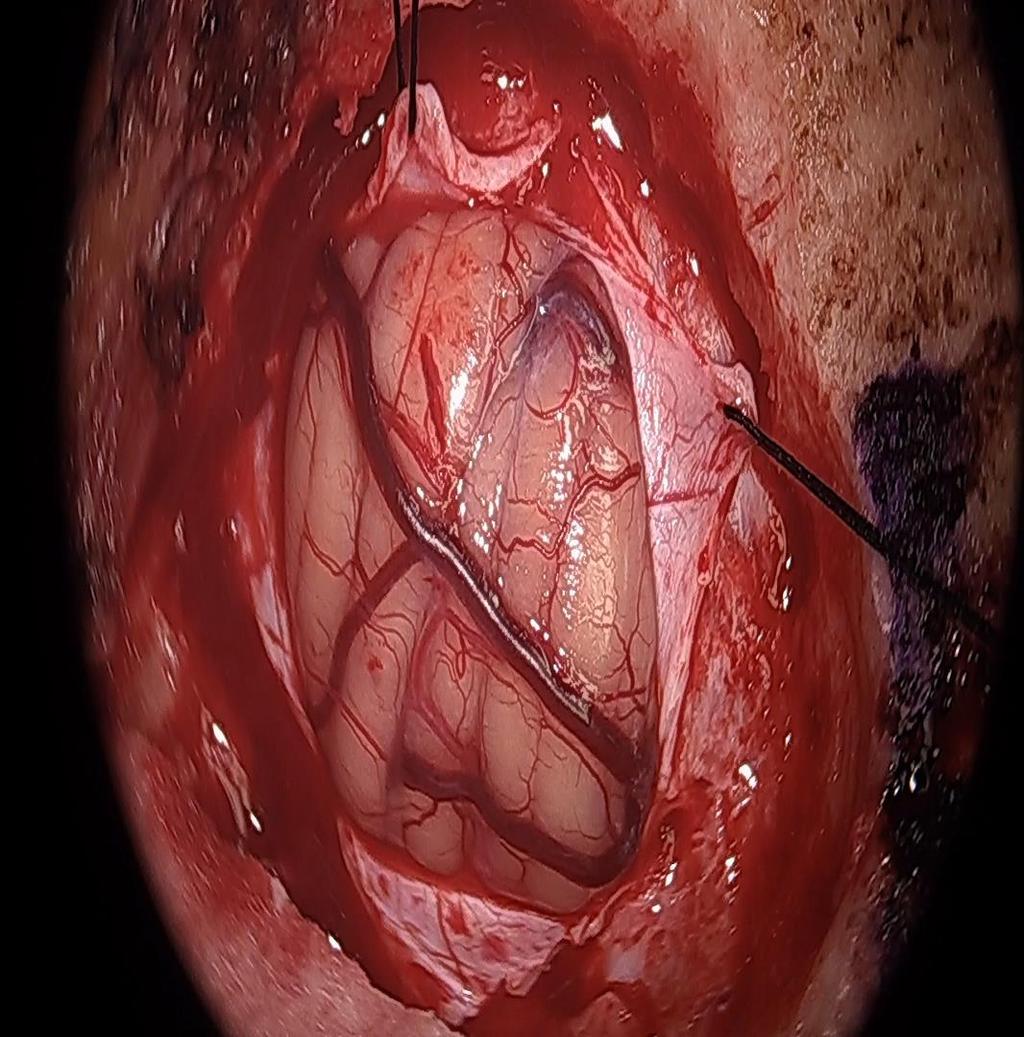
25 Colloid Cyst with Peel-Away Catheters 22 year-old female with history of colloid cyst that increased in size on serial imaging from 3 mm to 8mm, with increase in headaches. Right frontal, trans-cortical approach

26 Colloid Cyst with Peel-Away Catheters

27 Colloid Cyst with Peel-Away Catheters Final pathology: colloid cyst Discharged to home on POD 1


28 BrainPath TM Tubular Retractor Advantages Circumferential retraction Trans-sulcal or gyral Minimal collateral damage Microscope or exoscope Disadvantages Limited to 13.5 mm aperture Limited instruments Limited degree of freedom
29 Exoscopic Visualization

30 High resolution T2 axial and coronal MRI images Intraop navigation DTI/tractography Intra-operative monitoring Ultrasound Surgical Adjuncts
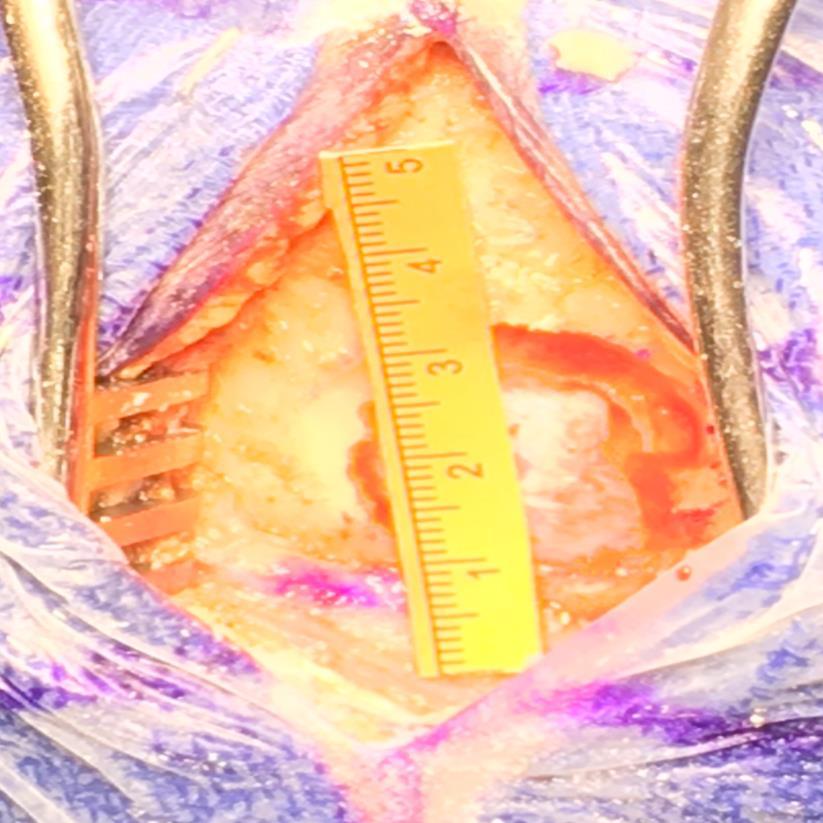
31 Tubular-Based Tumor Resection Case 1 CC: right UE/LE weakness HPI: 22 yo RH female presented with progressive right sided weakness PE: RUE/RLE 4-/5 Concerning areas Cortical: Broca s, motor Subcortical: SLF/AF, IFOF (anterior limb), FAT, internal capsule Chaichana et al, JNS-A, 178, Iyer et al, J Neurosurg, 2018, in press


32 Tubular-Based Tumor Resection Case 1 Chaichana et al, JNS-A, 178, 2017 Iyer et al, J Neurosurg, 2018, in press
, neuro intact 6 mo competing in archery competitions Chaichana et al, JNS-A, 178, 2017 Iyer et al, J Neurosurg, 2018,")
33 Tubular-Based Tumor Resection Case 1 Pathology: Glioblastoma (MGMT+/IDH-) Disposition: Home on POD 3 (18 months out without recurrence), neuro intact 6 mo competing in archery competitions Chaichana et al, JNS-A, 178, 2017 Iyer et al, J Neurosurg, 2018, in press


34 Tubular-Based Tumor Resection Case 2 CC: right weakness HPI: This is a 53 year-old RH male presented with progressive right upper and lower extremity weakness. PE: 1/5 RUE/RLE Concerning areas: Cortical: motor cortex, sensory cortex Subcortical: SLF/AF, IFOF, thalamus, basal ganglia, internal capsule Chaichana et al, JNS-A, 178, 2017 Iyer et al, J Neurosurg, 2018, in press
 Post-op 4/5 strength, discharged to home on POD")

35 Tubular-Based Tumor Resection Case 2 Pathology: glioblastoma (MGMT-/IDH-) Post-op 4/5 strength, discharged to home on POD 3 No recurrence at 7 months, slight left drift Chaichana et al, JNS-A, 178, 2017 Iyer et al, J Neurosurg, 2018, in press


36 Tubular-Based Tumor Resection Case 3 CC: left sided weakness HPI: This is a 32 yo RH male presented left hemiparesis PE: LUE/LLE 1-2/5 Concerning areas: Cortical: motor cortex, sensory cortex Subcortical: CST Chaichana et al, JNS-A, 178, Iyer et al, J Neurosurg, 2018, in press


37 Tubular-Based Tumor Resection Case 3 Pathology: GBM Disposition: Neuro intact, Discharged to home on POD3 No recurrence at 8 months Chaichana et al, JNS-A, 178, Iyer et al, J Neurosurg, 2018, in press

 and")
38 Tubular-Based Tumor Resection Case 4 CC: persistent emesis HPI: This is a 49 yo female who presented with persistent nausea and vomiting on two previous occasions from two hemorrhages of a right middle cerebellar peduncle cavernoma PE: neuro intact except right facial (HB1-2/6) and CNVI weakness
39 Tubular-Based Tumor Resection Case 4

40 Tubular-Based Tumor Resection Case 4 Pathology: cavernoma Disposition: Neuro intact, discharged to home on POD2
41 BrainPath Brain Tumor Case Totals - 53 Surgery 36 resections 17 excisional biopsies Locations Thalamus/basal ganglia - 24 Centrum semiovale/white matter tracts - 16 Optic pathways 4 Deep cerebellar nuclei 8 Pathology GBM/AA - 30 Metastatic - 10 Low grade glioma - 2 Cavernoma - 8 Other - 2 Outcomes Improved - 39 Stable 10 Worsened 3 (2 transient)


42 Conclusions Neurological function beyond the standard neurological exam is important to preserve Need to understand where the white matter tracts are and what they do You can either identify and avoid, or work around or displace these tracts Critical to be able to do both, which require a different set of tools
Telencephalon (Cerebral Hemisphere)
") Telencephalon (Cerebral Hemisphere) OUTLINE The Cortex - Lobes, Sulci & Gyri - Functional Subdivisions - Limbic Lobe & Limbic System The Subcortex - Basal Ganglia - White Matter (Internal Capsule) - Relations
Telencephalon (Cerebral Hemisphere) OUTLINE The Cortex - Lobes, Sulci & Gyri - Functional Subdivisions - Limbic Lobe & Limbic System The Subcortex - Basal Ganglia - White Matter (Internal Capsule) - Relations
fmri and Tractography in Preoperative Neurosurgical Planning Ronald L. Wolf, M.D., Ph.D. University of Pennsylvania Medical Center
 fmri and Tractography in Preoperative Neurosurgical Planning Ronald L. Wolf, M.D., Ph.D. University of Pennsylvania Medical Center Acknowledgements/Disclosures Radiology Ragini Verma Birkan Tunc Sumei
Fig.1: A, Sagittal 110x110 mm subimage close to the midline, passing through the cingulum. Note that the fibers of the corpus callosum run at a
 Fig.1 E Fig.1:, Sagittal 110x110 mm subimage close to the midline, passing through the cingulum. Note that the fibers of the corpus callosum run at a slight angle are through the plane (blue dots with
Seamless pre-surgical fmri and DTI mapping
 Seamless pre-surgical fmri and DTI mapping Newest release Achieva 3.0T X-series and Eloquence enable efficient, real-time fmri for brain activity mapping in clinical practice at Nebraska Medical Center
CEREBRUM Dr. Jamila Elmedany Dr. Essam Eldin Salama
 CEREBRUM Dr. Jamila Elmedany Dr. Essam Eldin Salama Objectives At the end of the lecture, the student should be able to: List the parts of the cerebral hemisphere (cortex, medulla, basal nuclei, lateral
Intraoperative Monitoring: Role in Epilepsy Based Tumor Surgery December 2, 2012
 Intraoperative Monitoring: Role in Epilepsy Based Tumor Surgery December 2, 2012 Aatif M. Husain, M.D. Duke University and Veterans Affairs Medical Centers, Durham, NC American Epilepsy Society Annual
Leah Militello, class of 2018
 Leah Militello, class of 2018 Objectives 1. Describe the general organization of cerebral hemispheres. 2. Describe the locations and features of the different functional areas of cortex. 3. Understand
Introduction to Neurosurgical Subspecialties:
 Introduction to Neurosurgical Subspecialties: Tumor and Skull Base Neurosurgery Brian L. Hoh, MD 1 and Gregory J. Zipfel, MD 2 1 University of Florida, 2 Washington University Tumor / Skull Base Neurosurgery
Diffusion Tensor Imaging in brain tumours
 Diffusion Tensor Imaging in brain tumours @MarionSmits, MD PhD Associate Professor of Neuroradiology Dept. of Radiology, Erasmus MC, Rotterdam (NL) Honorary Consultant and Reader UCLH National Hospital
P. Hitchcock, Ph.D. Department of Cell and Developmental Biology Kellogg Eye Center. Wednesday, 16 March 2009, 1:00p.m. 2:00p.m.
 Normal CNS, Special Senses, Head and Neck TOPIC: CEREBRAL HEMISPHERES FACULTY: LECTURE: READING: P. Hitchcock, Ph.D. Department of Cell and Developmental Biology Kellogg Eye Center Wednesday, 16 March
CEREBRUM & CEREBRAL CORTEX
 CEREBRUM & CEREBRAL CORTEX Seonghan Kim Dept. of Anatomy Inje University, College of Medicine THE BRAIN ANATOMICAL REGIONS A. Cerebrum B. Diencephalon Thalamus Hypothalamus C. Brain Stem Midbrain Pons
Anatomy and Physiology (Bio 220) The Brain Chapter 14 and select portions of Chapter 16
 The Brain Chapter 14 and select portions of Chapter 16") Anatomy and Physiology (Bio 220) The Brain Chapter 14 and select portions of Chapter 16 I. Introduction A. Appearance 1. physical 2. weight 3. relative weight B. Major parts of the brain 1. cerebrum 2.
Anatomy and Physiology (Bio 220) The Brain Chapter 14 and select portions of Chapter 16 I. Introduction A. Appearance 1. physical 2. weight 3. relative weight B. Major parts of the brain 1. cerebrum 2.
Auditory and Vestibular Systems
 Auditory and Vestibular Systems Objective To learn the functional organization of the auditory and vestibular systems To understand how one can use changes in auditory function following injury to localize
Stroke School for Internists Part 1
 Stroke School for Internists Part 1 November 4, 2017 Dr. Albert Jin Dr. Gurpreet Jaswal Disclosures I receive a stipend for my role as Medical Director of the Stroke Network of SEO I have no commercial
Brain Stem and cortical control of motor function. Dr Z Akbari
 Brain Stem and cortical control of motor function Dr Z Akbari Brain stem control of movement BS nuclear groups give rise to descending motor tracts that influence motor neurons and their associated interneurons
CEREBRUM. Dr. Jamila EL Medany
 CEREBRUM Dr. Jamila EL Medany Objectives At the end of the lecture, the student should be able to: List the parts of the cerebral hemisphere (cortex, medulla, basal nuclei, lateral ventricle). Describe
The Central Nervous System I. Chapter 12
 The Central Nervous System I Chapter 12 The Central Nervous System The Brain and Spinal Cord Contained within the Axial Skeleton Brain Regions and Organization Medical Scheme (4 regions) 1. Cerebral Hemispheres
Lab 2. we will look into several angled horizontal sections ( orbitomeatal plane ) i.e passing from the orbit into the ear
 i.e passing from the orbit into the ear") we will look into several angled horizontal sections ( orbitomeatal plane ) i.e passing from the orbit into the ear Figure I page 76 : looking at the key on the left side this section passed through the
we will look into several angled horizontal sections ( orbitomeatal plane ) i.e passing from the orbit into the ear Figure I page 76 : looking at the key on the left side this section passed through the
Human Paleoneurology and the Evolution of the Parietal Cortex
 PARIETAL LOBE The Parietal Lobes develop at about the age of 5 years. They function to give the individual perspective and to help them understand space, touch, and volume. The location of the parietal
SURGICAL MANAGEMENT OF BRAIN TUMORS
 SURGICAL MANAGEMENT OF BRAIN TUMORS LIGIA TATARANU, MD, Ph D NEUROSURGICAL CLINIC, BAGDASAR ARSENI CLINICAL HOSPITAL BUCHAREST, ROMANIA SURGICAL INDICATIONS CONFIRMING HISTOLOGIC DIAGNOSIS REDUCING TUMOR
Functional MRI and Diffusion Tensor Imaging
 Functional MRI and Diffusion Tensor Imaging Andrew Steven March 23, 2018 Ochsner Neuroscience Symposium None Disclosure 1 Objectives Review basic principles of BOLD fmri and DTI. Discuss indications and
Cerebral Cortex 1. Sarah Heilbronner
 Cerebral Cortex 1 Sarah Heilbronner heilb028@umn.edu Want to meet? Coffee hour 10-11am Tuesday 11/27 Surdyk s Overview and organization of the cerebral cortex What is the cerebral cortex? Where is each
Spinal Cord: Clinical Applications. Dr. Stuart Inglis
 Spinal Cord: Clinical Applications Dr. Stuart Inglis Tabes dorsalis, also known as syphilitic myelopathy, is a slow degeneration (specifically, demyelination) of the nerves in the dorsal funiculus of the
Define functional MRI. Briefly describe fmri image acquisition. Discuss relative functional neuroanatomy. Review clinical applications.
 Dr. Peter J. Fiester November 14, 2012 Define functional MRI. Briefly describe fmri image acquisition. Discuss relative functional neuroanatomy. Review clinical applications. Briefly discuss a few examples
Excellent Network Courses. Department of Neurology Affiliated hospital of Jiangsu University
 Excellent Network Courses Department of Neurology Affiliated hospital of Jiangsu University Agnosia Visual Agnosia Lissauer (1890) described 2 types: a) Apperceptive Cannot see objects b) Associative Does
Surgery for Medically Refractory Focal Epilepsy
 Surgery for Medically Refractory Focal Epilepsy Seth F Oliveria, MD PhD The Oregon Clinic Neurosurgery Director of Functional Neurosurgery: Providence Brain and Spine Institute Portland, OR Providence
Motor Functions of Cerebral Cortex
 Motor Functions of Cerebral Cortex I: To list the functions of different cortical laminae II: To describe the four motor areas of the cerebral cortex. III: To discuss the functions and dysfunctions of
Homework Week 2. PreLab 2 HW #2 Synapses (Page 1 in the HW Section)
") Homework Week 2 Due in Lab PreLab 2 HW #2 Synapses (Page 1 in the HW Section) Reminders No class next Monday Quiz 1 is @ 5:30pm on Tuesday, 1/22/13 Study guide posted under Study Aids section of website
Homework Week 2 Due in Lab PreLab 2 HW #2 Synapses (Page 1 in the HW Section) Reminders No class next Monday Quiz 1 is @ 5:30pm on Tuesday, 1/22/13 Study guide posted under Study Aids section of website
Psy /16 Human Communication. By Joseline
 Psy-302 11/16 Human Communication By Joseline Lateralization Left Hemisphere dominance in speech production in 95% of right handed and 70% of left handed people Left -> Timing, Sequence of events Right
Higher Cortical Function
 Emilie O Neill, class of 2016 Higher Cortical Function Objectives Describe the association cortical areas processing sensory, motor, executive, language, and emotion/memory information (know general location
Disclosures 2/10/2017. RAIN 2017 Difficult Cases Session. Patient MC, Original Diagnosis, 9/2006. MRI 9/5/06, pre-op
 Disclosures RAIN 2017 Difficult Cases Session Clinical trials research funding support from: Novartis Genentech/Roche Merck NEUROLOGY AND NEUROLOGICAL SURGERY Jennifer L. Clarke, MD, MPH Associate Professor
Prof. Saeed Abuel Makarem & Dr.Sanaa Alshaarawy
 Prof. Saeed Abuel Makarem & Dr.Sanaa Alshaarawy 1 Objectives By the end of the lecture, you should be able to: Describe the anatomy and main functions of the thalamus. Name and identify different nuclei
The Frontal Lobes. Anatomy of the Frontal Lobes. Anatomy of the Frontal Lobes 3/2/2011. Portrait: Losing Frontal-Lobe Functions. Readings: KW Ch.
 The Frontal Lobes Readings: KW Ch. 16 Portrait: Losing Frontal-Lobe Functions E.L. Highly organized college professor Became disorganized, showed little emotion, and began to miss deadlines Scores on intelligence
Geography of the Forehead
 5. Brain Areas Geography of the Forehead Everyone thinks the brain is so complicated, but let s look at the facts. The frontal lobe, for example, is located in the front! And the temporal lobe is where
correlates with social context behavioral adaptation.
 REVIEW OF FRONTAL LOBE STRUCTURES Main organization of frontal cortex: 1. Motor area (precentral gyrus). 2. Premotor & supplementary motor areas (immediately anterior to motor area). Includes premotor,
Stereotactic Diffusion Tensor Tractography For Gamma Knife Stereotactic Radiosurgery
 Disclosures The authors of this study declare that they have no commercial or other interests in the presentation of this study. This study does not contain any use of offlabel devices or treatments. Stereotactic
PARIETAL LOBE. Vasilios A. Zerris MD, MPH, MSc, FAANS
 PARIETAL LOBE Vasilios A. Zerris MD, MPH, MSc, FAANS Diplomate of the American Board of Neurological Surgery Fellow of the American Association of Neurological Surgeons Professor of Neurosurgery, European
Pearls and Pitfalls of MR Diffusion in Clinical Neurology
 Pearls and Pitfalls of MR Diffusion in Clinical Neurology Dr. Alberto Bizzi Neuroradiology Unit Fondazione IRCCS Istituto Neurologico Carlo Besta Milan, Italy Email: alberto_bizzi@fastwebnet.it Diffusion
Case Report Minimally Invasive Subcortical Parafascicular Transsulcal Access for Clot Evacuation (Mi SPACE) for Intracerebral Hemorrhage
 for Intracerebral Hemorrhage") Case Reports in Neurological Medicine, Article ID 102307, 4 pages http://dx.doi.org/10.1155/2014/102307 Case Report Minimally Invasive Subcortical Parafascicular Transsulcal Access for Clot Evacuation
Case Reports in Neurological Medicine, Article ID 102307, 4 pages http://dx.doi.org/10.1155/2014/102307 Case Report Minimally Invasive Subcortical Parafascicular Transsulcal Access for Clot Evacuation
Invasive Evaluation for Epilepsy Surgery Lesional Cases NO DISCLOSURES. Mr. Johnson. Seizures at 29 Years of Age. Dileep Nair, MD Juan Bulacio, MD
 Invasive Evaluation for Epilepsy Surgery Lesional Cases NO DISCLOSURES Dileep Nair, MD Juan Bulacio, MD Mr. Johnson Seizures at 29 Years of Age Onset of seizures at 16 years of age bed wetting episodes
Cerebral Cortex Structure, Function, Dysfunction Reading Ch 10 Waxman Dental Neuroanatomy Lecture. Suzanne Stensaas, Ph.D.
 Cerebral Cortex Structure, Function, Dysfunction Reading Ch 10 Waxman Dental Neuroanatomy Lecture Suzanne Stensaas, Ph.D. March 7, 2012 Anatomy Review Lobes and layers Brodmann s areas Vascular Supply
Introduction to the Central Nervous System: Internal Structure
 Introduction to the Central Nervous System: Internal Structure Objective To understand, in general terms, the internal organization of the brain and spinal cord. To understand the 3-dimensional organization
Exam 1 PSYC Fall 1998
 Exam 1 PSYC 2022 Fall 1998 (2 points) Briefly describe the difference between a dualistic and a materialistic explanation of brain-mind relationships. (1 point) True or False. George Berkely was a monist.
Introduction to Physiological Psychology Review
 Introduction to Physiological Psychology Review ksweeney@cogsci.ucsd.edu www.cogsci.ucsd.edu/~ksweeney/psy260.html n Learning and Memory n Human Communication n Emotion 1 What is memory? n Working Memory:
Diffusion Tensor Imaging 12/06/2013
 12/06/2013 Beate Diehl, MD PhD FRCP University College London National Hospital for Neurology and Neurosurgery Queen Square London, UK American Epilepsy Society Annual Meeting Disclosure None Learning
Learning Objectives.
 Emilie O Neill, class of 2016 Learning Objectives 1. Describe the types of deficits that occur with lesions in association areas including: prosopagnosia, neglect, aphasias, agnosia, apraxia 2. Discuss
Gross Morphology of the Brain
 Gross Morphology of the Brain Done by : Marah Marahleh & Razan Krishan *slides in bold Principal Parts of the Brain Cerebrum : largest part of the brain Diencephalon Thalamus & hypothalamus Cerebellum
Announcement. Danny to schedule a time if you are interested.
 Announcement If you need more experiments to participate in, contact Danny Sanchez (dsanchez@ucsd.edu) make sure to tell him that you are from LIGN171, so he will let me know about your credit (1 point).
Cerebrum-Cerebral Hemispheres. Cuneyt Mirzanli Istanbul Gelisim University
 Cerebrum-Cerebral Hemispheres Cuneyt Mirzanli Istanbul Gelisim University The largest part of the brain. Ovoid shape. Two incompletely separated cerebral hemispheres. The outer surface of the cerebral
Primary low-grade brain tumors in
 O R I G I N A L A R T I C L E S Copyright 2009, Barrow Neurological Institute Functional Cortical Mapping Using Subdural Grid Electrodes in Patients with Low-Grade Gliomas Presenting with Seizure Andrew
Chapter 14, Part 2! Chapter 14 Part 2 Brain/Cranial Nerves! The Cerebrum and Cranial Nerves! pp !
 Chapter 14, Part 2! The Cerebrum and Cranial pp. 482 505! SECTION 14-9! The cerebrum, the largest region of the brain, contains motor, sensory, and association areas! 2! White Matter of the Cerebrum! 1.
functional MRI everything you always wanted to know, but never dared to MD PhD
 functional MRI everything you always wanted to know, but never dared to ask @MarionSmits, MD PhD Associate Professor of Neuroradiology Dept. of Radiology, Erasmus MC, Rotterdam (NL) Honorary Consultant
Chapter 14, Part 2! The Cerebrum and Cranial Nerves! pp !
 Chapter 14, Part 2! The Cerebrum and Cranial pp. 482 505! SECTION 14-9! The cerebrum, the largest region of the brain, contains motor, sensory, and association areas! 2! 1! ! Chapter 14 Part 2 Brain/Cranial
Medical Neuroscience Tutorial Notes
 Medical Neuroscience Tutorial Notes Blood Supply to the Brain MAP TO NEUROSCIENCE CORE CONCEPTS 1 NCC1. The brain is the body's most complex organ. LEARNING OBJECTIVES After study of the assigned learning
Lecturer. Prof. Dr. Ali K. Al-Shalchy MBChB/ FIBMS/ MRCS/ FRCS 2014
 Lecturer Prof. Dr. Ali K. Al-Shalchy MBChB/ FIBMS/ MRCS/ FRCS 2014 Dorsal root: The dorsal root carries both myelinated and unmyelinated afferent fibers to the spinal cord. Posterior gray column: Long
CNS SESSION 3/8/ th Multidisciplinary Management of Cancers: A Case based Approach
 CNS SESSION Chair: Ruben Fragoso, MD/PhD UC Davis Fellow: Michael Cardenas, MD UC Davis Panel: Gordon Li, MD Stanford Seema Nagpal, MD Stanford Jennie Taylor, MD UCSF HPI: 46 yo right handed woman who
Nervous System: Part IV The Central Nervous System The Brain
 Nervous System: Part IV The Central Nervous System The Brain Can you survive when part of your brain is destroyed? 2 Essential Knowledge 3.D.2 2. Cells communicate with each other through direct contact
SWI including phase and magnitude images
 On-line Table: MRI imaging recommendation and summary of key features Sequence Pathologies Visible Key Features T1 volumetric high-resolution whole-brain reformatted in axial, coronal, and sagittal planes
OBJECTIVES. At the end of the lecture, students should be able to: List the cerebral arteries.
 DR JAMILA EL MEDANY OBJECTIVES At the end of the lecture, students should be able to: List the cerebral arteries. Describe the cerebral arterial supply regarding the origin, distribution and branches.
Sensorimotor Functioning. Sensory and Motor Systems. Functional Anatomy of Brain- Behavioral Relationships
 Sensorimotor Functioning Sensory and Motor Systems Understanding brain-behavior relationships requires knowledge of sensory and motor systems. Sensory System = Input Neural Processing Motor System = Output
SECOND ANNUAL 3D ADVANCED FIBER DISSECTION COURSE: ACQUIRING THE MENTAL IMAGERY NECESSARY TO OPERATE THE BRAIN Santander, 26 th and 27 th October
 SECOND ANNUAL 3D ADVANCED FIBER DISSECTION COURSE: ACQUIRING THE MENTAL IMAGERY NECESSARY TO OPERATE THE BRAIN Santander, 26 th and 27 th October 2018 The white matter of the cerebrum underlies the outer
I. Anatomy of the Brain A. Cranial Meninges and Ventricles of the Brain 1. Meninges a. Dura mater 1) Endosteal/Periosteal Layer - Outer 2) Meningeal
 Endosteal/Periosteal Layer - Outer 2) Meningeal") I. Anatomy of the Brain A. Cranial Meninges and Ventricles of the Brain 1. Meninges a. Dura mater 1) Endosteal/Periosteal Layer - Outer 2) Meningeal Layer - Inner 3) Falx cerebri a) Superior sagittal sinus
I. Anatomy of the Brain A. Cranial Meninges and Ventricles of the Brain 1. Meninges a. Dura mater 1) Endosteal/Periosteal Layer - Outer 2) Meningeal Layer - Inner 3) Falx cerebri a) Superior sagittal sinus
Pediatric Brain Tumors Pre, Intra & Post Op Evaluation and Management. Timothy M. George, MD, FACS, FAAP
 Pediatric Brain Tumors Pre, Intra & Post Op Evaluation and Management Timothy M. George, MD, FACS, FAAP PEDIATRIC BRAIN TUMORS BACKGROUND: Incidence: Third most common pediatric tumor type (leukemia, neuroblastoma,
The Nervous system is divided into 2 major divisions: 1) Central Nervous System (CNS): found within bones & consists of:
 Central Nervous System (CNS): found within bones & consists of:") The Nervous system is divided into 2 major divisions: 1) Central Nervous System (CNS): found within bones & consists of: - The Brain: within the skull, composed of cerebrum, cerebellum and brain stem.
The Nervous system is divided into 2 major divisions: 1) Central Nervous System (CNS): found within bones & consists of: - The Brain: within the skull, composed of cerebrum, cerebellum and brain stem.
 Spinal Cord Tracts DESCENDING SPINAL TRACTS: Are concerned with somatic motor function, modification of ms. tone, visceral innervation, segmental reflexes. Main tracts arise form cerebral cortex and others
Advances in Clinical Neuroimaging
 Advances in Clinical Neuroimaging Joseph I. Tracy 1, PhD, ABPP/CN; Gaelle Doucet 2, PhD; Xaiosong He 2, PhD; Dorian Pustina 2, PhD; Karol Osipowicz 2, PhD 1 Department of Radiology, Thomas Jefferson University,
Essentials of Clinical MR, 2 nd edition. 14. Ischemia and Infarction II
 14. Ischemia and Infarction II Lacunar infarcts are small deep parenchymal lesions involving the basal ganglia, internal capsule, thalamus, and brainstem. The vascular supply of these areas includes the
Brain anatomy tutorial. Dr. Michal Ben-Shachar 459 Neurolinguistics
 Brain anatomy tutorial Dr. Michal Ben-Shachar 459 Neurolinguistics The human brain Left hemisphere Right hemisphere http://www.brainmuseum.org/ Zoom out Zoom in Types of Brain Tissue Gray Matter: Cell
Brainstem. By Dr. Bhushan R. Kavimandan
 Brainstem By Dr. Bhushan R. Kavimandan Development Ventricles in brainstem Mesencephalon cerebral aqueduct Metencephalon 4 th ventricle Mylencephalon 4 th ventricle Corpus callosum Posterior commissure
Small and Big Operations: New Tools of the Trade for Brain Tumors. Disclosure. Incidence of Childhood Cancer
 Small and Big Operations: New Tools of the Trade for Brain Tumors Nalin Gupta MD PhD Chief, Division of Pediatric Neurosurgery Departments of Neurosurgery and Pediatrics University of California San Francisco
By Dr. Saeed Vohra & Dr. Sanaa Alshaarawy
 By Dr. Saeed Vohra & Dr. Sanaa Alshaarawy 1 By the end of the lecture, students will be able to : Distinguish the internal structure of the components of the brain stem in different levels and the specific
PRETERM BIRTH RESULTS IN ALTERATIONS IN NEURAL CONNECTIVITY AT AGE 16 YEARS
 Yale University EliScholar A Digital Platform for Scholarly Publishing at Yale Yale Medicine Thesis Digital Library School of Medicine 8-17-2010 PRETERM BIRTH RESULTS IN ALTERATIONS IN NEURAL CONNECTIVITY
Ch 13: Central Nervous System Part 1: The Brain p 374
 Ch 13: Central Nervous System Part 1: The Brain p 374 Discuss the organization of the brain, including the major structures and how they relate to one another! Review the meninges of the spinal cord and
Sectional Anatomy Head Practice Problems
 1. Which of the following is illustrated by #3? (Fig. 5-42) A) maxillary sinus B) vomer C) septal cartilage D) perpendicular plate of ethmoid bone 2. What number illustrates the cornea? (Fig. 5-42) A)
LIMBIC SYSTEM. Dr. Amani A. Elfaki Associate Professor Department of Anatomy
 LIMBIC SYSTEM Dr. Amani A. Elfaki Associate Professor Department of Anatomy Learning Objectives Define the limbic system Identify the parts of the limbic system Describe the circulation of the limbic system
DEVELOPMENT OF BRAIN
 Ahmed Fathalla OBJECTIVES At the end of the lecture, students should: List the components of brain stem. Describe the site of brain stem. Describe the relations between components of brain stem & their
This presentation is the intellectual property of the author. Contact them for permission to reprint and/or distribute.
 Medically Refractory Epilepsy with a Temporal Lobe Lesion Steven Ellis, MD Neurophysiology Fellow, PGY-5 UT Health San Antonio History No history of febrile seizures, meningitis, encephalitis, or head
Gross Organization I The Brain. Reading: BCP Chapter 7
 Gross Organization I The Brain Reading: BCP Chapter 7 Layout of the Nervous System Central Nervous System (CNS) Located inside of bone Includes the brain (in the skull) and the spinal cord (in the backbone)
10/3/2016. T1 Anatomical structures are clearly identified, white matter (which has a high fat content) appears bright.
 appears bright.") H2O -2 atoms of Hydrogen, 1 of Oxygen Hydrogen just has one single proton and orbited by one single electron Proton has a magnetic moment similar to the earths magnetic pole Also similar to earth in that
H2O -2 atoms of Hydrogen, 1 of Oxygen Hydrogen just has one single proton and orbited by one single electron Proton has a magnetic moment similar to the earths magnetic pole Also similar to earth in that
For Emergency Doctors. Dr Suzanne Smallbane November 2011
 For Emergency Doctors Dr Suzanne Smallbane November 2011 A: Orbit B: Sphenoid Sinus C: Temporal Lobe D: EAC E: Mastoid air cells F: Cerebellar hemisphere A: Frontal lobe B: Frontal bone C: Dorsum sellae
TRANSVERSE SECTION PLANE Scalp 2. Cranium. 13. Superior sagittal sinus
 TRANSVERSE SECTION PLANE 1 1. Scalp 2. Cranium 3. Superior sagittal sinus 4. Dura mater 5. Falx cerebri 6. Frontal lobes of the cerebrum 7. Middle meningeal artery 8. Cortex, grey matter 9. Cerebral vessels
Lecture 4 The BRAINSTEM Medulla Oblongata
 Lecture 4 The BRAINSTEM Medulla Oblongata Introduction to brainstem 1- Medulla oblongata 2- Pons 3- Midbrain - - - occupies the posterior cranial fossa of the skull. connects the narrow spinal cord
The NIHSS score is 4 (considering 2 pts for the ataxia involving upper and lower limbs.
 Neuroscience case 5 1. Speech comprehension, ability to speak, and word use were normal in Mr. Washburn, indicating that aphasia (cortical language problem) was not involved. However, he did have a problem
Epilepsy & Functional Neurosurgery
 Epilepsy & Functional Neurosurgery An Introduction The LSU-Shreveport Department of Neurosurgery Presenting Authors: Neurosurgery Residents & Faculty Epilepsy Neurosurgery What is a seizure? continuous
Neurosurgery Review. Mudit Sharma, MD May 16 th, 2008
 Neurosurgery Review Mudit Sharma, MD May 16 th, 2008 Dr. Mudit Sharma, Neurosurgeon Manassas, Fredericksburg, Virginia http://www.virginiaspinespecialists.com Phone: 1-855-SPINE FIX (774-6334) Fundamentals
ISCHEMIC STROKE IMAGING
 ISCHEMIC STROKE IMAGING ผศ.พญ พญ.จ ร ร ตน ธรรมโรจน ภาคว ชาร งส ว ทยา คณะแพทยศาสตร มหาว ทยาล ยขอนแก น A case of acute hemiplegia Which side is the abnormality, right or left? Early Right MCA infarction
2401 : Anatomy/Physiology
 Dr. Chris Doumen Week 7 2401 : Anatomy/Physiology The Cerebrum Central Nervous System TextBook Readings Pages 434-456 and 460-461 Make use of the figures in your textbook ; a picture is worth a thousand
Update on Pediatric Brain Tumors
 Update on Pediatric Brain Tumors David I. Sandberg, M.D. Director of Pediatric Neurosurgery & Associate Professor Dr. Marnie Rose Professorship in Pediatric Neurosurgery Pre-talk Questions for Audience
Insulo-opercular Gliomas: Four Different Natural Progression Patterns and Implications for Surgical Indications
 Neurol Med Chir (Tokyo) 50, 286 290, 2010 Insulo-opercular Gliomas: Four Different Natural Progression Patterns and Implications for Surgical Indications Ryuta SAITO, Toshihiro KUMABE, Masayuki KANAMORI,
The neurolinguistic toolbox Jonathan R. Brennan. Introduction to Neurolinguistics, LSA2017 1
 The neurolinguistic toolbox Jonathan R. Brennan Introduction to Neurolinguistics, LSA2017 1 Psycholinguistics / Neurolinguistics Happy Hour!!! Tuesdays 7/11, 7/18, 7/25 5:30-6:30 PM @ the Boone Center
Chronology of normal brain myelination in newborns with MR imaging
 Chronology of normal brain myelination in newborns with MR imaging Poster No.: C-0577 Congress: ECR 2012 Type: Authors: Keywords: DOI: Scientific Exhibit F. Fernandez Usagre; Sevilla/ES Neuroradiology
FRONTAL LOBE. Central Sulcus. Ascending ramus of the Cingulate Sulcus. Cingulate Sulcus. Lateral Sulcus
 FRONTAL LOBE Central Ascending ramus of the Cingulate Cingulate Lateral Lateral View Medial View Motor execution and higher cognitive functions (e.g., language production, impulse inhibition, reasoning
Neurosurgery. min. Minimally Invasive. Reprint. Minimally Invasive. Trans-Portal Resection of Deep Intracranial Lesions
 min Minimally Invasive Neurosurgery Reprint Minim Invas Neurosurg 2011 February Volume 54 Page 5 11 www.thieme-connect.com/ ejournals www.thieme.de/fz/min 2011 by Georg Thieme Verlag KG Rüdigerstraße 14
Is DTI Increasing the Connectivity Between the Magnet Suite and the Clinic?
 Current Literature In Clinical Science Is DTI Increasing the Connectivity Between the Magnet Suite and the Clinic? Spatial Patterns of Water Diffusion Along White Matter Tracts in Temporal Lobe Epilepsy.
Chapter 18: The Brain & Cranial Nerves. Origin of the Brain
 Chapter 18: The Brain & Cranial Nerves BIO 218 Fall 2015 Origin of the Brain The brain originates from a structure called the neural tube, which arises during a developmental stage called neurulation.
Brain plasticity and tumors
 Brain plasticity and tumors H. DUFFAU Department of Neurosurgery, H^opital Gui de Chauliac, CHU de Montpellier, Montpellier Cedex, France and Laboratoire de Psychologie et Neurosciences Cognitives (CNRS
Slide 1. Slide 2. Slide 3. Tomography vs Topography. Computed Tomography (CT): A simplified Topographical review of the Brain. Learning Objective
: A simplified Topographical review of the Brain. Learning Objective") Slide 1 Computed Tomography (CT): A simplified Topographical review of the Brain Jon Wheiler, ACNP-BC Slide 2 Tomography vs Topography Tomography: A technique for displaying a representation of a cross
Slide 1 Computed Tomography (CT): A simplified Topographical review of the Brain Jon Wheiler, ACNP-BC Slide 2 Tomography vs Topography Tomography: A technique for displaying a representation of a cross
A Comprehensive Overview of Intraoperative Language Assessment
 A Comprehensive Overview of Intraoperative Language Assessment Kelly L. Maatz, M.A., CCC-SLP Speech-Language Pathologist Kevin Reinard, M.D. Neurosurgery Resident Background Kelly Maatz Kent State University
 BRAIN AND ITS VITAL FUNCTIONS 1 Brain and Its Vital Functions Student s Name Institution Name Professor s Name Course Title BRAIN AND ITS VITAL FUNCTIONS 2 The brain is the integral organism and all its
ANATOMY & PHYSIOLOGY DISSECTION OF THE SHEEP BRAIN LAB GROUP:
 ANATOMY & PHYSIOLOGY DISSECTION OF THE SHEEP BRAIN LAB GROUP: Introduction The purpose of the sheep brain dissection is to familiarize you with the three dimensional structure of the brain and teach you