- Increased incidence? - Poor prognosis?
|
|
|
- Silas Marsh
- 6 years ago
- Views:
Transcription
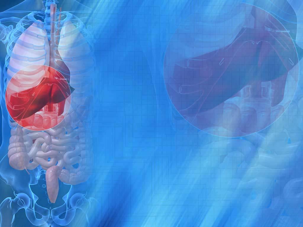

1 Imaging of Cholangiocarcinoma Amir Reza Radmard, MD Assistant Professor Shariati hospital Tehran University of medical sciences
2 Definitions Tumors that arise from the bile duct epithelium. Accounts 10-20% of primary hepatic malignancies. Second most common primary liver tumor. Increasing importance: - Increased incidence? - Poor prognosis? Microscopy (WHO classification, 2010): - Most are adenocarcinoma - Highly infiltrative - Contains areas of fibrosis, necrosis and mucin mdppath.com
3 Traditional classification: Location AJCC/UICC 7 th edition Surgical treatment Intrahepatic (10%) - Mass forming Hepatic resection Perihilar (50%) - Periductal infiltrative - Biliary confluence Resection of bile ducts and ipsilateral liver Distal (40%) - Periductal infiltrative Whipple s operation or PPPD Juntermanns B, Ann Surg Oncol. 2013; 20: Mansour JC, HBP (Oxford). 2015; 17:

 - 3yr DF survival, 60% -")

4 Traditional classification: Morphology Mass forming (20%) -3 yr DF survival, 29% -Mucosal spread Periductal infiltrative (>70%) -3 yr DF survival, 30% -Submucosal spread Intraductal growing (10%) - 3yr DF survival, 60% - Mucosal spread Lim JH, AJR. 2003; 181: Choi SB, Ann Surg Oncol. 2009;16: Cha JM, World J Gastroenterol. 2007;13:
5 Importance of classification of cholangiocarcinoma To understand biological behavior. Different mode of spread: - Longitudinal vs. vertical spread Different surgical technique. Different prognosis. - Intraductal growing >> Mass forming or periductal infiltrating Lim JH, AJR. 2003; 181:
6 Mode of spread: Periductal infiltrating vs. Intraductal growing Vertical extension Chung YE, Radiographics. 2009; 29: Usually defines resectability& curability Critical for prognosis PI: Submucosal, perivascular & perineural IDG: Submucosal
7 Recent classification based on cells of origin Canal of Hering: Lined by cholangiocytes and hepatocytes Association of intrahepatic CC and HCC with respect to the stem cells of origin Aishima et al. J Hepatobiliary Pancreat Sci. 2015; 22:
 Canals of Hering Hepatic progenitor cells The concepts of CCA and combined CC-HCC are evolving. Komuta M, et al. Hepatology.")
8 Recent classification based on cells of origin Large ducts 1-3 rd branch Peribiliary glands (+) Small ducts Interlobar/segmental ducts Peribiliary glands (-) Canals of Hering Hepatic progenitor cells The concepts of CCA and combined CC-HCC are evolving. Komuta M, et al. Hepatology. 2012;55:
9 1. Ultrasound: - > 3cm: Hyperechoic - < 3 cm: Hypo or isoechoic -Peripheral hypoechoic rim 2. CT scan Imaging of mass forming CCA Hypodense 3. MRI T1 low, T2 high 4. Peripheral arterial enhancement 5. Gradual centripetal enhancement Capsular retraction Satellite nodules Peripheral bile duct dilatation Vascular encasement Hepatolithiasis Cellular Fibrosis Chung YE, Radiographics. 2009; 29:
10 Imaging of mass forming CCA Madhusudhan KS, World J Radiol. 2015;28:28-44 Chung YE, Radiographics. 2009; 29:
11 Imaging of mass forming CCA
12 Imaging of mass forming CCA Target like diffusion restriction in 52 75% of mass-forming CCAs Seo N, AJR. 2017;209:W64-W75.
13 Imaging of mass forming CCA Lim J, Radiology. 2016;281: Poor prognosis Better prognosis Kim SA, Radiology. 2011;260: Prognostic role of preoperative DWI and on disease free survival and arterial enhancement
14 Imaging of Intraductal Growing CCA Ultrasound: - Localized or diffuse duct ectasia with or without an echogenic intraductal polypoid lesion. CT scan & MRI: 1- Diffuse and marked ductal dilatation with an intraductal mass that enhances at contrast-enhanced images. 2- Marked intrahepatic duct dilatation with no mass or stricture. 3- An intraductal polypoid mass within localized ductal dilatation. 4- An intraductal castlike lesion within a mildly dilated duct. Chung YE, Radiographics. 2009; 29:
15 Imaging of Intraductal Growing CCA Flat or fungating intraductal mass with proximal dilation. Mild arterial enhancement. No progressive enhancement in portal or delayed phase. Kim SY, ECR 2017.
16 Imaging of Intraductal Growing CCA Delgado Cordón F, Radiologia 2015;57:
17 Imaging of Intraductal Growing CCA Intraductal Papillary Neoplasm of the Bile ducts (IPNB) IPNB is a recognized precursor of invasive carcinoma. Share some histologic and clinical features with IPMNs of the pancreas. Dilated intrahepatic bile ducts filled with papillary or villous biliary neoplasm covering delicated fibrovascualr stalk. IPNB can progress to intraductal growing CCA. Ogawa H, Clin Radiol. 2012;67: Nakanuma Y, Best Pract Res Clin Gastroenterol 2015; 29: Rocha FG, HEPATOLOGY 2012;56:
18 Imaging of Intraductal Growing CCA Intraductal Papillary Neoplasm of the Bile ducts (IPNB) Dilatation of downstream bile duct consequent to mucin production is a characteristic feature. Intraductal linear or curvilinear hypointense striations (thread sign) in MRI highly specific for IPNB. Takanami K, Abdom Imaging 2011; 36: Hong GS, Eur Radiol 2016;26: Seo N, AJR. 2017;209:W64-W75.
19 Imaging of Periductal infiltrating CCA Ultrasound: Small, masslike lesion or diffuse bile duct thickening with or without obliteration of the bile duct CT scan & MRI: Diffuse periductal thickening with increased enhancement, abnormally dilated or irregularly narrowed duct Chung YE, Radiographics. 2009; 29:
20 Imaging of Periductal infiltrating CCA Kim SY, ECR 2017.
21 Imaging of Periductal infiltrating CCA Hilar periductal infiltrating CCA
22 Imaging of Periductal infiltrating CCA Hilar periductal infiltrating CCA
23 Imaging of Periductal infiltrating CCA Distal Periductal infiltrating CCA
24 Imaging of Periductal infiltrating CCA Biliary intraepithelial neoplasia (BilIN). Microscopic change in the biliary epithelium with abnormal epithelial cells, nuclear atypia and micropapillary projections. May be a precursor of periductal infiltrating CCA. Originates from peribiliary glands & chronic inflammation can induce it. Graded as BilIN-1 (low grade), BilIN-2 (intermediate grade), or BilIN-3 (high grade). Conventional imaging studies are limited for detection. Seo N, AJR. 2017;209:W64-W75. Sato Y, Int J Hepatol. 2014;2014:
25 Progression model of intrahepatic CCA Seo N, AJR. 2017;209:W64-W75.
 classification Longitudinal : Bismuth-Corlette +")
26 1. Bismuth-Corlette classification. 2. AJCC Staging of CCA 3. Memorial Sloan-Kettering Cancer Center (MSKCC) classification Longitudinal : Bismuth-Corlette + Vertical or axial: AJCC MSKCC classification
27 Bismuth-Corlette classification. Staging of CCA Useful in determining type of surgery. Not indicative of resectability. Not indicative of survival.
28 Staging of CCA Lee JM, ESGAR 2014.
29 Staging of CCA Left hemihepatectomy, Pancreatoduodectomy and RT. H jejunostomy Lee JM, ESGAR 2014.
30 AJCC (7 th edition) Hilar cholangiocarcinoma Staging of CCA Little information about longitudinal extent of tumor. Less helpful for surgical planning.
31 Staging of CCA MSKCC classification T1 T2 The tumor involves the biliary confluence with unilateral involvement up to secondary biliary radicles. There is no portal vein involvement or liver atrophy. The tumor involves the biliary confluence with unilateral involvement up to secondary biliary radicles. There is ipsilateral portal vein involvement or ipsilateral hepatic lobar atrophy. T3 The tumor involves : 1) Biliary confluence with bilateral involvement up to secondary biliary radicles, 2) Unilateral extension to secondary biliary radicles with contralateral portal vein involvement. 3) Unilateral involvement up to secondary biliary radicles with contralateral hepatic lobar atrophy, or main/bilateral portal vein involvement.
32 Staging of CCA MSKCC classification
33 MSKCC classification Staging of CCA Correlates well with: Resectability Survival Dedicated preop. imaging and radiologist experience No consideration of hepatic artery invasion or lymph node metastasis. William R, Ann Surg Oct; 234:
34 Staging of CCA Proximal tumor extent PV invasion Lobar atrophy HA invasion LN or distant mets. Size of tumor Bismuth-Corrlete AJCC MSKCC New system by De-Oliveira ML, et al Comparison of various preoperative staging systems De-Oliveira ML, Hepatology. 2011; 53:
35 Criteria for unresectability of CCA Lee HY, Radiology. 2006;239:113-21
36 Criteria for unresectable hilar CCA Unresectable cases Bilateral segmental ductal extension. Unilateral atrophy with either contralateral segmental ductal or vascular inflow involvement Unilateral segmental ductal extension with contralateral vascular inflow involvement. Main portal vein or common hepatic artery involvement. Mansour JC, HPB (Oxford) 2015; 17:
37 Criteria for unresectable hilar CCA Unresectable due to tumor infiltration along the hepatoduodenal ligament Lee HY, Radiology. 2006;239:113-21
38 Perioperative staging of CCA A checklist for radiologist report Longitudinal and radial tumor spread Vascular involvement Lymph node involvement Distant metastases Liver volume Biliary, arterial and portal anomalies Other co-existing disease Jhaveri KS, J Magn Reson Imaging. 2015;42:
39 MDCT: Imaging modalities Relationship between important structures and evaluation of distant mets. Accurate for tumor location and vertical invasion Ductal spread is underestimated. Combined with CT angiography, CT venography or CT cholangiography. Hyodo T. Br J Radiol. 2012; 85: Akamatsu N. J Gastroenterol Hepatol. 2010;25:731-7
40 2D-thick slab Imaging modalities-mrcp 2D-Multi slice 3D MRCP T2W:HASTE/SSFSE TE> 600 msec Coronal oblique/ axial 4-6 cm thick Gross morphology T2W:HASTE/SSFSE TE< 180 msec Coronal/ axial 2-5 mm thick Periductal anatomy 3D T2W TE>600 msec Coronal 1-2 mm thick No gap Post processing
41 Imaging modalities Combination of MRCP & Contrast enhanced MRI for differentiating malignant from benign biliary strictures Kim JY, J Magn Reson Imaging. 2007;26: Yu XR, Clin Radiol. 2014;69: Li N, Clin Radiol. 2012;67:
42 MDCT < % Imaging modalities Accuracy in longitudinal spread of tumor CT + Direct cholangiography ~ 80% MRI ~ 71-80% Causes of inaccuracy: 1. Underestimation: Superficial spread of papillary tumor Sub epithelial spread of infiltrative type Anatomic variation 2. Overestimation: Cholangitis, drainage catheter, intra ductal necrotic material Seo N, Korean J Radiol. 2016; 17: Ruys AT, BR J Radiol; 2012:85;
43 Imaging modalities Accuracy of CT: Overestimation of tumor spread Overestimated by CT: Bismuth 4 Direct cholangiography: Bismuth 3a Lee JM, ESGAR 2014.
44 Imaging modalities Direct cholangiography can provide higher spatial resolution than CT or MRI Lee JM, ESGAR 2014.
45 Accurate staging and determination of resectability Longitudinal spread (ductal extension) Multimodality approach Multidisciplinary team Gastroenterologist Radiologist Surgeon Oncologist Pathologist Vertical spread High resolution imaging Extrahepatic disease CT, PET/CT or PET/MR
46 Take home message Incidence of CCA is increasing across the world. Concepts and classifications of CCA have been evolving. Longitudinal and vertical spread of CCA must be assessed by evaluation of key anatomic landmarks. MDCT with 3D reconstruction and MRCP combined with dynamic MRI play major role in staging but there is a tendency toward underestimation. Multimodality approach seems to be reasonable.

47 Thank you for your kind attention
Contemporary Imaging of Biliary Malignancy and Preoperative Evaluation
 Contemporary Imaging of Biliary Malignancy and Preoperative Evaluation Linda Pantongrag-Brown, MD Advanced Diagnostic Imaging, Ramathibodi Hospital, Bangkok, Thailand Malignancy of biliary tract Cholangiocarcinoma
Hepatocellular carcinoma Cholangiocarcinoma. Jewels of hepatobiliary cancer imaging : what to look for? Imaging characteristics of HCC.
 Outline : Imaging Jewels Jewels of hepatobiliary cancer imaging : what to look for? Hepatocellular carcinoma Cholangiocarcinoma Surachate Siripongsakun, M.D. Chulabhorn Cancer Center Imaging characteristics
Intraductal papillary mucinous neoplasm of the bile ducts: a rare form of premalignant lesion of invasive cholangiocarcinoma
 Intraductal papillary mucinous neoplasm of the bile ducts: a rare form of premalignant lesion of invasive cholangiocarcinoma Authors: R. Revert Espí, Y. Fernandez Nuñez, I. Carbonell, D. P. Gómez valencia,
Hilar cholangiocarcinoma. Frank Wessels, Maarten van Leeuwen, UMCU utrecht
 Hilar cholangiocarcinoma Frank Wessels, Maarten van Leeuwen, UMCU utrecht Content Anatomy Biliary strictures (Hilar) Cholangiocarcinoom Staging Biliary tract 1 st order Ductus hepatica dextra Ductus hepaticus
State of the Art Imaging for Hepatic Malignancy: My Assignment
 State of the Art Imaging for Hepatic Malignancy: My Assignment CT vs MR vs MRCP Which one to choose for HCC vs Cholangiocarcinoma What special protocols to use for liver tumors Role of PET and Duplex US
Cholangiocarcinoma: appearances and mimics
 Cholangiocarcinoma: appearances and mimics Poster No.: C-1572 Congress: ECR 2011 Type: Educational Exhibit Authors: C. Cardenas Valencia, J. Fernandez Jara, J. Cubero Carralero, B. Corral Ramos, P. Perez
Cholangiocarcinoma. Judy Wyatt Dundee November 2010
 Cholangiocarcinoma Judy Wyatt Dundee November 2010 Making sense of cholangiocarcinoma Difficulties with diagnostic criteria How many entities within cholangiocarcinoma? Rapidly evolving Intrahepatic cholangiocarcinoma
RESEARCH ARTICLE. Morphological Classification of Intraductal Papillary Neoplasm of the Bile Duct with Survival Correlation
 DOI:10.22034/APJCP.2017.18.1.207 RESEARCH ARTICLE Morphological Classification of Intraductal Papillary Neoplasm of the Bile Duct with Survival Correlation Vor Luvira 1 *, Kulyada Somsap 2, Ake Pugkhem
Navigating the Biliary Tract with CT & MR: An Imaging Approach to Bile Duct Obstruction
 Navigating the Biliary Tract with CT & MR: An Imaging Approach to Bile Duct Obstruction Ann S. Fulcher, MD Medical College of Virginia Virginia Commonwealth University Richmond, Virginia Objectives To
Management of Cholangiocarcinoma. Roseanna Lee, MD PGY-5 Kings County Hospital
 Management of Cholangiocarcinoma Roseanna Lee, MD PGY-5 Kings County Hospital Case Presentation 37 year old male from Yemen presented with 2 week history of epigastric pain, anorexia, jaundice and puritis.
Spectrum of Cholangiocarcinoma
 Scholars Journal of Applied Medical Sciences (SJAMS) Sch. J. App. Med. Sci., 2013; 1(6):695-699 Scholars Academic and Scientific Publisher (An International Publisher for Academic and Scientific Resources)
Original article: new surgical approaches to the Klatskin tumour
 Alimentary Pharmacology & Therapeutics Original article: new surgical approaches to the Klatskin tumour T. M. VAN GULIK*, S. DINANT*, O. R. C. BUSCH*, E. A. J. RAUWS, H. OBERTOP* & D. J. GOUMA Departments
Multiple Primary Quiz
 Multiple Primary Quiz Case 1 A 72 year old man was found to have a 12 mm solid lesion in the pancreatic tail by computed tomography carried out during a routine follow up study of this patient with adult
Biliary tract tumors
 Short Course 2010 Annual Fall Meeting of the Korean Society for Pathologists Biliary tract tumors Joon Hyuk Choi, M.D., Ph.D. Professor, Department of Pathology, Yeungnam Univ. College of Medicine, Daegu,
Proximal Bile Duct Cancer: Contemporary Management. William R. Jarnagin, MD, FACS
 Proximal Bile Duct Cancer: Contemporary Management William R. Jarnagin, MD, FACS Biliary Tract Adenocarcinoma Spectrum of disease Intrahepatic (IHC) Hilar EH Gallbladder GB CBD Distal D PD Biliary Tract
Imaging in gastric cancer
 Imaging in gastric cancer Gastric cancer remains a deadly disease because of late diagnosis. Adenocarcinoma represents 90% of malignant tumors. Diagnosis is based on endoscopic examination with biopsies.
MRI Abdomen Protocol Pancreas/MRCP with Contrast
 MRI Abdomen Protocol Pancreas/MRCP with Contrast Reviewed By: Brett Mollard, MD; Anna Ellermeier, MD Last Reviewed: July 2018 Contact: (866) 761-4200 Standard uses: 1. Characterization of cystic and solid
Biliary cancers: imaging diagnosis. Study of 30 cases
 Biliary cancers: imaging diagnosis. Study of 30 cases N Hammoune, S Semlali, M Eddarai, T. Amil, M Zentar, S. El Kandri,, M Benameur,, S Chaouir. Radiology Department. Mohamed V Military Hospital. Rabat-
Personal Profile. Name: 劉 XX Gender: Female Age: 53-y/o Past history. Hepatitis B carrier
 Personal Profile Name: 劉 XX Gender: Female Age: 53-y/o Past history Hepatitis B carrier Chief complaint Fever on and off for 2 days Present illness 94.10.14 Sudden onset of epigastric pain 94.10.15 Fever
CHOLANGIOCARCINOMA (CCA)
") CHOLANGIOCARCINOMA (CCA) Deepak Hariharan MD (Research), FRCS, Locum Consultant HPB Surgeon AIM Outline essential facts & principles Present 4 cases Discuss Challenges /Controversies INTRODUCTION Most
CHOLANGIOCARCINOMA (CCA) Deepak Hariharan MD (Research), FRCS, Locum Consultant HPB Surgeon AIM Outline essential facts & principles Present 4 cases Discuss Challenges /Controversies INTRODUCTION Most
Effectiveness of additional resection of the invasive cancer-positive proximal bile duct margin in cases of hilar cholangiocarcinoma
 Original Article Effectiveness of additional resection of the invasive cancer-positive proximal bile duct margin in cases of hilar cholangiocarcinoma Wen-Jie Ma 1#, Zhen-Ru Wu 2#, Anuj Shrestha 1,3, Qin
Pancreas Case Scenario #1
 Pancreas Case Scenario #1 An 85 year old white female presented to her primary care physician with increasing abdominal pain. On 8/19 she had a CT scan of the abdomen and pelvis. This showed a 4.6 cm mass
Malignant Focal Liver Lesions
 Malignant Focal Liver Lesions Other Than HCC Pablo R. Ros, MD, MPH, PhD Departments of Radiology and Pathology University Hospitals Cleveland Medical Center Case Western Reserve University Pablo.Ros@UHhospitals.org
liver and upper GI cancers, Yorks and Humber Pub med Cholangiocarcinoma + pathology
 Update on Cholangiocarcinoma Update on Cholangiocarcinoma 1200 pa in England. Around 20% operable, around 10% 5 yr survival Liver resections in Leeds, 12 years 2005-2017 Judy Wyatt 7% cholangiocarcinoma
Post-operative complications following hepatobiliary surgery: imaging findings and current radiological treatment options
 Post-operative complications following hepatobiliary surgery: imaging findings and current radiological treatment options Poster No.: C-1501 Congress: ECR 2015 Type: Educational Exhibit Authors: A. Hadjivassiliou,
An Intraductal Papillary Neoplasm of the Bile Duct at the Duodenal Papilla
 Published online: July 2, 2014 1662 6575/14/0072 0417$39.50/0 This is an Open Access article licensed under the terms of the Creative Commons Attribution- NonCommercial 3.0 Unported license (CC BY-NC)
Newcastle HPB MDM updated radiology imaging protocol recommendations. Author Dr John Scott. Consultant Radiologist Freeman Hospital
 Newcastle HPB MDM updated radiology imaging protocol recommendations Author Dr John Scott. Consultant Radiologist Freeman Hospital This document is intended as a guide to aid radiologists and clinicians
MR imaging of primary sclerosing cholangitis (PSC) using the hepatobiliary specific contrast agent Gd-EOB-DTPA
 using the hepatobiliary specific contrast agent Gd-EOB-DTPA") MR imaging of primary sclerosing cholangitis (PSC) using the hepatobiliary specific contrast agent Gd-EOB-DTPA Poster No.: C-0019 Congress: ECR 2010 Type: Educational Exhibit Topic: Abdominal Viscera (Solid
MR imaging of primary sclerosing cholangitis (PSC) using the hepatobiliary specific contrast agent Gd-EOB-DTPA Poster No.: C-0019 Congress: ECR 2010 Type: Educational Exhibit Topic: Abdominal Viscera (Solid
Objectives. Intraoperative Consultation of the Whipple Resection Specimen. Pancreas Anatomy. Pancreatic ductal carcinoma 11/10/2014
 Intraoperative Consultation of the Whipple Resection Specimen Pathology Update Faculty of Medicine, University of Toronto November 15, 2014 John W. Wong, MD, FRCPC Department of Anatomical Pathology Sunnybrook
Protocol for the Examination of Specimens From Patients With Carcinoma of the Perihilar Bile Ducts
 Protocol for the Examination of Specimens From Patients With Carcinoma of the Perihilar Bile Ducts Version: Protocol Posting Date: June 2017 Includes ptnm requirements from the 8 th Edition, AJCC Staging
Intraductal papillary neoplasms in the bile ducts
 Intraductal papillary neoplasms in the bile ducts Seok Hwa Youn Myunghee Yoon Dong Hoon Shin Kosin University Gospel Hospital Department of general surgery Hepato-biliary-pancreatic division Introduction
Case Report Heterotopic Pancreas within the Proximal Hepatic Duct, Containing Intraductal Papillary Mucinous Neoplasm
 Case Reports in Surgery Volume 2015, Article ID 816960, 4 pages http://dx.doi.org/10.1155/2015/816960 Case Report Heterotopic Pancreas within the Proximal Hepatic Duct, Containing Intraductal Papillary
Overview of PSC Making the Diagnosis
 Overview of PSC Making the Diagnosis Tamar Taddei, MD Assistant Professor of Medicine Yale University School of Medicine Overview Definition Epidemiology Diagnosis Modes of presentation Associated diseases
Comparison of multidetector-row computed tomography findings of IgG4-related sclerosing cholangitis and cholangiocarcinoma
 Comparison of multidetector-row computed tomography findings of IgG4-related sclerosing cholangitis and cholangiocarcinoma Poster No.: C-0245 Congress: ECR 2014 Type: Scientific Exhibit Authors: M. Yata,
LIVER IMAGING TIPS IN VARIOUS MODALITIES. M.Vlychou, MD, PhD Assoc. Professor of Radiology University of Thessaly
 LIVER IMAGING TIPS IN VARIOUS MODALITIES M.Vlychou, MD, PhD Assoc. Professor of Radiology University of Thessaly Hepatocellular carcinoma is a common malignancy for which prevention, screening, diagnosis,
Pancreatic Adenocarcinoma: Everything You Need to Know From Cross-Sectional Imaging to Treatment
 Pancreatic Adenocarcinoma: Everything You Need to Know From Cross-Sectional Imaging to Treatment Andrew W. Bowman, MD PhD Assistant Professor of Radiology Mayo Clinic Florida SCBT-MR Annual Meeting Nashville,
X-ray Corner. Imaging of The Pancreas. Pantongrag-Brown L
 X-ray Corner 125 Imaging of The Pancreas Modern imaging modalities commonly used in pancreas include ultrasound (US), CT, and MRI. Pancreas is a retroperitoneal organ which makes it difficult to visualize
TREATMENT FOR HCC AND CHOLANGIOCARCINOMA. Shawn Pelletier, MD
 TREATMENT FOR HCC AND CHOLANGIOCARCINOMA Shawn Pelletier, MD Treatment for HCC Treatment strategies Curative first line therapy Thermal ablation vs Resection vs Transplant Other first line therapies TACE
IMAGING OF ACUTE AND CHRONIC PANCREATITIS, INCLUDING EXOCRINE FUNCTION
 IMAGING OF ACUTE AND CHRONIC PANCREATITIS, INCLUDING EXOCRINE FUNCTION Andrew T. Trout, MD @AndrewTroutMD Disclosures Grant support National Pancreas Foundation In-kind support - ChiRhoClin modified from:
Dr Claire Smith, Consultant Radiologist St James University Hospital Leeds
 Dr Claire Smith, Consultant Radiologist St James University Hospital Leeds Imaging in jaundice and 2ww pathway Image protocol Staging Limitations Pancreatic cancer 1.2.4 Refer people using a suspected
Common and unusual CT and MRI manifestations of pancreatic adenocarcinoma: a pictorial review
 Review Article Common and unusual CT and MRI manifestations of pancreatic adenocarcinoma: a pictorial review Min-Jie Yang, Su Li, Yong-Guang Liu, Na Jiao, Jing-Shan Gong Department of Radiology, Shenzhen
6 th August 2018 Day 1 - Gallbladder & Bile duct Topic
 Venue: Sterling Hospital Auditorium, Sterling Hospitals, Gurukul Road Ahmedabad, Gujarat 6 th August 2018 Day 1 - Gallbladder & Bile duct Registration(8:00am-8:15am) Inauguration(8:15am-8:30am) Welcome
Anatomical and Functional MRI of the Pancreas
 Anatomical and Functional MRI of the Pancreas MA Bali, MD, T Metens, PhD Erasme Hospital Free University of Brussels Belgium mbali@ulb.ac.be Introduction The use of MRI to investigate the pancreas has
ROLE OF RADIOLOGY IN INVESTIGATION OF JAUNDICE
 ROLE OF RADIOLOGY IN INVESTIGATION OF JAUNDICE Dr. Sohan kumar sah *, Dr. Liu Sibin, Dr. sumendra raj pandey, Dr. Prakashmaan shah, Dr. Gaurishankar pandit, Dr. Suraj kurmi and Dr. Sanjay kumar jaiswal
Management A Guideline Based Approach to the Incidental Pancreatic Cysts. Common Cystic Pancreatic Neoplasms.
 Management 2016 A Guideline Based Approach to the Incidental Pancreatic Cysts ISMRM 2016 Masoom Haider, MD, FRCP(C) Professor of Radiology, University of Toronto Clinician Scientist, Ontario Institute
저작권법에따른이용자의권리는위의내용에의하여영향을받지않습니다.
 저작자표시 - 비영리 - 변경금지. 대한민국 이용자는아래의조건을따르는경우에한하여자유롭게 이저작물을복제, 배포, 전송, 전시, 공연및방송할수있습니다. 다음과같은조건을따라야합니다 : 저작자표시. 귀하는원저작자를표시하여야합니다. 비영리. 귀하는이저작물을영리목적으로이용할수없습니다. 변경금지. 귀하는이저작물을개작, 변형또는가공할수없습니다. 귀하는, 이저작물의재이용이나배포의경우,
Predictive factors for invasive intraductal papillary mucinous neoplasm of the pancreas
 Korean J Hepatobiliary Pancreat Surg 2011;15:27-22 Original Article Predictive factors for invasive intraductal papillary mucinous neoplasm of the pancreas Dae Young Jun 1, Hyung Jun Kwon 2, Sang Geol
MR cholangiopancreatography; Predicting imaging findings for differentiation of malignant bile ductal obstruction versus benign lesion
 Acta Med Kindai Univ Vol.43, No.1 1-8, 2018 1 MR cholangiopancreatography; Predicting imaging findings for differentiation of malignant bile ductal obstruction versus benign lesion Shojiro Hidaka 1,2,
Pancreatobiliary Frozen Section Nightmares
 Pancreatobiliary Frozen Section Nightmares Aatur D. Singhi, MD PhD Assistant Professor University of Pittsburgh Medical Center Department of Pathology singhiad@upmc.edu Objectives Briefly give an overview
CT 101 :Pancreas and Spleen
 CT 101 :Pancreas and Spleen Shikha Khullar,, MD, MPH Division of Radiology University of South Alabama The Pancreas Normal Pancreas 3 Phase Pancreatic CT Non contrast Arterial phase : 30-35 35 second
Autoimmune Pancreatitis: A Great Imitator
 Massachusetts General Hospital Harvard Medical School Autoimmune Pancreatitis: A Great Imitator Dushyant V Sahani MD dsahani@partners.org Autoimmune Pancreatitis: Learning Objectives Clinical manifestations
5/17/2013. Pancreatic Cancer. Postgraduate Course in General Surgery CASE 1: CASE 1: Overview. Case presentation. Differential diagnosis
 Overview Case presentation Postgraduate Course in General Surgery Differential diagnosis Diagnosis and therapy Eric K. Nakakura Koloa, HI March 26, 2013 Outcomes CASE 1: CASE 1: A 78-year-old man developed
Case Report Cystic and Papillary Neoplasm at the Hepatic Hilum Possibly Originating in the Peribiliary Glands
 Case Reports in Pathology Volume 2016, Article ID 9130754, 7 pages http://dx.doi.org/10.1155/2016/9130754 Case Report Cystic and Papillary Neoplasm at the Hepatic Hilum Possibly Originating in the Peribiliary
HEPATOCYTE SPECIFIC CONTRAST MEDIA: WHERE DO WE STAND?
 HEPATOCYTE SPECIFIC CONTRAST MEDIA: WHERE DO WE STAND? Andrew T. Trout, MD @AndrewTroutMD Disclosures No relevant disclosures Outline Review of hepatocyte specific contrast media Review of hepatocellular
저작자표시 2.0 대한민국 이용자는아래의조건을따르는경우에한하여자유롭게 이저작물을복제, 배포, 전송, 전시, 공연및방송할수있습니다. 이차적저작물을작성할수있습니다. 이저작물을영리목적으로이용할수있습니다. 저작자표시. 귀하는원저작자를표시하여야합니다.
 저작자표시 2.0 대한민국 이용자는아래의조건을따르는경우에한하여자유롭게 이저작물을복제, 배포, 전송, 전시, 공연및방송할수있습니다. 이차적저작물을작성할수있습니다. 이저작물을영리목적으로이용할수있습니다. 다음과같은조건을따라야합니다 : 저작자표시. 귀하는원저작자를표시하여야합니다. 귀하는, 이저작물의재이용이나배포의경우, 이저작물에적용된이용허락조건을명확하게나타내어야합니다.
Cholangiocarcinoma (Bile Duct Cancer)
") Cholangiocarcinoma (Bile Duct Cancer) The Bile Duct System (Biliary Tract) A network of bile ducts (tubes) connects the liver and the gallbladder to the small intestine. This network begins in the liver
Cholangiocarcinoma (Bile Duct Cancer) The Bile Duct System (Biliary Tract) A network of bile ducts (tubes) connects the liver and the gallbladder to the small intestine. This network begins in the liver
Select problems in cystic pancreatic lesions
 Disclosure Select problems in cystic pancreatic lesions Five Prime Therapeutics shareholder Adicet Bio shareholder Bristol-Meyer Squibb advisory board grace.kim@ucsf.edu Pancreatic cystic lesions Intraductal
Liver imaging takes a step forward with Ingenia
 Publication for the Philips MRI Community ISSUE 49 2013 / 2 Liver imaging takes a step forward with Ingenia Lyon South Hospital strives to move from several studies first CT, then MR or PET to using just
Greater Manchester and Cheshire HPB Unit Guidelines for the Assessment & Management of Hepatobiliary and Pancreatic Disease Chapter 14
 Greater Manchester and Cheshire HPB Unit Guidelines for the Assessment & Management of Hepatobiliary and Pancreatic Disease Chapter 14 Contents 14. Neuroendocrine Tumours 161 14.1. Diagnostic algorithm
3/28/2012. Periampullary Tumors. Postgraduate Course in General Surgery CASE 1: CASE 1: Overview. Eric K. Nakakura Ko Olina, HI
 Overview Postgraduate Course in General Surgery Case presentation Differential diagnosis Diagnosis and therapy Outcomes Principles of palliative care Eric K. Nakakura Ko Olina, HI March 27, 2012 CASE 1:
Review. Computed tomography in the diagnosis and staging of cholangiocarcinoma and pancreatic carcinoma
 Annals of Oncology 10 Suppl. 4: S12-S17, 1999. 1999 Kluwer Academic Publishers. Printed in the Netherlands. Review Computed tomography in the diagnosis and staging of cholangiocarcinoma and pancreatic
Endoscopic Ultrasonography Assessment for Ampullary and Bile Duct Malignancy
 Diagnostic and Therapeutic Endoscopy, Vol. 3, pp. 35-40 Reprints available directly from the publisher Photocopying permitted by license only (C) 1996 OPA (Overseas Publishers Association) Amsterdam B.V.
Disclosure. Acknowledgement. What is the Best Workup for Rectal Cancer Staging: US/MRI/PET? Rectal cancer imaging. None
 What is the Best Workup for Rectal Cancer Staging: US/MRI/PET? Zhen Jane Wang, MD Assistant Professor in Residence UC SF Department of Radiology Disclosure None Acknowledgement Hueylan Chern, MD, Department
Imaging of liver and pancreas
 Imaging of liver and pancreas.. Disease of the liver Focal liver disease Diffusion liver disease Focal liver disease Benign Cyst Abscess Hemangioma FNH Hepatic adenoma HCC Malignant Fibrolamellar carcinoma
بسم هللا الرحمن الرحيم. Prof soha Talaat
 بسم هللا الرحمن الرحيم Ovarian tumors The leading indication for gynecologic surgery. Preoperative characterization of complex solid and cystic adnexal masses is crucial for informing patients about possible
Role of imaging in RCC. Ultrasonography. Solid lesion. Cystic RCC. Solid RCC 31/08/60. From Diagnosis to Treatment: the Radiologist Perspective
 Role of imaging in RCC From Diagnosis to Treatment: the Radiologist Perspective Diagnosis Staging Follow up Imaging modalities Limitations and pitfalls Duangkamon Prapruttam, MD Department of Therapeutic
Imaging of Neuroendocrine Metastases
 Imaging of Neuroendocrine Metastases Aoife Kilcoyne, Shaunagh McDermott, Colin McCarthy,Manuel Patino, Dushyant Sahani, Michael Blake Abdominal Imaging Division Massachusetts General Hospital Disclosure
Evidence based imaging of the pancreas
 Evidence based imaging of the pancreas D.Vanbeckevoort, D.Bielen, K.Op de beeck, R.Vanslembrouck Department of Radiology Chairman Prof. Dr. R.Oyen Non-invasive imaging tests available for the diagnosis
Management of Patients with Suspected Cholangiocarcinoma CLINICAL GUIDELINES
 London Cancer Hepatic Pancreatic and Biliary (HPB) Faculty Management of Patients with Suspected Cholangiocarcinoma CLINICAL GUIDELINES JULY 2014 This operational policy is agreed and accepted by: Designated
X-Ray Corner. Imaging Approach to Cystic Liver Lesions. Pantongrag-Brown L. Solitary cystic liver lesions. Hepatic simple cyst (Figure 1)
") THAI J 136 Imaging Approach to Cystic Liver Lesions GASTROENTEROL 2013 X-Ray Corner Imaging Approach to Cystic Liver Lesions Pantongrag-Brown L Cystic liver lesions are common findings in daily practice
THAI J 136 Imaging Approach to Cystic Liver Lesions GASTROENTEROL 2013 X-Ray Corner Imaging Approach to Cystic Liver Lesions Pantongrag-Brown L Cystic liver lesions are common findings in daily practice
Surgical Management of Pancreatic Cancer
 I Congresso de Oncologia D Or July 5-6, 2013 Surgical Management of Pancreatic Cancer Michael A. Choti, MD, MBA, FACS Department of Surgery Johns Hopkins University School of Medicine, Baltimore, MD Estimated
Anatomy of the biliary tract
 Harvard-MIT Division of Health Sciences and Technology HST.121: Gastroenterology, Fall 2005 Instructors: Dr. Jonathan Glickman Anatomy of the biliary tract Figure removed due to copyright reasons. Biliary
Financial Disclosure
 Benign Liver Masses Adil Abdalla, MBBS Creighton University-CHI Health August 25, 2018 Financial Disclosure Nothing to disclose Financial Disclosure 1 Objectives To assess patients with benign liver tumors
Double Intrahepatic and Extrahepatic Cholangiocarcinomas Arising from Biliary Papillomatosis: A Case Report
 Korean Journal of HBP Surgery 증 례 Vol. 14, No. 1, March 2010 Double Intrahepatic and Extrahepatic Cholangiocarcinomas Arising from Biliary Papillomatosis: A Case Report Biliary papillomatosis is a rare
R.Sotoudehmanesh, MD Professor of Gastroenterology Digestive Disease Research Institute Tehran University of Medical Sciences Pancreatobiliary
 R.Sotoudehmanesh, MD Professor of Gastroenterology Digestive Disease Research Institute Tehran University of Medical Sciences Pancreatobiliary /Advanced Endoscopy group Most common biliary malignancy and
Diffusion Weighted MR in Imaging Pancreatico-biliary & Gall Bladder Pathologies: friend or foe?
 Diffusion Weighted MR in Imaging Pancreatico-biliary & Gall Bladder Pathologies: friend or foe? Poster No.: C-0595 Congress: ECR 2014 Type: Educational Exhibit Authors: K. LIM; SINGAPORE/SG Keywords: Pancreas,
Radiologic and pathologic correlation of non-mass like breast lesions on US and MRI: Benign, high risk, versus malignant
 Radiologic and pathologic correlation of non-mass like breast lesions on US and MRI: Benign, high risk, versus malignant Poster No.: C-1161 Congress: ECR 2013 Type: Educational Exhibit Authors: J. Kwak,
Surgical management of HCC. Evangelos Prassas Hepatobiliary and Pancreatic Surgery / Liver Transplantation Kings College Hospital / London
 Surgical management of HCC Evangelos Prassas Hepatobiliary and Pancreatic Surgery / Liver Transplantation Kings College Hospital / London Global distribution of HCC and staging systems WEST 1. Italy (Milan,
Hepatobiliary and Pancreatic Malignancies
 Hepatobiliary and Pancreatic Malignancies Gareth Eeson MD MSc FRCSC Surgical Oncologist and General Surgeon Kelowna General Hospital Interior Health Consultant, Surgical Oncology BC Cancer Agency Centre
Management of Colorectal Liver Metastases
 Management of Colorectal Liver Metastases MM Bernon, JEJ Krige HPB Surgical Unit, Groote Schuur Hospital Department of Surgery, University of Cape Town 50% of patients with colorectal cancer develop liver
Pictorial review of Benign Biliary tract abnormality on MRCP/MRI Liver with Endoscopic (including splyglass) and Endoscopic Ultrasound correlation
 and Endoscopic Ultrasound correlation") Pictorial review of Benign Biliary tract abnormality on MRCP/MRI Liver with Endoscopic (including splyglass) and Endoscopic Ultrasound correlation Poster No.: C-2617 Congress: ECR 2015 Type: Educational
Pictorial review of Benign Biliary tract abnormality on MRCP/MRI Liver with Endoscopic (including splyglass) and Endoscopic Ultrasound correlation Poster No.: C-2617 Congress: ECR 2015 Type: Educational
Imaging of common diseases of hepatobiliary and GI system
 Imaging of common diseases of hepatobiliary and GI system Natthaporn Tanpowpong, M.D. Diagnostic radiology Faculty of Medicine, Chulalongkorn University Normal plain radiograph A = Common bile duct
Is Hepatic Resection Needed in the Patients with Peritoneal Side T2 Gallbladder Cancer?
 Is Hepatic Resection Needed in the Patients with Peritoneal Side T2 Gallbladder Cancer? Lee H, Park JY, Youn S, Kwon W, Heo JS, Choi SH, Choi DW Department of Surgery, Samsung Medical Center Sungkyunkwan
Occult mucin-producing cholangiocarcinoma in situ: a rare clinical case with difficult tumour staging
 Kiriyama et al. Surgical Case Reports (2017) 3:6 DOI 10.1186/s40792-016-0283-x CASE REPORT Open Access Occult mucin-producing cholangiocarcinoma in situ: a rare clinical case with difficult tumour staging
Radiological staging of lung cancer. Shukri Loutfi,MD,FRCR Consultant Thoracic Radiologist KAMC-Riyadh
 Radiological staging of lung cancer Shukri Loutfi,MD,FRCR Consultant Thoracic Radiologist KAMC-Riyadh Bronchogenic Carcinoma Accounts for 14% of new cancer diagnoses in 2012. Estimated to kill ~150,000
Hilar cholangiocarcinoma: diagnosis and staging
 HPB, 2005; 7: 244 251 REVIEW ARTICLES Hilar cholangiocarcinoma: diagnosis and staging WILLIAM JARNAGIN 1 & CORINNE WINSTON 2 Departments of 1 Surgery and 2 Radiology, Memorial Sloan-Kettering Cancer Center,
Upper GI Malignancies Imaging Guidelines for the Management of Gastric, Oesophageal & Pancreatic Cancers 2012
 Upper GI Malignancies Imaging Guidelines for the Management of Gastric, Oesophageal & Pancreatic Cancers 2012 Version Control This is a controlled document please destroy all previous versions on receipt
Overview. Disclosure. PRE INVASIVE NEOPLASIA OF BILIARY TREE New Perspectives on Old Themes. N. Volkan Adsay, MD
 PRE INVASIVE NEOPLASIA OF BILIARY TREE New Perspectives on Old Themes N. Volkan Adsay, MD Professor and Vice-Chair Director of Anatomic Pathology Emory University and Emory Winship Cancer Institute Atlanta,
Biliary Papillomatosis: case report
 Chin J Radiol 2003; 28: 407-412 407 Biliary Papillomatosis: case report CHUN-LIN HUANG WEN-PIN CHEN YU-BUN NG JOSEPH HANG LEUNG Department of Medical Imaging, Chiayi Christian Hospital Biliary papillomatosis
Biliary tree dilation - and now what?
 Biliary tree dilation - and now what? Poster No.: C-1767 Congress: ECR 2012 Type: Educational Exhibit Authors: I. Ferreira, A. B. Ramos, S. Magalhães, M. Certo; Porto/PT Keywords: Pathology, Diagnostic
Trans-abdominal ultrasound features of the newly named intraductal papillary neoplasm of the bile duct
 Original Article on Translational Imaging in Cancer Patient Care Trans-abdominal ultrasound features of the newly named intraductal papillary neoplasm of the bile duct Xian-Shui Fu 1 *, Meng-Na He 2 *,
Cholangiocellular carcinoma. Dr. med. Henrik Csaba Horváth PhD
 Cholangiocellular carcinoma Dr. med. Henrik Csaba Horváth PhD Acalculous biliary diseases April 12, 2017 2 Cholangiocarcinoma A slow growing malignancy of the biliary tract which tend - to infiltrate locally
Esophageal submucosal mass icd 10
 Esophageal submucosal mass icd 10 Search 6-6-2011 ICD-10; Risk Adjustment / HCC; Evaluation & Management (E/M). I'm hoping someone can help me with this DX, "soft tissue mass in. Upper gastrointestinal
Intrahepatic cholangiocarcinoma: the AJCC/UICC 8 th edition updates
 Review Article Page 1 of 5 Intrahepatic cholangiocarcinoma: the AJCC/UICC 8 th edition updates Andrew J. Lee, Yun Shin Chun Department of Surgical Oncology, The University of Texas MD Anderson Cancer Center,
Intrahepatic Sarcomatoid Cholangiocarcinoma with Portal Vein Thrombosis: A Case Report 1
 Intrahepatic Sarcomatoid Cholangiocarcinoma with Portal Vein Thrombosis: A Case Report 1 Jae-Hoon Lim, M.D., Jin Woong Kim, M.D., Suk Hee Heo, M.D., Yong Yeon Jeong, M.D., Heoung Keun Kang, M.D. A 53-year-old
Pancreas Quizzes c. Both A and B a. Directly into the blood stream (not using ducts)
") Pancreas Quizzes Quiz 1 1. The pancreas produces hormones. Which type of hormone producing organ is the pancreas? a. Endocrine b. Exocrine c. Both A and B d. Neither A or B 2. Endocrine indicates hormones
Pancreas Quizzes Quiz 1 1. The pancreas produces hormones. Which type of hormone producing organ is the pancreas? a. Endocrine b. Exocrine c. Both A and B d. Neither A or B 2. Endocrine indicates hormones
Embolotherapy for Cholangiocarcinoma: 2016 Update
 Embolotherapy for Cholangiocarcinoma: 2016 Update Igor Lobko,MD Chief, Division Vascular and Interventional Radiology Long Island Jewish Medical Center GEST 2016 Igor Lobko, M.D. No relevant financial
Classification of choledochal cyst with MR cholangiopancreatography in children and infants: special reference to type Ic and type IVa cyst
 Classification of choledochal cyst with MR cholangiopancreatography in children and infants: special reference to type Ic and type IVa cyst Poster No.: C-1333 Congress: ECR 2011 Type: Educational Exhibit
8. The polyp in the illustration can be described as (circle all that apply) a. Exophytic b. Pedunculated c. Sessile d. Frank
 a. Exophytic b. Pedunculated c. Sessile d. Frank") Quiz 1 Overview 1. Beginning with the cecum, which is the correct sequence of colon subsites? a. Cecum, ascending, splenic flexure, transverse, hepatic flexure, descending, sigmoid. b. Cecum, ascending,
Quiz 1 Overview 1. Beginning with the cecum, which is the correct sequence of colon subsites? a. Cecum, ascending, splenic flexure, transverse, hepatic flexure, descending, sigmoid. b. Cecum, ascending,