Whole-Body Dynamic Contrast- Enhanced (DCE) MR Imaging in patients with myeloma
|
|
|
- Lora Harrington
- 5 years ago
- Views:
Transcription

1 Whole-Body Dynamic Contrast- Enhanced (DCE) MR Imaging in patients with myeloma Alain Rahmouni, Department of Medical Imaging, Mondor Academic Hospital : Centre Hospitalo-Universitaire Henri Mondor-Assistance Publique-Hôpitaux de Paris et Université Paris Est-Créteil,

2 Imaging MM? Diffuse and multifocal disease W-B Increased angiogenesis Impact on prognosis and response to treatment Rationale : Extract functional parameters on a W-B scale Prognostic impact?
3 Plan Whole-Body MR imaging Biological aspects DCE MR imaging DCE WB MR imaging PET imaging


4 MRI in Myeloma Standard MRI direct, high contrast visualization of bone marrow: best imaging technique (T1&FatsatT2) Durie/Salmon PLUS 2006 T1WI T2WI 61y female patient? Durie/Salmon 1975 Rahmouni et al. AJR 1993, Baur-Melnyk A et al. EJR 2005
5 Plan Whole-Body MR imaging Biological aspects DCE MR imaging DCE WB MR imaging PET imaging
6 Myeloma, biological aspects 1. Myeloma cells produce angiogenic cytokines (VEGF ) inducing bone marrow neovascularization Vacca A et al. Blood 1999; Parameters of angiogenesis on bone marrow biopsy : Microvessel density (MVD) and total vascular area Higher in MM patients than controls (p < 0.001) Higher in nonresponders than complete responders (p <0.001) Bhatti SS et al. Am J Hematol 2006;81: MVD density: independent prognostic factor

:")
7 Angiogenesis in Myeloma Control Patient Microvessel density (MVD): anti-cd34 immunostain
8 Plan Whole-Body MR imaging Biological aspects DCE MR imaging DCE MR imaging PET imaging


9 Dynamic Contrast Enhanced DCE-MR Imaging Repeated imaging to track the entrance of diffusible paramagnetic contrast agents into tissue over time (reflecting angiogenesis) DCE parameters are related to flow, blood volume and capillary permeability


10 DCE-MR Imaging Infiltration grade /MVD/disease activity (serum markers) Low infiltration high infiltration Treatment response ** But only a single segment! 2D turboflash sequence single or 11 slices before after Norsas-Garcia S et al. J Magn Reson Imaging Rahmouni A et al. Radiology 2003.


11 Lecouvet FE et al. Br J Haematol 1999; 106:35-39.
12 Plan Whole-Body MR imaging Biological aspects DCE MR imaging DCE WB MR imaging PET imaging
13 Protocol design Cover most of the bone marrow space Where? Which planes to take? How many stations in total? WB temporal resolution (per repetition)? Factor of acceleration-parallel imaging? Resolution, number of slices Sequence, 2D vs. 3D? How long the duration? When to inject?


14 DCE WB MR Imaging Cover most of the bone marrow space Where? Which planes? How many stations in total? WB temporal resolution (per repetition)? Factor of acceleration-parallel imaging? Resolution, number of slices Sequence, 2D vs. 3D? How long the duration? When to inject?

15 DCE WB MR Imaging


16 WB 5-stations DCE-MRI


17 DCE WB MR Imaging
18 WB DCE-MRI sequences Goal < 1min 3D GRE fat sat VIBE with high spatial and temporal resolution GRAPPA parallel acquisition with acceleration factor 2 TR/TE 3/1.3 ms, angle de bascule de 20 to get heavy T1-weighted images 256 x 192 matrix (voxel: 2 x 2.6 x 3 mm sagittal and 2 x 2.6 x 5 mm coronal)
 Emax Timing")
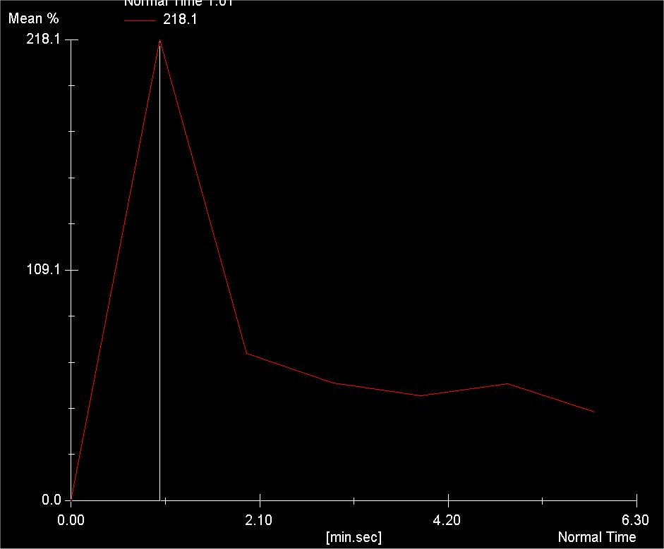
19 DCE WB MR Imaging Signal Emax (%) Emax Timing Time


20 DCE WB MR Imaging Injection Each sagittal station: 24 slices in 7 seconds (3mm/slice) Each coronal station: 40 slices in 9 seconds (5mm/slice) 152 slices/60 seconds


21 T1 SE T2 TSE FS Example: 64 ans / baseline 2ème 4ème 7ème 5ème 6ème 3ème 1ère répétition
 Bone marrow and focal lesion enhancement successfully")
22 Feasibility study: 21 patients with plasma cell disorders, 14 MM (66%) Bone marrow and focal lesion enhancement successfully evaluated


23 Without Diffuse infiltration With Diffuse infiltration Before Gd After Gd 1st repetition Before Gd After Gd 1st repetition
24 DCE WB MR Imaging for treatment evaluation 30 patients, prospective study Multiple myeloma Systemic therapy Assess post-treatment bone marrow changes and, Correlate with clinical response (based on international uniform criteria) Durie BGM et al. International uniform response criteria for multiple myeloma. Leukemia 2006; 20:
25 Timeline Baseline WB DCE-MRI 1 n = 30 Induction chemotherapy Pre-ASCT WB DCE-MRI 2 n = 30 High-dose therapy + ASCT Post-ASCT WB DCE-MRI 3 n = 20 ASCT: autologous stem cell transplantation
 of each station Maximal value among the 4 stations BMEmax (%) of each")
26 Bone marrow enhancement (BME) Stations I, II, IV, V 2 ROIs per station averaged Emax (%) of each station Maximal value among the 4 stations BMEmax (%) of each exam
 and FLEmax Timing of all target")
27 Focal lesion enhancement (FLE) 1 ROI per lesion 5 target lesions per exam FLEmax (%) and FLEmax Timing of all target lesions
 350 300 250 200 150 100 50 0 0 1 2 3")
 Timing of")
28 Female patient, 64y, before and after induction chemotherapy Responder Rehaussem ent (% ) Tem ps (m in) Timing of FLEmax (early) Rehaussem ent (% ) Tem ps (m in) Timing of FLEmax (late)
29 MR vs. clinical response Post-ASCT evaluation: concordant 65 (%) *4 good responders (uniform criteria) but poor responders on WB DCE-MRI relapsed

 2 months later After Gd 1st repetition After Gd 1st")
30 Female patient, 63y, good responder after ASCT on clinial and biological criteria but not WB DCE MRI M-protein IgA: from 37 g/l to 0g/L CR (uniform criteria) BMEmax < 97% Early-enhancing focal lesions (+) 2 months later After Gd 1st repetition After Gd 1st repetition
 Particular usefulness for patients with oligosecretory or nonsecretory")
31 WB DCE-MRI can be used to assess treatment response in patients with MM (superior to T1/T2WI) Particular usefulness for patients with oligosecretory or nonsecretory disease
32 Consensus statement MRI Dimopoulos et al, JCO 2015 Smoldering or asymptomatic myeloma: MRI indicated for >5mm focal lesion Is there a role for low-dose CT?

33

34


35
36 DCE WB-MRI in Myeloma 35 y/o man, non-secretory MM post autologous stem cell transplant in 2005 Newly-onset low back pain in Nov. 2006


37 Diffuse marrow infiltration + hepatic nodules (incidental findings)
38 DCE WB-MRI in Myeloma Potentials for evaluation of disease activity Same patient: after chemotherapy, before allogeneic stem cell transplant

39 Whole-spine MR (3 stations) T1, T2FS and T1FS+Gd difficult to know if the tiny lesion is active
40 Dynamic MR demonstrates its activity (another adjacent lesion only a central spot is active, other portions progressive enhancement).


41 3D-VIBE 0 3D-VIBE 1 3D-VIBE 6
42 57y/o woman, MM with right sacral mass. Bone marrow transplant 2 years ago Follow-up exam, clinically mild right shoulder pain


43 T1 SE T1 Gd 2ème répétition 1ère 4ème 7ème 2ème 3ème 6ème 5ème répétition
44 55y/o man, ASCT 2007 Ig 50 3 g/lgood response WB DCE MRI


45

46 3D-VIBE 2

47 3D-VIBE 0 3D-VIBE 1 All focal lesions appeared active on DCE-MRI!!
48 october Patient had back pain


49 Marked progression in the entire imaged marrow space with massive left pleural effusion

50 3D-VIBE 2


51 3D-VIBE 0 3D-VIBE 3
52 64y female new patient with stage 3 (Durie/Salmon) IgG Kappa MM, baseline WB-DCE MRI example of diffuse & focal infiltration

53 Diffuse infiltration Baseline, monoclonal peak at 20g/L After 3 cycles of chemotherapy, monoclonal peak decreased to 8g/L VIBE0 VIBE1 VIBE0 VIBE1


54 Right sacral mass Baseline, monoclonal peak at 20g/L After 3 cycles of chemotherapy, monoclonal peak decreased to 8g/L VIBE0 VIBE1 VIBE1
55 60y/o man, MM after 4 cycles Incidental findings


56


57
58 80y/o man with stage I MM and chronic hepatitis C Incidental findings


59

")
60 VIBE 0 VIBE 1 VIBE 3 VIBE 6 Last MRI of liver (décembre 06) negative!!
61 52y/o woman, MGUS Incidental findings


62 In favor of hemangiomas, no MR evidence of myeloma marrow infiltration, but.
63 Plan Whole-Body MR imaging Biological aspects DCE MR imaging DCE WB MR imaging PET imaging


64 A B C D E * 11 C-ACT 18 F-FDG 11 C-ACT PET/CT 18 F-FDG PET/CT WB-DCE-MRI Courtesy Dr Lin, Taiwan
65 Conclusion WB MRI should be used for myeloma staging but also plasmocytoma and MGUS On going WB DCE MRI evaluation for prognostic value As other hematologic malignancies, functional imaging is necessary for patient evaluation
Clinical Appropriateness Guidelines: Advanced Imaging
 Clinical Appropriateness Guidelines: Advanced Imaging Appropriate Use Criteria: Imaging of Bone Marrow Blood Supply Effective Date: September 5, 2017 Proprietary Date of Origin: 05/21/2007 Last revised:
MRI in lymphoma: where are we in 2016
 MRI in lymphoma: where are we in 2016 Alain Rahmouni Henri Mondor Academic Hospital Paris Est Créteil University, France Plan Requirements DWI signal in lymphoma ADC measurement SNR: Surface coils 1.5
Whole body MR in patients with multiple myeloma
 Whole body MR in patients with multiple myeloma Alina Piekarek, Piotr Sosnowski, Adam Nowicki, Mieczysław Komarnicki Received: 11.05.2009 Accepted: 13.07.2009 Subject: original article Clinical Radiology
The role of multimodality imaging in Multiple Myeloma: Past, Present and Future
 The role of multimodality imaging in Multiple Myeloma: Past, Present and Future Poster No.: C-1661 Congress: ECR 2015 Type: Educational Exhibit Authors: J. Niza, R. Gil, P. Pereira, C. Oliveira ; Setúbal/PT,
Multiple Myeloma: diagnosis and prognostic factors. N Meuleman May 2015
 Multiple Myeloma: diagnosis and prognostic factors N Meuleman May 2015 Diagnosis Diagnostic assessment of myeloma: what should we know? Is it really a myeloma? Is there a need for treatment? What is the
Quantitative Whole-Body MRI for Treatment Response Monitoring in Multiple Myeloma
 Quantitative Whole-Body MRI for Treatment Response Monitoring in Multiple Myeloma Arash Latifoltojar Arash.latifoltojar.10@ucl.ac.uk Background- Initial disease assessment - Whole body Imaging skeletal
FOR CMS (MEDICARE) MEMBERS ONLY NATIONAL COVERAGE DETERMINATION (NCD) FOR MAGNETIC RESONANCE IMAGING:
 MEMBERS ONLY NATIONAL COVERAGE DETERMINATION (NCD) FOR MAGNETIC RESONANCE IMAGING:") National Imaging Associates, Inc. Clinical guidelines BONE MARROW MRI Original Date: July 2008 Page 1 of 5 CPT Codes: 77084 Last Review Date: September 2014 NCD 220.2 MRI Last Effective Date: July 2011
National Imaging Associates, Inc. Clinical guidelines BONE MARROW MRI Original Date: July 2008 Page 1 of 5 CPT Codes: 77084 Last Review Date: September 2014 NCD 220.2 MRI Last Effective Date: July 2011
Imaging of bone metastases
 Imaging of bone metastases Antoine Feydy Service de Radiologie B Hôpital Cochin APHP Université Paris Descartes antoine.feydy@aphp.fr MEXICO 2016 INTRODUCTION Diagnostic Imaging Imaging Modalities Strengths,
Bone PET/MRI : Diagnostic yield in bone metastases and malignant primitive bone tumors
 Bone PET/MRI : Diagnostic yield in bone metastases and malignant primitive bone tumors Lars Stegger, Benjamin Noto Department of Nuclear Medicine University Hospital Münster, Germany Content From PET to
PET-MRI in malignant bone tumours. Lars Stegger Department of Nuclear Medicine University Hospital Münster, Germany
 PET-MRI in malignant bone tumours Lars Stegger Department of Nuclear Medicine University Hospital Münster, Germany Content From PET to PET/MRI General considerations Bone metastases Primary bone tumours
Criteria for Disease Assessment Joan Bladé
 Criteria for Disease Assessment Joan Bladé Unidad de Amiloidosis y Mieloma Servicio de Hematología Hospital Clínic de Barcelona COMy Meeting, París, May 4th, 2018 Response Evaluation EBMT, 1998 - CR and
Imaging of Pediatric MSK Tumors
 Imaging of Pediatric MSK Tumors Kirsten Ecklund, M.D. Boston Children s Hospital Harvard Medical School kirsten.ecklund@childrens.harvard.edu Tumor Imaging Goals Diagnosis Lesion characterization Benign
M-Protien, what to do next? Ismail A Sharif MD, FRCPc Internal Medicine Day 22 nd April 2016
 + M-Protien, what to do next? Ismail A Sharif MD, FRCPc Internal Medicine Day 22 nd April 2016 + Disclosures Advisory Boards: AMGEN, Lundbeck, NOVARTIS + Subtypes of Plasma Cell Disorders Increased Plasma
Innovations in HCC Imaging: MDCT/MRI
 Innovations in HCC Imaging: MDCT/MRI Anthony E. Cheng, M.D. Cardinal MRI Center Cardinal Santos Medical Center, Wilson Street, San Juan Innovations in HCC Imaging: Goals/Objectives MDCT/MRI Learn the diagnostic
Imaging in gastric cancer
 Imaging in gastric cancer Gastric cancer remains a deadly disease because of late diagnosis. Adenocarcinoma represents 90% of malignant tumors. Diagnosis is based on endoscopic examination with biopsies.
High Field MR of the Spine
 Department of Radiology University of California San Diego 3T for MR Applications Advantages High Field MR of the Spine Increased signal-to-noise Better fat suppression Increased enhancement with gadolinium
A.M.W. van Marion. H.M. Lokhorst. N.W.C.J. van de Donk. J.G. van den Tweel. Histopathology 2002, 41 (suppl 2):77-92 (modified)
:77-92 (modified)") chapter 4 The significance of monoclonal plasma cells in the bone marrow biopsies of patients with multiple myeloma following allogeneic or autologous stem cell transplantation A.M.W. van Marion H.M. Lokhorst
chapter 4 The significance of monoclonal plasma cells in the bone marrow biopsies of patients with multiple myeloma following allogeneic or autologous stem cell transplantation A.M.W. van Marion H.M. Lokhorst
This copy is for personal use only. To order printed copies, contact Purpose: Materials and Methods: Results: Conclusion:
 This copy is for personal use only. To order printed copies, contact reprints@rsna.org Quantitative Analysis of MR Imaging to Assess Treatment Response for Patients with Multiple Myeloma by Using Dynamic
Orthopedic Hardware Imaging Part II: MRI v. Metal
 Orthopedic Hardware Imaging Trent Roth, MD And Lauren Ladd, MD Indiana University School of Medicine IU Health Physicians-Radiology Recap: Imaging Techniques Radiography Standard for initial and surveillance
Mantle Cell Lymphoma
 Mantle Cell Lymphoma Clinical Case A 56 year-old woman complains of pain and fullness in the left superior abdominal quadrant for the last 8 months. She has lost 25 kg, and lately has had night sweats.
Smoldering Myeloma: Leave them alone!
 Smoldering Myeloma: Leave them alone! David H. Vesole, MD, PhD Co-Director, Myeloma Division Director, Myeloma Research John Theurer Cancer Center Hackensack University Medical Center Prevalence 1960 2002
Perfusion Physics. ICMRI2018 March 29-31, 2018 Grand Hilton Hotel, Seoul, Korea. Asian Forum Ⅱ: Perfusion MRI SY24-1.
 SY24-1 Perfusion Physics Hiroyuki Kabasawa MR Collaborations and Development, GE Healthcare, Tokyo, Japan Perfusion is referred as the blood supply to micro capillary in tissue. Perfusion parameter such
T2, T2*, ute. Yeo Ju Kim. Radiology, Inha University Hospital, Incheon, Korea
 SY28-1 T2, T2*, ute Yeo Ju Kim Radiology, Inha University Hospital, Incheon, Korea T2 relaxation times relate to the rate of transverse magnetization decay, caused by the loss of phase coherence induced
Case 1: Mrs. MG. ANCO Hematologic Malignancies Update: Acute leukemias, MDS and myeloma
 Partner Logo ANCO Hematologic Malignancies Update: Acute leukemias, MDS and myeloma Tim Campbell Research Fellow UCSF Division of Hematology and Oncology September 12 th, 2015 Case 1: Mrs. MG 65 yo woman
EXPERIMENTAL AND THERAPEUTIC MEDICINE 9: , 2015
 EXPERIMENTAL AND THERAPEUTIC MEDICINE 9: 1895-1900, 2015 Clinical characteristics of a group of patients with multiple myeloma who had two different λ light chains by immunofixation electrophoresis: A
Abdominal applications of DWI
 Postgraduate course, SPR San Antonio (Texas), May 14-15, 2013 Abdominal applications of DWI Rutger A.J. Nievelstein Wilhelmina Children s s Hospital, Utrecht (NL) Outline What is DWI? How to perform? Challenges
Monitoring bony metastases response with diffusion MRI
 Monitoring bony metastases response with diffusion MRI Anwar Padhani MD Mount Vernon Hospital Cancer Centre London, UK Objectives To illustrate the potential of whole body DWI in the therapy response assessment
Imaging techniques to characterize spleen involvement in patients with Hodgkin lymphoma
 Imaging techniques to characterize spleen involvement in patients with Hodgkin lymphoma Marco Picardi, MD Ematologia, Azienda Ospedaliera Universitaria Federico II, Naples, Italy 5th International Workshop
dgemric Effectively Predicts Cartilage Damage Associated with Femoroacetabular Impingement
 Riccardo Lattanzi 1,2 Catherine Petchprapa 2 Daniele Ascani 1 Roy I. Davidovitch 3 Thomas Youm 3 Robert J. Meislin 3 Michael. Recht 2 1 The Bernard and Irene Schwartz Center for Biomedical Imaging, New
Hematology 101. Rachid Baz, M.D. 5/16/2014
 Hematology 101 Rachid Baz, M.D. 5/16/2014 Florida 101 Epidemiology Estimated prevalence 8,000 individuals in U.S (compare with 80,000 MM patients) Annual age adjusted incidence 3-8/million-year 1 More
Cardiovascular Imaging
 Cardiovascular Imaging Cardiovascular Imaging Cardio and Vascular Imaging Vascularization / Angiogenesis Cardiovascular Imaging metabolic imaging of the heart myocardial perfusion imaging Cardiac CT Vascularization
Prof. Dr. NAGUI M. ABDELWAHAB,M.D.; MARYSE Y. AWADALLAH, M.D. AYA M. BASSAM, Ms.C.
 Role of Whole-body Diffusion MR in Detection of Metastatic lesions Prof. Dr. NAGUI M. ABDELWAHAB,M.D.; MARYSE Y. AWADALLAH, M.D. AYA M. BASSAM, Ms.C. Cancer is a potentially life-threatening disease,
Successful Breast MRI Program : The ingredients
 Successful Breast MRI Program : The ingredients Dr. Smriti Hari Associate Professor Deptt. Of Radiology All India Institute of Medical Sciences New Delhi How to perform Breast MRI Breast MRI descriptors
Multiple Myeloma Updates 2007
 Multiple Myeloma Updates 2007 Brian Berryman, M.D. Multiple Myeloma Updates 2007 Goals for today: Understand the staging systems for myeloma Understand prognostic factors in myeloma Review updates from
UPDATE Autologous Stem Cell Transplantation for Lymphoma and Myeloma
 UPDATE Autologous Stem Cell Transplantation for Lymphoma and Myeloma Supported by a grant from Supported by a grant from UPDATE Autologous Stem Cell Transplantation for Lymphoma and Myeloma Jonathan W.
Review of the recent publications from the French group of myeloma on urine vs serum FLC analysis in MM
 Review of the recent publications from the French group of myeloma on urine vs serum FLC analysis in MM Multiple myeloma Response evaluation Kumar Lancet Oncol 2016; 17: e328 46 Cumulative Proportion Surviving
Smouldering Myeloma: to treat or not to treat?
 Smouldering Myeloma: to treat or not to treat? Massimo Offidani Clinica di Ematologia Azienda Ospedaliero-Universitaria Ospedali Riuniti di Ancona Definitions and epidemiology 3000-5000 new SMM/year in
What Radiologists do?
 Multimodality Imaging in Oncology 2018 March 5 th 9th Diagnostic Imaging in Oncology What Radiologists do? Chikako Suzuki, MD, PhD Department of Diagnostic Radiology, KS Solna Department of Molecular Medicine
Management of high-risk diffuse large B cell lymphoma: case presentation
 Management of high-risk diffuse large B cell lymphoma: case presentation Daniel J. Landsburg, MD Assistant Professor of Clinical Medicine Perelman School of Medicine University of Pennsylvania January
Why Talk About Technique? MRI of the Knee:
 Why Talk About Technique? MRI of the Knee: Part 1 - Imaging Techniques Mark Anderson, M.D. University of Virginia Health Sciences Center Charlottesville, Virginia Always had an interest teach our fellows
Contrast-enhanced Breast MRI RSSA 2013
 Contrast-enhanced Breast MRI RSSA 2013 Prof. dr. Maurice van den Bosch University Medical Center Utrecht, the Netherlands Index 1) Breast cancer 2) Why MRI of the breast 3) Technique 4) Interpretation
Module 3: Multiple Myeloma Induction and Transplant Strategies Treatment Planning
 Module 3: Multiple Myeloma Induction and Transplant Strategies Treatment Planning Challenge Question: Role of Autologous Stem Cell Transplant Which of the following is true about eligibility for high-dose
Session Chair: Kenneth C. Anderson, MD Speakers: S. Vincent Rajkumar, MD; Peter Leif Bergsagel, MD; and Donna E. Reece, MD
 Myeloma Session Chair: Kenneth C. Anderson, MD Speakers: S. Vincent Rajkumar, MD; Peter Leif Bergsagel, MD; and Donna E. Reece, MD MGUS and Smoldering Multiple Myeloma: Update on Pathogenesis, Natural
MRI XR, CT, NM. Principal Modality (2): Case Report # 2. Date accepted: 15 March 2013
: Case Report # 2. Date accepted: 15 March 2013") Radiological Category: Musculoskeletal Principal Modality (1): Principal Modality (2): MRI XR, CT, NM Case Report # 2 Submitted by: Hannah Safia Elamir, D.O. Faculty reviewer: Naga R. Chinapuvvula, M.D.
Radiological Category: Musculoskeletal Principal Modality (1): Principal Modality (2): MRI XR, CT, NM Case Report # 2 Submitted by: Hannah Safia Elamir, D.O. Faculty reviewer: Naga R. Chinapuvvula, M.D.
Kymriah. Kymriah (tisagenlecleucel) Description
 Description") Federal Employee Program 1310 G Street, N.W. Washington, D.C. 20005 202.942.1000 Fax 202.942.1125 5.21.101 Subject: Kymriah Page: 1 of 5 Last Review Date: September 20, 2018 Kymriah Description Kymriah
Federal Employee Program 1310 G Street, N.W. Washington, D.C. 20005 202.942.1000 Fax 202.942.1125 5.21.101 Subject: Kymriah Page: 1 of 5 Last Review Date: September 20, 2018 Kymriah Description Kymriah
Multiple myeloma Biological & Clinical Aspects Isabelle Vande Broek, MD, PhD
 Multiple myeloma Biological & Clinical Aspects Isabelle Vande Broek, MD, PhD Department of Oncology & Hematology AZ Nikolaas Iridium Kanker Netwerk Introduction Multiple myeloma = Kahler s disease Dr.
Forms Revision: Myeloma Changes
 Sharing knowledge. Sharing hope. Forms Revision: Myeloma Changes J. Brunner, PA-C and A. Dispenzieri, MD February 2013 Disclosures Janet Brunner, PA-C I have no relevant conflicts of interest to disclose.
Vertebral and Paravertebral Diseases
 Department of Radiology University of California San Diego Vertebral and Paravertebral Diseases John R. Hesselink, M.D. Vertebral / Paravertebral Disease (Extradural) Metastatic disease Primary bone tumors
Whole body Diffusion Weighted MRI (WB-DWI) in the assessment and treatment response of multiple myeloma (MM).
 in the assessment and treatment response of multiple myeloma (MM).") Whole body Diffusion Weighted MRI (WB-DWI) in the assessment and treatment response of multiple myeloma (MM). Poster No.: C-1467 Congress: ECR 01 Type: Educational Exhibit Authors: A. Mahatma, A. Gogbashian,
Whole body Diffusion Weighted MRI (WB-DWI) in the assessment and treatment response of multiple myeloma (MM). Poster No.: C-1467 Congress: ECR 01 Type: Educational Exhibit Authors: A. Mahatma, A. Gogbashian,
MRI PEDIATRIC PROTOCOLS (Updated 6/19/2018)
") MRI PEDIATRIC PROTOCOLS (Updated 6/19/2018) *Please get or let us know where radiologist can review plain films. *For Texas Orthopedics and other Docs requesting only MSK section read for their pediatric
MRI PEDIATRIC PROTOCOLS (Updated 6/19/2018) *Please get or let us know where radiologist can review plain films. *For Texas Orthopedics and other Docs requesting only MSK section read for their pediatric
Multiple myeloma evolves from a clinically silent premalignant
 S. VINCENT RAJKUMAR Updated Diagnostic Criteria and Staging System for Multiple Myeloma S. Vincent Rajkumar, MD OVERVIEW There has been remarkable progress made in the diagnosis and treatment of multiple
Amyloidosis: What to do and how to diagnose: An Update 2017
 Amyloidosis: What to do and how to diagnose: An Update 2017 Jonathan L. Kaufman, MD Associate Professor Hematology & Oncology Winship Cancer Institute of Emory University Amyloidosis Protein Conformation/Deposition
MR Advance Techniques. Vascular Imaging. Class II
 MR Advance Techniques Vascular Imaging Class II 1 Vascular Imaging There are several methods that can be used to evaluate the cardiovascular systems with the use of MRI. MRI will aloud to evaluate morphology
Jo Abraham MD Division of Nephrology University of Utah
 Jo Abraham MD Division of Nephrology University of Utah 68 year old male presented 3 weeks ago with a 3 month history of increasing fatigue He reported a 1 week history of increasing dyspnea with a productive
Importancia del laboratorio en el seguimiento clínico de pacientes con mieloma múltiple
 Importancia del laboratorio en el seguimiento clínico de pacientes con mieloma múltiple Joan Bladé Unidad de Amiloidosis y Mieloma Servicio de Hematología Hospital Clínic de Barcelona Málaga, 16 de noviembre
Treatment of Waldenström s Macroglobulinemia Mayo Consensus
 Treatment of Waldenström s Macroglobulinemia Mayo Consensus Scottsdale, Arizona Rochester, Minnesota Jacksonville, Florida Mayo Clinic College of Medicine Mayo Clinic Comprehensive Cancer Center Mayo Clinic
Radiological staging of lung cancer. Shukri Loutfi,MD,FRCR Consultant Thoracic Radiologist KAMC-Riyadh
 Radiological staging of lung cancer Shukri Loutfi,MD,FRCR Consultant Thoracic Radiologist KAMC-Riyadh Bronchogenic Carcinoma Accounts for 14% of new cancer diagnoses in 2012. Estimated to kill ~150,000
Disclosure. Acknowledgement. What is the Best Workup for Rectal Cancer Staging: US/MRI/PET? Rectal cancer imaging. None
 What is the Best Workup for Rectal Cancer Staging: US/MRI/PET? Zhen Jane Wang, MD Assistant Professor in Residence UC SF Department of Radiology Disclosure None Acknowledgement Hueylan Chern, MD, Department
International Myeloma Foundation Patient and Family Seminar
 International Myeloma Foundation Patient and Family Seminar Vienna, Austria May 6 th, 2006 New Development in Diagnosis & Treatments Brian G.M. Durie, M.D., Chairman International Myeloma Foundation What
Various presentations of multiple myeloma MRI review: A Case series
 Mary Hazarika Bhuyan/ International Journal of Biomedical Research 2015; 6(07): 531-536. 531 International Journal of Biomedical Research ISSN: 0976-9633 (Online); 2455-0566 (Print) Journal DOI: 10.7439/ijbr
Human Herpes Virus-6 Limbic Encephalitis
 Case Studies [1] March 19, 2013 Case history: A 32-year-old Caucasian female with newly diagnosed acute myeloid leukemia (AML) was treated with induction chemotherapy and attained complete remission. She
Current Clinical Practice. MR Imaging Evaluations. MRI Anatomic Review. Imaging to Address Clinical Challenges. Prostate MR
 BETH ISRAEL DEACONESS MEDICAL CENTER Prostate MR Neil M. Rofsky, MD Harvard Medical School Current Clinical Practice DIGITAL RECTAL EXAMINATION PSA ( ~ 20% False negative) BIOPSY (18-25% False negative)
Case Workshop of Society for Hematopathology and European Association for Haematopathology
 Case 24 2007 Workshop of Society for Hematopathology and European Association for Haematopathology Aliyah Rahemtullah 1, Martin K Selig 1, Paola Dal Cin 2 and Robert P Hasserjian 1 Departments of Pathology,
Essentials of Clinical MR, 2 nd edition. 65. Benign Hepatic Masses
 65. Benign Hepatic Masses Pulse sequences acquired for abdominal MRI typically consist of fast acquisition schemes such as single-shot turbo spin echo (i.e. HASTE) and gradient echo schemes such as FLASH
Evaluation of Lung Cancer Response: Current Practice and Advances
 Evaluation of Lung Cancer Response: Current Practice and Advances Jeremy J. Erasmus I have no financial relationships, arrangements or affiliations and this presentation will not include discussion of
Case Reports: Tumor Detection by Diffusion-Weighted MRI and ADC-Mapping with Correlation to PET/CT Results
 Case Reports: Tumor Detection by Diffusion-Weighted MRI and ADC-Mapping with Correlation to PET/CT Results Matthias Philipp Lichy, M.D.; Philip Aschoff, M.D.; Christina Pfannenberg, M.D.; Schlemmer Heinz-Peter,
Value of true whole-body FDG- PET/CT scanning protocol in oncology and optimization of its use based on primary malignancy
 Value of true whole-body FDG- PET/CT scanning protocol in oncology and optimization of its use based on primary malignancy Ronnie Sebro MD, Ph.D Carina Mari Aparici MD, Miguel Hernandez Pampaloni MD, PhD
Liver imaging takes a step forward with Ingenia
 Publication for the Philips MRI Community ISSUE 49 2013 / 2 Liver imaging takes a step forward with Ingenia Lyon South Hospital strives to move from several studies first CT, then MR or PET to using just
Perfusion MRI. Youngkyoo Jung, PhD Associate Professor Radiology, Biomedical Engineering, and Clinical & Translational Science Institute
 Perfusion MRI Youngkyoo Jung, PhD Associate Professor Radiology, Biomedical Engineering, and Clinical & Translational Science Institute Perfusion The delivery of blood to a capillary bed in tissue Perfusion
Neuroradiology MR Protocols
 Neuroradiology MR Protocols Brain protocols N 1: Brain MRI without contrast N 2: Pre- and post-contrast brain MRI N 3 is deleted N 4: Brain MRI without or pre-/post-contrast (seizure protocol) N 5: Pre-
Lymphoma Read with the experts
 Lymphoma Read with the experts Marc Seltzer, MD Associate Professor of Radiology Geisel School of Medicine at Dartmouth Director, PET-CT Course American College of Radiology Learning Objectives Recognize
Multiparametric imaging in oncology
 Multiparametric imaging in oncology p1 T p2 p2 T T p3 p1 p3 T Marco Ravanelli Roberto Maroldi The goal of traditional imaging is high spatial and contrast resolution diagnosis, tumor extent treatment planning,
Whole-body-MRI with DWIBS for detecting distant metastases in patients with head and neck squamous cell carcinoma: a feasibility study
 Whole-body-MRI with DWIBS for detecting distant metastases in patients with head and neck squamous cell carcinoma: a feasibility study Poster No.: C-245 Congress: ECR 203 Type: Scientific Exhibit Authors:
2012 by American Society of Hematology
 2012 by American Society of Hematology Common Types of HIV-Associated Lymphomas DLBCL includes primary CNS lymphoma (PCNSL) Burkitt Lymphoma HIV-positive patients have a 60-200 fold increased incidence
Val M. Runge, MD Editor-in-Chief Investigative Radiology
 Val M. Runge, MD Editor-in-Chief Investigative Radiology Patients First RSNA 2012 The classical model of medical care which portrays the authoritative physician evaluating and treating an obedient, non-inquisitive
Dr Sneha Shah Tata Memorial Hospital, Mumbai.
 Dr Sneha Shah Tata Memorial Hospital, Mumbai. Topics covered Lymphomas including Burkitts Pediatric solid tumors (non CNS) Musculoskeletal Ewings & osteosarcoma. Neuroblastomas Nasopharyngeal carcinomas
Gemstone Spectral Imaging quantifies lesion characteristics for a confident diagnosis
 GE Healthcare Gemstone Spectral Imaging quantifies lesion characteristics for a confident diagnosis CT clinical case study lesion characterization Desiree Morgan, MD Vice Chair of Clinical Research Professor
Functional MRI in Oncology
 Functional MRI in Oncology J.O. Barentsz Chair for Research Department of Radiology Radboud University Medical Center, Nijmegen, NL j.barentsz@rad.umcn.nl Learning objectives to give an introduction of
TYPE 1 GAUCHER DISEASE PRESENTING AS PERSISTENT THROMBOCYTOPENIA, ASSOCIATED FACTOR XI DEFICIENCY & EMERGENT MYELOMA
 TYPE 1 GAUCHER DISEASE PRESENTING AS PERSISTENT THROMBOCYTOPENIA, ASSOCIATED FACTOR XI DEFICIENCY & EMERGENT MYELOMA Trish Hyland, Medical Scientist, Department of Haematology, Cork University Hospital
Los Angeles Radiological Society 62 nd Annual Midwinter Radiology Conference January 31, 2010
 Los Angeles Radiological Society 62 nd Annual Midwinter Radiology Conference January 31, 2010 Self Assessment Module on Nuclear Medicine and PET/CT Case Review FDG PET/CT IN LYMPHOMA AND MELANOMA Submitted
Evangelia E. Vassalou MD,PhD Radiologist Department of Medical Imaging, Heraklion University Hospital Department of Medical Imaging, Sitia General
 Evangelia E. Vassalou MD,PhD Radiologist Department of Medical Imaging, Heraklion University Hospital Department of Medical Imaging, Sitia General Hospital Osteonecrosis pathophysiology epidemiology imaging
Lugano classification: Role of PET-CT in lymphoma follow-up
 CAR Educational Exhibit: ID 084 Lugano classification: Role of PET-CT in lymphoma follow-up Charles Nhan 4 Kevin Lian MD Charlotte J. Yong-Hing MD FRCPC Pete Tonseth 3 MD FRCPC Department of Diagnostic
PET Imaging in Langerhans Cell Histiocytosis
 PET Imaging in Langerhans Cell Histiocytosis Christiane Franzius Bremen, Germany Histiocytosis Histiocytosis Idiopathic proliferation of histiocytes Two types of histiocytes macrophages: antigen processing
Should we treat Smoldering MM patients? María-Victoria Mateos University Hospital of Salamanca Salamanca. Spain
 Should we treat Smoldering MM patients? María-Victoria Mateos University Hospital of Salamanca Salamanca. Spain Should we treat some patients with Stage I MM? Len-dex is a promising and atractive option
RECENT ADVANCES IN CLINICAL MR OF ARTICULAR CARTILAGE
 In Practice RECENT ADVANCES IN CLINICAL MR OF ARTICULAR CARTILAGE By Atsuya Watanabe, MD, PhD, Director, Advanced Diagnostic Imaging Center and Associate Professor, Department of Orthopedic Surgery, Teikyo
Dr Prashant Tembhare
 Dr Prashant Tembhare docprt@gmail.com FCM very powerful technology in Identification and characterization of neoplastic plasma cells as it allows - simultaneous assessment of multiple antigens large numbers
6/23/2009. Inversion Recovery (IR) Techniques and Applications. Variations of IR Technique. STIR, FLAIR, TI and TI Null. Applications of IR
 Techniques and Applications. Variations of IR Technique. STIR, FLAIR, TI and TI Null. Applications of IR") The Anatomy of Basic R Pulse Sequences Inversion Recovery () Techniques and Applications Chen Lin, PhD Indiana University School of edicine & Clarian Health Partners agnetization Preparation Section Chemical
The Anatomy of Basic R Pulse Sequences Inversion Recovery () Techniques and Applications Chen Lin, PhD Indiana University School of edicine & Clarian Health Partners agnetization Preparation Section Chemical
Lung Cancer Imaging. Terence Z. Wong, MD,PhD. Department of Radiology Duke University Medical Center Durham, NC 9/9/09
 Lung Cancer Imaging Terence Z. Wong, MD,PhD Department of Radiology Duke University Medical Center Durham, NC 9/9/09 Acknowledgements Edward F. Patz, Jr., MD Jenny Hoang, MD Ellen L. Jones, MD, PhD Lung
Relapse of Multiple Myeloma in the Thyroid Gland
 Case Report Relapse of Multiple Myeloma in the Thyroid Gland Lopez Rojo I*, Gomez Valdazo A, Gomez Ramirez J Department of General Surgery, Jimenez Diaz University Hospital Foundation, Madrid, Spain *Corresponding
Blood Cancers. Blood Cells. Blood Cancers: Progress and Promise. Bone Marrow and Blood. Lymph Nodes and Spleen
 Blood Cancers: Progress and Promise Mike Barnett & Khaled Ramadan Division of Hematology Department of Medicine Providence Health Care & UBC Blood Cancers Significant health problem Arise from normal cells
Imaging of Articular Cartilage
 Clinical Imaging of Articular Cartilage Imaging of Articular Cartilage Prof. Dr. K. Verstraete Ghent University Introduction : Articular Cartilage Histology and biochemical composition Review of Imaging
Pediatric Lymphoma Update from the Children s Oncology Group
 Pediatric Lymphoma Update from the Children s Oncology Group Stephan D. Voss, MD, PhD Department of Radiology Boston Children s Hospital Harvard Medical School Staging Assessment Disclosures None Acknowledgements:
Prognostic value of bone marrow angiogenesis in patients with multiple myeloma undergoing high-dose therapy
 (2004) 34, 235 239 & 2004 Nature Publishing Group All rights reserved 0268-3369/04 $30.00 www.nature.com/bmt Prognostic value of bone marrow angiogenesis in patients with multiple myeloma undergoing high-dose
Extramedullary Multiple Myeloma in the Head and Neck: A Pictorial Essay
 Canadian Association of Radiologists Journal 64 (2013) 363e369 Neuroradiology / Neuroradiologie Extramedullary Multiple Myeloma in the Head and Neck: A Pictorial Essay Michael Chan, BHSc a, Eric Bartlett,
Oak foundation for donating the 3T Siemens Verio scanner. Board of directors BBH and Frh Hospitals for supporting the
 Knee pain and inflammation in the infrapatellar fat pad estimated by conventional and dynamic contrast-enhanced magnetic resonance imaging in obese patients with osteoarthritis: a crosssectional study
Using lesion washout volume fraction as a biomarker to improve suspicious breast lesion characterization
 JOURNAL OF APPLIED CLINICAL MEDICAL PHYSICS, VOLUME 16, NUMBER 5, 2015 Using lesion washout volume fraction as a biomarker to improve suspicious breast lesion characterization Jie Huang, a Sarah M. Schafer,
Imaging of Neuroendocrine Metastases
 Imaging of Neuroendocrine Metastases Aoife Kilcoyne, Shaunagh McDermott, Colin McCarthy,Manuel Patino, Dushyant Sahani, Michael Blake Abdominal Imaging Division Massachusetts General Hospital Disclosure
Linfoma de Hodgkin. Novos medicamentos. Otavio Baiocchi CRM-SP
 Linfoma de Hodgkin Novos medicamentos Otavio Baiocchi CRM-SP 96.074 Hodgkin Lymphoma Unique B-cell lymphoma HRS malignant cells Scattered malignant Hodgkin-Reed-Sternberg (RS) cells in a background of
ROLE OF MRI IN SCREENING, DIAGNOSIS AND MANAGEMENT OF BREAST CANCER. B.Zandi Professor of Radiology
 ROLE OF MRI IN SCREENING, DIAGNOSIS AND MANAGEMENT OF BREAST CANCER B.Zandi Professor of Radiology Introduction In the USA, Breast Cancer is : The Most Common Non-Skin Cancer The Second Leading cause of
PET/CT for Therapy Assessment in Oncology
 PET/CT for Therapy Assessment in Oncology Rodolfo Núñez Miller, M.D. Nuclear Medicine Section Division of Human Health International Atomic Energy Agency Vienna, Austria Clinical Applications of PET/CT