Oak foundation for donating the 3T Siemens Verio scanner. Board of directors BBH and Frh Hospitals for supporting the
|
|
|
- Silas Crawford
- 5 years ago
- Views:
Transcription
, Robert Riis (1), Henning Bliddal (1), Robin Christensen (1), Marius Henriksen (1), Else Bartels (1), Stefan Lohmander (2), David Hunter (3), Rasmus Bouert (4) and")
1 Knee pain and inflammation in the infrapatellar fat pad estimated by conventional and dynamic contrast-enhanced magnetic resonance imaging in obese patients with osteoarthritis: a crosssectional study Christine Ballegaard (1), Robert Riis (1), Henning Bliddal (1), Robin Christensen (1), Marius Henriksen (1), Else Bartels (1), Stefan Lohmander (2), David Hunter (3), Rasmus Bouert (4) and Mikael Boesen (1,4) 1) Parker Institute, Bispebjerg and Frederiksberg Hospitals, Copenhagen F, Denmark 2) Department of Orthopaedics, Clinical Sciences Lund, Lund University, Sweden 3) Department of Rheumatology, Royal North Shore Hospital, University of Sydney, Australia 4) Department of Radiology, Bispebjerg and Frederiksberg Hospitals, Copenhagen F, Denmark Bispebjerg og Frederiksberg Hospitals Presented by Mikael Boesen, MD Phd
2 Acknowledgements: Oak foundation for donating the 3T Siemens Verio scanner Board of directors BBH and Frh Hospitals for supporting the Olga Kubassova and her team at Image analysis, Ltd London for support using the software Dynamika Disclosures: Mikael Boesen and Rasmus Bouert has acted as Independent non-paid clinical advisors for Dynamika on the clinical advisory board of Image Analysis Ltd, London.
) seems to play a crusial, but not fully understood, role in Knee Osteoarthritis")
3 Background: In the knee, the intraarticular, extra synovial fat pad of Hoffa (infrapatellar fat pad (IPFP)) seems to play a crusial, but not fully understood, role in Knee Osteoarthritis (KOA)

4 Synovitis / inflammation in KOA Contrast enhanced(ce)-mri vs. non-ce MRI
 MRI vs.")

5 Synovitis: Non-Contrast Enhanced (CE) MRI vs. CE-MRI Proton weighted fat sat T1 weighted fat sat + Gadolinum
-mri in")


6 Dynamic contrast enhanced(dce)-mri in inflammtory arthritis Pre Contrast Post Contrast Boesen M et al Eur J Radiol Oct 26.


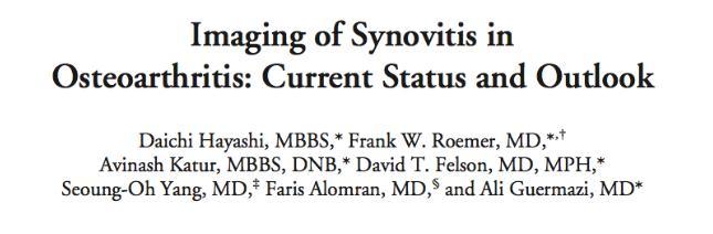
7 DCE-MRI voxel by voxel model-based time intensity analysis Boesen M et al Eur J Radiol. 2010


8 Number of enhancing voxels
9 Purpose: To investigate the association between knee pain and signs of inflammation in the infrapatellar fat pad (IPFP) in obese patients with knee osteoarthritis (KOA) using both conventional CE-MRI and DCE-MRI derived from a 3T MRI scanner. Methods: 95 KOA patients all participants in a weightloss trial Carot light (ClinicalTrials.gov identifier: NCT ) were included in the analysis
10 MRI Protocol MRI of the target knee was performed (Image time minutes) o 3T Siemens Verio system. o Supine position using a dedicated 16-channel send/receive knee coil. The following MRI-protocol was used: o Coronal T1-weighted (T1w) turbo spin echo (TSE) o Coronal and Sagittal short tau inversion recovery (STIR); o Sagittal 3D 0.6mm isotropic proton density weighted (PDw) FS TSE SPACE, o Sagittal GRE 3D T1w 0.5mm volumetric interpolated breath hold examination (VIBE) Simultaneously with the intravenous injection of 0.1 ml/kg body weight Gadolinium contrast (Prohance, Bracco Diagnostics Inc., Italy) using a power injector (2 ml/s): Axial DCE-MRI GRE T1w VIBE sequence 18, 5 mm slices every 9 s, with 30 repetitions TE 1.86, TR 5.51 FA 15 degrees, FOV 160x160, matrix resolution 192 x 138) Covering the supra-patellar recess to the insertion of the patella tendon on the tibia. Following this the static T1w sequences were repeated and used for MOAKS Hoffa synovitis scoring.
 for Hoffa synovitis using the 3D T1w CE-MRI DCE-MRI Ballegaard et al.")
11 Material and Metods: KOOS (knee injury and osteoarthritis score) is self-reported outcomes on pain, symptoms and quality of life Hoffa synovitis assessed according to the definitions in the MRI osteoarthritis knee score (MOAKS) for Hoffa synovitis using the 3D T1w CE-MRI DCE-MRI Ballegaard et al. (submitted)
 around the IPFP in all the axial DCE-MRI images from")
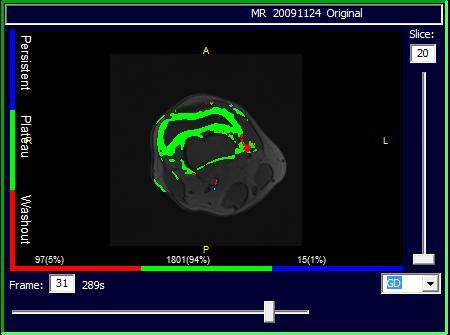
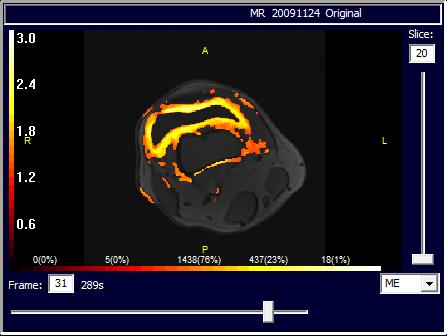
12 DCE-MRI Analysis Investigators blinded to the clinical data and the KOOS answers analysed the MR images. DCE-MRI images were analysed using Dynamika : o Motion correction to reduce the enhancement artefacts due to movement o Region of interest (ROI) around the IPFP in all the axial DCE-MRI images from the tip of the patella pole to the insertion of the patella tendon on tibia and summed into a volume ROI (VOI). o The most proximal slice chosen for IPFP-scoring corresponded to the slice in which the patella was still visible. Voxel-by-voxel time intensity curve analysis and Gadolinium (GD), ME, IRE map were computed in the VOI Perfusion variables used for further analyses: Σ IRE x ( N-plateau+N-washout ) ( Inflammation ), Σ ME x ( N-plateau+N-washout ), N-plateau+N-washout (number of enhancing voxels) Inflammation/volume of the IPFP within the drawn VOI


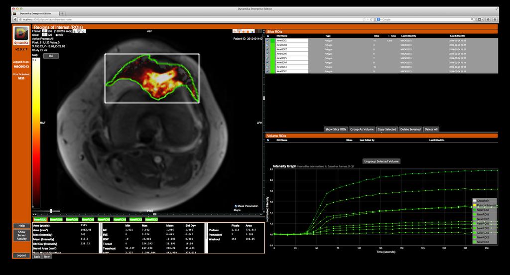

13 Hoffa s fat pad examples of a region of ROI Inter- and Intraclass coefficiency (ICC): DCE-MRI VOIs: MOAKS Hoffa synovitis: 0.58 (single reader only intra-icc)


14 Synovitis / DCE-MRI / Hoffa fat pad / pain / knee Pdw IRE Map ME Map 3D T1w + C

15 o Primary Outcome Measures (Spearman s rank correlation): KOOS pain and the perfusion variable, Inflammation (r=-0.42, p< ) o KOOS pain and MOAKS Hoffa-synovitis assessed on CE-MRI (r=-0.21, p<0.046) Conclusion: MOAKS Hoffa-synovitis assessed on CE-MRI and especially the perfusion variables from DCE-MRI reflecting inflammation in the IPFP, were related to pain and clinical symptoms in obese patients with KOA. DCE-MRI analysed with a dedicated software and a voxel by voxel time intensity curve method is a promising, and reproducible method to study the impact of inflammation in KOA
16 Thank you.
Assessing synovitis based on dynamic gadolinium-enhanced MRI and EULAR-OMERACT scores of the wrist in patients with rheumatoid arthritis
 Assessing synovitis based on dynamic gadolinium-enhanced MRI and EULAR-OMERACT scores of the wrist in patients with rheumatoid arthritis W. Wojciechowski 1,2, Z. Tabor 3, A. Urbanik 2 1 Medical Centre
The Effect of Lumbar Disc Degeneration on Positional Changes in the Lumbar Lordosis: a cross-sectional comparison with healthy controls
 The Effect of Lumbar Disc Degeneration on Positional Changes in the Lumbar Lordosis: a cross-sectional comparison with healthy controls Bjarke B Hansen, MD* Tom Bendix, MD, DMSc, Jacob Grindsted, MD Henning
Musculoskeletal Imaging at 3T with Simultaneous Use of Multipurpose Loop Coils
 Clinical Orthopedic Imaging Musculoskeletal Imaging at 3T with Simultaneous Use of Multipurpose Loop Coils Elena Ferrer 1 ; Rafael Coronado Santos 2 1 Radiology Department, Clínica Creu Blanca, Barcelona,
Why Talk About Technique? MRI of the Knee:
 Why Talk About Technique? MRI of the Knee: Part 1 - Imaging Techniques Mark Anderson, M.D. University of Virginia Health Sciences Center Charlottesville, Virginia Always had an interest teach our fellows
This presentation is the intellectual property of the author. Contact them at for permission to reprint and/or distribute.
 MRI of the Knee Jennifer Swart, M.D. Musculoskeletal Radiology South Texas Radiology Group Financial Disclosure Dr. Jennifer Swart has no relevant financial relationships with commercial interests to disclose.
Effects of body mass index, infrapatellar fat pad volume and age on patellar cartilage defect
 Acta Orthop. Belg., 2015, 81, 41-46 ORIGINAL STUDY Effects of body mass index, infrapatellar fat pad volume and age on patellar cartilage defect Semra Duran, Ertugrul Aksahin, Onur Kocadal, Cem Nuri Aktekin,
This presentation is the intellectual property of the author. Contact them for permission to reprint and/or distribute.
 MRI of the Knee Jennifer Swart, M.D. Musculoskeletal Radiology South Texas Radiology Group Outline Coils, Patient Positioning Acquisition Parameters, Planes and Pulse Sequences Knee Arthrography Normal
In vivo diffusion tensor imaging (DTI) of articular cartilage as a biomarker for osteoarthritis
 of articular cartilage as a biomarker for osteoarthritis") In vivo diffusion tensor imaging (DTI) of articular cartilage as a biomarker for osteoarthritis Jose G. Raya 1, Annie Horng 2, Olaf Dietrich 2, Svetlana Krasnokutsky 3, Luis S. Beltran 1, Maximilian F.
In vivo diffusion tensor imaging (DTI) of articular cartilage as a biomarker for osteoarthritis Jose G. Raya 1, Annie Horng 2, Olaf Dietrich 2, Svetlana Krasnokutsky 3, Luis S. Beltran 1, Maximilian F.
Synovial volume vs. synovial measurements from dynamic contrast enhanced MRI as
 Synovial volume vs. synovial measurements from dynamic contrast enhanced MRI as measures of response in osteoarthritis Andrew D Gait, PhD 1,, Richard Hodgson, BM, PhD,,, Matthew J Parkes, BSc,, Charles
RECENT ADVANCES IN CLINICAL MR OF ARTICULAR CARTILAGE
 In Practice RECENT ADVANCES IN CLINICAL MR OF ARTICULAR CARTILAGE By Atsuya Watanabe, MD, PhD, Director, Advanced Diagnostic Imaging Center and Associate Professor, Department of Orthopedic Surgery, Teikyo
Consortium of MS Centres Guidelines Revised Standardized MRI Protocol. for the Diagnosis and Follow-up of MS. David K.B.
 Consortium of MS Centres Guidelines Revised Standardized MRI Protocol for the Diagnosis and Follow-up of MS David K.B. Li MD FRCPC Indianapolis, Indiana May 27, 2015 Disclosure I have received research
Non Contrast MRA. Mayil Krishnam. Director, Cardiovascular and Thoracic Imaging University of California, Irvine
 Non Contrast MRA Mayil Krishnam Director, Cardiovascular and Thoracic Imaging University of California, Irvine No disclosures Non contrast MRA-Why? Limitations of CTA Radiation exposure Iodinated contrast
Numerous studies have demonstrated that magnetic resonance imaging (MRI) is more sensitive for detection of
 is more sensitive for detection of") Reducing Invasiveness, Duration, and Cost of Magnetic Resonance Imaging in Rheumatoid Arthritis by Omitting Intravenous Contrast Injection Does It Change the Assessment of Inflammatory and Destructive
Reducing Invasiveness, Duration, and Cost of Magnetic Resonance Imaging in Rheumatoid Arthritis by Omitting Intravenous Contrast Injection Does It Change the Assessment of Inflammatory and Destructive
Original Research JOURNAL OF MAGNETIC RESONANCE IMAGING 22: (2005)
") JOURNAL OF MAGNETIC RESONANCE IMAGING 22:788 793 (2005) Original Research STIR vs. T1-Weighted Fat-Suppressed Gadolinium- Enhanced MRI of Bone Marrow Edema of the Knee: Computer-Assisted Quantitative Comparison
JOURNAL OF MAGNETIC RESONANCE IMAGING 22:788 793 (2005) Original Research STIR vs. T1-Weighted Fat-Suppressed Gadolinium- Enhanced MRI of Bone Marrow Edema of the Knee: Computer-Assisted Quantitative Comparison
KNEE ALIGNMENT SYSTEM (KAS) MRI Protocol
 MRI Protocol") KNEE ALIGNMENT SYSTEM (KAS) MRI Protocol Sample referral sticker Referral Sticker Insert here Corin 17 Bridge Street Pymble NSW Australia 2073 P: +61 (0)2 9497 7400 F: +61 (0)2 9497 7498 E: KAS.customerservice@coringroup.com
KNEE ALIGNMENT SYSTEM (KAS) MRI Protocol Sample referral sticker Referral Sticker Insert here Corin 17 Bridge Street Pymble NSW Australia 2073 P: +61 (0)2 9497 7400 F: +61 (0)2 9497 7498 E: KAS.customerservice@coringroup.com
Case Report: Knee MR Imaging of Haemarthrosis in a Case of Haemophilia A
 Clinical > Pediatric Imaging Case Report: Knee MR Imaging of Haemarthrosis in a Case of Haemophilia A M. A. Weber, J. K. Kloth University Hospital Heidelberg, Department of Diagnostic and Interventional
High Field MR of the Spine
 Department of Radiology University of California San Diego 3T for MR Applications Advantages High Field MR of the Spine Increased signal-to-noise Better fat suppression Increased enhancement with gadolinium
MR Advance Techniques. Vascular Imaging. Class II
 MR Advance Techniques Vascular Imaging Class II 1 Vascular Imaging There are several methods that can be used to evaluate the cardiovascular systems with the use of MRI. MRI will aloud to evaluate morphology
MRI of Cartilage. D. BENDAHAN (PhD)
") MRI of Cartilage D. BENDAHAN (PhD) Centre de Résonance Magnétique Biologique et Médicale UMR CNRS 7339 Faculté de Médecine de la Timone 27, Bd J. Moulin 13005 Marseille France david.bendahan@univ-amu.fr
MRI of Cartilage D. BENDAHAN (PhD) Centre de Résonance Magnétique Biologique et Médicale UMR CNRS 7339 Faculté de Médecine de la Timone 27, Bd J. Moulin 13005 Marseille France david.bendahan@univ-amu.fr
Orthopedic Hardware Imaging Part II: MRI v. Metal
 Orthopedic Hardware Imaging Trent Roth, MD And Lauren Ladd, MD Indiana University School of Medicine IU Health Physicians-Radiology Recap: Imaging Techniques Radiography Standard for initial and surveillance
醫用磁振學 MRM 肌肉骨骼磁振造影簡介 肌肉骨骼磁振造影. 本週課程內容 General Technical Considerations 肌肉骨骼磁振造影簡介 盧家鋒助理教授國立陽明大學生物醫學影像暨放射科學系
 本週課程內容 http://www.ym.edu.tw/~cflu 肌肉骨骼磁振造影簡介 醫用磁振學 MRM 肌肉骨骼磁振造影 盧家鋒助理教授國立陽明大學生物醫學影像暨放射科學系 alvin4016@ym.edu.tw MRI of the musculoskeletal system (5th/6th edition) Editor: Thomas H. Berquist MD 2 General
Neuroradiology MR Protocols
 Neuroradiology MR Protocols Brain protocols N 1: Brain MRI without contrast N 2: Pre- and post-contrast brain MRI N 3 is deleted N 4: Brain MRI without or pre-/post-contrast (seizure protocol) N 5: Pre-
FieldStrength. Achieva 3.0T enables cutting-edge applications, best-in-class MSK images
 FieldStrength Publication for the Philips MRI Community Issue 33 December 2007 Achieva 3.0T enables cutting-edge applications, best-in-class MSK images Palo Alto Medical Clinic Sports Medicine Center employs
Can SCMR CMR protocol recommendations
 Can SCMR CMR protocol recommendations V1.3 - April 2009 CanSCMR CMR Protocol and SOP Recommendation 2009 (15 minutes) 2 Planning of LV fct. real time multiple axes Realtime 3 cine long axis 6 long axes
Magnetic Resonance Imaging. Basics of MRI in practice. Generation of MR signal. Generation of MR signal. Spin echo imaging. Generation of MR signal
 Magnetic Resonance Imaging Protons aligned with B0 magnetic filed Longitudinal magnetization - T1 relaxation Transverse magnetization - T2 relaxation Signal measured in the transverse plane Basics of MRI
Contrast-enhanced Breast MRI RSSA 2013
 Contrast-enhanced Breast MRI RSSA 2013 Prof. dr. Maurice van den Bosch University Medical Center Utrecht, the Netherlands Index 1) Breast cancer 2) Why MRI of the breast 3) Technique 4) Interpretation
Abdominal applications of DWI
 Postgraduate course, SPR San Antonio (Texas), May 14-15, 2013 Abdominal applications of DWI Rutger A.J. Nievelstein Wilhelmina Children s s Hospital, Utrecht (NL) Outline What is DWI? How to perform? Challenges
Knee, Ankle, and Foot: Normal and Abnormal Features with MRI and Ultrasound Correlation. Disclosures. Outline. Joint Effusion. Suprapatellar recess
 Knee, Ankle, and Foot: Normal and Abnormal Features with MRI and Ultrasound Correlation Jon A. Jacobson, M.D. Professor of Radiology Director, Division of Musculoskeletal Radiology University of Michigan
Anatomical and Functional MRI of the Pancreas
 Anatomical and Functional MRI of the Pancreas MA Bali, MD, T Metens, PhD Erasme Hospital Free University of Brussels Belgium mbali@ulb.ac.be Introduction The use of MRI to investigate the pancreas has
3D-Black-Blood 3T-MRI for the Diagnosis of thoracic large Vessel Vasculitis: A Feasibility Study
 3D-Black-Blood 3T-MRI for the Diagnosis of thoracic large Vessel Vasculitis: A Feasibility Study Tobias Saam 1, MD; Karla M. Treitl 1, MD; Stefan Maurus 1 ; Nora N. Kammer 1, MD; Hendrik Kooijman 2 ; PhD;
Personalized Solutions. MRI Protocol for PSI and Signature Guides
 Personalized Solutions MRI Protocol for PSI and Signature Guides 2 Personalized Solutions MRI Protocol for PSI and Signature Guides Purpose and Summary This protocol is applicable for the Zimmer Biomet
JMSCR Vol 05 Issue 07 Page July 2017
 www.jmscr.igmpublication.org Impact Factor 5.84 Index Copernicus Value: 83.27 ISSN (e)-2347-176x ISSN (p) 2455-0450 DOI: https://dx.doi.org/10.18535/jmscr/v5i7.84 Anatomical Differences Between T2 WI FSE
SMRT Student Scope Submission
 SMRT Student Scope Submission Title: Cardiac MRI Imaging Authors: Bridget Pomponio Title and Author(s) Supervisor Name: Anthony Festa R.T. (R) (MR) Hospital of the University of Pennsylvania Date of Submission:
Aortic Vessel Wall Imaging Using 3D Phase Sensitive Inversion Recovery in Children and Young Adults
 Aortic Vessel Wall Imaging Using 3D Phase Sensitive Inversion Recovery in Children and Young Adults Animesh Tandon, MD, MS 1,2, Tarique Hussain, MD, PhD 1,2, Andrew Tran, MD, MS 3, René M Botnar, PhD 4,
MRI Abdomen Protocol Pancreas/MRCP with Contrast
 MRI Abdomen Protocol Pancreas/MRCP with Contrast Reviewed By: Brett Mollard, MD; Anna Ellermeier, MD Last Reviewed: July 2018 Contact: (866) 761-4200 Standard uses: 1. Characterization of cystic and solid
Assessment and multiparametric functional MRI evaluation of Arthritis
 Assessment and multiparametric functional MRI evaluation of Arthritis 1 2 3 4 T. Martin Noguerol, MD 1 ; A. Luna, MD 1 M. Gomez Cabrera 3, MD; J Vilanova 2, MD, PhD; M. Romero Rivera MD 3 ; F Caro Mateo
OASIS 1.2T: MULTIPARAMETRIC MRI OF PROSTATE CANCER
 OASIS 1.2T: MULTIPARAMETRIC MRI OF PROSTATE CANCER By Dr. John Feller, MD, Radiologist Desert Medical Imaging, Palm Springs, CA MRI is clinically accepted as the best imaging modality for displaying anatomical
Changes in bone marrow lesions in response to weight-loss in obese knee osteoarthritis patients: a prospective cohort study
 Gudbergsen et al. BMC Musculoskeletal Disorders 2013, 14:106 RESEARCH ARTICLE Open Access Changes in bone marrow lesions in response to weight-loss in obese knee osteoarthritis patients: a prospective
Concise report RHEUMATOLOGY
 RHEUMATOLOGY Rheumatology 2012;51:2034 2038 doi:10.1093/rheumatology/kes124 Advance Access publication 30 July 2012 Concise report Head-to-head comparison of quantitative and semi-quantitative ultrasound
Magnetic Resonance Imaging of Inflammatory Lesions in the Spine in Ankylosing Spondylitis Clinical Trials: Is Paramagnetic Contrast Medium Necessary?
 Magnetic Resonance Imaging of Inflammatory Lesions in the Spine in Ankylosing Spondylitis Clinical Trials: Is Paramagnetic Contrast Medium Necessary? KAY-GEERT A. HERMANN, ROBERT B.M. LANDEWÉ, JÜRGEN BRAUN,
Effect of intravenous contrast medium administration on prostate diffusion-weighted imaging
 Effect of intravenous contrast medium administration on prostate diffusion-weighted imaging Poster No.: C-1766 Congress: ECR 2015 Type: Authors: Keywords: DOI: Scientific Exhibit J. Bae, C. K. Kim, S.
Knee Articular Cartilage in an Asymptomatic Population : Comparison of T1rho and T2 Mapping
 TR_002 Technical Reports Knee Articular Cartilage in an Asymptomatic Population : Comparison of T1rho and T2 Mapping Min A Yoon 1,*, Suk-Joo Hong 1, Chang Ho Kang 2, Baek Hyun Kim 3 1 Korea University
MRI PEDIATRIC PROTOCOLS (Updated 6/19/2018)
") MRI PEDIATRIC PROTOCOLS (Updated 6/19/2018) *Please get or let us know where radiologist can review plain films. *For Texas Orthopedics and other Docs requesting only MSK section read for their pediatric
MRI PEDIATRIC PROTOCOLS (Updated 6/19/2018) *Please get or let us know where radiologist can review plain films. *For Texas Orthopedics and other Docs requesting only MSK section read for their pediatric
6/23/2009. Inversion Recovery (IR) Techniques and Applications. Variations of IR Technique. STIR, FLAIR, TI and TI Null. Applications of IR
 Techniques and Applications. Variations of IR Technique. STIR, FLAIR, TI and TI Null. Applications of IR") The Anatomy of Basic R Pulse Sequences Inversion Recovery () Techniques and Applications Chen Lin, PhD Indiana University School of edicine & Clarian Health Partners agnetization Preparation Section Chemical
The Anatomy of Basic R Pulse Sequences Inversion Recovery () Techniques and Applications Chen Lin, PhD Indiana University School of edicine & Clarian Health Partners agnetization Preparation Section Chemical
MR imaging of the knee in marathon runners before and after competition
 Skeletal Radiol (2001) 30:72 76 International Skeletal Society 2001 ARTICLE W. Krampla R. Mayrhofer J. Malcher K.H. Kristen M. Urban W. Hruby MR imaging of the knee in marathon runners before and after
Supplementary Online Content
 Supplementary Online Content Hooshmand B, Magialasche F, Kalpouzos G, et al. Association of vitamin B, folate, and sulfur amino acids with brain magnetic resonance imaging measures in older adults: a longitudinal
Correspondence should be addressed to Thomas Kurien;
 Case Reports in Orthopedics Volume 2016, Article ID 6043497, 5 pages http://dx.doi.org/10.1155/2016/6043497 Case Report Resection and Resolution of Bone Marrow Lesions Associated with an Improvement of
Tissue-engineered medical products Evaluation of anisotropic structure of articular cartilage using DT (Diffusion Tensor)-MR Imaging
-MR Imaging") Provläsningsexemplar / Preview TECHNICAL REPORT ISO/TR 16379 First edition 2014-03-01 Tissue-engineered medical products Evaluation of anisotropic structure of articular cartilage using DT (Diffusion Tensor)-MR
Provläsningsexemplar / Preview TECHNICAL REPORT ISO/TR 16379 First edition 2014-03-01 Tissue-engineered medical products Evaluation of anisotropic structure of articular cartilage using DT (Diffusion Tensor)-MR
Available online at
 Original Research Article Evaluation of knee joint by MRI in 65 patients Gulamus sibtain asad *, Himanshu Singla, Ankit vasoya, P. J. Jhala 2 2 nd year Resident, 2 Professor Radiology Department, SBKS
Iron deposits in the knee joints of a thalassemic patient
 Iron deposits in the knee joints of a thalassemic patient The Harvard community has made this article openly available. Please share how this access benefits you. Your story matters Citation Economides,
dgemric Effectively Predicts Cartilage Damage Associated with Femoroacetabular Impingement
 Riccardo Lattanzi 1,2 Catherine Petchprapa 2 Daniele Ascani 1 Roy I. Davidovitch 3 Thomas Youm 3 Robert J. Meislin 3 Michael. Recht 2 1 The Bernard and Irene Schwartz Center for Biomedical Imaging, New
Cover Page. The handle holds various files of this Leiden University dissertation.
 Cover Page The handle http://hdl.handle.net/1887/40654 holds various files of this Leiden University dissertation. Author: Stomp, W. Title: MR imaging in early rheumatoid arthritis : techniques and applications
Musculoskeletal MR Protocols
 Musculoskeletal MR Protocols Joint-based protocols MSK 1: Shoulder MRI MSK 1A: Shoulder MR arthrogram MSK 1AB: Shoulder MR arthrogram (instability protocol) MSK 2: Elbow MRI MSK 2A: Elbow MR arthrogram
How Much Tesla Is Too Much?
 How Much Tesla Is Too Much? Johnny U. V. Monu, MB, BS; MSc Professor of Radiology and Orthopedics University of Rochester School of Medicine Rochester, New York Historical Timeline Clinical Imaging 1970
Sensitivity and Specificity in Detection of Labral Tears with 3.0-T MRI of the Shoulder
 Magee and Williams MRI for Detection of Labral Tears Musculoskeletal Imaging Clinical Observations C M E D E N T U R I C L I M G I N G JR 2006; 187:1448 1452 0361 803X/06/1876 1448 merican Roentgen Ray
Revised Dec Spine MR Protocols
 Spine MR Protocols Sp 1: Cervical spine MRI without contrast Sp 2: Pre- and post-contrast cervical spine MRI Sp 3: Pre- and post-contrast cervical spine MRI (multiple sclerosis protocol) Sp 4: Thoracic
Fully-Automatic Determination of the Arterial Input Function for Dynamic Contrast-Enhanced Pulmonary MR Imaging (DCE-pMRI)
") Fully-Automatic Determination of the Arterial Input Function for Dynamic Contrast-Enhanced Pulmonary MR Imaging (DCE-pMRI) Kohlmann P. 1, Laue H. 1, Anjorin A. 2, Wolf U. 3, Terekhov M. 3, Krass S. 1,
Fully-Automatic Determination of the Arterial Input Function for Dynamic Contrast-Enhanced Pulmonary MR Imaging (DCE-pMRI) Kohlmann P. 1, Laue H. 1, Anjorin A. 2, Wolf U. 3, Terekhov M. 3, Krass S. 1,
MRI of Acute Abdominal Pain in Pregnancy
 MRI of Acute Abdominal Pain in Pregnancy Ivan Pedrosa, M.D. Chief of MRI Jack Reynolds MD Chair in Radiology Associate Professor of Radiology UT Southwestern Medical Center Advanced Imag ing Research Center
Case Reports: Tumor Detection by Diffusion-Weighted MRI and ADC-Mapping with Correlation to PET/CT Results
 Case Reports: Tumor Detection by Diffusion-Weighted MRI and ADC-Mapping with Correlation to PET/CT Results Matthias Philipp Lichy, M.D.; Philip Aschoff, M.D.; Christina Pfannenberg, M.D.; Schlemmer Heinz-Peter,
The Low Sensitivity of Fluid-Attenuated Inversion-Recovery MR in the Detection of Multiple Sclerosis of the Spinal Cord
 The Low Sensitivity of Fluid-Attenuated Inversion-Recovery MR in the Detection of Multiple Sclerosis of the Spinal Cord Mark D. Keiper, Robert I. Grossman, John C. Brunson, and Mitchell D. Schnall PURPOSE:
Meniscus T2 Relaxation Time at Various Stages of Knee Joint Degeneration
 Meniscus T2 Relaxation Time at Various Stages of Knee Joint Degeneration Richard Kijowski, Michael Fazio, Benjamin Beduhn, and Fang Liu Department of Radiology University of Wisconsin School of Medicine
DTI fiber tracking at 3T MR using b-1000 value in the depiction of periprostatic nerve before and after nervesparing prostatectomy
 DTI fiber tracking at 3T MR using b-1000 value in the depiction of periprostatic nerve before and after nervesparing prostatectomy Poster No.: C-2328 Congress: ECR 2012 Type: Scientific Paper Authors:
Imaging of Articular Cartilage
 Clinical Imaging of Articular Cartilage Imaging of Articular Cartilage Prof. Dr. K. Verstraete Ghent University Introduction : Articular Cartilage Histology and biochemical composition Review of Imaging
Tissue perfusion in knee osteoarthritis Implications for exercise therapy
 U N I V E R S I T Y O F C O P E N H A G E N F A C U L T Y O F H E A L T H A N D M E D I C A L S C I E N C E S Tissue perfusion in knee osteoarthritis Implications for exercise therapy PhD thesis Elisabeth
Ultrasound Evaluation of Masses
 Ultrasound Evaluation of Masses Jon A. Jacobson, M.D. Professor of Radiology Director, Division of Musculoskeletal Radiology University of Michigan Disclosures: Consultant: Bioclinica Advisory Panel: GE,
Methods of MR Fat Quantification and their Pros and Cons
 Methods of MR Fat Quantification and their Pros and Cons Michael Middleton, MD PhD UCSD Department of Radiology International Workshop on NASH Biomarkers Washington, DC April 29-30, 2016 1 Disclosures
Liver Fat Quantification
 Liver Fat Quantification Jie Deng, PhD, DABMP Department of Medical Imaging May 18 th, 2017 Disclosure Research agreement with Siemens Medical Solutions 2 Background Non-alcoholic fatty liver diseases
The influence of preserving Infrapatellar fatpad in ACL reconstruction
 The influence of preserving Infrapatellar fatpad in ACL reconstruction Kazuki Asai 1), Junsuke Nakase 1), Kengo Shimozaki 1), Kazu Toyooka 1), Hiroyuki Tsuchiya 1) 1) Department of Orthopaedic Surgery,
CAN SOFT TISSUES STRUCTURES DIFFERENTIATE BETWEEN DYSPLASIA AND CAM-FAI OF THE HIP?
 CAN SOFT TISSUES STRUCTURES DIFFERENTIATE BETWEEN DYSPLASIA AND CAM-FAI OF THE HIP? A Le Bouthillier, KS Rakhra 1, PE Beaulé 2, RCB Foster 1 1 Department of Medical Imaging 2 Division of Orthopaedic Surgery
Viviane Khoury, MD. Assistant Professor Department of Radiology University of Pennsylvania
 U Penn Diagnostic Imaging: On the Cape Chatham, MA July 11-15, 2016 Viviane Khoury, MD Assistant Professor Department of Radiology University of Pennsylvania Hip imaging has changed in recent years: new
This copy is for personal use only. To order printed copies, contact Purpose: Materials and Methods: Results: Conclusion:
 This copy is for personal use only. To order printed copies, contact reprints@rsna.org Diagnostic Accuracy of a Fluidattenuated Inversion-Recovery Sequence with Fat Suppression for Assessment of Peripatellar
Ultrasound of the Knee
 Ultrasound of the Knee Jon A. Jacobson, M.D. Professor of Radiology Director, Division of Musculoskeletal Radiology University of Michigan Disclosures: Consultant: Bioclinica Book Royalties: Elsevier Advisory
Perfusion MRI. Youngkyoo Jung, PhD Associate Professor Radiology, Biomedical Engineering, and Clinical & Translational Science Institute
 Perfusion MRI Youngkyoo Jung, PhD Associate Professor Radiology, Biomedical Engineering, and Clinical & Translational Science Institute Perfusion The delivery of blood to a capillary bed in tissue Perfusion
Cover Page. The handle holds various files of this Leiden University dissertation.
 Cover Page The handle http://hdl.handle.net/1887/35124 holds various files of this Leiden University dissertation. Author: Wokke, Beatrijs Henriette Aleid Title: Muscle MRI in Duchenne and Becker muscular
Unenhanced and dynamic contrast enhanced (DCE) MRI in assessment of scaphoid fracture non-union revisited: role in pre-operative planning
 MRI in assessment of scaphoid fracture non-union revisited: role in pre-operative planning") Unenhanced and dynamic contrast enhanced (DCE) MRI in assessment of scaphoid fracture non-union revisited: role in pre-operative planning Poster No.: B-0440 Congress: ECR 2014 Type: Authors: Keywords:
Unenhanced and dynamic contrast enhanced (DCE) MRI in assessment of scaphoid fracture non-union revisited: role in pre-operative planning Poster No.: B-0440 Congress: ECR 2014 Type: Authors: Keywords:
Magnetic Resonance Angiography
 Magnetic Resonance Angiography 1 Magnetic Resonance Angiography exploits flow enhancement of GR sequences saturation of venous flow allows arterial visualization saturation of arterial flow allows venous
MR Tumor Staging for Treatment Decision in Case of Wilms Tumor
 MR Tumor Staging for Treatment Decision in Case of Wilms Tumor G. Schneider, M.D., Ph.D.; P. Fries, M.D. Dept. of Diagnostic and Interventional Radiology, Saarland University Hospital, Homburg/Saar, Germany
Chronic sport injuries of the knee
 Chronic sport injuries of the knee 5th Musculoskeletal MRI meeting 2018: Knee MRI Gustav Andreisek, MD, MBA Saturday, May 5th, 2018; 14:00-16:00 Professor of Radiology, University of Zurich and Head of
Quantitative evaluation of endolymphatic hydrops and acceptance testing in patients with Menière's disease
 Quantitative evaluation of endolymphatic hydrops and acceptance testing in patients with Menière's disease Award: Certificate of Merit Poster No.: C-0185 Congress: ECR 2014 Type: Scientific Exhibit Authors:
Localized anterior arthrofibrosis (Cyclops lesion) with no history of anterior cruciate ligament reconstruction: MRI findings.
 with no history of anterior cruciate ligament reconstruction: MRI findings.") Localized anterior arthrofibrosis (Cyclops lesion) with no history of anterior cruciate ligament reconstruction: MRI findings. Poster No.: P-0145 Congress: ESSR 2013 Type: Scientific Exhibit Authors: L.
Localized anterior arthrofibrosis (Cyclops lesion) with no history of anterior cruciate ligament reconstruction: MRI findings. Poster No.: P-0145 Congress: ESSR 2013 Type: Scientific Exhibit Authors: L.
Fat Suppression in the Abdomen
 Clinical How I do it? Fat Suppression in the Abdomen Wilhelm Horger Siemens Medical Solutions, Erlangen, Germany Introduction Due to the different chemical environment, hydrogen nuclei in - and in -tissue
Crohn s Disease. bowel. Two picks of diagnosis - 2nd and 6th decades of life. Multifactorial - Genetic and Environmental.
 Crohn s Disease Chronic and Inflammatory disease of the small and or large bowel. Two picks of diagnosis - 2nd and 6th decades of life. Multifactorial - Genetic and Environmental. In Israel: 40,000 patients.
Visualization of Endolymphatic Hydrops after Intratympanic Injection of Gd-DTPA: Comparison of 2D and 3D Real Inversion Recovery Imaging
 Magn Reson Med Sci, Vol. 10, No. 2, pp. 101 106, 2011 MAJOR PAPER Visualization of Endolymphatic Hydrops after Intratympanic Injection of Gd-DTPA: Comparison of 2D and 3D Real Inversion Recovery Imaging
Cone-Beam CT for MSK Extremities
 8/6/0 Diagnostic Image Quality Evaluation of an Extremity Cone-Beam CT Scanner: Pre-Clinical and First Clinical Results Abdullah Muhit Wojciech Zbijewski, J Webster Stayman John Yorkston, Nathan Packard,
Clinical Applications
 C H A P T E R 16 Clinical Applications In selecting pulse sequences and measurement parameters for a specific application, MRI allows the user tremendous flexibility to produce variations in contrast between
Repeatability of 2D FISP MR Fingerprinting in the Brain at 1.5T and 3.0T
 Repeatability of 2D FISP MR Fingerprinting in the Brain at 1.5T and 3.0T Guido Buonincontri 1,2, Laura Biagi 1,3, Alessandra Retico 2, Michela Tosetti 1,3, Paolo Cecchi 4, Mirco Cosottini 1,4,5, Pedro
Real-Time MRI of Joint Movement With TrueFISP
 JOURNAL OF MAGNETIC RESONANCE IMAGING 15:710 715 (2002) Technical Note Real-Time MRI of Joint Movement With TrueFISP Harald H. Quick, MSc, 1 * Mark E. Ladd, PhD, 1 Matthias Hoevel, MD, 2 Silke Bosk, RT,
UPPER EXTREMITY
 UPPER EXTREMITY 11-24-17 MSK TIPS: Ensure extremity of interest is as isocenter as possible SHIM all Fat sat scans!! Only use 4ch wrist if additional coverage needed If Contrast needed: Multihance.1mmol/kg
Supplementary Online Content
 Supplementary Online Content Schlaeger R, Papinutto N, Zhu AH, et al. Association between thoracic spinal cord gray matter atrophy and disability in multiple sclerosis. JAMA Neurol. Published online June
Comparative study of high resolusion ultrasonography and magnetic resonance imaging in diagnosing traumatic knee injuries & pathologies
 Original article: Comparative study of high resolusion ultrasonography and magnetic resonance imaging in diagnosing traumatic knee injuries & pathologies Dr. Rakesh Gujjar*, Dr. R. P. Bansal, Dr. Sandeep
Essentials of Clinical MR, 2 nd edition. 99. MRA Principles and Carotid MRA
 99. MRA Principles and Carotid MRA As described in Chapter 12, time of flight (TOF) magnetic resonance angiography (MRA) is commonly utilized in the evaluation of the circle of Willis. TOF MRA allows depiction
Supplementary Online Content
 Supplementary Online Content Gregg NM, Kim AE, Gurol ME, et al. Incidental cerebral microbleeds and cerebral blood flow in elderly individuals. JAMA Neurol. Published online July 13, 2015. doi:10.1001/jamaneurol.2015.1359.
Objectives 8/17/2011. Challenges in Cardiac Imaging. Challenges in Cardiac Imaging. Basic Cardiac MRI Sequences
 8/17/2011 Traditional Protocol Model for Tomographic Imaging Cardiac MRI Sequences and Protocols Frandics Chan, M.D., Ph.D. Stanford University Medical Center Interpretation Lucile Packard Children s Hospital
High-resolution diffusion-weighted MRI of the breast using readout-segmented EPI and single-shot EPI
 High-resolution diffusion-weighted MRI of the breast using readout-segmented EPI and single-shot EPI Objective: Compared to dynamic contrast enhanced MRI (DCE-MRI), image quality in diffusion-weighted
MRI Assessment of the Right Ventricle and Pulmonary Blood Flow, Perfusion and Ventilation
 MRI Assessment of the Right Ventricle and Pulmonary Blood Flow, Perfusion and Ventilation Dr. Richard Thompson Department of Biomedical Engineering University of Alberta Heart and Lung Imaging Many Constantly
Prevalence of Meniscal Radial Tears of the Knee Revealed by MRI After Surgery
 Downloaded from www.ajronline.org by 46.3.207.114 on 12/22/17 from IP address 46.3.207.114. Copyright RRS. For personal use only; all rights reserved Thomas Magee 1 Marc Shapiro David Williams Received
Magnetic resonance (MR) diffusion-weighted imaging (DWI) is
 diffusion-weighted imaging (DWI) is") Diagn Interv Radiol DOI 10.4261/1305-3825.DIR.3892-10.1 Turkish Society of Radiology 2010 ABDOMINAL IMAGING ORIGINAL ARTICLE Diffusion tensor imaging of the kidney at 3 Tesla: normative values and repeatability
Diagn Interv Radiol DOI 10.4261/1305-3825.DIR.3892-10.1 Turkish Society of Radiology 2010 ABDOMINAL IMAGING ORIGINAL ARTICLE Diffusion tensor imaging of the kidney at 3 Tesla: normative values and repeatability
MRI/MRS Biomarkers. Robert E. Lenkinski, Ph.D.
 MRI/MRS Biomarkers Robert E. Lenkinski, Ph.D. Disclosure GE Healthcare-Research Grant Aspect MR-Scientific Advisor Aposense-Scientific Advisor Brainwatch-Scientific Advisor I will be discussing off-label
Advances in MRI for Radiation Therapy
 Advances in MRI for Radiation Therapy Jing Cai, PhD, DABR Associate Professor Department of Radiation Oncology Duke University Medical Center, Durham NC Advances in MRI Structural Imaging Fast Imaging