Diffusione e spettroscopia
|
|
|
- Beatrice White
- 6 years ago
- Views:
Transcription
 Unit of Diagnostic")

1 Institute for Cancer Research and Treatment Candiolo -Torino (Italy) Unit of Diagnostic Imaging - Director Dr. Daniele Regge Diffusione e spettroscopia Laura Martincich Parma Aprile 2011
2 What is Molecular Diffusion? How can we detect Molecular Diffusion on MRI? Which information provides DWI? How can we apply DWI on the breast?

3 What is Molecular Diffusion? Anisotropic Diffusion Biological tissues are highly heterogeneous media that consist of o various compartments and barriers with different diffusivity The movement of water molecules during diffusion random displacement is impeded by compartmental boundaries and other molecular barriers, that the diffusion distance is reduced compared to unrestricted diffusion Hagmann, Radiographics 2006 O Flynn, Breast Cancer Research 2011
4 How can we detect Molecular Diffusion on MRI? b value =0 and b value >0 Short acquisition time QuickTime and a Cinepak decompressor are needed to see this picture. Courtesy of Dr. Bracco, Medical Physics IRCC


5 Which information provides DWI? High diffusion = Major loss of signal e.g. liquid content Low diffusion = Minor loss of signal e.g. high cellular density tissues


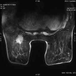
6 Qualitative information: ADC maps + = b 0 s/mm 2 b 900s/mm 2


7 Quantitative information: ADC value ADC= -(1/b)In(S/S 0 ) S and S 0 = Signal intensity with and without diffusion sensitisation b = level of sensitisation to diffusion of the sequence Related to cellular density and extracellular volume Lee, Clin Cancer Res 2007 ADC ± = 0.96±0.3x10 0.3x10⁻³ mm 2 /s
8 How can we apply DWI on the breast? Characterisation of Breast Lesion Improving specificity and PPV of DCE-MRI Monitoring the tumoural response to medical treatment
9 Characterisation of Breast Lesions Studies included if: 1.5T Diagnostic criteria clearly stated Calculation of ADC and SD Tsushima, JMRI 2009
10 Characterisation of Breast Lesions ADC Malignant mm 2 /s ADC Benign mm 2 /s ADC Normal mm 2 /s Tsushima, JMRI 2009
11 Characterisation of Breast Lesions Lesion Presentation Partridge, AJR 2009
12 Characterisation of Breast Lesions Lesion Size Partridge, AJR 2009

13 Characterisation of Breast Lesions Woodhams, AJR 2009


14 CARCINOMA INFILTRANTE DELLA MAMMELLA. Istotipo: mucinoso pt2 pn0(i-)(sn) G2 ER 98%, PgR 99%, HER2 0+, Ki-67 21%


15 CARCINOMA DUTTALE INFILTRANTE ER-,, PgR-,, HER 3+
16 How can we apply DWI on the breast? Characterisation of Breast Lesion Improving the specificity and PPV of DCE-MRI Monitoring the tumoural response to medical treatment
17 Improving specificity and PPV of DCE-MRI

18 Improving specificity and PPV of DCE-MRI 83 lesions recommended for biopsy (BIRADS 4,5) PPV DCE-MRI alone 37% lesions <1cm 35% PPV DWI + DCE-MRI 47% lesions <1 1 cm 52% prevention of benign biopsy 17% Partridge, AJR 2009
19 How can we apply DWI on the breast? Characterisation of Breast Lesion Improving specificity and PPV of DCE-MRI Monitoring the tumoural response to medical treatment
20 Monitoring the tumoural response to medical treatment Background based on Preclinical models Higher ADC values were observed in treated cancers with respect to untreated DWI has the potential: to detect and quantify cellular changes that occur in response to therapeutic intervention prior to macroscopic changes in size to provide a window to peer into the temporal dynamics of drugs action to be an imaging surrogate for predicting the tumoral response Author Cell line Therapy Timing from baseline DWI ADC increase in treated cancer ADC in control group p Galons 1999 MCF7/S Taxane 2 days 50% Resistant Cells MCF7/D40 0% na Lee 2007 MX-1 Human BC Cyclophosphamide 4-77 days 94% Untreated cancer 17% Galons, Neoplasia 1999 Ross, Mol Cancer Therapy 2003 Theilmann, Neoplasia 2004 Lee, Clin Cancer Res 2007
 Thoeny HC, JMRI")
21 ADC value as biomarkers in the tumour response Water motion within a tumour increases over time after medical treatment, t as a result of membrane damage with subsequent reduction of cells s density (apoptotic cell death) Thoeny HC, JMRI 2010
 d Sensitivity 97% Specificity 89% Accuracy 96% Sensitivity 93% Specificity 56% Accuracy 89% Woodhams, Radiology")
22 Monitoring tumour response to neoadjuvant medical treatment 70 locally advanced breast cancer in 69 patients Imaging: before and after PCT Standard of Reference: histhopatology (pcr vs size of residual disease) d Sensitivity 97% Specificity 89% Accuracy 96% Sensitivity 93% Specificity 56% Accuracy 89% Woodhams, Radiology 2010

23 CDI ER 90%; PgR 80%; Herceptest 3+, Ki 67 48% p r e p o s t


24 p r e p o s t PARENCHIMA MAMMARIO CON AREA DI FIBROPLASIA CICATRIZIALE, VEROSIMILE ESITO DI PRECEDENTE TRATTAMENTO CHEMIOTERAPICO Grado di risposta istopatologica a pregressa CT neoadiuvante: 5/5 (risposta completa) sec. Smith et al [JCO 2002 ].
25 Monitoring the tumoural response to medical treatment Clinical applications PCT: 3 cycles of Epirubicin and Ciclophosphamide Imaging: at Baseline and after 1 st and 2 nd cycles p=0.05 p=0.04 n.s. n.s.


26 Pre PCT During PCT Histopathological grade of response: 5/5 No invasive tumour cells identifiable in the sections from the site of the previous tumour Core-biopsy: IDC


27 Pre PCT During PCT Histopathological grade of response: 1/5 Some alteration to individual malignant cells but no reduction in overall numbers as compared with the pretreatment core-biopsy Core-biopsy: ILC

28 Diffusion Weighted Imaging Cancer gene pathways Yenkeelov, Magn Reson Imaging 2007 Vogelstein, Nature Med 2004

![Those included tissue VEGF, activated VEGFR2 status (phosphorylated VEGFR2 [p- VEGFR2]), total VEGFR2, tumor](/docs-images/73/69168785/images/29-2.jpg "MVD, tumor cell apoptosis, and proliferation.")
29 Potential tool in Monitoring the tumoral response to targheted-therapies therapies The objectives were to access molecular changes with bevacizumab alone and in combination with chemotherapy. Those included tissue VEGF, activated VEGFR2 status (phosphorylated VEGFR2 [p- VEGFR2]), total VEGFR2, tumor MVD, tumor cell apoptosis, and proliferation. The increase of apoptosis again persisted with addition of chemotherapy (72.7%; P=.013)
30 A c q u i s i t i o n P o s t P r o c e s s i n g? Which and how many b-value? b? Standardization and Reproducibility? ADC value thresholds? Pitfalls and False cases
31 Which and how many b-value? b Diffusion weighting is expressed by the b-value b (s/mm 2 ) The loss of signal due solely to diffusion is described by the equatione quation: S = S 0 e -badc S and S 0 = signal intensity with and without diffusion sensitisation ADC = Apparent Diffusion Coefficient b = level of sensitisation to diffusion of the sequence b depends on: Time interval between the two pulse gradients Intensity of the two pulse gradients Duration of the two pulse gradients As b increases, so too does the weighting diffusion of the sequence

32 Which and how many b-value? b Perfusion-insensitive insensitive ADC values calculation should include low b value to extinguish flow signal For ADC quantification two b values should be used ( 100sec/mm( 2 and 500 but 1000sec/mm 2 ) with a monexponential decay. Padhani, Neoplasia 2009
33 Which and how many b-value? b b 0, 250, 500, 750 and 1000 s/mm 2 (scanner GEHC) ADC calculation using 5 b-values (0 and >0) These findings suggest that the higher b values are useful to distinguish benign from malignant lesions and that there is no need to use multiple b values in the DWI sequence, saving examination time Arantes Pereira, AJR 2009
34 Which and how many b-value? b Large maximum b value b 0 s/mm 2 Advantages elimination of signals from normal (noncancerous) tissue, better detectability of malignant lesions Disadvantages Decreased SNR b 900s/mm 2 Arantes Pereira, AJR 2009 Tsushima, JMRI 2009


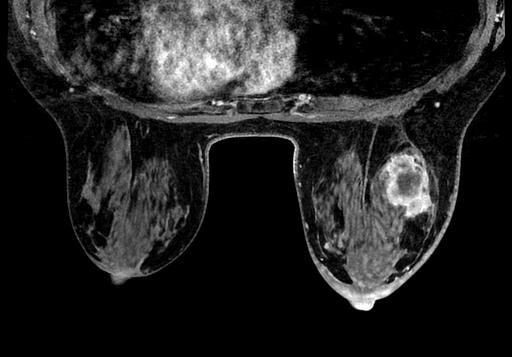
35 Which and how many b-value? b 500s/mm s/mm 2 Multicentric LIC. pt3(m) pn3a
36 Drawing the ROI Avoiding necrosis? Within the lesion margins Ideally, the region of interest (ROI) is contoured around lesions using images with the highest contrast between lesion and normal tissue. Subjective placement of smaller ROIs within lesions is not recommended particularly for response assessment studies Padhani AR, Neoplasia 2009 Liquefactive necrosis is characteristic of bacterial or fungal infections, because microbes stimulate the accumulation of inflammatory cells. Whatever the pathogenesis, liquefaction completely digests the dead cells.. The end result is transformation of the tissue into a liquid viscous mass. Coagulative necrosis implies preservation of the basic outline of the coagulated cell. The affected tissues exhibit a firm texture. Presumably, the injury or the subsequent increasing intracellular acidosis denatures not only structural proteins but also enzymes and so blocks the proteolysis of the cell. The process of coagulative necrosis, with preservation of the general tissue architecture, is characteristic of hypoxic death of cells in all tissues Robbins, Elsevier 2007


37 Drawing the ROI CDI G3 ER- PgR- HER2 3+

38 ADC value cut off? Sasaki, Radiology 2008
39 ADC value cut off? ADC Malignant mm 2 /s ADC Benign mm 2 /s ADC Normal mm 2 /s Tsushima, JMRI 2009
40


41 Pitfalls and false cases


42 Pitfalls and false cases 2008 BCS for right DCI 2011 Left nodal MTS


43 Pitfalls and false cases


44 Pitfalls and false cases Microfocolai multipli di DCIS. Istotipo: piatto e cribriforme; Grado nucleare: alto; Necrosi comedonica: presente


45 Pitfalls and false cases Microfocolai multipli di DCIS. Istotipo: piatto e cribriforme; Grado nucleare: alto; Necrosi comedonica: presente


46 Characterisation of Breast Lesions 13 subjects with breast cancer 16 Malignant lesions Intraindividual study Small lesions were visible more clearly at 3.0 T
47 Conclusion DWI is a promising but still investigational MR technique Limitation DCE-MRI guidance for (small) lesion detection Advantages for the application in clinical practice Short acquisition and post processing time Quantitative approach No contrast media administration required
48 Conclusion It could be a way To improve the diagnostic performance of breast MRI in the characterization of small MRI enhancing areas To detect tumoural changes due PCT Monitoring response to PCT in association with DCE- MRI before and after PCT and as a single technique during PCT Effectiveness of target-therapy therapy High quality equipment/technical consinstency
49 Spectroscopy
50 H 1 Magnetic Resonance Spectroscopy (MRS) Imaging of molecules, which generates detectable peaks at MRS MRS does not generate an image of the tumour directly, but the spectroscopic data can be obtained from a well localised area (voxel) Provides biochemical information about biological tissues by a generation of images of metabolites distribution (spectrum)
 ν Space Chemical structure")

51 H 1 Magnetic Resonance Spectroscopy (MRS) ν Space Chemical structure (metabolites distribution) Magnetic Resonance Imaging Magnetic Resonance Spectroscopy
52 H 1 Magnetic Resonance Spectroscopy (MRS) Metabolites Spectral Decomposition Water 4.7 ppm ( ) Lipid 1.3 ppm (0-2.5)
53 H 1 Magnetic Resonance Spectroscopy (MRS) Metabolites Choline compounds peak at 3.2ppm is considered a marker of active tumour Spectral Decomposition I D C Choline Compounds 3.2 ppm ( ) 3.34) Choline, phosphocoline, glyceroposphocholine, myoinositol, taurine Stanwell, Eur Radiol 2005 Sardanelli, Eur Radiol 2006
54 H 1 Magnetic Resonance Spectroscopy (MRS) Metabolites Imbalance between biosynthesis and catabolism of phospatidyl-choline Podo, NMR Biomed 1999

55 H 1 Magnetic Resonance Spectroscopy (MRS) Technique (1,5T) Single Voxel Spectroscopy The acquisition of a spectrum related to a single volume of interest (VOI) including the lesion detected Voxel shimming PRESS sequence Point Resolved Spectroscopy STEAM sequence Stimulated Echo Acquisition Mode Lesion size influences MRS


56 H 1 Magnetic Resonance Spectroscopy (MRS) Technique Lipid Sidebands from spurious echoes from mobile lipids Water Reduced spectra quality Obscuration of choline peak Sardanelli, Eur Radiol 2006 Bartella, Radiographics 2007

")


57 H 1 Magnetic Resonance Spectroscopy (MRS) Technique IDC Fibroadenoma Sidebands from spurious echoes from mobile lipids


58 H 1 Magnetic Resonance Spectroscopy (MRS) Technique Single Voxel Spectroscopy Water Unsuppressed and suppressed data
59 H 1 Magnetic Resonance Spectroscopy (MRS) Technique Single Voxel Spectroscopy PRESS sequence STEAM sequence Set up (5-10min) Water suppression Voxel shimming Check of field homogeneity and efficacy of prepulses + Inclusion of prepulses for water and fat suppression Echo time 135ms TR 1,5-3sec Signals acquired Acquisition Time 3-12min Bartella, Radiographics 2007
60 H 1 Magnetic Resonance Spectroscopy (MRS) Post Processing Reference Data r[n] Phase Correction DC Mixing Zero Phasing Linear Phase Correction Phase Spline Smoothing Phase Correction Vector c[n] Water Subtraction Water-suppressed Data s[n] Phase Correction Non quantitative/semiquantitative information Detection vs Non Detection of Choline peak Choline Signal (SNR) FFT S[k] Sardanelli, Eur Radiol 2006 Bartella, Radiographics 2007
61 H 1 Magnetic Resonance Spectroscopy (MRS) Clinical Application at 1,5T Characterization of Breast Lesions Potential obviation of biopsy in 57% of the cases Bartella, Radiographics 2007
62 H 1 Magnetic Resonance Spectroscopy (MRS) Clinical Application at 1,5T
63 Conclusion MRS may help in improving the diagnostic value of Breast MRI Evaluation of one lesion at a time MRS Established technique Approval by USA FDA Limitations Selection of the cases Lesion size: voxel >1cm 3 Presence of hematoma or metallic clip Long acquisition time Non quantitative method Dedicated team

64 DWI vs MRS H1 MRS was useful for characterizing breast lesions measuring 15mm or larger, and Diffusion-weighted imaging was useful for characterizing lesions of any size
65
Successful Breast MRI Program : The ingredients
 Successful Breast MRI Program : The ingredients Dr. Smriti Hari Associate Professor Deptt. Of Radiology All India Institute of Medical Sciences New Delhi How to perform Breast MRI Breast MRI descriptors
Breast MRI Update. Jeffrey C. Weinreb, MD, FACR Yale University School of Medicine
 Breast MRI Update Jeffrey C. Weinreb, MD, FACR jeffrey.weinreb@yale.edu Yale University School of Medicine I disclose the following financial relationships with relevant commercial interests: Bracco Bayer
MRI/MRS Biomarkers. Robert E. Lenkinski, Ph.D.
 MRI/MRS Biomarkers Robert E. Lenkinski, Ph.D. Disclosure GE Healthcare-Research Grant Aspect MR-Scientific Advisor Aposense-Scientific Advisor Brainwatch-Scientific Advisor I will be discussing off-label
A NEW BIOMARC TISSUE MARKER FOR BREAST BIOPSY: CLINICAL EVALUATION IN ULTRASOUND, MAMMOGRAPHY, CT SCANNING AND BREAST MRI
 A NEW BIOMARC TISSUE MARKER FOR BREAST BIOPSY: CLINICAL EVALUATION IN ULTRASOUND, MAMMOGRAPHY, CT SCANNING AND BREAST MRI Michael T. Nelson, M.D. 2 Sina Meisamy, M.D. 1,2 Adeka D. McIntosh, M.D. 1,2 Patrick
Pitfalls and Limitations of Breast MRI. Susan Orel Roth, MD Professor of Radiology University of Pennsylvania
 Pitfalls and Limitations of Breast MRI Susan Orel Roth, MD Professor of Radiology University of Pennsylvania Objectives Review the etiologies of false negative breast MRI examinations Discuss the limitations
Spiculated breast masses on MRI: Which category should we choose, 4 or 5?
 Spiculated breast masses on MRI: Which category should we choose, 4 or 5? Poster No.: C-1394 Congress: ECR 2015 Type: Scientific Exhibit Authors: N. Onishi, S. Kanao, M. Kataoka, M. Kawai, M. Iima, A.
Methods of MR Fat Quantification and their Pros and Cons
 Methods of MR Fat Quantification and their Pros and Cons Michael Middleton, MD PhD UCSD Department of Radiology International Workshop on NASH Biomarkers Washington, DC April 29-30, 2016 1 Disclosures
Mammographic imaging of nonpalpable breast lesions. Malai Muttarak, MD Department of Radiology Chiang Mai University Chiang Mai, Thailand
 Mammographic imaging of nonpalpable breast lesions Malai Muttarak, MD Department of Radiology Chiang Mai University Chiang Mai, Thailand Introduction Contents Mammographic signs of nonpalpable breast cancer
Abdominal applications of DWI
 Postgraduate course, SPR San Antonio (Texas), May 14-15, 2013 Abdominal applications of DWI Rutger A.J. Nievelstein Wilhelmina Children s s Hospital, Utrecht (NL) Outline What is DWI? How to perform? Challenges
Clinical application of 3.0 T proton MR spectroscopy in evaluation of pancreatic diseases
 Clinical application of 3.0 T proton MR spectroscopy in evaluation of pancreatic diseases Award: Cum Laude Poster No.: C-1762 Congress: ECR 2012 Type: Scientific Paper Authors: T. Su, E. Jin; Beijing/CN
Diffusion Weighted Imaging in Prostate Cancer
 Diffusion Weighted Imaging in Prostate Cancer Disclosure Information Vikas Kundra, M.D, Ph.D. No financial relationships to disclose. Education Goals and Objectives To describe the utility of diffusion-weighted
Outline. Why Image Animals?
 Small Animal Magnetic Resonance Imaging: Current Trends, Challenges and Perspectives for Pathological Imaging C. Chad Quarles Vanderbilt University Institute of Imaging Science Outline Why image animals?
T2, T2*, ute. Yeo Ju Kim. Radiology, Inha University Hospital, Incheon, Korea
 SY28-1 T2, T2*, ute Yeo Ju Kim Radiology, Inha University Hospital, Incheon, Korea T2 relaxation times relate to the rate of transverse magnetization decay, caused by the loss of phase coherence induced
Multiparametric imaging in oncology
 Multiparametric imaging in oncology p1 T p2 p2 T T p3 p1 p3 T Marco Ravanelli Roberto Maroldi The goal of traditional imaging is high spatial and contrast resolution diagnosis, tumor extent treatment planning,
Emerging Techniques in Breast Imaging: Contrast-Enhanced Mammography and Fast MRI
 Emerging Techniques in Breast Imaging: Contrast-Enhanced Mammography and Fast MRI Lilian Wang, M.D. Breast Imaging Section Department of Radiology Northwestern Medicine Overview Rationale for new imaging
The follow-up of uterine fibroids treated with HIFU: role of DWI and Dynamic contrast-study MRI
 The follow-up of uterine fibroids treated with HIFU: role of DWI and Dynamic contrast-study MRI Poster No.: C-1137 Congress: ECR 2011 Type: Authors: Keywords: DOI: Scientific Exhibit V. Zampa, V. Vallini,
Contrast-enhanced Breast MRI RSSA 2013
 Contrast-enhanced Breast MRI RSSA 2013 Prof. dr. Maurice van den Bosch University Medical Center Utrecht, the Netherlands Index 1) Breast cancer 2) Why MRI of the breast 3) Technique 4) Interpretation
Essentials of Clinical MR, 2 nd edition. 73. Urinary Bladder and Male Pelvis
 73. Urinary Bladder and Male Pelvis Urinary bladder carcinoma is best locally staged with MRI. It is important however to note that a thickened wall (> 5 mm) is a non-specific finding seen in an underfilled
1) Diffusion weighted imaging DWI is a term used to describe moving molecules due to random thermal motion. This motion is restricted by boundaries
 Diffusion weighted imaging DWI is a term used to describe moving molecules due to random thermal motion. This motion is restricted by boundaries") 1) Diffusion weighted imaging DWI is a term used to describe moving molecules due to random thermal motion. This motion is restricted by boundaries such as ligaments, membranes and macro molecules. Diffusion
1) Diffusion weighted imaging DWI is a term used to describe moving molecules due to random thermal motion. This motion is restricted by boundaries such as ligaments, membranes and macro molecules. Diffusion
Effects of magnetic field strength and b value on the sensitivity and specificity of quantitative breast diffusion-weighted MRI
 Original Article Effects of magnetic field strength and b value on the sensitivity and specificity of quantitative breast diffusion-weighted MRI Mohammad Eghtedari 1, Jingfei Ma 2, Patricia Fox 3, Inanc
Radiological assessment of neoadjuvent chemotherapy for breast cancer
 XV th Balkan Congress of Radiology Budapest, Hungary, October 12 15, 2017 Radiological assessment of neoadjuvent chemotherapy for breast cancer V. Bešlagić C l i n i c o f R a d i o l o g y, U n i v e
Functional aspects of anatomical imaging techniques
 Functional aspects of anatomical imaging techniques Nilendu Purandare Associate Professor & Consultant Radiologist Tata Memorial Centre Functional/metabolic/molecular imaging (radioisotope scanning) PET
MR imaging of FIGO stage I uterine cervical cancer: The diagnostic impact of 3T-MRI
 MR imaging of FIGO stage I uterine cervical cancer: The diagnostic impact of 3T-MRI Poster No.: C-1191 Congress: ECR 2010 Type: Educational Exhibit Topic: Genitourinary Authors: M. Takeuchi, K. Matsuzaki,
Role of Proton MR Spectroscopy in Differentiation of Benign and Malignant Breast Lesions
 Med. J. Cairo Univ., Vol. 82, No. 1, June: 341-349, 2014 www.medicaljournalofcairouniversity.net Role of Proton MR Spectroscopy in Differentiation of Benign and Malignant Breast Lesions GALAL EL HAWARY,
How to Use MRI Following Neoadjuvant Chemotherapy (NAC) in Locally Advanced Breast Cancer
 in Locally Advanced Breast Cancer") Global Breast Cancer Conference 2016 & 5 th International Breast Cancer Symposium April 29 th 2016, 09:40-10:50 How to Use MRI Following Neoadjuvant Chemotherapy (NAC) in Locally Advanced Breast Cancer
Global Breast Cancer Conference 2016 & 5 th International Breast Cancer Symposium April 29 th 2016, 09:40-10:50 How to Use MRI Following Neoadjuvant Chemotherapy (NAC) in Locally Advanced Breast Cancer
Armed Forces Institute of Pathology.
 Armed Forces Institute of Pathology www.radpath.com Armed Forces Institute of Pathology Breast Disease www.radpath.org Armed Forces Institute of Pathology Interpretation of Breast MRI Leonard M. Glassman
Breast MRI: Friend or Foe?
 Breast : Friend or Foe? APPLEGATE HAS DOUBLE MASTECTOMY IN CANCER SCARE DIAGNOSED WITH CANCER IN ONE BREAST Comments: 0 ASSOCIATED PRESS 8/19/2008 UCSF Postgraduate Course March 19, 2009 E. Shelley Hwang
Monitoring bony metastases response with diffusion MRI
 Monitoring bony metastases response with diffusion MRI Anwar Padhani MD Mount Vernon Hospital Cancer Centre London, UK Objectives To illustrate the potential of whole body DWI in the therapy response assessment
Liver Fat Quantification
 Liver Fat Quantification Jie Deng, PhD, DABMP Department of Medical Imaging May 18 th, 2017 Disclosure Research agreement with Siemens Medical Solutions 2 Background Non-alcoholic fatty liver diseases
Diffusion-weighted Imaging of the Breast: Principles and Clinical Applications 1
 Note: This copy is for your personal non-commercial use only. To order presentation-ready copies for distribution to your colleagues or clients, contact us at www.rsna.org/rsnarights. BREAST IMAGING Diffusion-weighted
BREAST MRI. VASILIKI FILIPPI RADIOLOGIST CT MRI & PET/CT Departments Hygeia Hospital, Athens, Greece
 BREAST MRI VASILIKI FILIPPI RADIOLOGIST CT MRI & PET/CT Departments Hygeia Hospital, Athens, Greece Breast ΜR Imaging (MRM) Breast MR imaging is an extremely powerful diagnostic tool, that when used in
Monitoring neo-adjuvant chemotherapy: comparison of contrast-enhanced spectral mammography (CESM) and MRI versus breast cancer characteristics
 and MRI versus breast cancer characteristics") Monitoring neo-adjuvant chemotherapy: comparison of contrast-enhanced spectral mammography (CESM) and MRI versus breast cancer characteristics Poster No.: B-1062 Congress: ECR 2016 Type: Scientific Paper
Monitoring neo-adjuvant chemotherapy: comparison of contrast-enhanced spectral mammography (CESM) and MRI versus breast cancer characteristics Poster No.: B-1062 Congress: ECR 2016 Type: Scientific Paper
Correlation between Diffusion-Weighted Imaging and Apparent Diffusion Coefficient with Breast Cancer Histopathological Grading
 Med. J. Cairo Univ., Vol. 85, No. 3, June: 1087-1093, 2017 www.medicaljournalofcairouniversity.net Correlation between Diffusion-Weighted Imaging and Apparent Diffusion Coefficient with Breast Cancer Histopathological
The role of apparent diffusion coefficient (ADC) and relative ADC in the evaluation of breast masses
 and relative ADC in the evaluation of breast masses") The role of apparent diffusion coefficient (ADC) and relative ADC in the evaluation of breast masses Poster No.: C-1749 Congress: ECR 2014 Type: Scientific Exhibit Authors: U. Aksoy Ozcan 1, A. Öz 2, S.
The role of apparent diffusion coefficient (ADC) and relative ADC in the evaluation of breast masses Poster No.: C-1749 Congress: ECR 2014 Type: Scientific Exhibit Authors: U. Aksoy Ozcan 1, A. Öz 2, S.
DOI: / The Egyptian Journal of Hospital Medicine (October 2013) Vol. 53, Page
 Vol. 53, Page") The Egyptian Journal of Hospital Medicine (October 2013) Vol. 53, Page 923 934 Role of MR Spectroscopy in Characterization of Breast Masses Sherif T Gamal El Din *, Mohammed A Darwish *, Ahmed M Monib
The Egyptian Journal of Hospital Medicine (October 2013) Vol. 53, Page 923 934 Role of MR Spectroscopy in Characterization of Breast Masses Sherif T Gamal El Din *, Mohammed A Darwish *, Ahmed M Monib
Radiologic and pathologic correlation of non-mass like breast lesions on US and MRI: Benign, high risk, versus malignant
 Radiologic and pathologic correlation of non-mass like breast lesions on US and MRI: Benign, high risk, versus malignant Poster No.: C-1161 Congress: ECR 2013 Type: Educational Exhibit Authors: J. Kwak,
Using lesion washout volume fraction as a biomarker to improve suspicious breast lesion characterization
 JOURNAL OF APPLIED CLINICAL MEDICAL PHYSICS, VOLUME 16, NUMBER 5, 2015 Using lesion washout volume fraction as a biomarker to improve suspicious breast lesion characterization Jie Huang, a Sarah M. Schafer,
Surgical Pathology Issues of Practical Importance
 Surgical Pathology Issues of Practical Importance Anne Moore, MD Medical Oncology Syed Hoda, MD Surgical Pathology The pathologist is central to the team approach needed to manage the patient with breast
11/10/2015. Prostate cancer in the U.S. Multi-parametric MRI of Prostate Diagnosis and Treatment Planning. NIH estimates for 2015.
 Multi-parametric MRI of Prostate Diagnosis and Treatment Planning Temel Tirkes, M.D. Associate Professor of Radiology Director, Genitourinary Radiology Indiana University School of Medicine Department
Diffusion Tensor Imaging in brain tumours
 Diffusion Tensor Imaging in brain tumours @MarionSmits, MD PhD Associate Professor of Neuroradiology Dept. of Radiology, Erasmus MC, Rotterdam (NL) Honorary Consultant and Reader UCLH National Hospital
MRI-Based Biomarkers of Therapeutic Response in Triple-Negative Breast Cancer
 MRI-Based Biomarkers of Therapeutic Response in Triple-Negative Breast Cancer Daniel Golden Postdoctoral Scholar (Radiology) Stanford University Daniel Rubin Laboratory NCI Cancer Imaging Fellowship Seminar
Differentiation of osteoporosis from metastasis in the vertebral fracture using chemical shift and diffusion weighted imaging
 Differentiation of osteoporosis from metastasis in the vertebral fracture using chemical shift and diffusion weighted imaging Poster No.: C-0444 Congress: ECR 2012 Type: Educational Exhibit Authors: H.
Aims and objectives. Page 2 of 10
 Diagnostic performance of automated breast volume scanner (ABVS) versus hand-held ultrasound (HHUS) as second look for breast lesions detected only on magnetic resonance imaging. Poster No.: C-1701 Congress:
Diffusion Weighted Imaging in IBD: An Update Ethan A. Smith, MD
 Diffusion Weighted Imaging in IBD: An Update Ethan A. Smith, MD Section of Pediatric Radiology C.S. Mott Children s Hospital University of Michigan ethans@med.umich.edu Disclosures Royalties from Elsevier
Imaging and radiotherapy physics topics for project and master thesis
 Imaging and radiotherapy physics topics for project and master thesis Supervisors: Assoc. Professor Kathrine Røe Redalen and PhD candidates Franziska Knuth and Kajsa Fridström. Contact: kathrine.redalen@ntnu.no,
Disclosures. Diffusion and Perfusion Imaging in the Head and Neck. Learning objectives ???
 Disclosures No relevant financial disclosures Diffusion and Perfusion Imaging in the Head and Neck Ashok Srinivasan, MD Associate Professor Director of Neuroradiology University of Michigan Health System
Johns Hopkins Medicine - eform A
 Johns Hopkins Medicine - eform A Use the section headings to write the eform A, inserting the appropriate material in each. If a section is not applicable, leave heading in and insert N/A. When submitting
Diffusion Magnetic Resonance Imaging of the Breast
 Diffusion Magnetic Resonance Imaging of the Breast Fernanda Philadelpho Arantes Pereira, MD a,b, *, Gabriela Martins, MD a,c, Raquel de Vasconcellos Carvalhaes de Oliveira, MSc d KEYWORDS Breast cancer
PREPARED FOR: U.S. Army Medical Research and Materiel Command Fort Detrick, Maryland
 AWARD NUMBER: W81XWH-16-1-0524 TITLE: Non-Uniformly Sampled MR Correlated Spectroscopic Imaging in Breast Cancer and Nonlinear Reconstruction PRINCIPAL INVESTIGATOR: Michael Albert Thomas CONTRACTING ORGANIZATION:
Perfusion Physics. ICMRI2018 March 29-31, 2018 Grand Hilton Hotel, Seoul, Korea. Asian Forum Ⅱ: Perfusion MRI SY24-1.
 SY24-1 Perfusion Physics Hiroyuki Kabasawa MR Collaborations and Development, GE Healthcare, Tokyo, Japan Perfusion is referred as the blood supply to micro capillary in tissue. Perfusion parameter such
PURPOSE IMAGE-GUIDANCE MODALITIES IMAGE-GUIDED BREAST BIOPSY. US-Techniques. Ultrasound. US guided NLOBB. TH. Helbich
 IMAGE-GUIDED BREAST BIOPSY PURPOSE TH. Helbich Department of Radiology Division of Molecular & Gender Imaging Medical University of Vienna Imaging techniques Interventional procedures Quality management
Diffusion weighted MRI in evaluation of transplanted kidney: Preliminary clinical experience
 African Journal of Nephrology (2009) 13: 26-30 Original Article AJN Diffusion weighted MRI in evaluation of transplanted kidney: Preliminary clinical experience Mohamed Abou El-Ghar; M.D, Huda Refaie;
What Radiologists do?
 Multimodality Imaging in Oncology 2018 March 5 th 9th Diagnostic Imaging in Oncology What Radiologists do? Chikako Suzuki, MD, PhD Department of Diagnostic Radiology, KS Solna Department of Molecular Medicine
Effect of intravenous contrast medium administration on prostate diffusion-weighted imaging
 Effect of intravenous contrast medium administration on prostate diffusion-weighted imaging Poster No.: C-1766 Congress: ECR 2015 Type: Authors: Keywords: DOI: Scientific Exhibit J. Bae, C. K. Kim, S.
Quantification of liver steatosis in MRI: available techniques and use of transverse magnetization decay curve in patients with iron overload
 Quantification of liver steatosis in MRI: available techniques and use of transverse magnetization decay curve in patients with iron overload Poster No.: C-1302 Congress: ECR 2013 Type: Educational Exhibit
UK Interdisciplinary Breast Cancer Symposium. Should lobular phenotype be considered when deciding treatment? Michael J Kerin
 UK Interdisciplinary Breast Cancer Symposium Should lobular phenotype be considered when deciding treatment? Michael J Kerin Professor of Surgery National University of Ireland, Galway and Galway University
Vacuum-assisted breast biopsy using computer-aided 3.0 T- MRI guidance: diagnostic performance in 173 lesions
 Vacuum-assisted breast biopsy using computer-aided 3.0 T- MRI guidance: diagnostic performance in 173 lesions Poster No.: C-2870 Congress: ECR 2017 Type: Scientific Exhibit Authors: A. Pozzetto, L. Camera,
Innovations in HCC Imaging: MDCT/MRI
 Innovations in HCC Imaging: MDCT/MRI Anthony E. Cheng, M.D. Cardinal MRI Center Cardinal Santos Medical Center, Wilson Street, San Juan Innovations in HCC Imaging: Goals/Objectives MDCT/MRI Learn the diagnostic
Role of Conventional and Diffusion Weighted MRI in the Evaluation of Pediatric Musculoskeletal Tumors
 Med. J. Cairo Univ., Vol. 85, No. 3, June: 1029-1037, 2017 www.medicaljournalofcairouniversity.net Role of Conventional and Diffusion Weighted MRI in the Evaluation of Pediatric Musculoskeletal Tumors
Breast Imaging: Multidisciplinary Approach. Madelene Lewis, MD Assistant Professor Associate Program Director Medical University of South Carolina
 Breast Imaging: Multidisciplinary Approach Madelene Lewis, MD Assistant Professor Associate Program Director Medical University of South Carolina No Disclosures Objectives Discuss a multidisciplinary breast
Carcinoma mammario: le istologie non frequenti. Valentina Guarneri Università di Padova IOV-IRCCS
 Carcinoma mammario: le istologie non frequenti Valentina Guarneri Università di Padova IOV-IRCCS Histological diversity of breast adenocarcinomas Different histological types are defined according to specific
In vivo diffusion tensor imaging (DTI) of articular cartilage as a biomarker for osteoarthritis
 of articular cartilage as a biomarker for osteoarthritis") In vivo diffusion tensor imaging (DTI) of articular cartilage as a biomarker for osteoarthritis Jose G. Raya 1, Annie Horng 2, Olaf Dietrich 2, Svetlana Krasnokutsky 3, Luis S. Beltran 1, Maximilian F.
In vivo diffusion tensor imaging (DTI) of articular cartilage as a biomarker for osteoarthritis Jose G. Raya 1, Annie Horng 2, Olaf Dietrich 2, Svetlana Krasnokutsky 3, Luis S. Beltran 1, Maximilian F.
Breast cancer tumor size: Correlation between MRI and histopathology
 Breast cancer tumor size: Correlation between MRI and histopathology Poster No.: C-0409 Congress: ECR 2010 Type: Topic: Scientific Exhibit Breast Authors: H. Khan, M. Hoosein, M. Alattar, S. Tenant, L.
Predicting and Monitoring Cancer Treatment Response with Diffusion-Weighted MRI
 JOURNAL OF MAGNETIC RESONANCE IMAGING 32:2 16 (2010) Review Predicting and Monitoring Cancer Treatment Response with Diffusion-Weighted MRI Harriet C. Thoeny, MD 1 * and Brian D. Ross, PhD 2 An imaging
Multi-parametric MRI for Radiotherapy Response Prediction in Rectal Cancer
 Multi-parametric MRI for Radiotherapy Response Prediction in Rectal Cancer Dr Trang Pham Radiation Oncologist PhD Supervisors: Prof Barton, A/Prof Liney, Dr K Wong Current Status in Locally Advanced Rectal
Functional Chest MRI in Children Hyun Woo Goo
 Functional Chest MRI in Children Hyun Woo Goo Department of Radiology and Research Institute of Radiology Asan Medical Center, University of Ulsan College of Medicine, Seoul, Korea No ionizing radiation
Role of MRI Apparent Diffusion Coefficient Quantification in the Differentiation between Benign and Malignant Mediastinal and Pulmonary Lesions
 Med. J. Cairo Univ., Vol. 82, No. 2, March: 153-158, 2014 www.medicaljournalofcairouniversity.net Role of MRI Apparent Diffusion Coefficient Quantification in the Differentiation between Benign and Malignant
Imaging Endpoints for Neoadjuvant Therapy
 Imaging Endpoints for Neoadjuvant Therapy Sarah J Vinnicombe Clinical Senior Lecturer Ninewells Hospital and Medical School University of Dundee s.vinnicombe@dundee.ac.uk Objectives What clinical questions
Experimental Assessment of Infarct Lesion Growth in Mice using Time-Resolved T2* MR Image Sequences
 Experimental Assessment of Infarct Lesion Growth in Mice using Time-Resolved T2* MR Image Sequences Nils Daniel Forkert 1, Dennis Säring 1, Andrea Eisenbeis 2, Frank Leypoldt 3, Jens Fiehler 2, Heinz Handels
Computer-aided IVIM/Kurtosis Diffusion MRI for breast lesions: comparison with BI-RADS MRI categories
 Computer-aided IVIM/Kurtosis Diffusion MRI for breast lesions: comparison with BI-RADS MRI categories Poster No.: C-1494 Congress: ECR 2014 Type: Scientific Exhibit Authors: M. Iima, M. Kataoka, Y. Nakanishi,
Diagnostic improvement from average image in acute ischemic stroke
 Diagnostic improvement from average image in acute ischemic stroke N. Magne (1), E.Tollard (1), O. Ozkul- Wermester (2), V. Macaigne (1), J.-N. Dacher (1), E. Gerardin (1) (1) Department of Radiology,
Diffusion-weighted MR imaging for Diagnosis of Uterine Leiomyomas
 Diffusion-weighted MR imaging for Diagnosis of Uterine Leiomyomas Poster No.: C-0111 Congress: ECR 2015 Type: Scientific Exhibit Authors: A. Er 1, G. Pekindil 2, M. Gök 3, A. R. Kandiloglu 2, A. G. Tamay
Breast MRI: Friend or Foe?
 Breast MRI: Friend or Foe? UCSF Postgraduate Course May 18, 2013 Cheryl Ewing, MD Clinical Professor of Surgery UCSF Department of Surgery APPLEGATE HAS DOUBLE MASTECTOMY IN CANCER SCARE DIAGNOSED WITH
Fully-Automatic Determination of the Arterial Input Function for Dynamic Contrast-Enhanced Pulmonary MR Imaging (DCE-pMRI)
") Fully-Automatic Determination of the Arterial Input Function for Dynamic Contrast-Enhanced Pulmonary MR Imaging (DCE-pMRI) Kohlmann P. 1, Laue H. 1, Anjorin A. 2, Wolf U. 3, Terekhov M. 3, Krass S. 1,
Fully-Automatic Determination of the Arterial Input Function for Dynamic Contrast-Enhanced Pulmonary MR Imaging (DCE-pMRI) Kohlmann P. 1, Laue H. 1, Anjorin A. 2, Wolf U. 3, Terekhov M. 3, Krass S. 1,
Does elastography change the indication to biopsy? IBDC
 Does elastography change the indication to biopsy? A LEXANDRA A THANASIOU, M D DEPARTMENT OF RADIOLOGY CURIE INSTITUTE PARIS, FRANCE IBDC Ultrasound Detected Cancers Physician-performed ultrasound increases
Newborn Hypoxic Ischemic Brain Injury. Hisham Dahmoush, MBBCh FRCR Lucile Packard Children s Hospital at Stanford
 Newborn Hypoxic Ischemic Brain Injury Hisham Dahmoush, MBBCh FRCR Lucile Packard Children s Hospital at Stanford NO DISCLOSURES INTRODUCTION Neonatal hypoxic-ischemic encephalopathy (HIE) is a major cause
Post Neoadjuvant therapy: issues in interpretation
 Post Neoadjuvant therapy: issues in interpretation Disclosure: Overview D Prognostic features in assessment of post treatment specimens: Tumor size Cellularity Grade Receptors LN Neoadjuvant chemotherapy:
Detailed Program of the second BREAST IMAGING AND INTERVENTIONS PROGRAM am am : Clinician s requirements from breast imaging
 Detailed Program of the second BREAST IMAGING AND INTERVENTIONS PROGRAM 2012 Day one, 2 nd November BREAST IMAGING AND INTERVENTIONS PROGRAM 2012 9.00 AM 9.10 am Introduction 9.10 am - 9.30 am : Clinician
The latest developments - Automated Breast Volume Scanning. Dr. med. M. Golatta
 The latest developments - Automated Breast Volume Scanning Dr. med. M. Golatta Automated Breast Volume US: Why? o Mammography is limited in dense breasts: high false negative rate o Many of these tumors
P2 Visual - Perception
 P2 Visual - Perception 2014 SOSE Neuroimaging of high-level visual functions gyula.kovacs@uni-jena.de 11/09/06 Functional magnetic resonance imaging (fmri) The very basics What is fmri? What is MRI? The
Prostate MRI. Overview. Introduction 2/20/2015. Prostate cancer is most frequently diagnosed noncutaneous cancer in males (25%)
") Prostate MRI John Bell, MD Introduction Prostate Cancer Screening Staging Anatomy Prostate MRI overview Functional MRI Multiparametric Approach Indications Example Cases Overview Introduction Prostate
Prostate MRI John Bell, MD Introduction Prostate Cancer Screening Staging Anatomy Prostate MRI overview Functional MRI Multiparametric Approach Indications Example Cases Overview Introduction Prostate
PI-RADS V2 IN PRACTICE A PICTORIAL REVIEW
 PI-RADS V2 IN PRACTICE A PICTORIAL REVIEW KP Murphy, A Walsh, C Donagh, R Aljurayyan, AC Harris, SD Chang Department of Abdominal and GU Radiology, Vancouver General Hospital & University of British Columbia,
K. M. Sorensen Utah State University, Logan, Utah
 K. M. Sorensen Utah State University, Logan, Utah T. E. Doyle, B. D. Borget, M. Cervantes, J. A. Chappell, B. J. Curtis, M. A. Grover, J. E. Roring, J. E. Stiles, and L. A. Thompson Utah Valley University,
OPTO-ACOUSTIC BREAST IMAGING
 OPTO-ACOUSTIC BREAST IMAGING A Novel Fusion of Functional and Morphologic Imaging Reni S. Butler, MD A. Thomas Stavros, MD F. Lee Tucker, MD Michael J. Ulissey, MD PURPOSE 1. Explain opto-acoustic (OA)
A quality control program for MR-guided focused ultrasound ablation therapy
 JOURNAL OF APPLIED CLINICAL MEDICAL PHYSICS, VOLUME 3, NUMBER 2, SPRING 2002 A quality control program for MR-guided focused ultrasound ablation therapy Tao Wu* and Joel P. Felmlee Department of Radiology,
Outline. Neuroradiology. Diffusion Imaging in. Clinical Applications of. Basics of Diffusion Imaging. Basics of Diffusion Imaging
 Clinical Applications of Diffusion Imaging in Neuroradiology No disclosures Stephen F. Kralik Assistant Professor of Radiology Indiana University School of Medicine Department of Radiology and Imaging
40 th Annual Meeting of the SCBT/MR
 Liver Fat and Iron Quantification 40 th Annual Meeting of the SCBT/MR Nashville, TN September 11, 2017 Scott B. Reeder, MD, PhD 100% 0% Department of Radiology University of Wisconsin Madison, WI Disclosures
RSNA, /radiol Appendix E1. Methods
 RSNA, 2016 10.1148/radiol.2016151097 Appendix E1 Methods US and Near-infrared Data Acquisition Four optical wavelengths (740 nm, 780 nm, 808 nm, and 830 nm) were used to sequentially deliver the light
The Paul Evans Memorial Lecture Functional radiotherapy targeting using focused dose escalation. Roberto Alonzi Mount Vernon Cancer Centre
 The Paul Evans Memorial Lecture Functional radiotherapy targeting using focused dose escalation Roberto Alonzi Mount Vernon Cancer Centre Overview Introduction and rationale for focused dose escalation
IBCM 2, April 2009, Sarajevo, Bosnia and Herzegovina
 Preoperative diagnosis and treatment planning in breast cancer The pathologist s perspective L. Mazzucchelli Istituto Cantonale di Patologia Locarno, Switzerland IBCM 2, 23-25 April 2009, Sarajevo, Bosnia
PET-CT for radiotherapy planning in lung cancer: current recommendations and future directions
 PET-CT for radiotherapy planning in lung cancer: current recommendations and future directions Gerry Hanna Centre for Cancer Research and Cell Biology Queen s University of Belfast @gerryhanna Talk Outline
Introduction 1. Executive Summary 5
 Roman_pages 20-09-2005 21:01 Pagina IX Table of contents Introduction 1 Executive Summary 5 1. Epidemiological guidelines for quality assurance in breast cancer screening 15 1.10 Introduction 17 1.20 Local
First Clinical Experiences with Simultaneous Multi-Slice Accelerated Diffusion-Weighted Imaging Throughout the Body
 Clinical Oncological Imaging First Clinical Experiences with Simultaneous Multi-Slice Accelerated Diffusion-Weighted Imaging Throughout the Body Valentin Tissot, M.D. 1 ; Olivier Legeas, M.D. 1 ; Isabelle
Emerging Referral Patterns for Whole-Body Diffusion Weighted Imaging (WB-DWI) in an Oncology Center
 in an Oncology Center") Emerging Referral Patterns for Whole-Body Diffusion Weighted Imaging (WB-DWI) in an Oncology Center Poster No.: C-1296 Congress: ECR 2014 Type: Scientific Exhibit Authors: G. Petralia 1, G. Conte 1, S.
Emerging Referral Patterns for Whole-Body Diffusion Weighted Imaging (WB-DWI) in an Oncology Center Poster No.: C-1296 Congress: ECR 2014 Type: Scientific Exhibit Authors: G. Petralia 1, G. Conte 1, S.
Magnetic Resonance Spectroscopic Imaging (MRSI) Study of Breast Cancer
 Study of Breast Cancer") Magnetic Resonance Spectroscopic Imaging (MRSI) Study of Breast Cancer K. B. Ashok Vol. 3 No. 5 (May 2011) International Journal of Collaborative Research on Internal Medicine & Public Health (IJCRIMPH)
Magnetic Resonance Spectroscopic Imaging (MRSI) Study of Breast Cancer K. B. Ashok Vol. 3 No. 5 (May 2011) International Journal of Collaborative Research on Internal Medicine & Public Health (IJCRIMPH)
Dynamic contrast-enhanced (DCE) magnetic resonance imaging
 magnetic resonance imaging") ORIGINAL ARTICLE Improved Diagnostic Accuracy With Multiparametric Magnetic Resonance Imaging of the Breast Using Dynamic Contrast-Enhanced Magnetic Resonance Imaging, Diffusion-Weighted Imaging, and 3-Dimensional
ORIGINAL ARTICLE Improved Diagnostic Accuracy With Multiparametric Magnetic Resonance Imaging of the Breast Using Dynamic Contrast-Enhanced Magnetic Resonance Imaging, Diffusion-Weighted Imaging, and 3-Dimensional
MRI OF TESTICULAR MALIGNANCIES
 ATHENS 4-6 October 2018 European Society of Urogenital Radiology MRI OF TESTICULAR MALIGNANCIES Effrosyni I. Styliara, Athina C. Tsili, Alexia Psichou, Nikolaos Sofikitis, Maria I. Argyropoulou Department
Histopathology: Cell necrosis and cytoplasmic accumulations
 Histopathology: Cell necrosis and cytoplasmic accumulations These presentations are to help you identify basic histopathological features. They do not contain the additional factual information that you
Mammography is a most effective imaging modality in early breast cancer detection. The radiographs are searched for signs of abnormality by expert
 Abstract Methodologies for early detection of breast cancer still remain an open problem in the Research community. Breast cancer continues to be a significant problem in the contemporary world. Nearly