A NEW BIOMARC TISSUE MARKER FOR BREAST BIOPSY: CLINICAL EVALUATION IN ULTRASOUND, MAMMOGRAPHY, CT SCANNING AND BREAST MRI
|
|
|
- Damian Powers
- 5 years ago
- Views:
Transcription
1 A NEW BIOMARC TISSUE MARKER FOR BREAST BIOPSY: CLINICAL EVALUATION IN ULTRASOUND, MAMMOGRAPHY, CT SCANNING AND BREAST MRI Michael T. Nelson, M.D. 2 Sina Meisamy, M.D. 1,2 Adeka D. McIntosh, M.D. 1,2 Patrick J. Bolan, Ph.D. 1,2 Tim Emory, M.D. 2 Nathan Braun, B.S. 2 Michael Garwood, Ph.D. 1,2 The Center for Magnetic Resonance Research, University of Minnesota 1 Department of Radiology, University of Minnesota 2
2 Background: Breast Radiographic Markers (RMs) Surgical localization after biopsy Follow up neoadjuvant chemotherapy Marking lesion site for boost field irradiation RM composition - Stainless Steel Alloy or Titanium Alloy Diagnosis and management of breast cancer Mammography / Ultrasound MRI in vivo 1 H MR Spectroscopy
3 Background: H 2 O 1 H MR spectrum choline compounds (tcho) Lipids Neoplastic breast tissue produces a signal from tcho Roebuck et al. Anticancer Res 1999 Kvistad et al. J Magn Reson Imaging 1999 Gribbestad et al. Anticancer Res Katz-Brull et al. Natl Cancer Inst 2002 Bolan et al. Mag Res Med. 2003
4 Background: Stainless Steel and Titanium Alloy RM Movement Rosen et al Radiology 2001 Harris et al Radiology 2003 Birdwell et al Radiology 2003 Esserman et al Radiographics 2004 Breast MRI - void artifact Lanner et al AJR 2004 Kubota et al Breast Cancer 2004 Breast 1 H MRS - spectral artifact U of Minnesota / Center for Magnetic Resonance Research

5 Background: Sample Case Metallic RM ppm ppm
6 Purpose: Evaluate the effects of a new FDA approved radiographic marker on: Mammography Ultrasound Breast MRI (1.5 T and 4T) Breast MRS (1.5 T and 4T)
7 Materials and Methods: New FDA approved breast RM Biomarc (Carbon Medical Inc.) Composition: Pyrolytic Carbon Zirconium Oxide Size: 3 mm X 1 mm and 4 mm x 2 mm Color: Black 70 Biomarc RM in 55 patients (BIRAD 4/5) 49 ultrasound guidance (14 gauge trocar) 17 stereotactic guidance (11 gauge needle biopsy) 4 MRI guidance (Daum titanium trocar)
8 Materials and Methods: All RM evaluated for visibility at time of deployment: Mammography Ultrasound 15 RM evaluated for migration and visibility on follow up: 5 mammography at 6 month follow up 10 ultrasound at 3 wk, 6wk, and 24 wks 5 RM were check for void and spectral artifact at 1.5 T and 4T [tcho] quantified with 1 H MRS at 4T (Bolan et al. MRM 2003)

9 BiomarC Pre-loaded Sideport Dry Delivery Device


10
11 Results: All 55 pts with 70 RM placed within 4 mm of biopsy site All 55 pts with 70 RM visible on mammography / ultrasound - deployment 5/5 RM visible at 6 month follow up on mammography 8/10 RM visible on follow up ultrasound 3/5 RM visible on MRI at 1.5T 5/5 RM visible on MRI at 4T 5/5 RM adequate MRS at 4T


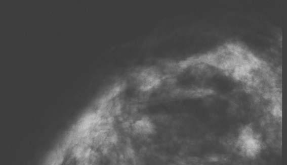


12 BiomarC - Mammography 38 year old female with invasive ductal carcinoma


13 BiomarC - Ultrasound 38 year old female with invasive ductal carcinoma IDC


14 BiomarC - Ultrasound 38 year old female with invasive ductal carcinoma 7 day follow-up

15 BiomarC - Computed Tomography 38-year-old female with invasive ductal carcinoma

16 BiomarC - 1.5T MRI / MRS 38 year old female with invasive ductal carcinoma (tcho)

17 BiomarC - 4T MRI / MRS 38 year old female with invasive ductal carcinoma ppm [tcho] = 4.85 ±.07 Highly consistent with malignancy
18 BiomarC - PET Scan 38 year old female with invasive ductal carcinoma

19 BiomarC - 4T MRI / MRS 47 year old female with invasive ductal carcinoma ppm [tcho] = 7.63 ± 0.5 Highly consistent with malignancy
20 Large BiomarC Breast Marker 1. 4x 2 mm size 2. 3 x mass versus 1x3 marker 3. Easier to see on - Mammography - Ultrasound - MRI - Does not interfere with MR spectroscopy


21 45 year-old female with left breast mass on mammography showing atypia Left Breast MLO view Left Breast CC view

22 45 year-old female with left breast mass on ultrasound showing atypia Large BiomarC

23 CT scan of the chest showing two BiomarCs in left breast in 45-year old female L


24 45-year-old female with right breast atypical ductal hyperplasia Large BiomarC SenorX, titanium Right Breast CC view Right Breast MLO view

25 45-year-old female right breast atypical ductal hyperplasia seen on ultrasound Large BiomarC
26 61-year-old female with right breast mass seen on mammography showing invasive lobular carcinoma Small BiomarC Large BiomarC Case #48 -Right Breast CC view Right Breast ML view

27 61 year old female with invasive lobular carcinoma of the right breast seen on ultrasound Small BiomarC
28 Conclusion: RM (BiomarC ) - deployed with accuracy (4mm) and was clearly visible under mammography, ultrasound, and MRI Follow up studies after 12 weeks demonstrated no evidence of migration RM did not interfere with 1 H MR spectroscopy (1.5 T or 4T) or PET scanning.
Breast Cancer Screening Clinical Practice Guideline. Kaiser Permanente National Breast Cancer Screening Guideline Development Team
 NATIONAL CLINICAL PRACTICE GUIDELINE Breast Cancer Screening Clinical Practice Guideline Kaiser Permanente National Breast Cancer Screening Guideline Development Team This guideline is informational only.
3-marker technique for the localisation and delineation of residual tumour bed following neoadjuvant chemotherapy in patients within the I-SPY 2 trial
 3-marker technique for the localisation and delineation of residual tumour bed following neoadjuvant chemotherapy in patients within the I-SPY 2 trial Poster No.: C-2125 Congress: ECR 2016 Type: Authors:
Atypical Ductal Hyperplasia and Papillomas: A Comparison of Ultrasound Guided Breast Biopsy and Stereotactic Guided Breast Biopsy
 Atypical Ductal Hyperplasia and Papillomas: A Comparison of Ultrasound Guided Breast Biopsy and Stereotactic Guided Breast Biopsy Breast Cancer is the most common cancer diagnosed in women in the United
Index. Surg Oncol Clin N Am 16 (2007) Note: Page numbers of article titles are in boldface type.
 Note: Page numbers of article titles are in boldface type.") Surg Oncol Clin N Am 16 (2007) 465 469 Index Note: Page numbers of article titles are in boldface type. A Adjuvant therapy, preoperative for gastric cancer, staging and, 339 B Breast cancer, metabolic
Surg Oncol Clin N Am 16 (2007) 465 469 Index Note: Page numbers of article titles are in boldface type. A Adjuvant therapy, preoperative for gastric cancer, staging and, 339 B Breast cancer, metabolic
Stereotactic vacuum-assisted biopsy (SVAB) of Nonpalpable Breast Microcalcifications: Advantage of clip placement (Prospective study)
 of Nonpalpable Breast Microcalcifications: Advantage of clip placement (Prospective study)") Department of Radiology, University of Ulm Director: Prof. Dr. med. HJ Brambs Stereotactic vacuum-assisted biopsy (SVAB) of Nonpalpable Breast Microcalcifications: Advantage of clip placement (Prospective
Department of Radiology, University of Ulm Director: Prof. Dr. med. HJ Brambs Stereotactic vacuum-assisted biopsy (SVAB) of Nonpalpable Breast Microcalcifications: Advantage of clip placement (Prospective
Macrobiopsy under X-Ray Guidance
 Macrobiopsy under X-Ray Guidance C. Balleyguier, B. Boyer Radiology Gustave Roussy, Villejuif, France Breast Intervention Imaging Major domain in breast imaging European guidelines recommend a pre surgical
Breast Cancer. Most common cancer among women in the US. 2nd leading cause of death in women. Mortality rates though have declined
 Breast Cancer Most common cancer among women in the US 2nd leading cause of death in women Mortality rates though have declined 1 in 8 women will develop breast cancer Breast Cancer Breast cancer increases
Breast Cancer. Saima Saeed MD
 Breast Cancer Saima Saeed MD Breast Cancer Most common cancer among women in the US 2nd leading cause of death in women 1 in 8 women will develop breast cancer Incidence/mortality rates have declined Breast
Mammographic imaging of nonpalpable breast lesions. Malai Muttarak, MD Department of Radiology Chiang Mai University Chiang Mai, Thailand
 Mammographic imaging of nonpalpable breast lesions Malai Muttarak, MD Department of Radiology Chiang Mai University Chiang Mai, Thailand Introduction Contents Mammographic signs of nonpalpable breast cancer
Breast Lesion Excision System-Intact (BLES): A Stereotactic Method of Biopsy of Suspicius Non-Palpable Mammographic Lesions.
: A Stereotactic Method of Biopsy of Suspicius Non-Palpable Mammographic Lesions.") Breast Lesion Excision System-Intact (BLES): A Stereotactic Method of Biopsy of Suspicius Non-Palpable Mammographic Lesions. Poster No.: C-1595 Congress: ECR 2014 Type: Authors: Scientific Exhibit I. Georgiou
Breast Lesion Excision System-Intact (BLES): A Stereotactic Method of Biopsy of Suspicius Non-Palpable Mammographic Lesions. Poster No.: C-1595 Congress: ECR 2014 Type: Authors: Scientific Exhibit I. Georgiou
DISCLOSURE. Relevant Financial Relationship(s) Off Label Usage
 Off Label Usage") DISCLOSURE Relevant Financial Relationship(s) 1. Medical Advisory Board GE, Siemens, Hologic, Bayer Medical Inc 2. CEO Breast-Med Inc, Consulting and Development of Breast Markers Off Label Usage University
DISCLOSURE Relevant Financial Relationship(s) 1. Medical Advisory Board GE, Siemens, Hologic, Bayer Medical Inc 2. CEO Breast-Med Inc, Consulting and Development of Breast Markers Off Label Usage University
Vacuum-assisted breast biopsy using computer-aided 3.0 T- MRI guidance: diagnostic performance in 173 lesions
 Vacuum-assisted breast biopsy using computer-aided 3.0 T- MRI guidance: diagnostic performance in 173 lesions Poster No.: C-2870 Congress: ECR 2017 Type: Scientific Exhibit Authors: A. Pozzetto, L. Camera,
Breast Cancer Diagnosis, Treatment and Follow-up
 Breast Cancer Diagnosis, Treatment and Follow-up What is breast cancer? Each of the body s organs, including the breast, is made up of many types of cells. Normally, healthy cells grow and divide to produce
Ana Sofia Preto 19/06/2013
 Ana Sofia Preto 19/06/2013 Understanding the underlying pathophysiologic processes leading to the various types of calcifications Description and illustration of the several types of calcifications, according
Feasibility of MRI-guided large-core-needle biopsy of suspiscious breast lesions at 3T
 Eur Radiol (2009) 19: 1639 1644 DOI 10.1007/s00330-009-1310-0 BREAST Nicky H. G. M. Peters Carla Meeuwis Chris J. G. Bakker Willem P. Th. M. Mali Arancha M. Fernandez-Gallardo Richard van Hillegersberg
ADI Procedure Codes. August 2016 Revised April 2017 Page 1 of 7 ADI Procedure Codes
 Code Description 70450 CT Head without contrast 70460 CT Head with contrast 70470 CT Head with & without contrast 70480 CT Orbit, et al without contrast 70481 CT Orbit, et al with contrast 70482 CT Orbit,
Here are examples of bilateral analog mammograms from the same patient including CC and MLO projections.
 Good afternoon. It s my pleasure to be discussing Diagnostic Breast Imaging over the next half hour. I m Wei Yang, Professor of Diagnostic Radiology and Chief, the Section of Breast Imaging as well as
Excisional biopsy or long term follow-up results in breast high-risk lesions diagnosed at core needle biopsy
 Excisional biopsy or long term follow-up results in breast high-risk lesions diagnosed at core needle biopsy Poster No.: C-2515 Congress: ECR 2015 Type: Authors: Scientific Exhibit Ö. S. Okcu 1, A. Oktay
MR spectroscopy in breast cancer: retrospective study of a population with high prevalence of disease
 MR spectroscopy in breast cancer: retrospective study of a population with high prevalence of disease Poster No.: C-2025 Congress: ECR 2015 Type: Scientific Exhibit Authors: D. De Falco Alfano 1, R. N.
Radioactive Seed Localization of Nonpalpable Breast Lesions
 Radioactive Seed Localization of Nonpalpable Breast Lesions Policy Number: 6.01.57 Last Review: 11/2017 Origination: 11/2016 Next Review: 11/2018 Policy Blue Cross and Blue Shield of Kansas City (Blue
Breast Cancer Imaging
 Breast Cancer Imaging I. Policy University Health Alliance (UHA) will cover breast imaging when such services meet the medical criteria guidelines (subject to limitations and exclusions) indicated below.
Screening Mammograms: Questions and Answers
 CANCER FACTS N a t i o n a l C a n c e r I n s t i t u t e N a t i o n a l I n s t i t u t e s o f H e a l t h D e p a r t m e n t o f H e a l t h a n d H u m a n S e r v i c e s Screening Mammograms:
PURPOSE IMAGE-GUIDANCE MODALITIES IMAGE-GUIDED BREAST BIOPSY. US-Techniques. Ultrasound. US guided NLOBB. TH. Helbich
 IMAGE-GUIDED BREAST BIOPSY PURPOSE TH. Helbich Department of Radiology Division of Molecular & Gender Imaging Medical University of Vienna Imaging techniques Interventional procedures Quality management
Diagnostic benefits of ultrasound-guided. CNB) versus mammograph-guided biopsy for suspicious microcalcifications. without definite breast mass
 versus mammograph-guided biopsy for suspicious microcalcifications. without definite breast mass") Volume 118 No. 19 2018, 531-543 ISSN: 1311-8080 (printed version); ISSN: 1314-3395 (on-line version) url: http://www.ijpam.eu ijpam.eu Diagnostic benefits of ultrasound-guided biopsy versus mammography-guided
Volume 118 No. 19 2018, 531-543 ISSN: 1311-8080 (printed version); ISSN: 1314-3395 (on-line version) url: http://www.ijpam.eu ijpam.eu Diagnostic benefits of ultrasound-guided biopsy versus mammography-guided
Role of Proton MR Spectroscopy in Differentiation of Benign and Malignant Breast Lesions
 Med. J. Cairo Univ., Vol. 82, No. 1, June: 341-349, 2014 www.medicaljournalofcairouniversity.net Role of Proton MR Spectroscopy in Differentiation of Benign and Malignant Breast Lesions GALAL EL HAWARY,
Case study 1. Rie Horii, M.D., Ph.D. Division of Pathology Cancer Institute Hospital, Japanese Foundation for Cancer Research
 NCCN/JCCNB Seminar in Japan April 15, 2012 Case study 1 Rie Horii, M.D., Ph.D. Division of Pathology Cancer Institute Hospital, Japanese Foundation for Cancer Research Present illness: A 50y.o.premenopausal
Quality ID #263: Preoperative Diagnosis of Breast Cancer National Quality Strategy Domain: Effective Clinical Care
 Quality ID #263: Preoperative Diagnosis of Breast Cancer National Quality Strategy Domain: Effective Clinical Care 2018 OPTIONS FOR INDIVIDUAL MEASURES: REGISTRY ONLY MEASURE TYPE: Process DESCRIPTION:
Successful Breast MRI Program : The ingredients
 Successful Breast MRI Program : The ingredients Dr. Smriti Hari Associate Professor Deptt. Of Radiology All India Institute of Medical Sciences New Delhi How to perform Breast MRI Breast MRI descriptors
Improving Methods for Breast Cancer Detection and Diagnosis. The National Cancer Institute (NCI) is funding numerous research projects to improve
 is funding numerous research projects to improve") CANCER FACTS N a t i o n a l C a n c e r I n s t i t u t e N a t i o n a l I n s t i t u t e s o f H e a l t h D e p a r t m e n t o f H e a l t h a n d H u m a n S e r v i c e s Improving Methods for
CANCER FACTS N a t i o n a l C a n c e r I n s t i t u t e N a t i o n a l I n s t i t u t e s o f H e a l t h D e p a r t m e n t o f H e a l t h a n d H u m a n S e r v i c e s Improving Methods for
Detailed Program of the second BREAST IMAGING AND INTERVENTIONS PROGRAM am am : Clinician s requirements from breast imaging
 Detailed Program of the second BREAST IMAGING AND INTERVENTIONS PROGRAM 2012 Day one, 2 nd November BREAST IMAGING AND INTERVENTIONS PROGRAM 2012 9.00 AM 9.10 am Introduction 9.10 am - 9.30 am : Clinician
Prof. Dr. NAGUI M. ABDELWAHAB,M.D.; MARYSE Y. AWADALLAH, M.D. AYA M. BASSAM, Ms.C.
 Role of Whole-body Diffusion MR in Detection of Metastatic lesions Prof. Dr. NAGUI M. ABDELWAHAB,M.D.; MARYSE Y. AWADALLAH, M.D. AYA M. BASSAM, Ms.C. Cancer is a potentially life-threatening disease,
Radioactive Seed Localization of Nonpalpable Breast Lesions
 Subject: Radioactive Seed Localization of Page: 1 of 7 Last Review Status/Date: March 2017 Radioactive Seed Localization of Description Radioactive seed localization is used to detect nonpalpable breast
Breast MR biopsy. I Thomassin-Naggara, A.Jalaguier-Coudray, J Chopier
 Breast MR biopsy I Thomassin-Naggara, A.Jalaguier-Coudray, J Chopier Background EUSOBI When a radiologist perform a MR breast imaging he has to be able to realize or to be apart of a network who is able
Breast Cancer. What is breast cancer?
 Scan for mobile link. Breast Cancer Breast cancer is a malignant tumor in or around breast tissue. It usually begins as a lump or calcium deposit that develops from abnormal cell growth. Most breast lumps
PET/CT in breast cancer staging
 PET/CT in breast cancer staging Anni Morsing Consultant, PhD, DMSc Rigshospitalet 1 18F- FDG PET/CT for breastcancer staging Where is the clinical impact? To which women should 18F- FDG PET/CT be offered?
Multicenter Evaluation of the Breast Lesion Excision System, a Percutaneous, Vacuum-Assisted, Intact-Specimen Breast Biopsy Device
 945 Multicenter Evaluation of the Breast Lesion Excision System, a Percutaneous, Vacuum-Assisted, Intact-Specimen Breast Biopsy Device Angela Sie, MD 1 David C. Bryan, MD 2 Victor Gaines, MD 3 Larry K.
Breast density: imaging, risks and recommendations
 Breast density: imaging, risks and recommendations Maureen Baxter, MD Radiologist Director of Ruth J. Spear Breast Center Providence St. Vincent Medical Center Alison Conlin, MD/MPH Medical Oncologist
Digital breast tomosynthesis (DBT) occult breast cancers: clinical, radiological and histopathological features.
 occult breast cancers: clinical, radiological and histopathological features.") Digital breast tomosynthesis (DBT) occult breast cancers: clinical, radiological and histopathological features. Poster No.: C-1707 Congress: ECR 2015 Type: Scientific Exhibit Authors: V. Vinci 1, A. Iqbal
Digital breast tomosynthesis (DBT) occult breast cancers: clinical, radiological and histopathological features. Poster No.: C-1707 Congress: ECR 2015 Type: Scientific Exhibit Authors: V. Vinci 1, A. Iqbal
Imaging Surveillance in Women with a History of Treated Breast Cancer. Wei Tse Yang, M.D.
 Imaging Surveillance in Women with a History of Treated Breast Cancer Wei Tse Yang, M.D. Breast Cancer 1. Extent 2. Response 3. Recurrence Surveillance Breast Cancer 1. Extent 2. Response Surveillance
MRI/MRS Biomarkers. Robert E. Lenkinski, Ph.D.
 MRI/MRS Biomarkers Robert E. Lenkinski, Ph.D. Disclosure GE Healthcare-Research Grant Aspect MR-Scientific Advisor Aposense-Scientific Advisor Brainwatch-Scientific Advisor I will be discussing off-label
Breast Health and Imaging Glossary
 Contact: Lorna Vaughan HerSpace Breast Imaging & Biopsy Associates 300 State Route 35 South W. Long Branch, NJ 07764 732-571-9100, ext. 104 lorna@breast-imaging.com Breast Health and Imaging Glossary Women
Atypical proliferative lesions diagnosed on core biopsy - 6 year review
 Atypical proliferative lesions diagnosed on core biopsy - 6 year review Dr Angela Harris, Dr Julie Weigner & Dr Ricardo Vilain NSW Health Pathology Pathology North, Hunter Anatomical Pathology & Cytology
DCIS of the Breast--MRI findings with mammographic correlation.
 DCIS of the Breast--MRI findings with mammographic correlation. Poster No.: C-1560 Congress: ECR 2013 Type: Educational Exhibit Authors: N. B. Ibrahim, P. Morris, S. ANANDAN; Burlington, MA/US Keywords:
Jeddah Breast Cancer Pilot Screening Program, KSA
 Jeddah Breast Cancer Pilot Screening Program, KSA 7 th Global Summit on Cancer Therapy, Oct 5-7, 2015 Dubai, Crown Plaza Hotel Muna Baslaim, MD Consultant Surgeon Head of the Breast Unit, King Fahd General
Interpretation of Breast Pathology in the Era of Minimally Invasive Procedures
 Shahla Masood, M.D. Professor and Chair Department of Pathology and Laboratory Medicine University of Florida College of Medicine Jacksonville Medical Director, UF Health Breast Center Chief of Pathology
Impact of value based breast cancer care pathway implementation on pre-operative breast magnetic resonance imaging utilization
 Original Article Impact of value based breast cancer care pathway implementation on pre-operative breast magnetic resonance imaging utilization Devina K. S. McCray, Stephen R. Grobmyer, Holly J. Pederson
Breast Cancer. What is breast cancer?
 Scan for mobile link. Breast Cancer Breast cancer is a malignant tumor in or around breast tissue. It usually begins as a lump or calcium deposit that develops from abnormal cell growth. Most breast lumps
Correlation between lesion type and the additional value of digital breast tomosynthesis
 Correlation between lesion type and the additional value of digital breast tomosynthesis Poster No.: C-1604 Congress: ECR 2011 Type: Scientific Exhibit Authors: C. Van Ongeval, L. Cockmartin, A. Van Steen,
Certified Breast Care Nurse (CBCN ) Test Content Outline (Effective 2018)
 Test Content Outline (Effective 2018)") Certified Breast Care Nurse (CBCN ) Test Content Outline (Effective 2018) I. Coordination of Care - 26% A. Breast health, screening, early detection, risk assessment and reduction 1. Issues related to
Certified Breast Care Nurse (CBCN ) Test Content Outline (Effective 2018) I. Coordination of Care - 26% A. Breast health, screening, early detection, risk assessment and reduction 1. Issues related to
Presented by: Lillian Erdahl, MD
 Presented by: Lillian Erdahl, MD Learning Objectives What is Breast Cancer Types of Breast Cancer Risk Factors Warning Signs Diagnosis Treatment Options Prognosis What is Breast Cancer? A disease that
Surgical Therapy: Sentinel Node Biopsy and Breast Conservation
 Surgical Therapy: Sentinel Node Biopsy and Breast Conservation Stephen B. Edge, MD Professor of Surgery and Oncology Roswell Park Cancer Institute University at Buffalo Dr. Roswell Park: Tradition in Cancer
ROLE OF MRI IN SCREENING, DIAGNOSIS AND MANAGEMENT OF BREAST CANCER. B.Zandi Professor of Radiology
 ROLE OF MRI IN SCREENING, DIAGNOSIS AND MANAGEMENT OF BREAST CANCER B.Zandi Professor of Radiology Introduction In the USA, Breast Cancer is : The Most Common Non-Skin Cancer The Second Leading cause of
BREAST MRI. Elizabeth A. Rafferty, M.D. Avon Comprehensive Breast Center Massachusetts General Hospital Harvard Medical School
 BREAST MRI Elizabeth A. Rafferty, M.D. Avon Comprehensive Breast Center Massachusetts General Hospital Harvard Medical School BREAST MRI Any assessment of the breast parenchyma requires the administration
Tomosynthesis and breast imaging update. Dr Michael J Michell Consultant Radiologist King's College Hospital NHS Foundation Trust
 Tomosynthesis and breast imaging update Dr Michael J Michell Consultant Radiologist King's College Hospital NHS Foundation Trust Breast imaging new technology BREAST CANCER FLT PET shows different grades
Radiologic and pathologic correlation of non-mass like breast lesions on US and MRI: Benign, high risk, versus malignant
 Radiologic and pathologic correlation of non-mass like breast lesions on US and MRI: Benign, high risk, versus malignant Poster No.: C-1161 Congress: ECR 2013 Type: Educational Exhibit Authors: J. Kwak,
Effective Health Care Program
 Comparative Effectiveness Review Number 19 Effective Health Care Program Comparative Effectiveness of Core-Needle and Open Surgical Biopsy for the Diagnosis of Breast Lesions Executive Summary Background
Breast Cancer Screening
 Scan for mobile link. Breast Cancer Screening What is breast cancer screening? Screening examinations are tests performed to find disease before symptoms begin. The goal of screening is to detect disease
BREAST IMAGING and NEW IMAGING MODALITIES- A Surgeons view
 BREAST IMAGING and NEW IMAGING MODALITIES- A Surgeons view DR CHANTEL THORNTON SPECIALIST BREAST CANCER SURGEON BMSc (hons) MBBS (hons) FRACS Epworth Hospital, Richmond- Agora Centre for Women s Health
MANAGEMENT OF DENSE BREASTS. Nichole K Ingalls, MD, MPH NW Surgical Specialists September 25, 2015
 MANAGEMENT OF DENSE BREASTS Nichole K Ingalls, MD, MPH NW Surgical Specialists September 25, 2015 No financial disclosures National Cancer Institute National Cancer Institute Increased Cancer Risk... DENSITY
Breast MRI Update. Jeffrey C. Weinreb, MD, FACR Yale University School of Medicine
 Breast MRI Update Jeffrey C. Weinreb, MD, FACR jeffrey.weinreb@yale.edu Yale University School of Medicine I disclose the following financial relationships with relevant commercial interests: Bracco Bayer
In Vivo Quantification of Choline Compounds in the Breast With 1 H MR Spectroscopy
 Magnetic Resonance in Medicine 50:000 000 (2003) In Vivo Quantification of Choline Compounds in the Breast With 1 H MR Spectroscopy Patrick J. Bolan, 1,2 Sina Meisamy, 1,2 Eva H. Baker, 1,2 Joseph Lin,
MP Magnetic Resonance Imaging for Detection and Diagnosis of Breast Cancer
 Medical Policy MP 6.01.29 BCBSA Ref. Policy: 6.01.29 Last Review: 09/19/2018 Effective Date: 09/19/2018 Section: Radiology Related Policies 6.01.45 Computer-Aided Evaluation of Malignancy With Magnetic
Current Status of Supplementary Screening With Breast Ultrasound
 Current Status of Supplementary Screening With Breast Ultrasound Stephen A. Feig, M.D., FACR Fong and Jean Tsai Professor of Women s Imaging Department of Radiologic Sciences University of California,
Mammography and Other Screening Tests. for Breast Problems
 301.681.3400 OBGYNCWC.COM Mammography and Other Screening Tests What is a screening test? for Breast Problems A screening test is used to find diseases, such as cancer, in people who do not have signs
ANNEX 1 OBJECTIVES. At the completion of the training period, the fellow should be able to:
 1 ANNEX 1 OBJECTIVES At the completion of the training period, the fellow should be able to: 1. Breast Surgery Evaluate and manage common benign and malignant breast conditions. Assess the indications
Educational Goals and Objectives for Rotations on: Breast, Wound and Plastic Surgery
 Educational Goals and Objectives for Rotations on: Breast, Wound and Plastic Surgery Goal The goal of the Breast Surgery rotation is to develop the knowledge, skills and attitudes necessary to evaluate,
BREAST MRI. Elizabeth A. Rafferty, M.D. Avon Comprehensive Breast Center Massachusetts General Hospital Harvard Medical School
 BREAST MRI Elizabeth A. Rafferty, M.D. Avon Comprehensive Breast Center Massachusetts General Hospital Harvard Medical School BREAST MRI Any assessment of the breast parenchyma requires the administration
Evaluation & Management of PowerPoint Cover Title. the High Risk Population. High Risk Clinic
 Evaluation & Management of PowerPoint Cover Title the High Risk Population High Risk Clinic Subtitle Joanna Would Springman, Go Here PA-C Assessment Genetics Known genetic mutation Family history with
Cairo/EG, Khartoum/SD, London/UK Biological effects, Diagnostic procedure, Ultrasound, Mammography, Breast /ecr2015/C-0107
 Role of sono-mammography in the evaluation of clinically palapble breast masses during pregnancy & lactation with differentaition between true patholgical & false physiological lobular hyperlpasia.sudanese
Monitoring neo-adjuvant chemotherapy: comparison of contrast-enhanced spectral mammography (CESM) and MRI versus breast cancer characteristics
 and MRI versus breast cancer characteristics") Monitoring neo-adjuvant chemotherapy: comparison of contrast-enhanced spectral mammography (CESM) and MRI versus breast cancer characteristics Poster No.: B-1062 Congress: ECR 2016 Type: Scientific Paper
Monitoring neo-adjuvant chemotherapy: comparison of contrast-enhanced spectral mammography (CESM) and MRI versus breast cancer characteristics Poster No.: B-1062 Congress: ECR 2016 Type: Scientific Paper
BI-RADS 3 category, a pain in the neck for the radiologist which technique detects more cases?
 BI-RADS 3 category, a pain in the neck for the radiologist which technique detects more cases? Poster No.: B-0966 Congress: ECR 2013 Type: Scientific Paper Authors: J. Etxano Cantera, I. Simon-Yarza, G.
Tissue Breast Density
 Tissue Breast Density Reporting breast density within the letter to the patient is now mandated by VA law. Therefore, this website has been established by Peninsula Radiological Associates (PRA), the radiologists
Contrast Enhanced Spectral Mammography (CESM) Updates
 Updates") Contrast Enhanced Spectral Mammography (CESM) Updates Georgeta Mihai, PhD, DABR Medical Physicist, BIDMC, Boston Assistant Professor, Harvard Medical School, Boston Disclosures None Acknowledgments: Da
Contrast Enhanced Spectral Mammography (CESM) Updates Georgeta Mihai, PhD, DABR Medical Physicist, BIDMC, Boston Assistant Professor, Harvard Medical School, Boston Disclosures None Acknowledgments: Da
STEREOTACTIC BREAST BIOPSY: CORRELATION WITH HISTOLOGY
 3-rd Baltic Congress of Radiology, October 8-9, 2010 Riga Rūta Briedienė, Rūta Grigienė, Raimundas Meškauskas Institute of Oncology Vilnius University, National Centre of Pathology STEREOTACTIC BREAST
Sheri Saluga A/P 2 May 1, Breast Cancer
 Sheri Saluga A/P 2 May 1, 2010 Breast Cancer Cancer, one word common to the word and one word no one wants to hear. There are many forms of cancer but of particular interest is stomach cancer. So what
Contrast-enhanced Breast MRI RSSA 2013
 Contrast-enhanced Breast MRI RSSA 2013 Prof. dr. Maurice van den Bosch University Medical Center Utrecht, the Netherlands Index 1) Breast cancer 2) Why MRI of the breast 3) Technique 4) Interpretation
5101: of 5 APPENDIX B. Revenue Center Codes Requiring CPT or HCPCS Coding
 ACTION: Final ENACTED Appendix 5101:3-2-21 DATE: 03/17/2011 1:51 PM 5101:3-2-21 1 of 5 IV Therapy 0260 General Classification 0261 Infusion Pump 0269 Other IV Therapy Oncology 0280 General Classification
CNB vs Surgical Excision
 Update on Core Needle Biopsy of Non-palpable Breast Lesions Nour Sneige, M.D. UT MD Anderson Cancer Center Houston, Tx Image-Guided CNB of Breast Lesions An alternative to surgical biospy CNB vs Surgical
Guidance on the management of B3 lesions
 Guidance on the management of B3 lesions Lesion diagnosed on 14g or vacuumassisted biopsy (VAB) Risk of upgrade Recommended investigation Suggested approach for follow-up if no malignancy on VAE awaiting
Prediction of Postoperative Tumor Size in Breast Cancer Patients by Clinical Assessment, Mammography and Ultrasonography
 Prediction of Postoperative Tumor Size in Breast Cancer Patients by Clinical Assessment, Mammography and Ultrasonography Eyad Fawzi AlSaeed 1 and Mutahir A. Tunio 2* 1 Consultant Radiation Oncology, Chairman
04/10/2018 HIGH RISK BREAST LESIONS. Pathology Perspectives of High Risk Breast Lesions ELEVATED RISK OF BREAST CANCER HISTORICAL PERSPECTIVES
 Pathology Perspectives of High Risk Breast Lesions Savitri Krishnamurthy MD Professor of Pathology Deputy Division Head Director of Clinical Trials, Research and Development The University of Texas MD
Role of PEM in Breast Cancer Management. Judy Kalinyak, MD, PhD Chief Medical Officer Naviscan, Inc (San Diego, CA)
") Role of PEM in Breast Cancer Management Judy Kalinyak, MD, PhD Chief Medical Officer Naviscan, Inc (San Diego, CA) Role of PEM in Breast Cancer Management Introduction to Positron Emission Mammography
Role of PEM in Breast Cancer Management Judy Kalinyak, MD, PhD Chief Medical Officer Naviscan, Inc (San Diego, CA) Role of PEM in Breast Cancer Management Introduction to Positron Emission Mammography
Correlation Between BIRADS Classification and Ultrasound -guided Tru-Cut Biopsy Results of Breast Lesions: Retrospective Analysis of 285 Patients
 Correlation Between BIRADS Classification and Ultrasound -guided Tru-Cut Biopsy Results of Breast Lesions: Retrospective Analysis of 285 Patients Poster No.: C-1433 Congress: ECR 2014 Type: Scientific
Non-mass Enhancement on Breast MRI. Aditi A. Desai, MD Margaret Ann Mays, MD
 Non-mass Enhancement on Breast MRI Aditi A. Desai, MD Margaret Ann Mays, MD Breast MRI Important screening and diagnostic tool, given its high sensitivity for breast cancer detection Breast MRI - Indications
Clinical Experience with MRI-Guided Vacuum-Assisted Breast Biopsy
 Lehman et al. MRI-Guided Vacuum-Assisted Breast Biopsy Constance D. Lehman 1,2 Elizabeth R. DePeri 3 Sue Peacock 1,2 Michelle D. McDonough 3 Wendy B. DeMartini 1,2 Jennifer Shook 1,2 Lehman CD, DePeri
Contrast-Enhanced Spectral Mammography
 Contrast-Enhanced Spectral Mammography Illuminating Breast Cancer Detection SenoBright HD TM gehealthcare.com/senobright Mammography is the most reliable imaging technique for breasts, but limitations
Case Report Tubular Carcinoma of the Breast: Advantages and Limitations of Breast Tomosynthesis
 Case Reports in Radiology Volume 2016, Article ID 3906195, 4 pages http://dx.doi.org/10.1155/2016/3906195 Case Report Tubular Carcinoma of the Breast: Advantages and Limitations of Breast Tomosynthesis
Related Policies None
 Medical Policy MP 6.01.57 BCBSA Ref. Policy: 6.01.57 Last Review: 09/19/2018 Effective Date: 09/19/2018 Section: Radiology Related Policies None DISCLAIMER Our medical policies are designed for informational
National Diagnostic Imaging Symposium 2013 SAM - Breast MRI 1
 National Diagnostic Imaging Symposium 2013 December 8-12, 2013 Disney s Yacht Club Resort Lake Buena Vista, Florida Self Assessment Module Questions, Answers and References Day SAM Title - Each SAM title
BREAST CANCER SCREENING IS A CHOICE
 BREAST CANCER SCREENING IS A CHOICE by ELAINE SCHATTNER, MD no financial disclosures (ES) American Association for Cancer Research Typical headlines focus on controversy 2 Data: Breast Cancer Incidence
Molecular Imaging and Breast Cancer
 Molecular Imaging and Breast Cancer Breast cancer forms in tissues of the breast usually in the ducts, tubes that carry milk to the nipple, and lobules, the glands that make milk. It occurs in both men
Breast ultrasound appearances after Mammotome vacuumassisted
 Breast ultrasound appearances after Mammotome vacuumassisted biopsy. Poster No.: C-1924 Congress: ECR 2011 Type: Educational Exhibit Authors: R. Patel 1, G. R. Kaplan 2 ; 1 London/UK, 2 Herts/UK Keywords:
Brain Tumors. What is a brain tumor?
 Scan for mobile link. Brain Tumors A brain tumor is a collection of abnormal cells that grows in or around the brain. It poses a risk to the healthy brain by either invading or destroying normal brain
Sonographic Detection and Sonographically Guided Biopsy of Breast Microcalcifications
 Sonographic Detection and Sonographically Guided Biopsy of Breast Microcalcifications Mary Scott Soo 1 Jay A. Baker Eric L. Rosen OBJECTIVE. The purpose of this study was to evaluate the ability of sonography
Poster No.: C-0466 Congress: ECR 2010 Scientific Exhibit
 Up-right stereotactic vacuum-assisted biopsy (UP-VAB) of non palpable breast lesions: Results and correlations with radiological suspicion (BI-RADS classification) Poster No.: C-0466 Congress: ECR 2010
Emerging Techniques in Breast Imaging: Contrast-Enhanced Mammography and Fast MRI
 Emerging Techniques in Breast Imaging: Contrast-Enhanced Mammography and Fast MRI Lilian Wang, M.D. Breast Imaging Section Department of Radiology Northwestern Medicine Overview Rationale for new imaging
What is Cancer? Petra Ketterl, MD Medical Oncology and Functional Medicine
 What is Cancer? Petra Ketterl, MD Medical Oncology and Functional Medicine What is Cancer? Layman s terms: cancer starts when cells grow out of control (in any place in the body) and crowd out normal cells
Lesion Imaging Characteristics Mass, Favoring Benign Circumscribed Margins Intramammary Lymph Node
 Lesion Imaging Characteristics Mass, Favoring Benign Circumscribed Margins Intramammary Lymph Node Oil Cyst Mass, Intermediate Concern Microlobulated Margins Obscured Margins Mass, Favoring Malignant Indistinct
Breast Imaging: Multidisciplinary Approach. Madelene Lewis, MD Assistant Professor Associate Program Director Medical University of South Carolina
 Breast Imaging: Multidisciplinary Approach Madelene Lewis, MD Assistant Professor Associate Program Director Medical University of South Carolina No Disclosures Objectives Discuss a multidisciplinary breast