GTC 2018, San Jose, USA
|
|
|
- Toby Copeland
- 5 years ago
- Views:
Transcription


1 Automated Segmentation of Suspicious Breast Masses from Ultrasound Images Viksit Kumar, Jeremy Webb, Adriana Gregory, Mostafa Fatemi, Azra Alizad GTC 2018, San Jose, USA

2 SIGNIFICANCE Breast cancer is most common and leading cause of death in American women* Segmentation and classification of suspicious breast masses can avoid unnecessary biopsies Conventional segmentation algorithm requires an initial seed Sørensen Dice performance of , a measure of similarity of segmentation Core Needle biopsy for suspicious breast masses *Siegel RL, Miller KD, Jemal A. Cancer statistics, CA: a cancer journal for clinicians *Shulman LN, Willett W, Sievers A, Knaul FM. Breast Cancer in Developing Countries: Opportunities for Improved Survival. Journal of Oncology. 2010;2010. doi: /2010/
3 GOALS Develop an automated real-time detection and segmentation of suspicious breast masses Currently ultrasonographers use the morphological and textural features to identify suspicious masses based on suggestions from American College of Radiology The suspicious masses are then scored based on the Breast Imaging Reporting and Data System (BI-RADS) scale and is the basis for recommending core needle biopsy Automated detection process can aid in rapid localization of suspicious masses Automated segmentation and classification process can assist in assigning BI-RADS score and recommending biopsy


4 ULTRASOUND IMAGING Transducer Raw channel RF data Beam formed RF data In-phase quadrature data Post processed B-mode image B-mode image 4
5 MATERIALS AND METHODS 258 patients, 433 images from multiple cross-sections, 148 cases of BI-RADS malignant and 134 benign cases Equal number of images from General Electric LOGIQ E9 and Philips IU22 Algorithm: Multi U-net with majority voting Loss function: negative dice coefficient Testing in real-time on Titan xp (13.83 ms/image)

6 ARCHITECTURE Ronneberger O, Fischer P, Brox T, editors. U-net: Convolutional networks for biomedical image segmentation. International Conference on Medical Image Computing and Computer-Assisted Intervention; 2015: Springer 6

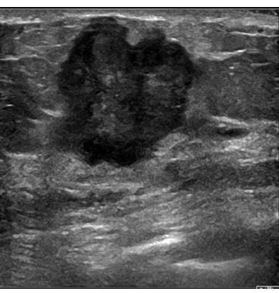
7 SEGMENTATION RESULTS-CONTD. Red outline = Expert segmentation 2(b) Blue outline = Predicted segmentation Dice coefficient = 0.94 B-mode image of a benign-cellular fibro epithelial mass. The mass has typical smooth boundaries of a benign mass and is oval in shape

8 SEGMENTATION RESULTS-CONTD. Red outline = Expert segmentation 3(a) 3(b) Blue outline = Predicted segmentation Dice coefficient = 0.88 B-mode image for benign fat necrosis with dystrophic calcifications. Notice the posterior acoustic shadowing beneath the benign mass

9 SEGMENTATION RESULTS-CONTD. Red outline = Expert segmentation 4(a) 4(b) Blue outline = Predicted segmentation Dice coefficient = 0.88 The biopsy result from the suspicious mass was malignant invasive/infiltrating ductal carcinoma, grade III. The irregular boundaries of the mass are typical of malignant masses which are usually challenging cases for segmentation algorithms

10 RESULTS: CINE CLIP 1 Benign fibroadenoma

11 CONTD. CINE CLIP 2 Invasive Ductal carcinoma G.II/III
12 CONCLUSION Mean Sørensen Dice coefficient of 0.82 Real-time application with no need for initial seed Can be used for segmentation and detection of suspicious breast masses Segmentation masks can be used for classification of suspicious masses Reduce localization time and aid sonographer in categorizing the suspicious mass Applications in point of care, mobile health monitoring, assisting sonographers Provides the expertise of an expert sonographer to sonographers in training
13 ACKNOWLEDGEMENT We gratefully acknowledge the support of NVIDIA Corporation with the donation of the Titan X Pascal GPU used for this research We gratefully acknowledge the support of Amazon web services for the donation of credits used for this research This work was supported by National Institute of health grants R01CA from the National Cancer Institute 13
Innovations in Ultrasound & Breast Cancer Imaging
 Innovations in Ultrasound & Breast Cancer Imaging Azra Alizad, MD Department of Radiology Mayo Clinic College of Medicine 2018 AAPM Annual meeting 2012 MFMER slide-1 Disclosure Mayo Clinic and some investigators
OPTO-ACOUSTIC BREAST IMAGING
 OPTO-ACOUSTIC BREAST IMAGING A Novel Fusion of Functional and Morphologic Imaging Reni S. Butler, MD A. Thomas Stavros, MD F. Lee Tucker, MD Michael J. Ulissey, MD PURPOSE 1. Explain opto-acoustic (OA)
Leonard M. Glassman MD
 BI-RADS The New BI-RADS Leonard M. Glassman MD FACR Former Chief of Breast Imaging American Institute for Radiologic Pathology Washington Radiology Associates, PC Breast Imaging Reporting and Data System
UW Radiology Review Course Breast Calcifications. BI-RADS 5 th Edition
 UW Radiology Review Course Breast Calcifications Grace Kalish, MD Vantage Radiology BI-RADS 5 th Edition Benign Skin Vascular Large rod like Coarse popcorn Suspicious Amorphous Coarse heterogenous Fine
ISSN X (Print) Research Article. *Corresponding author Dr. Amlendu Nagar
 Research Article. *Corresponding author Dr. Amlendu Nagar") Scholars Journal of Applied Medical Sciences (SJAMS) Sch. J. App. Med. Sci., 2015; 3(3A):1069-1073 Scholars Academic and Scientific Publisher (An International Publisher for Academic and Scientific Resources)
Scholars Journal of Applied Medical Sciences (SJAMS) Sch. J. App. Med. Sci., 2015; 3(3A):1069-1073 Scholars Academic and Scientific Publisher (An International Publisher for Academic and Scientific Resources)
AMSER Case of the Month: November 2018
 AMSER Case of the Month: November 2018 52 year old female with an abnormal screening mammogram Areeg Rehman, MS 4 Nova Southeastern University Rebecca T. Sivarajah, MD Penn State University College of
Atypical ductal hyperplasia diagnosed at ultrasound guided biopsy of breast mass
 Atypical ductal hyperplasia diagnosed at ultrasound guided biopsy of breast mass Poster No.: C-1483 Congress: ECR 2014 Type: Authors: Keywords: DOI: Scientific Exhibit J. Cho, J. Chung, E. S. Cha, J. E.
Leonard M. Glassman MD Analysis of Breast Calcifications
 Importance of Calcification Leonard M. Glassman MD FACR American Institute for Radiologic Pathology Washington Radiology Associates, PC Washington DC 45% of all breast cancers present as calcification
The latest developments - Automated Breast Volume Scanning. Dr. med. M. Golatta
 The latest developments - Automated Breast Volume Scanning Dr. med. M. Golatta Automated Breast Volume US: Why? o Mammography is limited in dense breasts: high false negative rate o Many of these tumors
The radiologic workup of a palpable breast mass
 Imaging in Practice CME CREDIT EDUCTIONL OJECTIVE: The reader will consider which breast masses require further workup and which imaging study is most appropriate Lauren Stein, MD Imaging Institute, Cleveland
Amammography report is a key component of the breast
 Review Article Writing a Mammography Report Amammography report is a key component of the breast cancer diagnostic process. Although mammographic findings were not clearly differentiated between benign
OPTO-ACOUSTIC BREAST IMAGING
 OPTO-ACOUSTIC BREAST IMAGING Imaging-Pathology Correlation of Opto-Acoustic Features in Benign and Malignant Breast Masses Reni Butler, M.D. F. Lee Tucker, M.D. Philip Lavin, Ph.D. Erin Neuschler, M.D.
Breast Imaging Lexicon
 9//201 200 BI RADS th Edition 201 BI RADS th Edition Breast Imaging Lexicon Mammographic Pathology and Assessment Categories Deborah Thames, R.T.(R)(M)(QM) The Advanced Health Education Center Nonmember:
Imaging in breast cancer. Mammography and Ultrasound Donya Farrokh.MD Radiologist Mashhad University of Medical Since
 Imaging in breast cancer Mammography and Ultrasound Donya Farrokh.MD Radiologist Mashhad University of Medical Since A mammogram report is a key component of the breast cancer diagnostic process. A mammogram
Overview of Biomedical Applica2ons of Vibro- acoustography
 Overview of Biomedical Applica2ons of Vibro- acoustography Mostafa Fatemi Department of Physiology and Biomedical Engineering Mayo Clinic College of Medicine Credits, Acknowledgements, and Disclosure Colleagues:
Triple Negative Breast Cancer: Clinical Presentation and Multimodality Imaging Characteristics
 Triple Negative Breast Cancer: Clinical Presentation and Multimodality Imaging Characteristics Poster No.: R-0141 Congress: RANZCR-AOCR 2012 Type: Scientific Exhibit Authors: O. H. Woo, S. Jang, K. R.
A-005 US DIAGNOSIS OF NONPALPABLE BREAST LESIONS
 A-005 US DIAGNOSIS OF NONPALPABLE BREAST LESIONS Hideaki Shirai M.D., M. Sakurai M.D., K. Yoshida M.D., N. Usuda M.D., H. Masuoka M.D., I. Shimokawara M.D, K. Asaishi M.D. Sapporo Kotoni Breast Clinic,
AMSER Case of the Month: September 2018
 AMSER Case of the Month: September 2018 60-year-old woman with a left breast mass noted on screening mammography. Catherine McNulty, MS4 Tulane University School of Medicine Dr. Robin Sobolewski Breast
arxiv: v2 [cs.cv] 8 Mar 2018
![arxiv: v2 [cs.cv] 8 Mar 2018](/thumbs/87/97094636.jpg "arxiv: v2 [cs.cv] 8 Mar 2018") Automated soft tissue lesion detection and segmentation in digital mammography using a u-net deep learning network Timothy de Moor a, Alejandro Rodriguez-Ruiz a, Albert Gubern Mérida a, Ritse Mann a, and
Automated soft tissue lesion detection and segmentation in digital mammography using a u-net deep learning network Timothy de Moor a, Alejandro Rodriguez-Ruiz a, Albert Gubern Mérida a, Ritse Mann a, and
Atypical Ductal Hyperplasia and Papillomas: A Comparison of Ultrasound Guided Breast Biopsy and Stereotactic Guided Breast Biopsy
 Atypical Ductal Hyperplasia and Papillomas: A Comparison of Ultrasound Guided Breast Biopsy and Stereotactic Guided Breast Biopsy Breast Cancer is the most common cancer diagnosed in women in the United
Pitfalls and Limitations of Breast MRI. Susan Orel Roth, MD Professor of Radiology University of Pennsylvania
 Pitfalls and Limitations of Breast MRI Susan Orel Roth, MD Professor of Radiology University of Pennsylvania Objectives Review the etiologies of false negative breast MRI examinations Discuss the limitations
Benign, Reactive and Inflammatory Lesions of the Breast
 Benign, Reactive and Inflammatory Lesions of the Breast Marilin Rosa, MD Associate Member Section Head of Breast Pathology Department of Anatomic Pathology Program Director, Breast Pathology Fellowship
Real Time Spatial Compound Imaging in breast ultrasound: technology and early clinical experience
 R. Entrekin 1, P. Jackson 1, J.R. Jago 1 and B.A. Porter 2 Real Time Spatial Compound Imaging in breast ultrasound: technology and early clinical experience In current clinical practice, high-resolution
Does elastography change the indication to biopsy? IBDC
 Does elastography change the indication to biopsy? A LEXANDRA A THANASIOU, M D DEPARTMENT OF RADIOLOGY CURIE INSTITUTE PARIS, FRANCE IBDC Ultrasound Detected Cancers Physician-performed ultrasound increases
MEDICAL IMAGING AND BREAST DISEASE HOW CAN WE HELP YOU?
 MEDICAL IMAGING AND BREAST DISEASE HOW CAN WE HELP YOU? Barbara M. Preston, M.D. SCREENING MAMMOGRAPHY AVERAGE RISK PATIENTS KAISER RECOMMENDATION: ALL WOMEN (INCLUDING TRANSGENDER FEMALES) Every 1-21
Armed Forces Institute of Pathology.
 Armed Forces Institute of Pathology www.radpath.com Armed Forces Institute of Pathology Breast Disease www.radpath.org Armed Forces Institute of Pathology Evaluation of Breast Calcifications Leonard M.
ACRIN 6666 IM Additional Evaluation: Additional Views/Targeted US
 Additional Evaluation: Additional Views/Targeted US For revised or corrected form check box and fax to 215-717-0936. Instructions: The form is completed based on recommendations (from ID form) for additional
Real-time opto-acoustic imaging system for clinical assessment of breast lesions
 Real-time opto-acoustic imaging system for clinical assessment of breast lesions Photons plus Ultrasound: Imaging and Sensing, SPIE/BiOS, Photonics West Symposium, San Francisco, California February 3,
Aims and objectives. Page 2 of 10
 Diagnostic performance of automated breast volume scanner (ABVS) versus hand-held ultrasound (HHUS) as second look for breast lesions detected only on magnetic resonance imaging. Poster No.: C-1701 Congress:
Evolution of diagnostic ultrasound systems Current achievements in breast ultrasound
 Evolution of diagnostic ultrasound systems Current achievements in breast ultrasound Dr. Ayumi Izumori, M. D. Department of Breast Surgery, Takamatsu Heiwa Hospital Tokushima Breast Care Clinic, Japan
BI-RADS Update. Martha B. Mainiero, MD, FACR, FSBI Brown University Rhode Island Hospital
 BI-RADS Update Martha B. Mainiero, MD, FACR, FSBI Brown University Rhode Island Hospital No Disclosures BI-RADS History 1980s Quality Issues ACR Accreditation BI-RADS 1994 2003 4 th Edition MRI, US January
BI-RADS Categorization As a Predictor of Malignancy 1
 Susan G. Orel, MD Nicole Kay, BA Carol Reynolds, MD Daniel C. Sullivan, MD BI-RADS Categorization As a Predictor of Malignancy 1 Index terms: Breast, biopsy, 00.1261 Breast neoplasms, localization, 00.125,
Diagnostic benefits of ultrasound-guided. CNB) versus mammograph-guided biopsy for suspicious microcalcifications. without definite breast mass
 versus mammograph-guided biopsy for suspicious microcalcifications. without definite breast mass") Volume 118 No. 19 2018, 531-543 ISSN: 1311-8080 (printed version); ISSN: 1314-3395 (on-line version) url: http://www.ijpam.eu ijpam.eu Diagnostic benefits of ultrasound-guided biopsy versus mammography-guided
Volume 118 No. 19 2018, 531-543 ISSN: 1311-8080 (printed version); ISSN: 1314-3395 (on-line version) url: http://www.ijpam.eu ijpam.eu Diagnostic benefits of ultrasound-guided biopsy versus mammography-guided
Validation of the fifth edition BI-RADS ultrasound lexicon with comparison of fourth and fifth edition diagnostic performance using video clips
 Validation of the fifth edition BI-RADS ultrasound lexicon with comparison of fourth and fifth edition diagnostic performance using video clips Jung Hyun Yoon 1, Min Jung Kim 1, Hye Sun Lee 2, Sung Hun
Criteria of Malignancy. Evaluation Score
 30 5 Diagnostic Criteria Criteria of Malignancy Table 5.2 lists criteria in contrast-enhancing MR mammography that strongly indicate the presence of malignancy or are unspecific. Unifactorial evaluation
Radiologic Findings of Mucocele-like Tumors of the breast: Can we differentiate pure benign from associated with high risk lesions?
 Radiologic Findings of Mucocele-like Tumors of the breast: Can we differentiate pure benign from associated with high risk lesions? Poster No.: C-0332 Congress: ECR 2014 Type: Educational Exhibit Authors:
Ana Sofia Preto 19/06/2013
 Ana Sofia Preto 19/06/2013 Understanding the underlying pathophysiologic processes leading to the various types of calcifications Description and illustration of the several types of calcifications, according
National Diagnostic Imaging Symposium 2013 SAM - Breast MRI 1
 National Diagnostic Imaging Symposium 2013 December 8-12, 2013 Disney s Yacht Club Resort Lake Buena Vista, Florida Self Assessment Module Questions, Answers and References Day SAM Title - Each SAM title
Ultrasound of the Breast BASICS FOR THE ORDERING CLINICIAN
 Ultrasound of the Breast BASICS FOR THE ORDERING CLINICIAN Breast Ultrasound Anatomy Skin Breast Parenchyma Pectoralis Fascia Pectoralis Breast Ultrasound Anatomy Indications for Breast Ultrasound Palpable
Tips and Tricks to performing Magnetic Resonance Imaging Guided Breast Interventional Procedures Habib Rahbar, MD, FSBI October 23, 2018, 7:00pm ET
 Tips and Tricks to performing Magnetic Resonance Imaging Guided Breast Interventional Procedures Habib Rahbar, MD, FSBI October 23, 2018, 7:00pm ET SAM Questions/Answers/Rationales/References 1. Below
Current Imaging Diagnosis of the Breast Tumors
 Breast Cancer Current Imaging Diagnosis of the Breast Tumors JMAJ 45(6): 258 264, 2002 Tokiko ENDO Director of the Department of Radiology, National Nagoya Hospital Abstract: Breast masses include the
University of Washington Radiology Review Course: Strange and Specific Diagnoses. Case #1
 University of Washington Radiology Review Course: Strange and Specific Diagnoses Katherine E. Dee, MD Seattle Breast Center Via Radiology 2014 Case #1 37 year old presents with bilateral palpable lumps.
Observer Agreement Using the ACR Breast Imaging Reporting and Data System (BI-RADS)-Ultrasound, First Edition (2003)
-Ultrasound, First Edition (2003)") Observer Agreement Using the ACR Breast Imaging Reporting and Data System (BI-RADS)-Ultrasound, First Edition (2003) Chang Suk Park, MD 1 Jae Hee Lee, MD 2 Hyeon Woo Yim, MD 3 Bong Joo Kang, MD 4 Hyeon
Observer Agreement Using the ACR Breast Imaging Reporting and Data System (BI-RADS)-Ultrasound, First Edition (2003) Chang Suk Park, MD 1 Jae Hee Lee, MD 2 Hyeon Woo Yim, MD 3 Bong Joo Kang, MD 4 Hyeon
Metachronic solitary breast metastasis from renal cell carcinoma: case report
 Metachronic solitary breast metastasis from renal cell carcinoma: case report Abstract We describe the case of a patient with solitary and metachronic breast metastasis, 3 years after nephrectomy for renal
Step up to the iu22 The key reasons are now even more compelling
 Step up to the iu22 The key reasons are now even more compelling The reasons to step up to the iu22 are now more compelling than ever Reason #1 Reduce failed ultrasound exams on your technically difficult
Radiologic and pathologic correlation of non-mass like breast lesions on US and MRI: Benign, high risk, versus malignant
 Radiologic and pathologic correlation of non-mass like breast lesions on US and MRI: Benign, high risk, versus malignant Poster No.: C-1161 Congress: ECR 2013 Type: Educational Exhibit Authors: J. Kwak,
Index words: Breast US Breast neoplasm Breast cancer
 Index words: Breast US Breast neoplasm Breast cancer 125 47.. 53. (),, taller than wide. 50.. 126 Table 1. + 34 24-106 145,, + 139 167-1 2 + 65 37-75 132 47. duct extension. 127 taller than wide + 62 95-78
Imaging the Symptomatic Patient. Avice M.O Connell MD,FACR,FSBI Professor of Imaging Sciences Director, Women s Imaging University of Rochester
 Imaging the Symptomatic Patient Avice M.O Connell MD,FACR,FSBI Professor of Imaging Sciences Director, Women s Imaging University of Rochester The four most common symptoms Mass Pain Discharge Infection
Over the recent decades, breast ultrasonography (US) has
 has") ORIGINAL RESEARCH Application of Computer-Aided Diagnosis on Breast Ultrasonography Evaluation of Diagnostic Performances and Agreement of Radiologists According to Different Levels of Experience Eun Cho,
ORIGINAL RESEARCH Application of Computer-Aided Diagnosis on Breast Ultrasonography Evaluation of Diagnostic Performances and Agreement of Radiologists According to Different Levels of Experience Eun Cho,
Armed Forces Institute of Pathology.
 Armed Forces Institute of Pathology www.radpath.com Armed Forces Institute of Pathology Breast Disease www.radpath.org Armed Forces Institute of Pathology Interpretation of Breast MRI Leonard M. Glassman
Evaluation of BI-RADS 3 lesions in women with a high risk of hereditary breast cancer.
 Evaluation of BI-RADS 3 lesions in women with a high risk of hereditary breast cancer. Poster No.: C-0346 Congress: ECR 2014 Type: Scientific Exhibit Authors: A. Thomas 1, R. Dominguez Oronoz 1, S. Roche
ANNEX 1 OBJECTIVES. At the completion of the training period, the fellow should be able to:
 1 ANNEX 1 OBJECTIVES At the completion of the training period, the fellow should be able to: 1. Breast Surgery Evaluate and manage common benign and malignant breast conditions. Assess the indications
Case study 1. Rie Horii, M.D., Ph.D. Division of Pathology Cancer Institute Hospital, Japanese Foundation for Cancer Research
 NCCN/JCCNB Seminar in Japan April 15, 2012 Case study 1 Rie Horii, M.D., Ph.D. Division of Pathology Cancer Institute Hospital, Japanese Foundation for Cancer Research Present illness: A 50y.o.premenopausal
AB MR Interpretation Overview
 AB MR Interpretation Overview Goal of AB MR interpretation is to maintain high sensitivity and specificity In order to minimize false positives and short term follow ups, it is fundamental to focus only
Mammographic imaging of nonpalpable breast lesions. Malai Muttarak, MD Department of Radiology Chiang Mai University Chiang Mai, Thailand
 Mammographic imaging of nonpalpable breast lesions Malai Muttarak, MD Department of Radiology Chiang Mai University Chiang Mai, Thailand Introduction Contents Mammographic signs of nonpalpable breast cancer
Triple-negative breast cancer: which typical features can we identify on conventional and MRI imaging?
 Triple-negative breast cancer: which typical features can we identify on conventional and MRI imaging? Poster No.: C-1862 Congress: ECR 2013 Type: Educational Exhibit Authors: V. Bertani 1, A. Gualano
Table 1. Classification of US Features Based on BI-RADS for US in Benign and Malignant Breast Lesions US Features Benign n(%) Malignant n(%) Odds
 Malignant n(%) Odds") 215 Table 1. Classification of US Features Based on BI-RADS for US in Benign and Malignant Breast Lesions US Features Benign n(%) Malignant n(%) Odds ratio 719 (100) 305(100) Shape Oval 445 (61.9) 019
215 Table 1. Classification of US Features Based on BI-RADS for US in Benign and Malignant Breast Lesions US Features Benign n(%) Malignant n(%) Odds ratio 719 (100) 305(100) Shape Oval 445 (61.9) 019
Treatment options for the precancerous Atypical Breast lesions. Prof. YOUNG-JIN SUH The Catholic University of Korea
 Treatment options for the precancerous Atypical Breast lesions Prof. YOUNG-JIN SUH The Catholic University of Korea Not so benign lesions? Imaging abnormalities(10% recall) lead to diagnostic evaluation,
Breast imaging in general practice
 Breast series CLINICAL PRACTICE Breast imaging in general practice Nehmat Houssami, MBBS, FAFPHM, FASBP, PhD, is Associate Clinical Director, NSW Breast Cancer Institute, Westmead Hospital, Honorary Senior
MRI BI-RADS: How to make it out?
 MRI BI-RADS: How to make it out? Poster No.: C-1850 Congress: ECR 2016 Type: Educational Exhibit Authors: M. Ben Ammar, A. Ben Miled, O. Ghdes, S. Harguem, A. Gaja, N. Mnif; Tunis/TN Keywords: Breast,
Non-mass Enhancement on Breast MRI. Aditi A. Desai, MD Margaret Ann Mays, MD
 Non-mass Enhancement on Breast MRI Aditi A. Desai, MD Margaret Ann Mays, MD Breast MRI Important screening and diagnostic tool, given its high sensitivity for breast cancer detection Breast MRI - Indications
Look differently. Invenia ABUS. Automated Breast Ultrasound
 Look differently. Invenia ABUS Automated Breast Ultrasound InveniaTM ABUS from GE Healthcare offers a view beyond mammography, with breast screening technology that looks differently. 40 % The unseen risk.
Guidance on the management of B3 lesions
 Guidance on the management of B3 lesions Lesion diagnosed on 14g or vacuumassisted biopsy (VAB) Risk of upgrade Recommended investigation Suggested approach for follow-up if no malignancy on VAE awaiting
Automatic Prostate Cancer Classification using Deep Learning. Ida Arvidsson Centre for Mathematical Sciences, Lund University, Sweden
 Automatic Prostate Cancer Classification using Deep Learning Ida Arvidsson Centre for Mathematical Sciences, Lund University, Sweden Outline Autoencoders, theory Motivation, background and goal for prostate
Rapid Diagnosis in Breast oncology: The One Stop Clinic model. Suzette Delaloge Breast Oncologist Gustave Roussy, Villejuif, France
 Rapid Diagnosis in Breast oncology: The One Stop Clinic model Suzette Delaloge Breast Oncologist Gustave Roussy, Villejuif, France Disclosures Amgen Consulting/ expert Conferences/ formations Research
Ultrasonography. Methods. Brief Description. Indications. Device-related Prerequisites. Technical Requirements. Evaluation Criteria
 1 Ultrasonography Brief Description Imaging modality using sound waves Tissue-specific wave reflection. Indications Evaluation of palpable breast nodules Evaluation of clinically occult mammographic findings
Clinical feasibility of co-registered opto-acoustic and ultrasonic imaging for differentiation of breast tumors
 Clinical feasibility of co-registered opto-acoustic and ultrasonic imaging for differentiation of breast tumors Pamela Otto 1, Kenneth Kist 1, N. Carol Dornbluth 1, Don Herzog 2, Bryan Clingman 2, Sergey
Current Status of Supplementary Screening With Breast Ultrasound
 Current Status of Supplementary Screening With Breast Ultrasound Stephen A. Feig, M.D., FACR Fong and Jean Tsai Professor of Women s Imaging Department of Radiologic Sciences University of California,
RSNA, /radiol Appendix E1. Methods
 RSNA, 2016 10.1148/radiol.2016151097 Appendix E1 Methods US and Near-infrared Data Acquisition Four optical wavelengths (740 nm, 780 nm, 808 nm, and 830 nm) were used to sequentially deliver the light
Breast Pathology in Men: Radiologic-Pathologic Correlation
 Breast Pathology in Men: Radiologic-Pathologic Correlation Poster No.: C-0243 Congress: ECR 2012 Type: Scientific Exhibit Authors: G. Garrido; Málaga/ES Keywords: Breast, Ultrasound, Mammography, Biopsy,
Correlation Between BIRADS Classification and Ultrasound -guided Tru-Cut Biopsy Results of Breast Lesions: Retrospective Analysis of 285 Patients
 Correlation Between BIRADS Classification and Ultrasound -guided Tru-Cut Biopsy Results of Breast Lesions: Retrospective Analysis of 285 Patients Poster No.: C-1433 Congress: ECR 2014 Type: Scientific
M Wani, M Khan, N Ul Gani, S Sangeen, B Singh, M Shafi, A Bilal, S Umer
 ISPUB.COM The Internet Journal of Surgery Volume 12 Number 1 Juvenile Fibroadenoma With Fibroadenomatoid Hyperplasia M Wani, M Khan, N Ul Gani, S Sangeen, B Singh, M Shafi, A Bilal, S Umer Citation M Wani,
Lesion Imaging Characteristics Mass, Favoring Benign Circumscribed Margins Intramammary Lymph Node
 Lesion Imaging Characteristics Mass, Favoring Benign Circumscribed Margins Intramammary Lymph Node Oil Cyst Mass, Intermediate Concern Microlobulated Margins Obscured Margins Mass, Favoring Malignant Indistinct
PAAF vs Core Biopsy en Lesiones Mamarias Case #1
 5/19/2014 PAAF vs Core Biopsy en Lesiones Mamarias Case #1 Fine Needle Aspiration Cytology of Breast: Correlation with Needle Core Biopsy 64-year-old woman Mass in breast Syed Hoda, MD CD31 Post-Radiation
8/15/2011. Quantitative Ultrasound Imaging: A Historical Perspective. Motivation. Motivation & Applications. Motivation & Applications
 Motivation Quantitative Ultrasound Imaging: A Historical Perspective Timothy J Hall Medical Physics Department University of Wisconsin-Madison This work was funded in part by NIH R21HD061896 and R01CA111289
Malignant transformation of fibroadenomas
 Malignant transformation of fibroadenomas Poster No.: C-2503 Congress: ECR 2013 Type: Educational Exhibit Authors: L. N. Elias, M. A. Rudner, L. M. Yano, P. C. Moraes, Y. 1 1 1 1 1 1 2 1 2 Chang, M. B.
Cytyc Corporation - Case Presentation Archive - March 2002
 FirstCyte Ductal Lavage History: 68 Year Old Female Gail Index: Unknown Clinical History: Negative Mammogram in 1995 6 yrs. later presents with bloody nipple discharge Subsequent suspicious mammogram Suspicious
Image guided core biopsies:
 Recommendations on the Surgical, Radiologic and Pathologic Approaches to Breast Disease: Using best practices based on multidisciplinary methodologies developed through the Allina Breast Committee. Image
Case Scenario 1 History and Physical 3/15/13 Imaging Pathology
 Case Scenario 1 History and Physical 3/15/13 The patient is an 84 year old white female who presented with an abnormal mammogram. The patient has a five year history of refractory anemia with ringed sideroblasts
ORIGINAL ARTICLE EVALUATION OF BREAST LESIONS USING X-RAY MAMMOGRAM WITH HISTOPATHOLOGICAL CORRELATION
 Available online at www.journalijmrr.com INTERNATIONAL JOURNAL OF MODERN RESEARCH AND REVIEWS IJMRR ISSN: 2347-8314 Int. J. Modn. Res. Revs. Volume 3, Issue 10, pp 807-814, October, 2015 ORIGINAL ARTICLE
EARLY DETECTION: MAMMOGRAPHY AND SONOGRAPHY
 EARLY DETECTION: MAMMOGRAPHY AND SONOGRAPHY Elizabeth A. Rafferty, M.D. Avon Comprehensive Breast Center Massachusetts General Hospital Harvard Medical School Breast Cancer Screening Early detection of
BREAST MRI. VASILIKI FILIPPI RADIOLOGIST CT MRI & PET/CT Departments Hygeia Hospital, Athens, Greece
 BREAST MRI VASILIKI FILIPPI RADIOLOGIST CT MRI & PET/CT Departments Hygeia Hospital, Athens, Greece Breast ΜR Imaging (MRM) Breast MR imaging is an extremely powerful diagnostic tool, that when used in
Standard Breast Imaging Modalities. Lilian Wang, M.D. Breast Imaging Section Department of Radiology Northwestern Medicine
 Standard Breast Imaging Modalities Lilian Wang, M.D. Breast Imaging Section Department of Radiology Northwestern Medicine Overview Standard breast imaging modalities Mammography Ultrasound MRI Imaging
CPC 4 Breast Cancer. Rochelle Harwood, a 35 year old sales assistant, presents to her GP because she has noticed a painless lump in her left breast.
 CPC 4 Breast Cancer Rochelle Harwood, a 35 year old sales assistant, presents to her GP because she has noticed a painless lump in her left breast. 1. What are the most likely diagnoses of this lump? Fibroadenoma
K. M. Sorensen Utah State University, Logan, Utah
 K. M. Sorensen Utah State University, Logan, Utah T. E. Doyle, B. D. Borget, M. Cervantes, J. A. Chappell, B. J. Curtis, M. A. Grover, J. E. Roring, J. E. Stiles, and L. A. Thompson Utah Valley University,
BIRADS 3 and 4 lesions viewed by ultrasound and not seen in digital mammograms and tomosynthesis.
 Original article Anales de Radiología México 2016 Jul;15(3):205-213. BIRADS 3 and 4 lesions viewed by ultrasound and not seen in digital mammograms and tomosynthesis. García-Quintanilla JF 1, González-Coronado
Sonographic Detection and Sonographically Guided Biopsy of Breast Microcalcifications
 Sonographic Detection and Sonographically Guided Biopsy of Breast Microcalcifications Mary Scott Soo 1 Jay A. Baker Eric L. Rosen OBJECTIVE. The purpose of this study was to evaluate the ability of sonography
Cervical Lymph Nodes
 Cervical Lymph Nodes Diana Gaitini, MD Unit of Ultrasound, Department of Medical Imaging Rambam Medical Center and Faculty of Medicine Technion, Israel Institute of Technology Haifa, Israel Learning Targets
BI-RADS and Breast MRI. Kathy Borovicka, M.D. Thursday February 15, 2018
 BI-RADS and Breast MRI Kathy Borovicka, M.D. Thursday February 15, 2018 Learning Objectives Be familiar with the Breast Imaging Reporting and Data System (BI-RADS) Understand the components of a breast
Evaluation of breast lesions with ultrasonography and colour doppler
 ORIGINAL ARTICLE Evaluation of breast lesions with USG and colour Doppler Evaluation of breast lesions with ultrasonography and colour doppler Falguni Shah 1, Hemangi Patel 2*, Nila Gandhi 3, Sudhir Singh
Atypical proliferative lesions diagnosed on core biopsy - 6 year review
 Atypical proliferative lesions diagnosed on core biopsy - 6 year review Dr Angela Harris, Dr Julie Weigner & Dr Ricardo Vilain NSW Health Pathology Pathology North, Hunter Anatomical Pathology & Cytology
BI-RADS 3 category, a pain in the neck for the radiologist which technique detects more cases?
 BI-RADS 3 category, a pain in the neck for the radiologist which technique detects more cases? Poster No.: B-0966 Congress: ECR 2013 Type: Scientific Paper Authors: J. Etxano Cantera, I. Simon-Yarza, G.
Breast calcification: Management and Pictorial Review
 Breast calcification: Management and Pictorial Review Poster No.: C-0692 Congress: ECR 2014 Type: Educational Exhibit Authors: V. de Lara Bendahan, M. F. Ramos Solis, A. Amador Gil, C. 1 2 3 2 4 4 Gómez
Mammographic evaluation of palpable breast masses with pathological correlation: a tertiary care centre study in Nepal
 Original article 21 Mammographic evaluation of palpable breast masses with pathological correlation: a tertiary care centre study in Nepal G. Gurung, R. K. Ghimire, B. Lohani Department of Radiology and
The role of the cytologist in breast cancer screening
 The role of the cytologist in breast cancer screening I.Seili-Bekafigo, MD, PhD Clinical cytologist KBC Rijeka Croatian Society for Clinical Cytology Fine needle aspiration (FNA, FNAB, FNAC) Fine needle
Ultrasound Assessment of Invasive Breast Cancer:
 Ultrasound Assessment of Invasive Breast Cancer: Correlation with Histologic Grade 1 Joo Hee Cha, M.D., Woo Kyung Moon, M.D. 2, Nariya Cho, M.D. 2, Sun Mi Kim, M.D. 2, Seung Ja Kim, M.D. 2, Seong Ho Park,
Mammography Education, Inc.
 Mammography Education, Inc. 2018 3D image of the breast tissue BREAST SEMINAR SERIES Faculty LÁSZLÓ TABÁR, MD, FACR (Hon) Professor emeritus of Radiology The Critical Role of the Breast Imaging Technologists
STEREOTACTIC BREAST BIOPSY: CORRELATION WITH HISTOLOGY
 3-rd Baltic Congress of Radiology, October 8-9, 2010 Riga Rūta Briedienė, Rūta Grigienė, Raimundas Meškauskas Institute of Oncology Vilnius University, National Centre of Pathology STEREOTACTIC BREAST
Categorical Classification of Spiculated Mass on Breast MRI
 Categorical Classification of Spiculated Mass on Breast MRI Poster No.: C-1974 Congress: ECR 2013 Type: Authors: Scientific Exhibit Y. Kanda 1, S. Kanao 2, M. Kataoka 2, K. Togashi 2 ; 1 Kyoto City/JP,