Correlation between MRI & biopsies under second look Ultrasound
|
|
|
- Russell Andrews
- 5 years ago
- Views:
Transcription


1 Correlation between MRI & biopsies under second look Ultrasound Anne de Roquancourt, Service d anatomopathologie Cédric de Bazelaire, Service de radiologie Hôpital Saint-Louis Paris
2 Introduction Correlation between MRI & biopsies under second look US 2

3 CMS Saint-Louis Retrospective study 100 patients nd look US + Biopsy Follow-up 2-4 years 3
4 Displacement? Correlation between MRI & biopsies under second look US 4


5 Switch MRI Utrasound Displacement of the target High in anterior-posterior axis: 30 à 60 mm 1 (K=0.55) Moderate in other axis : 10 mm Carbonaro LA. Eur J Radiol. 2012

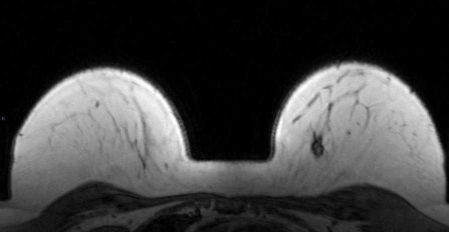
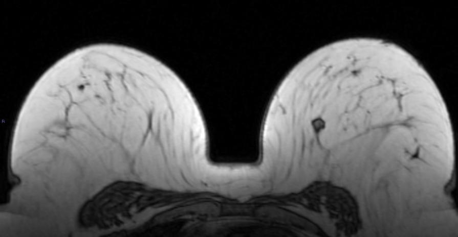
6 Displacement Breast cancer history, new microcalcifications of the left lower External Quadrant BIRADS 4. 6


7 Displacement MPR provides good showing of the distances between the lesion and the skin/muscle/scar 7
8 Agreement between MRI and US Location of the target Anteror/posterior displacement Fisher, p= 0.55 Cranio caudal displacement Quadrant Superior/lower: Kappa=0.97 Lateral displacement Quadrant Internal/External : Kappa=0.93 The hour topography Kappa=0.52 8


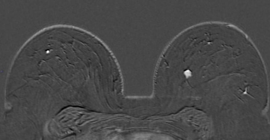
9 Displacement MIP provides good showing of the location of the lesions 9
10 Morphological findings Correlation between MRI & biopsies under second look US 10
11 Agreement between MRI and US Morphological findings Shape: benign vs suspicious Kappa=0.09 Margin: benign vs suspicious Kappa=0.23 Size T-test, p= BIRADS 3 vs 4 &5 Kappa=


12 Agreement between MRI and US: morphological findings 46 yo, history of breast cancer Houssami N. J Clin Oncol Off J Am Soc Clin Oncol. 2008


13 Agreement between MRI and US: morphological findings 46 yo, history of breast cancer Houssami N. J Clin Oncol Off J Am Soc Clin Oncol. 2008

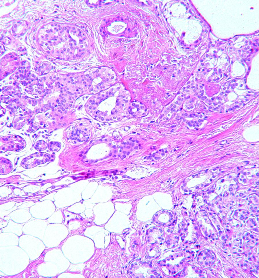
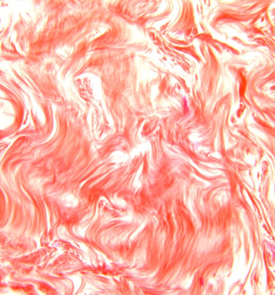
14 Pathology IDC SBR grade 1 14
15 Succes rate according morphological findings Correlation between MRI & biopsies under second look US 15


16 Success rate of second look US: Mass versus non-mass 60 yo, staging of ILC of the right breast 63 yo, history of breast cancer, follow-up 16


17 Success rate of second look US: Mass versus non-mass 60 yo, staging of ILC of the right breast 63 yo, history of breast cancer, follow-up LaTrenta LR. Radiology. 2003; 2. Trop I. Current problems in diagnostic radiology. 2010; 3. Wiratkapun C. Acad Radiol. 2008; 4. Meissnitzer M. Am J Roentgenol. 2009

18 Pathology Dystrophy: adenosis Scare sclerosis 18


19 Success rate of second look US: Suspicions versus benign 60 yo, left nipple retraction 57 yo, history of breast cancer, lymph nodes in left axilla Wiratkapun C. Acad Radiol. 2008; 2. Meissnitzer M. Am J Roentgenol. 2009


20 Success rate of second look US: Suspicions versus benign 60 yo, left nipple retraction 57 yo, history of breast cancer, lymph nodes in left axilla Wiratkapun C. Acad Radiol. 2008; 2. Meissnitzer M. Am J Roentgenol. 2009


21 Pathology Desmoïde fibroma IDC, SBR grade II Arrighi * H
22 Succes rate according to the size Size Masses Non-Masses <5 mm 50%? 5-10 mm 56% 13% mm 72% 25% >15 mm 86% 42% Meissnitzer M. Am J Roentgenol. 2009
23 Depth Correlation between MRI & biopsies under second look US 23

24 Breast cancer risk according depth Chi2, p=0,066 24


25 Breast cancer risk according depth 45 yo, left breast cancer staging Houssami N. J Clin Oncol Off J Am Soc Clin Oncol. 2008


26 Breast cancer risk according depth 45 yo, left breast cancer staging Houssami N. J Clin Oncol Off J Am Soc Clin Oncol. 2008

27 Pathology IDC SBR grade II 27
28 Cancer rates according to risk factor Correlation between MRI & biopsies under second look US 28
29 Cancer rate according to risk factors High risk versus no risk patient Fisher test, p=0.79 History of breast cancer in young patient Fisher, p=0.34 During staging Fisher, p=0.80 Be careful with suspicious Clinical findings 29


30 Risk factors 37 yo, BRCA1 mutation, history of right breast cancer, screening 30

31 Risk factors 37 yo, BRCA1 mutation, history of right breast cancer, screening 31
32 Risk factors 37 yo, BRCA1 mutation, history of right breast cancer, screening 32

33 Pathology Fibrous dystrophy 33
34 Cancer rates according to morphological findings in MRI Correlation between MRI & biopsies under second look US 34
35 Suspicious findings in MRI Mass Margins: NPV = 0.86 (Fisher test, p=0.03) Enhancement curves: NPV = 1 (Fisher test, p=0.01) T1, T2, Shape, internal enhancement : (Fisher test, p>0.072) Size (Student s t-test, p = 0.89). Non-Mass Distribution, internal enhancement: NPV < 0.85 (Fisher test, p>0.56) BIRADS BIRADS 3 : NPV = 0.94 (Fisher, p=0,068) 35


36 Enhancement curves 69 yo, breast cancer metastasis in axillary lymph nodes 36

37 Enhancement curves 69 yo, breast cancer metastasis in axillary lymph nodes 37


38 Enhancement curves 69 yo, breast cancer metastasis in axillary lymph nodes 38

39 Pathology IDC SBR grade III Triple negative Milagre, H
40 Cancer rates according to morphological findings in US Correlation between MRI & biopsies under second look US 40
41 Suspicious findings in US Shape: NPV = 0.90 (Fisher, p=0.025) Margin: NPV = 0.91 (Fisher, p=0,0046) Orientation: NPV = 0.87 (Fisher, p=0,0018) Depth, echogenicity, posterior US Beam (Fisher, p=0,53) Taille : t-test, p=0,65 BIRADS : NPV = 0.95%, (Fisher, p=0,039) 41


42 Vertical orientation 44 yo, discrepancy between luminal breast cancer and triple negative lymph node metastasis 42


43 Vertical orientation 44 yo, discrepancy between luminal breast cancer and triple negative lymph node metastasis 43


44 Vertical orientation 44 yo, discrepancy between luminal breast cancer and triple negative lymph node metastasis 44

45 Vertical orientation 44 yo, discrepancy between luminal breast cancer and triple negative lymph node metastasis 45

46 Pathology IDC SBR Grade III Inflammatory stroma 46
47 Conclusion Correlation between MRI & biopsies under second look US 47
48 Take Home Messages Risk factors were not reliable criteria for establishing an indication for second look ultrasound Displacement in anterior-posterior axis Masses are found more frequently than non-mass BIRADS 5 are found more frequently than BIRADS 4 Circumscribed contours and a progressive enhancement curve for masses on MRI had the strongest NPV (>0.85) Round or oval shape, circumscribed contours and the parallel orientation on US had the strongest NPV (>0.85) Correlation between abnormalities detected on MRI and US is sometimes delicate, biopsy and clip placement should be easily recommended 48
49 49
50 Litterature review 1. Houssami N, Ciatto S, Macaskill P, Lord SJ, Warren RM, Dixon JM, et al. Accuracy and surgical impact of magnetic resonance imaging in breast cancer staging: systematic review and meta-analysis in detection of multifocal and multicentric cancer. J Clin Oncol Off J Am Soc Clin Oncol Jul 1;26(19): Monticciolo DL. Postbiopsy Confirmation of MR-Detected Lesions Biopsied Using Ultrasound. Am J Roentgenol Jun;198(6):W618 W Carbonaro LA, Tannaphai P, Trimboli RM, Verardi N, Fedeli MP, Sardanelli F. Contrast enhanced breast MRI: Spatial displacement from prone to supine patient s position. Preliminary results. Eur J Radiol Jun;81(6):e771 e LaTrenta LR, Menell JH, Morris EA, Abramson AF, Dershaw DD, Liberman L. Breast lesions detected with MR imaging: utility and histopathologic importance of identification with US. Radiology Jun;227(3): Berg WA, Blume JD, Cormack JB, Mendelson EB, Madsen EL. Lesion detection and characterization in a breast US phantom: results of the ACRIN 6666 Investigators. Radiology Jun;239(3): Trop I, Labelle M, David J, Mayrand MH, Lalonde L. Second-look targeted studies after breast magnetic resonance imaging: practical tips to improve lesion identification. Current problems in diagnostic radiology Sep;39: Wiratkapun C, Duke D, Nordmann AS, Lertsithichai P, Narra V, Barton PT, et al. Indeterminate or suspicious breast lesions detected initially with MR imaging: value of MRI-directed breast ultrasound. Acad Radiol May;15(5): Meissnitzer M, Dershaw DD, Lee CH, Morris EA. Targeted Ultrasound of the Breast in Women With Abnormal MRI Findings for Whom Biopsy Has Been Recommended. Am J Roentgenol Oct;193(4): Abe H, Schmidt RA, Shah RN, Shimauchi A, Kulkarni K, Sennett CA, et al. MR-Directed ( Second-Look ) Ultrasound Examination for Breast Lesions Detected Initially on MRI: MR and Sonographic Findings. Am J Roentgenol Feb;194(2): Sim LSJ, Hendriks JHCL, Bult P, Fook-Chong SMC. US correlation for MRI-detected breast lesions in women with familial risk of breast cancer. Clin Radiol Jul;60(7): Fiaschetti V, Salimbeni C, Gaspari E, Dembele GK, Bolacchi F, Cossu E, et al. The role of second-look ultrasound of BIRADS-3 mammary lesions detected by breast MR imaging. Eur J Radiol Nov;81(11): Trop I, David J, Lalonde L. Postbiopsy confirmation of adequate targeting after second-look biopsy of MRI-enhancing breast lesions. Ajr Am J Roentgenol Jan;200(1):W Elshof LE, Rutgers EJT, Deurloo EE, Loo CE, Wesseling J, Pengel KE, et al. A practical approach to manage additional lesions at preoperative breast MRI in patients eligible for breast conserving therapy: results. Breast Cancer Res Treat Jul 22;124(3): Holland R, Hendriks JH, Vebeek AL, Mravunac M, Schuurmans Stekhoven JH. Extent, distribution, and mammographic/histological correlations of breast ductal carcinoma in situ. Lancet Mar 3;335: Sardanelli F, Boetes C, Borisch B, Decker T, Federico M, Gilbert FJ, et al. Magnetic resonance imaging of the breast: recommendations from the EUSOMA working group. Eur J Cancer May;46: Nakano S, Kousaka J, Fujii K, Yorozuya K, Yoshida M, Mouri Y, et al. Impact of real-time virtual sonography, a coordinated sonography and MRI system that uses an image fusion technique, on the sonographic evaluation of MRI-detected lesions of the breast in second-look sonography. Breast Cancer Res Treat Jul 24;134(3):
51 Pathology Correlation between MRI & biopsies under second look US 51
52 Pathology 52


53 Fibrosis changes 59 yo, history of right breast cancer, right nipple retraction 46 yo, BRCA 2, history of breast cancer, follow up 53


54 Fibrosis changes 59 yo, history of right breast cancer, right nipple retraction 46 yo, BRCA 2, history of breast cancer, follow up 54


55 Pathology Fibrous Dystophy Fibrosis, nuclear dystrophy post radiotherapy SARRAZY, H Margot, H


56 MRI Findings 44 yo, staging of a right breast cancer 53 yo, distorsion in the upper quadrants of the left breast 56


57 MRI Findings 44 yo, staging of a right breast cancer 53 yo, distorsion in the upper quadrants of the left breast 57


58 Pathology Fibrous Dystophy Dystrophy with atypical ductal hyperplasia De Saint Ours, H PASSAS, H
TÍTULO The role of FUSION between MRI and PET-CT as preoperative staging in breast cancer
 TÍTULO The role of FUSION between MRI and PET-CT as preoperative staging in breast cancer Páramo M, Zalazar R, Elizalde A, Pina-Insausti L, Vigil C, Hernández M, Rodríguez-Fraile M Clínica Universidad
Tips and Tricks to performing Magnetic Resonance Imaging Guided Breast Interventional Procedures Habib Rahbar, MD, FSBI October 23, 2018, 7:00pm ET
 Tips and Tricks to performing Magnetic Resonance Imaging Guided Breast Interventional Procedures Habib Rahbar, MD, FSBI October 23, 2018, 7:00pm ET SAM Questions/Answers/Rationales/References 1. Below
National Diagnostic Imaging Symposium 2013 SAM - Breast MRI 1
 National Diagnostic Imaging Symposium 2013 December 8-12, 2013 Disney s Yacht Club Resort Lake Buena Vista, Florida Self Assessment Module Questions, Answers and References Day SAM Title - Each SAM title
Is Probably Benign Really Just Benign? Peter R Eby, MD, FSBI Virginia Mason Medical Center Seattle, WA
 Is Probably Benign Really Just Benign? Peter R Eby, MD, FSBI Virginia Mason Medical Center Seattle, WA Disclosures: CONSULTANT FOR DEVICOR MEDICAL ARS Question 1 Is probably benign really just benign?
Journal of Breast Cancer
 Journal of Breast Cancer ORIGINAL ARTICLE J Breast Cancer 2011 September; 14(3): 213-218 Clinical Outcome of Magnetic Resonance Imaging-Detected Additional Lesions in Breast Cancer Patients Gi-Won Ha,
Table 1. Classification of US Features Based on BI-RADS for US in Benign and Malignant Breast Lesions US Features Benign n(%) Malignant n(%) Odds
 Malignant n(%) Odds") 215 Table 1. Classification of US Features Based on BI-RADS for US in Benign and Malignant Breast Lesions US Features Benign n(%) Malignant n(%) Odds ratio 719 (100) 305(100) Shape Oval 445 (61.9) 019
215 Table 1. Classification of US Features Based on BI-RADS for US in Benign and Malignant Breast Lesions US Features Benign n(%) Malignant n(%) Odds ratio 719 (100) 305(100) Shape Oval 445 (61.9) 019
Medical Policy An independent licensee of the Blue Cross Blue Shield Association
 CAE of Malignancy with MRI of the Breast Page 1 of 9 Medical Policy An independent licensee of the Blue Cross Blue Shield Association Title: See also: Computer-Aided Evaluation of Malignancy with Magnetic
Ductal carcinoma in situ, underestimation, ultrasound-guided core needle biopsy
 Ductal carcinoma in situ diagnosed after an ultrasoundguided 14-gauge core needle biopsy of breast masses: Can underestimation be predicted preoperatively? Poster No.: C-0442 Congress: ECR 2010 Type: Scientific
BREAST MRI. Elizabeth A. Rafferty, M.D. Avon Comprehensive Breast Center Massachusetts General Hospital Harvard Medical School
 BREAST MRI Elizabeth A. Rafferty, M.D. Avon Comprehensive Breast Center Massachusetts General Hospital Harvard Medical School BREAST MRI Any assessment of the breast parenchyma requires the administration
Patient Outcomes in Canceled MRI-Guided Breast Biopsies
 Women s Imaging Original Research Outcomes After Canceled MRI-Guided Breast Biopsies Women s Imaging Original Research Bethany L. Niell 1 Janie M. Lee 1, 2 Christopher Johansen 3 Elkan F. Halpern 4 Elizabeth
Management of Palpable Abnormalities in the Breast Katerina Dodelzon, MD July 31, 2018, 7:00pm ET
 Management of Palpable Abnormalities in the Breast Katerina Dodelzon, MD July 31, 2018, 7:00pm ET SAM Questions 1. 21 year old female presenting with left breast palpable mass, what is the most appropriate
Current Status of Supplementary Screening With Breast Ultrasound
 Current Status of Supplementary Screening With Breast Ultrasound Stephen A. Feig, M.D., FACR Fong and Jean Tsai Professor of Women s Imaging Department of Radiologic Sciences University of California,
When do you need PET/CT or MRI in early breast cancer?
 When do you need PET/CT or MRI in early breast cancer? Elizabeth A. Morris MD FACR Chief, Breast Imaging Service Memorial Sloan-Kettering Cancer Center NY, NY Objectives What is the role of MRI in initial
Computer-Aided Evaluation of Malignancy with Magnetic Resonance Imaging of the Breast. Original Policy Date
 MP 6.01.36 Computer-Aided Evaluation of Malignancy with Magnetic Resonance Imaging of the Breast Medical Policy Section Radiology Issue 12:2013 Original Policy Date 12:2013 Last Review Status/Date Reviewed
Pitfalls and Limitations of Breast MRI. Susan Orel Roth, MD Professor of Radiology University of Pennsylvania
 Pitfalls and Limitations of Breast MRI Susan Orel Roth, MD Professor of Radiology University of Pennsylvania Objectives Review the etiologies of false negative breast MRI examinations Discuss the limitations
ORIGINAL PAPER. Background parenchymal enhancement in preoperative breast MRI
 Nagoya J. Med. Sci. 77. 373 ~ 382, 2015 ORIGINAL PAPER Background parenchymal enhancement in preoperative breast MRI Satoko Kohara 1, Satoko Ishigaki 1, Hiroko Satake 1, Akiko Kawamura 1, Hisashi Kawai
Aims and objectives. Page 2 of 10
 Diagnostic performance of automated breast volume scanner (ABVS) versus hand-held ultrasound (HHUS) as second look for breast lesions detected only on magnetic resonance imaging. Poster No.: C-1701 Congress:
Second-look ultrasonography for MRI-detected suspicious breast lesions in patients with breast cancer
 Second-look ultrasonography for MRI-detected suspicious breast lesions in patients with breast cancer Min Ji Hong 1, Joo Hee Cha 1, Hak Hee Kim 1, Hee Jung Shin 1, Eun Young Chae 1, Ji Eun Shin 1,2, Woo
Contrast-enhanced magnetic resonance imaging (MRI) is a complementary
 is a complementary") Diagn Interv Radiol 2012; 18:460 467 Turkish Society of Radiology 2012 BREAST IMAGING ORIGINAL ARTICLE The role of breast MRI in planning the surgical treatment of breast cancer Gökhan Duygulu, Ayşenur
Diagn Interv Radiol 2012; 18:460 467 Turkish Society of Radiology 2012 BREAST IMAGING ORIGINAL ARTICLE The role of breast MRI in planning the surgical treatment of breast cancer Gökhan Duygulu, Ayşenur
ACRIN 6666 IM Additional Evaluation: Additional Views/Targeted US
 Additional Evaluation: Additional Views/Targeted US For revised or corrected form check box and fax to 215-717-0936. Instructions: The form is completed based on recommendations (from ID form) for additional
Newly Diagnosed Breast Cancer: Preoperative Imaging and Localization
 Newly Diagnosed Breast Cancer: Preoperative Imaging and Localization Debra Monticciolo, MD Professor of Radiology Texas A&M University no disclosures Debra Monticciolo, MD Professor of Radiology Texas
MEDICAL POLICY SUBJECT: MAGNETIC RESONANCE IMAGING (MRI) OF THE BREAST. POLICY NUMBER: CATEGORY: Technology Assessment
 OF THE BREAST. POLICY NUMBER: CATEGORY: Technology Assessment") MEDICAL POLICY SUBJECT: MAGNETIC RESONANCE IMAGING (MRI) OF THE BREAST PAGE: 1 OF: 9 If the member's subscriber contract excludes coverage for a specific service it is not covered under that contract.
MEDICAL POLICY SUBJECT: MAGNETIC RESONANCE IMAGING (MRI) OF THE BREAST PAGE: 1 OF: 9 If the member's subscriber contract excludes coverage for a specific service it is not covered under that contract.
BI-RADS and Breast MRI. Kathy Borovicka, M.D. Thursday February 15, 2018
 BI-RADS and Breast MRI Kathy Borovicka, M.D. Thursday February 15, 2018 Learning Objectives Be familiar with the Breast Imaging Reporting and Data System (BI-RADS) Understand the components of a breast
Mammographic imaging of nonpalpable breast lesions. Malai Muttarak, MD Department of Radiology Chiang Mai University Chiang Mai, Thailand
 Mammographic imaging of nonpalpable breast lesions Malai Muttarak, MD Department of Radiology Chiang Mai University Chiang Mai, Thailand Introduction Contents Mammographic signs of nonpalpable breast cancer
Detailed Program of the second BREAST IMAGING AND INTERVENTIONS PROGRAM am am : Clinician s requirements from breast imaging
 Detailed Program of the second BREAST IMAGING AND INTERVENTIONS PROGRAM 2012 Day one, 2 nd November BREAST IMAGING AND INTERVENTIONS PROGRAM 2012 9.00 AM 9.10 am Introduction 9.10 am - 9.30 am : Clinician
Computer-Aided Evaluation of Malignancy with Magnetic Resonance Imaging of the Breast
 Page: 1 of 11 Last Review Status/Date: March 2015 Magnetic Resonance Imaging of the Breast Description The use of computer-aided evaluation (CAE) may assist radiologists interpretation of contrastenhanced
Vacuum-assisted breast biopsy using computer-aided 3.0 T- MRI guidance: diagnostic performance in 173 lesions
 Vacuum-assisted breast biopsy using computer-aided 3.0 T- MRI guidance: diagnostic performance in 173 lesions Poster No.: C-2870 Congress: ECR 2017 Type: Scientific Exhibit Authors: A. Pozzetto, L. Camera,
Rate of Malignancy in MRI-Detected Probably Benign (BI-RADS 3) Lesions
 Lesions") Women s Imaging Original Research Spick et al. Malignancy in MRI BI-RADS 3 Lesions Women s Imaging Original Research Claudio Spick 1,2 Dieter H. M. Szolar 1 Pascal A. Baltzer 2 Manfred Tillich 1 Pia Reittner
Women s Imaging Original Research Spick et al. Malignancy in MRI BI-RADS 3 Lesions Women s Imaging Original Research Claudio Spick 1,2 Dieter H. M. Szolar 1 Pascal A. Baltzer 2 Manfred Tillich 1 Pia Reittner
Atypical ductal hyperplasia diagnosed at ultrasound guided biopsy of breast mass
 Atypical ductal hyperplasia diagnosed at ultrasound guided biopsy of breast mass Poster No.: C-1483 Congress: ECR 2014 Type: Authors: Keywords: DOI: Scientific Exhibit J. Cho, J. Chung, E. S. Cha, J. E.
J Clin Oncol 26: by American Society of Clinical Oncology INTRODUCTION
 VOLUME 26 NUMBER 3 JANUARY 20 2008 JOURNAL OF CLINICAL ONCOLOGY O R I G I N A L R E P O R T Relationship of Breast Magnetic Resonance Imaging to Outcome After Breast-Conservation Treatment With Radiation
E94. Aims and Objectives To assess whether it is possible to establish. received revised accepted
 Can Cut-Off-Values for Tumor Size or Patient Age in Breast Ultrasound Reduce Unnecessary Biopsies or is it all About Bi-rads? A Retrospective Analysis of 763 Biopsied T1-Sized Lesions Authors Laura Holzer-Fruehwald
Non-mass Enhancement on Breast MRI. Aditi A. Desai, MD Margaret Ann Mays, MD
 Non-mass Enhancement on Breast MRI Aditi A. Desai, MD Margaret Ann Mays, MD Breast MRI Important screening and diagnostic tool, given its high sensitivity for breast cancer detection Breast MRI - Indications
Lesion Imaging Characteristics Mass, Favoring Benign Circumscribed Margins Intramammary Lymph Node
 Lesion Imaging Characteristics Mass, Favoring Benign Circumscribed Margins Intramammary Lymph Node Oil Cyst Mass, Intermediate Concern Microlobulated Margins Obscured Margins Mass, Favoring Malignant Indistinct
Clinical Practice Guideline for the Indications for Use of Breast Magnetic Resonance Imaging (MRI)
") CIHRT Exhibit P-2595 Page 1 Question: Clinical Practice Guideline for the Indications for Use of Breast Magnetic Resonance Imaging (MRI) Eastern Health Breast Disease Site Group What are the current indications
CIHRT Exhibit P-2595 Page 1 Question: Clinical Practice Guideline for the Indications for Use of Breast Magnetic Resonance Imaging (MRI) Eastern Health Breast Disease Site Group What are the current indications
Triple-negative breast cancer: which typical features can we identify on conventional and MRI imaging?
 Triple-negative breast cancer: which typical features can we identify on conventional and MRI imaging? Poster No.: C-1862 Congress: ECR 2013 Type: Educational Exhibit Authors: V. Bertani 1, A. Gualano
Imaging the Symptomatic Patient. Avice M.O Connell MD,FACR,FSBI Professor of Imaging Sciences Director, Women s Imaging University of Rochester
 Imaging the Symptomatic Patient Avice M.O Connell MD,FACR,FSBI Professor of Imaging Sciences Director, Women s Imaging University of Rochester The four most common symptoms Mass Pain Discharge Infection
BR 1 Palpable breast lump
 BR 1 Palpable breast lump Palpable breast lump in patient 40 years of age or above MMG +/- spot compression or digital breast tomosynthesis over palpable findings Suspicious or malignant findings (BIRADS
Radiologic Findings of Mucocele-like Tumors of the breast: Can we differentiate pure benign from associated with high risk lesions?
 Radiologic Findings of Mucocele-like Tumors of the breast: Can we differentiate pure benign from associated with high risk lesions? Poster No.: C-0332 Congress: ECR 2014 Type: Educational Exhibit Authors:
Angela Gilliam, MD University of Colorado Surgical Grand Rounds November 3, 2008
 Angela Gilliam, MD University of Colorado Surgical Grand Rounds November 3, 2008 Breast Cancer Most common cancer in American women 180,000 new cases per year Second most common cause of cancer death 44,000
Triple Negative Breast Cancer
 Triple Negative Breast Cancer Prof. Dr. Pornchai O-charoenrat Division of Head-Neck & Breast Surgery Department of Surgery Faculty of Medicine Siriraj Hospital Breast Cancer Classification Traditional
Here are examples of bilateral analog mammograms from the same patient including CC and MLO projections.
 Good afternoon. It s my pleasure to be discussing Diagnostic Breast Imaging over the next half hour. I m Wei Yang, Professor of Diagnostic Radiology and Chief, the Section of Breast Imaging as well as
Feasibility of MRI-guided large-core-needle biopsy of suspiscious breast lesions at 3T
 Eur Radiol (2009) 19: 1639 1644 DOI 10.1007/s00330-009-1310-0 BREAST Nicky H. G. M. Peters Carla Meeuwis Chris J. G. Bakker Willem P. Th. M. Mali Arancha M. Fernandez-Gallardo Richard van Hillegersberg
Impact of value based breast cancer care pathway implementation on pre-operative breast magnetic resonance imaging utilization
 Original Article Impact of value based breast cancer care pathway implementation on pre-operative breast magnetic resonance imaging utilization Devina K. S. McCray, Stephen R. Grobmyer, Holly J. Pederson
What the surgeon wants from the radiologist before breast cancer surgery. Erica Patocskai Isabelle Trop
 What the surgeon wants from the radiologist before breast cancer surgery Erica Patocskai Isabelle Trop Centre Hospitalier de l université de Montréal CAR, April 2013 Plan What is the role of MRI for breast
Date of Preparation: December 8, 2017 A. GENERAL INFORMATION. 1. Name: Eun Sook KO Citizenship: Republic of Korea
 Date of Preparation: December 8, 2017 A. GENERAL INFORMATION 1. Name: Eun Sook KO 2. Email: mathilda0330@gmail.com 3. Citizenship: Republic of Korea B. EDUCATIONAL & TRAINING BACKGROUND Degree Institution
How to Use MRI Following Neoadjuvant Chemotherapy (NAC) in Locally Advanced Breast Cancer
 in Locally Advanced Breast Cancer") Global Breast Cancer Conference 2016 & 5 th International Breast Cancer Symposium April 29 th 2016, 09:40-10:50 How to Use MRI Following Neoadjuvant Chemotherapy (NAC) in Locally Advanced Breast Cancer
Global Breast Cancer Conference 2016 & 5 th International Breast Cancer Symposium April 29 th 2016, 09:40-10:50 How to Use MRI Following Neoadjuvant Chemotherapy (NAC) in Locally Advanced Breast Cancer
Mammographically non-calcified ductal carcinoma in situ: sonographic features with pathological correlation in 35 patients
 Clinical Radiology (2009) 64, 628e636 ORIGINAL PAPER Mammographically non-calcified ductal carcinoma in situ: sonographic features with pathological correlation in 35 patients B. Mesurolle a, *, M. El-Khoury
AMSER Case of the Month: September 2018
 AMSER Case of the Month: September 2018 60-year-old woman with a left breast mass noted on screening mammography. Catherine McNulty, MS4 Tulane University School of Medicine Dr. Robin Sobolewski Breast
Spiculated breast masses on MRI: Which category should we choose, 4 or 5?
 Spiculated breast masses on MRI: Which category should we choose, 4 or 5? Poster No.: C-1394 Congress: ECR 2015 Type: Scientific Exhibit Authors: N. Onishi, S. Kanao, M. Kataoka, M. Kawai, M. Iima, A.
Correlation Between BIRADS Classification and Ultrasound -guided Tru-Cut Biopsy Results of Breast Lesions: Retrospective Analysis of 285 Patients
 Correlation Between BIRADS Classification and Ultrasound -guided Tru-Cut Biopsy Results of Breast Lesions: Retrospective Analysis of 285 Patients Poster No.: C-1433 Congress: ECR 2014 Type: Scientific
SBI/ACR Breast Imaging Symposium April 7-10, 2016 Austin, TX
 SBI/ACR Breast Imaging Symposium April 7-10, 2016 Austin, TX SAM Session 2 Friday, April 8 2.5 SAM Credits Thank you for completing this SAM activity. Below you will find correct responses, rationales,
MP Magnetic Resonance Imaging for Detection and Diagnosis of Breast Cancer
 Medical Policy MP 6.01.29 BCBSA Ref. Policy: 6.01.29 Last Review: 09/19/2018 Effective Date: 09/19/2018 Section: Radiology Related Policies 6.01.45 Computer-Aided Evaluation of Malignancy With Magnetic
BREAST MRI. VASILIKI FILIPPI RADIOLOGIST CT MRI & PET/CT Departments Hygeia Hospital, Athens, Greece
 BREAST MRI VASILIKI FILIPPI RADIOLOGIST CT MRI & PET/CT Departments Hygeia Hospital, Athens, Greece Breast ΜR Imaging (MRM) Breast MR imaging is an extremely powerful diagnostic tool, that when used in
MEDICAL POLICY SUBJECT: MAMMOGRAPHY: COMPUTER- AIDED DETECTION (CAD) POLICY NUMBER: CATEGORY: Technology Assessment
 POLICY NUMBER: CATEGORY: Technology Assessment") MEDICAL POLICY SUBJECT: MAMMOGRAPHY: COMPUTER- PAGE: 1 OF: 5 If a product excludes coverage for a service, it is not covered, and medical policy criteria do not apply. If a commercial product, including
MEDICAL POLICY SUBJECT: MAMMOGRAPHY: COMPUTER- PAGE: 1 OF: 5 If a product excludes coverage for a service, it is not covered, and medical policy criteria do not apply. If a commercial product, including
ACR Appropriateness Criteria on Nonpalpable Mammographic Findings (Excluding Calcifications)
") ACR Appropriateness Criteria on Nonpalpable Mammographic Findings (Excluding Calcifications) Mary S. Newell, MD a, Robyn L. Birdwell, MD b, Carl J. D Orsi, MD c, Lawrence W. Bassett, MD d, Mary C. Mahoney,
ACR Appropriateness Criteria on Nonpalpable Mammographic Findings (Excluding Calcifications) Mary S. Newell, MD a, Robyn L. Birdwell, MD b, Carl J. D Orsi, MD c, Lawrence W. Bassett, MD d, Mary C. Mahoney,
Breast MRI: Friend or Foe?
 Breast MRI: Friend or Foe? UCSF Postgraduate Course May 18, 2013 Cheryl Ewing, MD Clinical Professor of Surgery UCSF Department of Surgery APPLEGATE HAS DOUBLE MASTECTOMY IN CANCER SCARE DIAGNOSED WITH
RSNA, /radiol Appendix E1. Methods
 RSNA, 2016 10.1148/radiol.2016151097 Appendix E1 Methods US and Near-infrared Data Acquisition Four optical wavelengths (740 nm, 780 nm, 808 nm, and 830 nm) were used to sequentially deliver the light
BARC/2013/E/019 BARC/2013/E/019. AUDIT OF MAMMOGRAPHY PERFORMED IN OUR HOSPITAL by Surita Kantharia Medical Division
 BARC/2013/E/019 BARC/2013/E/019 AUDIT OF MAMMOGRAPHY PERFORMED IN OUR HOSPITAL by Surita Kantharia Medical Division BARC/2013/E/019 GOVERNMENT OF INDIA ATOMIC ENERGY COMMISSION BARC/2013/E/019 AUDIT OF
Breast Pathology in Men: Radiologic-Pathologic Correlation
 Breast Pathology in Men: Radiologic-Pathologic Correlation Poster No.: C-0243 Congress: ECR 2012 Type: Scientific Exhibit Authors: G. Garrido; Málaga/ES Keywords: Breast, Ultrasound, Mammography, Biopsy,
Role of PEM in Breast Cancer Management. Judy Kalinyak, MD, PhD Chief Medical Officer Naviscan, Inc (San Diego, CA)
") Role of PEM in Breast Cancer Management Judy Kalinyak, MD, PhD Chief Medical Officer Naviscan, Inc (San Diego, CA) Role of PEM in Breast Cancer Management Introduction to Positron Emission Mammography
Role of PEM in Breast Cancer Management Judy Kalinyak, MD, PhD Chief Medical Officer Naviscan, Inc (San Diego, CA) Role of PEM in Breast Cancer Management Introduction to Positron Emission Mammography
CDIS: what's beyond microcalcifications? - Pictorial essay
 CDIS: what's beyond microcalcifications? - Pictorial essay Poster No.: C-1096 Congress: ECR 2014 Type: Educational Exhibit Authors: R. N. Lucas, C. A. S. Ruano, I. Oliveira, J. M. G. Lourenco, Z. 1 1 1
PLACE LABEL HERE. ACRIN 6657 MRI Form: Pre-Treatment (MRI-1)
") M3 ACRIN 6657 MRI Form: Pre-Treatment (MRI-1) If this is a revised or corrected form,indicate by checking box. ACRIN Study 6657 Case # Instructions: In accordance with the protocol, four MRI exams are
M3 ACRIN 6657 MRI Form: Pre-Treatment (MRI-1) If this is a revised or corrected form,indicate by checking box. ACRIN Study 6657 Case # Instructions: In accordance with the protocol, four MRI exams are
MR Imaging of the Ipsilateral Breast in Women with Percutaneously Proven Breast Cancer
 Laura Liberman 1 Elizabeth A. Morris 1 D. David Dershaw 1 Andrea F. Abramson 1 Lee K. Tan 2 Received July 23, 2002; accepted after revision September 18, 2002. 1 Breast Imaging Section, Department of Radiology,
MRI Occult Invasive Breast Cancer
 MRI Occult Invasive Breast Cancer Poster No.: C-1573 Congress: ECR 2015 Type: Educational Exhibit Authors: R. Patel, N. Chhaya, K. Stafford, B. Holloway, D. Tsukagoshi, A. Malhotra; London/ Keywords: Cancer,
Introduction ORIGINAL ARTICLE. 170 Ultrasonography 33(3), July 2014 e-ultrasonography.org
, July 2014 e-ultrasonography.org") Positive predictive value of additional synchronous breast lesions in wholebreast ultrasonography at the diagnosis of breast cancer: clinical and imaging factors Ah Hyun Kim 1 *, Min Jung Kim 1, Eun-Kyung
Positive predictive value of additional synchronous breast lesions in wholebreast ultrasonography at the diagnosis of breast cancer: clinical and imaging factors Ah Hyun Kim 1 *, Min Jung Kim 1, Eun-Kyung
BI-RADS CATEGORIZATION AND BREAST BIOPSY categorization in the selection of appropriate breast biopsy technique is also discussed. Patients and method
 Original Article Positive Predictive Value of BI-RADS Categorization in an Asian Population Yah-Yuen Tan, Siew-Bock Wee, Mona P.C. Tan and Bee-Kiang Chong, 1 Departments of General Surgery and 1Diagnostic
MRI BI-RADS: How to make it out?
 MRI BI-RADS: How to make it out? Poster No.: C-1850 Congress: ECR 2016 Type: Educational Exhibit Authors: M. Ben Ammar, A. Ben Miled, O. Ghdes, S. Harguem, A. Gaja, N. Mnif; Tunis/TN Keywords: Breast,
Title: Ultrasound for Breast Cancer Screening: Clinical Effectiveness. Date: 4 January Context and policy issues:
 Title: Ultrasound for Breast Cancer Screening: Clinical Effectiveness Date: 4 January 2008 Context and policy issues: It is estimated that 22,300 women will be diagnosed with breast cancer and approximately
Role of positron emission mammography (PEM) for assessment of axillary lymph node status in patients with breast cancer
 for assessment of axillary lymph node status in patients with breast cancer") Role of positron emission mammography (PEM) for assessment of axillary lymph node status in patients with breast cancer Poster No.: C-1260 Congress: ECR 2011 Type: Scientific Paper Authors: K. M. Kulkarni,
Role of positron emission mammography (PEM) for assessment of axillary lymph node status in patients with breast cancer Poster No.: C-1260 Congress: ECR 2011 Type: Scientific Paper Authors: K. M. Kulkarni,
Case series: imaging features of intraductal papillomas in patients presenting as nipple discharge
 International Journal of Research in Medical Sciences Dhull V et al. Int J Res Med Sci. 20 Jul;4():2882882 www.msjonline.org pissn 2200 eissn 22002 Research Article DOI: http://dx.doi.org/0.820/22002.ijrms2099
EVALUATION OF AXILLARY LYMPH NODES AFTER NEOADJUVANT SYSTEMIC THERAPY KIM, MIN JUNG SEVERANCE HOSPITAL, YONSEI UNIVERSITY
 EVALUATION OF AXILLARY LYMPH NODES AFTER NEOADJUVANT SYSTEMIC THERAPY KIM, MIN JUNG SEVERANCE HOSPITAL, YONSEI UNIVERSITY AXILLARY LYMPH NODE METASTASIS Axillary lymph node metastasis is one of the most
Case study 1. Rie Horii, M.D., Ph.D. Division of Pathology Cancer Institute Hospital, Japanese Foundation for Cancer Research
 NCCN/JCCNB Seminar in Japan April 15, 2012 Case study 1 Rie Horii, M.D., Ph.D. Division of Pathology Cancer Institute Hospital, Japanese Foundation for Cancer Research Present illness: A 50y.o.premenopausal
Mammographic evaluation of palpable breast masses with pathological correlation: a tertiary care centre study in Nepal
 Original article 21 Mammographic evaluation of palpable breast masses with pathological correlation: a tertiary care centre study in Nepal G. Gurung, R. K. Ghimire, B. Lohani Department of Radiology and
Multicenter Evaluation of the Breast Lesion Excision System, a Percutaneous, Vacuum-Assisted, Intact-Specimen Breast Biopsy Device
 945 Multicenter Evaluation of the Breast Lesion Excision System, a Percutaneous, Vacuum-Assisted, Intact-Specimen Breast Biopsy Device Angela Sie, MD 1 David C. Bryan, MD 2 Victor Gaines, MD 3 Larry K.
Breast Cancers Not Detected at MRI: Review of False-Negative Lesions
 Women s Imaging Original Report Shimauchi et al. MRI-Occult Breast s Women s Imaging Original Report WOMEN S IMAGING Breast s Not Detected at MRI: Review of False-Negative Lesions Akiko Shimauchi 1 Sanaz
Pathologic outcomes of coarse heterogeneous calcifications detected on mammography
 Pathologic outcomes of coarse heterogeneous calcifications detected on mammography Poster No.: C-1957 Congress: ECR 2011 Type: Scientific Paper Authors: H. J. Lim, K. R. Cho, K. W. Hwang, B. K. Seo, O.
Emerging Techniques in Breast Imaging: Contrast-Enhanced Mammography and Fast MRI
 Emerging Techniques in Breast Imaging: Contrast-Enhanced Mammography and Fast MRI Lilian Wang, M.D. Breast Imaging Section Department of Radiology Northwestern Medicine Overview Rationale for new imaging
Underestimation of Atypical Ductal Hyperplasia at Sonographically Guided Core Biopsy of the Breast
 Women s Imaging Original Research Jang et al. Sonographic Breast Biopsy Women s Imaging Original Research WOMEN S IMAGING Underestimation of Atypical Ductal Hyperplasia at Sonographically Guided Core Biopsy
MR sin plass i brystkreftdiagnostikk, dagens anbefalinger og fremtidsperspektiver
 MR sin plass i brystkreftdiagnostikk, dagens anbefalinger og fremtidsperspektiver Kathinka Kurz, MD, PhD, seksjonsoverlege SUS, kathinka.dehli.kurz@sus.no Technique - Subtraction Without contrast agent
Breast Magnetic Resonance Imaging Indications in Current Practice
 DOI:http://dx.doi.org/10.7314/APJCP.2014.15.2.569 MINI-REVIEW Breast Magnetic Resonance Imaging Indications in Current Practice Sawsan Abdulkareem Taif Abstract Although mammography is the primary imaging
Evaluation of BI-RADS 3 lesions in women with a high risk of hereditary breast cancer.
 Evaluation of BI-RADS 3 lesions in women with a high risk of hereditary breast cancer. Poster No.: C-0346 Congress: ECR 2014 Type: Scientific Exhibit Authors: A. Thomas 1, R. Dominguez Oronoz 1, S. Roche
Successful Breast MRI Program : The ingredients
 Successful Breast MRI Program : The ingredients Dr. Smriti Hari Associate Professor Deptt. Of Radiology All India Institute of Medical Sciences New Delhi How to perform Breast MRI Breast MRI descriptors
BI-RADS Categorization As a Predictor of Malignancy 1
 Susan G. Orel, MD Nicole Kay, BA Carol Reynolds, MD Daniel C. Sullivan, MD BI-RADS Categorization As a Predictor of Malignancy 1 Index terms: Breast, biopsy, 00.1261 Breast neoplasms, localization, 00.125,
Breast MR biopsy. I Thomassin-Naggara, A.Jalaguier-Coudray, J Chopier
 Breast MR biopsy I Thomassin-Naggara, A.Jalaguier-Coudray, J Chopier Background EUSOBI When a radiologist perform a MR breast imaging he has to be able to realize or to be apart of a network who is able
MEDICAL IMAGING AND BREAST DISEASE HOW CAN WE HELP YOU?
 MEDICAL IMAGING AND BREAST DISEASE HOW CAN WE HELP YOU? Barbara M. Preston, M.D. SCREENING MAMMOGRAPHY AVERAGE RISK PATIENTS KAISER RECOMMENDATION: ALL WOMEN (INCLUDING TRANSGENDER FEMALES) Every 1-21
Does elastography change the indication to biopsy? IBDC
 Does elastography change the indication to biopsy? A LEXANDRA A THANASIOU, M D DEPARTMENT OF RADIOLOGY CURIE INSTITUTE PARIS, FRANCE IBDC Ultrasound Detected Cancers Physician-performed ultrasound increases
Correlation between lesion type and the additional value of digital breast tomosynthesis
 Correlation between lesion type and the additional value of digital breast tomosynthesis Poster No.: C-1604 Congress: ECR 2011 Type: Scientific Exhibit Authors: C. Van Ongeval, L. Cockmartin, A. Van Steen,
PLACE LABEL HERE BASELINE / PRE-TREATMENT. ACRIN 6657 Extension MRI Form: Baseline / Pre-Treatment MRI 1. o Unknown
 T1 ACRIN 6657 Extension MRI Form: Baseline / Pre-Treatment MRI 1 If this is a revised or corrected form, please box. ACRIN Study 6657 No. Instructions: In accordance with the protocol, four MRI exams are
Magnetic resonance imaging (MRI) in high risk women: benefits and problems
 in high risk women: benefits and problems") Magnetic resonance imaging (MRI) in high risk women: benefits and problems Poster No.: C-2466 Congress: ECR 2013 Type: Scientific Exhibit Authors: A. Pecchi, V. Marchesi, R. Battista, B. Canossi, L. Cortesi,
Magnetic resonance imaging (MRI) in high risk women: benefits and problems Poster No.: C-2466 Congress: ECR 2013 Type: Scientific Exhibit Authors: A. Pecchi, V. Marchesi, R. Battista, B. Canossi, L. Cortesi,
Monitoring neo-adjuvant chemotherapy: comparison of contrast-enhanced spectral mammography (CESM) and MRI versus breast cancer characteristics
 and MRI versus breast cancer characteristics") Monitoring neo-adjuvant chemotherapy: comparison of contrast-enhanced spectral mammography (CESM) and MRI versus breast cancer characteristics Poster No.: B-1062 Congress: ECR 2016 Type: Scientific Paper
Monitoring neo-adjuvant chemotherapy: comparison of contrast-enhanced spectral mammography (CESM) and MRI versus breast cancer characteristics Poster No.: B-1062 Congress: ECR 2016 Type: Scientific Paper
Invasive lobular carcinoma of the breast; spectrum of imaging findings.
 Invasive lobular carcinoma of the breast; spectrum of imaging findings. Poster No.: C-0847 Congress: ECR 2014 Type: Educational Exhibit Authors: D. Mandich, T. Diaz de Bustamante, L. Koren, M. Arroyo,
BREAST IMAGING and NEW IMAGING MODALITIES- A Surgeons view
 BREAST IMAGING and NEW IMAGING MODALITIES- A Surgeons view DR CHANTEL THORNTON SPECIALIST BREAST CANCER SURGEON BMSc (hons) MBBS (hons) FRACS Epworth Hospital, Richmond- Agora Centre for Women s Health
Radiologic and pathologic correlation of non-mass like breast lesions on US and MRI: Benign, high risk, versus malignant
 Radiologic and pathologic correlation of non-mass like breast lesions on US and MRI: Benign, high risk, versus malignant Poster No.: C-1161 Congress: ECR 2013 Type: Educational Exhibit Authors: J. Kwak,
DCIS of the Breast--MRI findings with mammographic correlation.
 DCIS of the Breast--MRI findings with mammographic correlation. Poster No.: C-1560 Congress: ECR 2013 Type: Educational Exhibit Authors: N. B. Ibrahim, P. Morris, S. ANANDAN; Burlington, MA/US Keywords:
Study on Efficacy of Preoperative Ultrasonography for Axillary Lymph Node Involvement In Breast Carcinoma
 IOSR Journal of Dental and Medical Sciences (IOSR-JDMS) e-issn: 2279-0853, p-issn: 2279-0861. Volume 13, Issue 4 Ver. II. (Apr. 2014), PP 01-05 Study on Efficacy of Preoperative Ultrasonography for Axillary
ISSN X (Print) Research Article. *Corresponding author Dr. Amlendu Nagar
 Research Article. *Corresponding author Dr. Amlendu Nagar") Scholars Journal of Applied Medical Sciences (SJAMS) Sch. J. App. Med. Sci., 2015; 3(3A):1069-1073 Scholars Academic and Scientific Publisher (An International Publisher for Academic and Scientific Resources)
Scholars Journal of Applied Medical Sciences (SJAMS) Sch. J. App. Med. Sci., 2015; 3(3A):1069-1073 Scholars Academic and Scientific Publisher (An International Publisher for Academic and Scientific Resources)
Pharmacokinetic evaluation of DCIS
 Pharmacokinetic evaluation of DCIS Poster No.: C-0412 Congress: ECR 2010 Type: Topic: Authors: Keywords: DOI: Scientific Exhibit Breast H. G. Toonen, R. Mann, H. Huisman, J. Veltman, C. Boetes; Nijmegen/NL
Leonard M. Glassman MD
 BI-RADS The New BI-RADS Leonard M. Glassman MD FACR Former Chief of Breast Imaging American Institute for Radiologic Pathology Washington Radiology Associates, PC Breast Imaging Reporting and Data System