Breast MR biopsy. I Thomassin-Naggara, A.Jalaguier-Coudray, J Chopier
|
|
|
- Lydia Caldwell
- 5 years ago
- Views:
Transcription
1 Breast MR biopsy I Thomassin-Naggara, A.Jalaguier-Coudray, J Chopier
 2 Heywang-Köbrunner SH, Eur J Radiol.")
2 Background EUSOBI When a radiologist perform a MR breast imaging he has to be able to realize or to be apart of a network who is able to realize MR biopsy European regulations : Team with an experience in percutaneous core biopsy and in MRI More than 50 procédures/year with 10 under MRI Learning curve : 15 procedures (EUSOBI) 2 Heywang-Köbrunner SH, Eur J Radiol. 2009


3 Indications BI-RADS 4 or BI-RADS 5 lesion not seen on mammography and breast ultrasonography BI-RADS 3 lesion in a specific context (proven associated cancer in the same or in contralateral breast, high-risk women BRCA1/2) 3


 DCIS A")

4 Indications MR imaging abnormality without any target on conventional imaging (2 nd look mammography and ultrasonography) DCIS A NMLE>2cm associated with calcifications = PPV>90% of malignancy 4 Thomassin-Naggara et al. Radiology 2011
 Perlet, 2006 517/538 (96%) Gebauer, 2006 42/42 (100%) Orel, 2006 85/85 (100%) Liberman, 2005 95/98 (97%)")
5 Breast MR biopsy : Is it technically accurate? Success rate Main studies Technical success rate Fisher, /389 (96%) Perlet, /538 (96%) Gebauer, /42 (100%) Orel, /85 (100%) Liberman, /98 (97%) Lehman, /38 (100%) 5 Fisher Rofo 2009, Perlet Cancer 2006, Gebauer Act Radiol 2006, Orel Radiology 2006, Liberman AJR 2005, Lehman AJR 2005


6 Breast MR biopsy : Is it technically accurate? Failures 6 Failure rate : 10-15% Causes : Accessibility Bleeding Vanishing lesion Background enhancement Glandular density Size <1cm Over compressed breastt Low experience Cancer rate: 2% Recommandation : Short follow up Diagnostic MRI The day of the biopsy, 1 month later Axial T1 fat-suppressed early contrastenhanced subtracted MRI RSNA 2013 Trop and Thomassin-Naggara et al.
7 Breast MR biopsy : Is it technically accurate? Malignancy rate Main studies Malignancy rate Fisher, /365 (27%) Perlet, /517 (27%) Gebauer, /42 (26%) Orel, /85 (61%) Liberman, /98 (25%) Lehman, /38 (37%) 7 Fisher Rofo 2009, Perlet Cancer 2006, Gebauer Act Radiol 2006, Orel Radiology 2006, Liberman AJR 2005, Lehman AJR 2005
8 Tenon Hospital Experience n Malignat Borderline Benign ACR ACR ACR (angiome) n Malignat Borderline Benign Mass 21 24% (5) 14% (3) 62%(13) Non Mass like enhancement 88 19% (17) 10% (9) 71% (60) 8
9 Breast MR biopsy : Is there any risk? 9 Usual secondary effects (50%) : Hematoma Air in the cavity Secondary effect with interruption of the procedure (10%) : Bleeding Vaso-vagal reaction Pain Rare effects (1%) : Infection Hyperventilation Liberman AJR 2005 Permet Cancer 2006 Hauth Eur radiol 2010 Mahoney JMRI 2008 Perretta breast radiology 2008


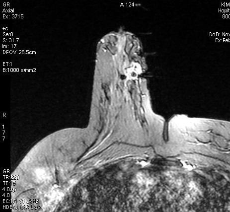
10 Routine clinical case MRI : Extension of an invasive lobular carcinoma Report Left breast: BI-RADS 2 Right breast : BI-RADS 6 : Proven malignant lesion Another mass of 15 mm in UEQ, BI-RADS 4c Strategy: Distance with index cancer : 4 cm 2 nd look ultrasound Percutaneous biopsy under sonography with a marker : non contributive Breast MR biopsy BI-RADS 6 BI-RADS 4c BI-RADS 4c 10
 A biopsy guidance")
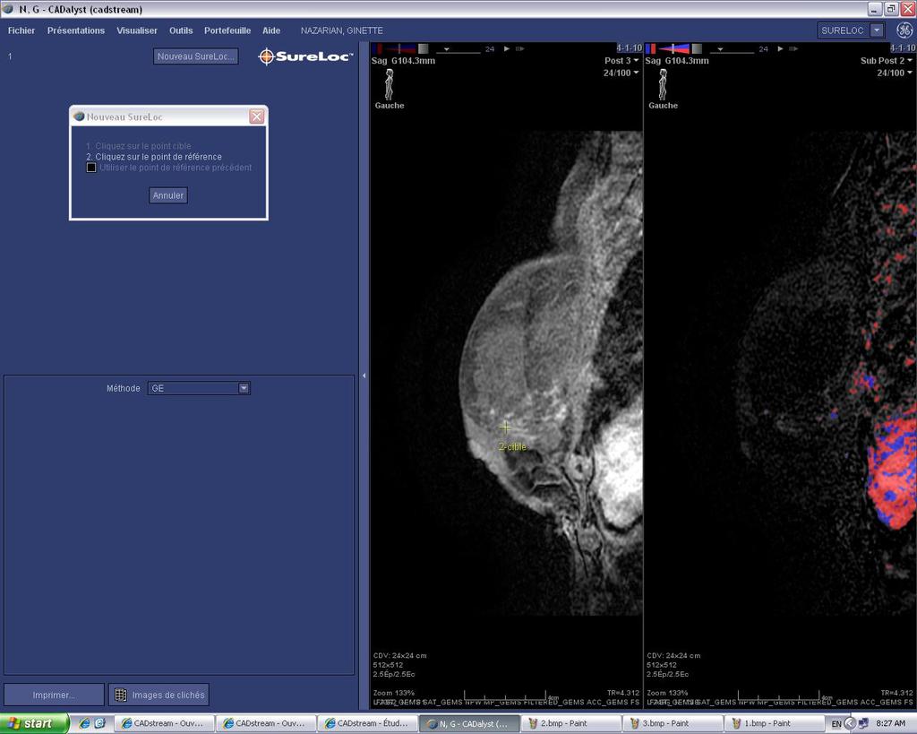
 11 Procubitus, Head outside from the")
11 MR machine : 1,5 T Step 1 : Positioning Dedicated breast coil (ideally the same than for breast MR diagnosis) A biopsy guidance system compatible with the breast coil and tissue sampling device (compression with a grid) 11 Procubitus, Head outside from the coil


12 Lateral access Coil 12 Medial access

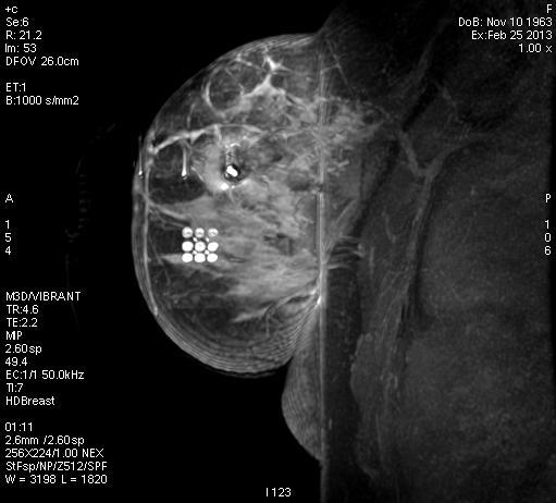
13 Step 2 : Localization After breast compression with the grid Positionning a cube visible on T1 sequence (Parafin or vitamin D) 13


14 Step 2 : Localization Positionning of the cube is reported in a schema The patient goes in the coil Cube Schema 14 Cube positionned on N 18
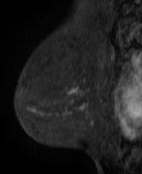
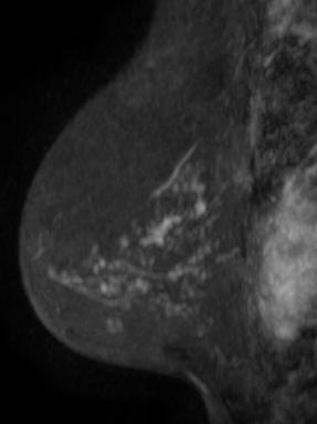
15 Step 2 : Localization High resolution T1 DCE-MR sequences with Fat saturation in Sagittal or Axial plane Before injection After injection on 3 phases with : Complete dose: 0.2 ml/kg with 20 ml saline, débit 2 à 3ml/s (No reinjection at the end of the procédure) 15
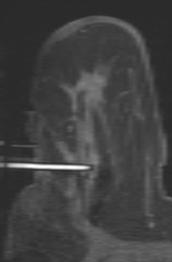
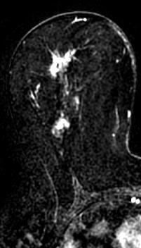
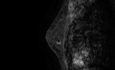
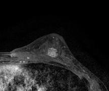
16 T1FS gadolinium in sagital plane 16 Find the target ++++
17 17
18 18
19 19
20 20
21 21
22 22
23 23
24 24
25 25
26 26
27 27
28 28
29 29
30 30
31 31
32 32
33 33
34 BI-RADS 6 BI-RADS ACR4 c Indication of breast MR biopsy 34

35 Step 2 : Localization Objective : To localize the target in comparison with the cube Lésion ACR4 c A biopsier sous IRM 35 Lésion ACR6 Biospiée sous écho
36 BI-RADS 4 c Breast MR biopsy target 36
37 Projection of BI-RADS 4 c Breast MR biopsy target 37
38 Projection of BI-RADS 4 c Breast MR biopsy target 38
39 Projection of BI-RADS 4 c Breast MR biopsy target 39
40 Projection of BI-RADS 4 c Breast MR biopsy target 40
41 Projection of BI-RADS 4 c Breast MR biopsy target 41
42 Projection of BI-RADS 4 c Breast MR biopsy target 42
43 Projection of BI-RADS 4 c Breast MR biopsy target 43
44 Projection of BI-RADS 4 c Breast MR biopsy target 44
45 Projection of BI-RADS 4 c Breast MR biopsy target 45
46 Projection of BI-RADS 4 c Breast MR biopsy target 46

47 Grid on the breast compressed Projection of BI-RADS 4 c Breast MR biopsy target Cube 47

48 Breast Schema On MR images Entrance point Breast schema During MR biopsy Cube 48


49 Step 2 : Localization We need to calculate the depth of the lesion from the skin : substraction ( : 27mm) ou simple mesurement on reformatted image BI-RADS 4c Breast MR biopsy target External side of the breast with the grid 27 mm 49
50 Step 2 : Localization x and y Breast MR biopsy entrance point 50 Z (depth) Z = 27mm


51 Step 3 : Samples Material : MR-compatible tissue sampling device (<11G) Xylocaine (20cc) Betadine Trocar tip Needle guide 51 Visible MR obturator

52 Step 3 : Samples Topical anaesthesic: Breast MR biopsy Entrance point 52

53 Step 3 : Samples Cutaneous section To put the cube in the grid Adjust the depth of the lesion : 27 mm under the skin According to constructor needles : SenoRx : adding 20 mm Bard : adding 10 mm Mammotome : adding 0 mm To put the trocar inside the breast 53
 DCE MR sequence to check the")
54 Step 3 : Samples When the position is well: It s necessary to replace the trocart by a MRI visible obturator (high T1FS) DCE MR sequence to check the position 54

55 Step 3 : Samples 55


56 End of the procedure of biopsy Minimum number of samples : 18 56


57 Step 4 : Quality insurance To mark the biopsy site with a marker To perform a control sequence 57
58 Usefulness of T2* sequence to visualize the marker


59 Technical challenges Sternal edge of breast coil * Localization of lesions Posterior lesion or axillary lesion Axial T1 fat-suppressed early contrast-enhancedmri Lateral approach is sometimes the only one issue Internal and superior approach are usually more difficult to realize. Visualization of lesions MR Biopsy - introductor in place Vanishing lesion Impossibility to confirm sampling 59 RSNA 2013 MR post-biopsy - introductor in place


60 Tips and tricks Depth lesion: To put the arms along the body To remove the foam of the coil To elevate the opposite side to down the breast in the coil 60


61 After removing the foam of the coil Apocrine metaplasia
62 Tips and tricks Small breats : Use a technique «Wonderbra» to pump the breast Use a «biopsy cover» to limit cutaneous lesion Cover 62


63 Screening MRI Tip and thricks pad Technical tip Padding around the least supported breast surfaces can help provide stability during the procedure. Axial T1 fat-suppressed early contrastenhanced subtracted MRI MR Biopsy - introductor in place History: 53 year old high-risk woman. MR biopsy of a 7 mm enhancing mass in anterior central left breast. Challenge: The anterior location of the lesion results in little breast support far anteriorly. A pad was placed to maintain the breast in place, and a blunt tip needle was used to avoid damage to the inner breast skin surface. Histology: Inconclusive histology. No follow-up available. Technical tip Use of a blunt tip needle is indicated for sampling of lesions close to the far surface of the breast to avoid skin damage. RSNA 2013 Trop and ThomassinNaggara et al.

64 Tips and techniques CAD system Actual position of the cube Precise localization of the entrance point 64 Depth of the target Schema to insert biopsy device


65 Breast MR biopsy Of a segmental NMLE 65
66 66
67 67
68 68
69 69
70 70
71 Patho radiological correlation: Underestimation rate Higher than those under stereotactic guidance About 38% in case of ADH at MR biopsy versus 20% with stereotactic biopsy Liberman et al. AJR Understimation is partially due to a partial sampling of target lesion 342 biopsied benign lesions 24 discordant lesions(7 %), with 6 cancers (30%) These lesions was more frequently simply sampled than removed Lee et al. AJR


72 How to follow patients after breast MR biopsy? Few data Radial scar MSK hospital : 177 benign biopsied lesions 8 increased during follow up. 17 re-biopsied et 4 were malignant Good radiopathological correlation is crucial 72 Systematic follow up 5 months if benign Liberman L et al.. AJR 2007 Li J, Dershaw DD, et al. AJR 2009


73 A 34 years old women, MR screening, BRCA mutation

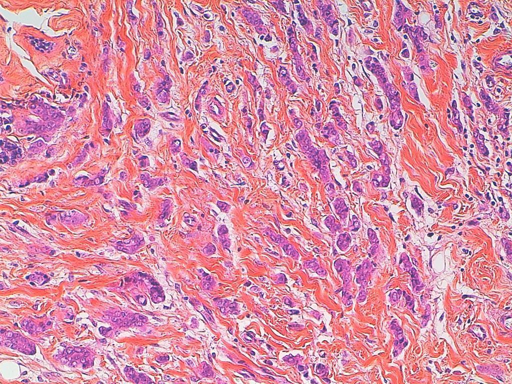
74 Pathology : Fibrosis Normal breast parenchyma with fibrosis with acini without any atypia Courtesy A.Jalaguier, IPC

")
75 MR follow up (6 monthes later)

76 Biopsy under sonography Invasive ductal carcinoma


77 Take home messages A coil Only for MR lesion not seen on conventional imaging Be careful : If benign results, follow up at 5 months +++ A grid Psychological take care Better results! A cube

78 Thank you for your attention
Macrobiopsy under X-Ray Guidance
 Macrobiopsy under X-Ray Guidance C. Balleyguier, B. Boyer Radiology Gustave Roussy, Villejuif, France Breast Intervention Imaging Major domain in breast imaging European guidelines recommend a pre surgical
National Diagnostic Imaging Symposium 2013 SAM - Breast MRI 1
 National Diagnostic Imaging Symposium 2013 December 8-12, 2013 Disney s Yacht Club Resort Lake Buena Vista, Florida Self Assessment Module Questions, Answers and References Day SAM Title - Each SAM title
Pitfalls and Limitations of Breast MRI. Susan Orel Roth, MD Professor of Radiology University of Pennsylvania
 Pitfalls and Limitations of Breast MRI Susan Orel Roth, MD Professor of Radiology University of Pennsylvania Objectives Review the etiologies of false negative breast MRI examinations Discuss the limitations
Vacuum-assisted breast biopsy using computer-aided 3.0 T- MRI guidance: diagnostic performance in 173 lesions
 Vacuum-assisted breast biopsy using computer-aided 3.0 T- MRI guidance: diagnostic performance in 173 lesions Poster No.: C-2870 Congress: ECR 2017 Type: Scientific Exhibit Authors: A. Pozzetto, L. Camera,
PURPOSE IMAGE-GUIDANCE MODALITIES IMAGE-GUIDED BREAST BIOPSY. US-Techniques. Ultrasound. US guided NLOBB. TH. Helbich
 IMAGE-GUIDED BREAST BIOPSY PURPOSE TH. Helbich Department of Radiology Division of Molecular & Gender Imaging Medical University of Vienna Imaging techniques Interventional procedures Quality management
Tips and Tricks to performing Magnetic Resonance Imaging Guided Breast Interventional Procedures Habib Rahbar, MD, FSBI October 23, 2018, 7:00pm ET
 Tips and Tricks to performing Magnetic Resonance Imaging Guided Breast Interventional Procedures Habib Rahbar, MD, FSBI October 23, 2018, 7:00pm ET SAM Questions/Answers/Rationales/References 1. Below
Patient Outcomes in Canceled MRI-Guided Breast Biopsies
 Women s Imaging Original Research Outcomes After Canceled MRI-Guided Breast Biopsies Women s Imaging Original Research Bethany L. Niell 1 Janie M. Lee 1, 2 Christopher Johansen 3 Elkan F. Halpern 4 Elizabeth
BREAST MRI. VASILIKI FILIPPI RADIOLOGIST CT MRI & PET/CT Departments Hygeia Hospital, Athens, Greece
 BREAST MRI VASILIKI FILIPPI RADIOLOGIST CT MRI & PET/CT Departments Hygeia Hospital, Athens, Greece Breast ΜR Imaging (MRM) Breast MR imaging is an extremely powerful diagnostic tool, that when used in
Radiologic and pathologic correlation of non-mass like breast lesions on US and MRI: Benign, high risk, versus malignant
 Radiologic and pathologic correlation of non-mass like breast lesions on US and MRI: Benign, high risk, versus malignant Poster No.: C-1161 Congress: ECR 2013 Type: Educational Exhibit Authors: J. Kwak,
Atypical Ductal Hyperplasia and Papillomas: A Comparison of Ultrasound Guided Breast Biopsy and Stereotactic Guided Breast Biopsy
 Atypical Ductal Hyperplasia and Papillomas: A Comparison of Ultrasound Guided Breast Biopsy and Stereotactic Guided Breast Biopsy Breast Cancer is the most common cancer diagnosed in women in the United
Treatment options for the precancerous Atypical Breast lesions. Prof. YOUNG-JIN SUH The Catholic University of Korea
 Treatment options for the precancerous Atypical Breast lesions Prof. YOUNG-JIN SUH The Catholic University of Korea Not so benign lesions? Imaging abnormalities(10% recall) lead to diagnostic evaluation,
Spiculated breast masses on MRI: Which category should we choose, 4 or 5?
 Spiculated breast masses on MRI: Which category should we choose, 4 or 5? Poster No.: C-1394 Congress: ECR 2015 Type: Scientific Exhibit Authors: N. Onishi, S. Kanao, M. Kataoka, M. Kawai, M. Iima, A.
Atypical ductal hyperplasia diagnosed at ultrasound guided biopsy of breast mass
 Atypical ductal hyperplasia diagnosed at ultrasound guided biopsy of breast mass Poster No.: C-1483 Congress: ECR 2014 Type: Authors: Keywords: DOI: Scientific Exhibit J. Cho, J. Chung, E. S. Cha, J. E.
Usefulness of magnetic resonance imaging-guided vacuum-assisted breast biopsy in Korean women: a pilot study
 An et al. World Journal of Surgical Oncology 2013, 11:200 WORLD JOURNAL OF SURGICAL ONCOLOGY RESEARCH Open Access Usefulness of magnetic resonance imaging-guided vacuum-assisted breast biopsy in Korean
Diagnostic benefits of ultrasound-guided. CNB) versus mammograph-guided biopsy for suspicious microcalcifications. without definite breast mass
 versus mammograph-guided biopsy for suspicious microcalcifications. without definite breast mass") Volume 118 No. 19 2018, 531-543 ISSN: 1311-8080 (printed version); ISSN: 1314-3395 (on-line version) url: http://www.ijpam.eu ijpam.eu Diagnostic benefits of ultrasound-guided biopsy versus mammography-guided
Volume 118 No. 19 2018, 531-543 ISSN: 1311-8080 (printed version); ISSN: 1314-3395 (on-line version) url: http://www.ijpam.eu ijpam.eu Diagnostic benefits of ultrasound-guided biopsy versus mammography-guided
Feasibility of MRI-guided large-core-needle biopsy of suspiscious breast lesions at 3T
 Eur Radiol (2009) 19: 1639 1644 DOI 10.1007/s00330-009-1310-0 BREAST Nicky H. G. M. Peters Carla Meeuwis Chris J. G. Bakker Willem P. Th. M. Mali Arancha M. Fernandez-Gallardo Richard van Hillegersberg
Large health system benefits from multiple scanners for breast MRI
 Publication for the Philips MRI Community Issue 42 DECEMBER 2010 Large health system benefits from multiple scanners for breast MRI Breast MR and MR-guided biopsy are daily practice at WellStar Health
Breast Cancer Screening
 Scan for mobile link. Breast Cancer Screening What is breast cancer screening? Screening examinations are tests performed to find disease before symptoms begin. The goal of screening is to detect disease
Armed Forces Institute of Pathology.
 Armed Forces Institute of Pathology www.radpath.com Armed Forces Institute of Pathology Breast Disease www.radpath.org Armed Forces Institute of Pathology Interpretation of Breast MRI Leonard M. Glassman
Non-mass Enhancement on Breast MRI. Aditi A. Desai, MD Margaret Ann Mays, MD
 Non-mass Enhancement on Breast MRI Aditi A. Desai, MD Margaret Ann Mays, MD Breast MRI Important screening and diagnostic tool, given its high sensitivity for breast cancer detection Breast MRI - Indications
Breast Health and Imaging Glossary
 Contact: Lorna Vaughan HerSpace Breast Imaging & Biopsy Associates 300 State Route 35 South W. Long Branch, NJ 07764 732-571-9100, ext. 104 lorna@breast-imaging.com Breast Health and Imaging Glossary Women
Poster No.: C-0466 Congress: ECR 2010 Scientific Exhibit
 Up-right stereotactic vacuum-assisted biopsy (UP-VAB) of non palpable breast lesions: Results and correlations with radiological suspicion (BI-RADS classification) Poster No.: C-0466 Congress: ECR 2010
This is the point of Accuracy
 This is the point of Accuracy Mammotome EX is also available to you, your patients with benign breast disease and the ones they love. Please contact your Breast Care Specialist for more information. This
Mammographic features and correlation with biopsy findings using 11-gauge stereotactic vacuum-assisted breast biopsy (SVABB)
") Original article Annals of Oncology 14: 450 454, 2003 DOI: 10.1093/annonc/mdh088 Mammographic features and correlation with biopsy findings using 11-gauge stereotactic vacuum-assisted breast biopsy (SVABB)
Original article Annals of Oncology 14: 450 454, 2003 DOI: 10.1093/annonc/mdh088 Mammographic features and correlation with biopsy findings using 11-gauge stereotactic vacuum-assisted breast biopsy (SVABB)
Post & Pillar Biopsy Method With a vacuum assisted device
 Post & Pillar Biopsy Method With a vacuum assisted device * Images to the right will help demonstrate the steps and supplies needed. The images were taken on a BBC coil, using an apple in place of the
Current Status of Supplementary Screening With Breast Ultrasound
 Current Status of Supplementary Screening With Breast Ultrasound Stephen A. Feig, M.D., FACR Fong and Jean Tsai Professor of Women s Imaging Department of Radiologic Sciences University of California,
CNB vs Surgical Excision
 Update on Core Needle Biopsy of Non-palpable Breast Lesions Nour Sneige, M.D. UT MD Anderson Cancer Center Houston, Tx Image-Guided CNB of Breast Lesions An alternative to surgical biospy CNB vs Surgical
MRI in breast cancer: diagnosis and intervention. Dr Sue Barter Addenbrookes Hospital, Cambridge UK
 MRI in breast cancer: diagnosis and intervention Dr Sue Barter Addenbrookes Hospital, Cambridge UK Intervention will be discussed in High Risk Screening! Indications UK and Europe: Breast MRI is well established
Aims and objectives. Page 2 of 10
 Diagnostic performance of automated breast volume scanner (ABVS) versus hand-held ultrasound (HHUS) as second look for breast lesions detected only on magnetic resonance imaging. Poster No.: C-1701 Congress:
Excisional biopsy or long term follow-up results in breast high-risk lesions diagnosed at core needle biopsy
 Excisional biopsy or long term follow-up results in breast high-risk lesions diagnosed at core needle biopsy Poster No.: C-2515 Congress: ECR 2015 Type: Authors: Scientific Exhibit Ö. S. Okcu 1, A. Oktay
Underestimation of Atypical Ductal Hyperplasia at Sonographically Guided Core Biopsy of the Breast
 Women s Imaging Original Research Jang et al. Sonographic Breast Biopsy Women s Imaging Original Research WOMEN S IMAGING Underestimation of Atypical Ductal Hyperplasia at Sonographically Guided Core Biopsy
Clinical Experience with MRI-Guided Vacuum-Assisted Breast Biopsy
 Lehman et al. MRI-Guided Vacuum-Assisted Breast Biopsy Constance D. Lehman 1,2 Elizabeth R. DePeri 3 Sue Peacock 1,2 Michelle D. McDonough 3 Wendy B. DeMartini 1,2 Jennifer Shook 1,2 Lehman CD, DePeri
BI-RADS CATEGORIZATION AND BREAST BIOPSY categorization in the selection of appropriate breast biopsy technique is also discussed. Patients and method
 Original Article Positive Predictive Value of BI-RADS Categorization in an Asian Population Yah-Yuen Tan, Siew-Bock Wee, Mona P.C. Tan and Bee-Kiang Chong, 1 Departments of General Surgery and 1Diagnostic
Emerging Techniques in Breast Imaging: Contrast-Enhanced Mammography and Fast MRI
 Emerging Techniques in Breast Imaging: Contrast-Enhanced Mammography and Fast MRI Lilian Wang, M.D. Breast Imaging Section Department of Radiology Northwestern Medicine Overview Rationale for new imaging
Overview of vacuum-assisted breast biopsy
 Overview of vacuum-assisted breast biopsy Poster No.: C-1571 Congress: ECR 2014 Type: Educational Exhibit Authors: P. Roels, B. Claikens ; Ostend/BE, Oostende/BE Keywords: Vacuum assisted biopsy, Ultrasound,
Sonographic Detection and Sonographically Guided Biopsy of Breast Microcalcifications
 Sonographic Detection and Sonographically Guided Biopsy of Breast Microcalcifications Mary Scott Soo 1 Jay A. Baker Eric L. Rosen OBJECTIVE. The purpose of this study was to evaluate the ability of sonography
Here are examples of bilateral analog mammograms from the same patient including CC and MLO projections.
 Good afternoon. It s my pleasure to be discussing Diagnostic Breast Imaging over the next half hour. I m Wei Yang, Professor of Diagnostic Radiology and Chief, the Section of Breast Imaging as well as
Evaluation of BI-RADS 3 lesions in women with a high risk of hereditary breast cancer.
 Evaluation of BI-RADS 3 lesions in women with a high risk of hereditary breast cancer. Poster No.: C-0346 Congress: ECR 2014 Type: Scientific Exhibit Authors: A. Thomas 1, R. Dominguez Oronoz 1, S. Roche
Breast MRI Update. Jeffrey C. Weinreb, MD, FACR Yale University School of Medicine
 Breast MRI Update Jeffrey C. Weinreb, MD, FACR jeffrey.weinreb@yale.edu Yale University School of Medicine I disclose the following financial relationships with relevant commercial interests: Bracco Bayer
Case study 1. Rie Horii, M.D., Ph.D. Division of Pathology Cancer Institute Hospital, Japanese Foundation for Cancer Research
 NCCN/JCCNB Seminar in Japan April 15, 2012 Case study 1 Rie Horii, M.D., Ph.D. Division of Pathology Cancer Institute Hospital, Japanese Foundation for Cancer Research Present illness: A 50y.o.premenopausal
Value of the BI-RADS classification in MR-Mammography for diagnosis of benign and malignant breast tumors
 Eur Radiol (2011) 21:2475 2483 DOI 10.1007/s00330-011-2210-7 BREAST Value of the BI-RADS classification in MR-Mammography for diagnosis of benign and malignant breast tumors Christian Sohns & Martin Scherrer
Imaging in breast cancer. Mammography and Ultrasound Donya Farrokh.MD Radiologist Mashhad University of Medical Since
 Imaging in breast cancer Mammography and Ultrasound Donya Farrokh.MD Radiologist Mashhad University of Medical Since A mammogram report is a key component of the breast cancer diagnostic process. A mammogram
The latest developments - Automated Breast Volume Scanning. Dr. med. M. Golatta
 The latest developments - Automated Breast Volume Scanning Dr. med. M. Golatta Automated Breast Volume US: Why? o Mammography is limited in dense breasts: high false negative rate o Many of these tumors
Percutaneous Biopsy of the Breast
 Percutaneous Biopsy of the Breast Expires Tuesday, March 31, 2020 Radiology Brooke A. Caldwell, M.D. Objectives 1. Describe the pros and cons of surgical biopsy and the reasoning behind the development
EARLY DETECTION: MAMMOGRAPHY AND SONOGRAPHY
 EARLY DETECTION: MAMMOGRAPHY AND SONOGRAPHY Elizabeth A. Rafferty, M.D. Avon Comprehensive Breast Center Massachusetts General Hospital Harvard Medical School Breast Cancer Screening Early detection of
EARLY DETECTION: MAMMOGRAPHY AND SONOGRAPHY
 EARLY DETECTION: MAMMOGRAPHY AND SONOGRAPHY Elizabeth A. Rafferty, M.D. Avon Comprehensive Breast Center Massachusetts General Hospital Harvard Medical School Breast Cancer Screening Early detection of
BI-RADS Categorization As a Predictor of Malignancy 1
 Susan G. Orel, MD Nicole Kay, BA Carol Reynolds, MD Daniel C. Sullivan, MD BI-RADS Categorization As a Predictor of Malignancy 1 Index terms: Breast, biopsy, 00.1261 Breast neoplasms, localization, 00.125,
Stereotactic 11-Gauge Vacuum- Assisted Breast Biopsy: A Validation Study
 Georg Pfarl 1 Thomas H. Helbich 1 Christopher C. Riedl 1 Teresa Wagner 2 Michael Gnant 3 Margaretha Rudas 4 Laura Liberman 5 Received March 11, 2002; accepted after revision May 17, 2002. 1 Department
Categorical Classification of Spiculated Mass on Breast MRI
 Categorical Classification of Spiculated Mass on Breast MRI Poster No.: C-1974 Congress: ECR 2013 Type: Authors: Scientific Exhibit Y. Kanda 1, S. Kanao 2, M. Kataoka 2, K. Togashi 2 ; 1 Kyoto City/JP,
Successful Breast MRI Program : The ingredients
 Successful Breast MRI Program : The ingredients Dr. Smriti Hari Associate Professor Deptt. Of Radiology All India Institute of Medical Sciences New Delhi How to perform Breast MRI Breast MRI descriptors
ROLE OF MRI IN SCREENING, DIAGNOSIS AND MANAGEMENT OF BREAST CANCER. B.Zandi Professor of Radiology
 ROLE OF MRI IN SCREENING, DIAGNOSIS AND MANAGEMENT OF BREAST CANCER B.Zandi Professor of Radiology Introduction In the USA, Breast Cancer is : The Most Common Non-Skin Cancer The Second Leading cause of
Contrast-enhanced Breast MRI RSSA 2013
 Contrast-enhanced Breast MRI RSSA 2013 Prof. dr. Maurice van den Bosch University Medical Center Utrecht, the Netherlands Index 1) Breast cancer 2) Why MRI of the breast 3) Technique 4) Interpretation
CURRENT METHODS IN IMAGE GUIDED BREAST BIOPSY
 CURRENT METHODS IN IMAGE GUIDED BREAST BIOPSY Stuart Silver April 24, 2004 OBJECTIVES Review development of current techniques Discuss stereotactic breast biopsy Discuss US guided breast biopsy 1 OBJECTIVES
DCIS of the Breast--MRI findings with mammographic correlation.
 DCIS of the Breast--MRI findings with mammographic correlation. Poster No.: C-1560 Congress: ECR 2013 Type: Educational Exhibit Authors: N. B. Ibrahim, P. Morris, S. ANANDAN; Burlington, MA/US Keywords:
Using lesion washout volume fraction as a biomarker to improve suspicious breast lesion characterization
 JOURNAL OF APPLIED CLINICAL MEDICAL PHYSICS, VOLUME 16, NUMBER 5, 2015 Using lesion washout volume fraction as a biomarker to improve suspicious breast lesion characterization Jie Huang, a Sarah M. Schafer,
BI-RADS and Breast MRI. Kathy Borovicka, M.D. Thursday February 15, 2018
 BI-RADS and Breast MRI Kathy Borovicka, M.D. Thursday February 15, 2018 Learning Objectives Be familiar with the Breast Imaging Reporting and Data System (BI-RADS) Understand the components of a breast
Imaging-Guided Core Needle Biopsy of Papillary Lesions of the Breast
 Eric L. Rosen 1 Rex C. Bentley 2 Jay A. Baker 1 Mary Scott Soo 1 Received January 30, 2002; accepted after revision April 12, 2002. 1 Department of Radiology, Breast Imaging Division, Duke University Medical
Can magnetic resonance imaging obviate the need for biopsy for microcalcifications?
 Original Article Can magnetic resonance imaging obviate the need for biopsy for microcalcifications? Shinya Yamamoto, Takashi Chishima Department of Breast Surgery, Yokohama Rosai Hospital, Yokohama 222-0036,
Improving Methods for Breast Cancer Detection and Diagnosis. The National Cancer Institute (NCI) is funding numerous research projects to improve
 is funding numerous research projects to improve") CANCER FACTS N a t i o n a l C a n c e r I n s t i t u t e N a t i o n a l I n s t i t u t e s o f H e a l t h D e p a r t m e n t o f H e a l t h a n d H u m a n S e r v i c e s Improving Methods for
CANCER FACTS N a t i o n a l C a n c e r I n s t i t u t e N a t i o n a l I n s t i t u t e s o f H e a l t h D e p a r t m e n t o f H e a l t h a n d H u m a n S e r v i c e s Improving Methods for
Breast Imaging Update: Old Dog New Tricks
 Breast Imaging Update: Old Dog New Tricks Claire McKay, DO M&S Imaging Assoc. San Antonio, TX cmckayhart@juno.com Goals Describe modalities available, old and new Provide understanding of pros and cons
Breast MRI: Friend or Foe?
 Breast MRI: Friend or Foe? UCSF Postgraduate Course May 18, 2013 Cheryl Ewing, MD Clinical Professor of Surgery UCSF Department of Surgery APPLEGATE HAS DOUBLE MASTECTOMY IN CANCER SCARE DIAGNOSED WITH
Sonographically Guided Core Biopsy of the Breast: Comparison of 14-Gauge Automated Gun and 11-Gauge Directional Vacuum-Assisted Biopsy Methods
 Sonographically Guided Core Biopsy of the Breast: Comparison of 14-Gauge Automated Gun and 11-Gauge Directional Vacuum-Assisted Biopsy Methods Nariya Cho, MD 1 Woo Kyung Moon, MD 1 Joo Hee Cha, MD 1 Sun
Ana Sofia Preto 19/06/2013
 Ana Sofia Preto 19/06/2013 Understanding the underlying pathophysiologic processes leading to the various types of calcifications Description and illustration of the several types of calcifications, according
Standard Breast Imaging Modalities. Lilian Wang, M.D. Breast Imaging Section Department of Radiology Northwestern Medicine
 Standard Breast Imaging Modalities Lilian Wang, M.D. Breast Imaging Section Department of Radiology Northwestern Medicine Overview Standard breast imaging modalities Mammography Ultrasound MRI Imaging
A NEW BIOMARC TISSUE MARKER FOR BREAST BIOPSY: CLINICAL EVALUATION IN ULTRASOUND, MAMMOGRAPHY, CT SCANNING AND BREAST MRI
 A NEW BIOMARC TISSUE MARKER FOR BREAST BIOPSY: CLINICAL EVALUATION IN ULTRASOUND, MAMMOGRAPHY, CT SCANNING AND BREAST MRI Michael T. Nelson, M.D. 2 Sina Meisamy, M.D. 1,2 Adeka D. McIntosh, M.D. 1,2 Patrick
Mammography. What is Mammography? What are some common uses of the procedure?
 Mammography What is Mammography? Mammography is a specific type of imaging that uses a low-dose x-ray system to examine breasts. A mammography exam, called a mammogram, is used to aid in the early detection
Invasive lobular carcinoma of the breast; spectrum of imaging findings.
 Invasive lobular carcinoma of the breast; spectrum of imaging findings. Poster No.: C-0847 Congress: ECR 2014 Type: Educational Exhibit Authors: D. Mandich, T. Diaz de Bustamante, L. Koren, M. Arroyo,
MR sin plass i brystkreftdiagnostikk, dagens anbefalinger og fremtidsperspektiver
 MR sin plass i brystkreftdiagnostikk, dagens anbefalinger og fremtidsperspektiver Kathinka Kurz, MD, PhD, seksjonsoverlege SUS, kathinka.dehli.kurz@sus.no Technique - Subtraction Without contrast agent
Updates in Mammography. Dr. Yang Faridah A. Aziz Department of Biomedical Imaging University Malaya Medical Centre
 Updates in Mammography Dr. Yang Faridah A. Aziz Department of Biomedical Imaging University Malaya Medical Centre Updates in Mammography Breast Imaging Dr. Yang Faridah A. Aziz Department of Biomedical
Screening with Abbreviated Breast MRI (AB-MR)
") Screening with Abbreviated Breast MRI (AB-MR) Christopher Comstock, MD, FACR, FSBI Department of Radiology Memorial Sloan-Kettering Cancer Center New York, NY Outline History of our approach to screening
Screening with Abbreviated Breast MRI (AB-MR) Christopher Comstock, MD, FACR, FSBI Department of Radiology Memorial Sloan-Kettering Cancer Center New York, NY Outline History of our approach to screening
BREAST IMAGING and NEW IMAGING MODALITIES- A Surgeons view
 BREAST IMAGING and NEW IMAGING MODALITIES- A Surgeons view DR CHANTEL THORNTON SPECIALIST BREAST CANCER SURGEON BMSc (hons) MBBS (hons) FRACS Epworth Hospital, Richmond- Agora Centre for Women s Health
Breast Imaging! Ravi Adhikary, MD!
 Breast Imaging! Ravi Adhikary, MD! ACS Estimated Cancers Statistics 2014! Breast! New Cases in Women! 232,670 (+67,570 in situ)! Deaths in Women! 40,000! Colon! 48,380! 24,040! Cervical! 12,360! 4,020!
Ductal carcinoma in situ, underestimation, ultrasound-guided core needle biopsy
 Ductal carcinoma in situ diagnosed after an ultrasoundguided 14-gauge core needle biopsy of breast masses: Can underestimation be predicted preoperatively? Poster No.: C-0442 Congress: ECR 2010 Type: Scientific
Stereotactic vacuum-assisted breast biopsy under lateral decubitus position
 ORIGINAL ARTICLE pissn 2288-6575 eissn 2288-6796 http://dx.doi.org/10.4174/astr.2016.90.1.16 Annals of Surgical Treatment and Research Stereotactic vacuum-assisted breast biopsy under lateral decubitus
S. Murgo, MD. Chr St-Joseph, Mons Erasme Hospital, Brussels
 S. Murgo, MD Chr St-Joseph, Mons Erasme Hospital, Brussels? Introduction Mammography reports are sometimes ambiguous and indecisive. ACR has developped the BIRADS. BIRADS consists of a lexicon in order
Effective Health Care Program
 Comparative Effectiveness Review Number 19 Effective Health Care Program Comparative Effectiveness of Core-Needle and Open Surgical Biopsy for the Diagnosis of Breast Lesions Executive Summary Background
Anyone can get breast cancer BREAST MRI BREAST CANCER. The incidence of getting breast cancer is 1:19 in Malaysia
 Anyone can get breast cancer BREAST MRI KATE Datin Dr Fatimah Moosa Sunway Medical Centre DATIN SERI ENDON KYLIE SIZE DOES NOT MAKE A DIFFERENCE BREAST CANCER The incidence of getting breast cancer is
High-Risk Lesions at MRI-Guided Breast Biopsy: Frequency and Rate of Underestimation
 Women s Imaging Original Research Lourenco et al. MRI-Guided Biopsy of High-Risk Breast Lesions Women s Imaging Original Research Ana P. Lourenco Hanan Khalil Matthew Sanford,2 Linda Donegan Lourenco AP,
Sonographically-Guided 14-Gauge Core Needle Biopsy for Papillary Lesions of the Breast
 Sonographically-Guided 14-Gauge Core Needle Biopsy for Papillary Lesions of the Breast Eun Sook Ko, MD Nariya Cho, MD Joo Hee Cha, MD Jeong Seon Park, MD Sun Mi Kim, MD Woo Kyung Moon, MD Index terms:
Disclosures. Breast Cancer. Breast Imaging Modalities. Breast Cancer Screening. Breast Cancer 6/4/2014
 : Information for the Primary Care Physician Disclosures No financial relationships with commercial entities producing health care products/services. Roxsann Roberts, MD Section Chief, MRI Erlanger/EmCare
BREAST MRI. Elizabeth A. Rafferty, M.D. Avon Comprehensive Breast Center Massachusetts General Hospital Harvard Medical School
 BREAST MRI Elizabeth A. Rafferty, M.D. Avon Comprehensive Breast Center Massachusetts General Hospital Harvard Medical School BREAST MRI Any assessment of the breast parenchyma requires the administration
Ultrasonography-guided 14-gauge core biopsy of the breast: results of 7 years of experience
 Ultrasonography-guided 14-gauge core biopsy of the breast: results of 7 years of experience Inha Jung, Min Jung Kim, Hee Jung Moon, Jung Hyun Yoon, Eun-Kyung Kim Department of Radiology and Research Institute
Mammographic imaging of nonpalpable breast lesions. Malai Muttarak, MD Department of Radiology Chiang Mai University Chiang Mai, Thailand
 Mammographic imaging of nonpalpable breast lesions Malai Muttarak, MD Department of Radiology Chiang Mai University Chiang Mai, Thailand Introduction Contents Mammographic signs of nonpalpable breast cancer
Stereotactic Breast Biopsy
 Scan for mobile link. Stereotactic Breast Biopsy Stereotactic breast biopsy uses mammography a specific type of breast imaging that uses low-dose x-rays to help locate a breast abnormality and remove a
Consensus Guideline on Image-Guided Percutaneous Biopsy of Palpable and Nonpalpable Breast Lesions
 Consensus Guideline on Image-Guided Percutaneous Biopsy of Palpable and Nonpalpable Breast Lesions Purpose: To outline the use of minimally invasive biopsy techniques (MIBT) for palpable and nonpalpable
Digital breast tomosynthesis (DBT) occult breast cancers: clinical, radiological and histopathological features.
 occult breast cancers: clinical, radiological and histopathological features.") Digital breast tomosynthesis (DBT) occult breast cancers: clinical, radiological and histopathological features. Poster No.: C-1707 Congress: ECR 2015 Type: Scientific Exhibit Authors: V. Vinci 1, A. Iqbal
Digital breast tomosynthesis (DBT) occult breast cancers: clinical, radiological and histopathological features. Poster No.: C-1707 Congress: ECR 2015 Type: Scientific Exhibit Authors: V. Vinci 1, A. Iqbal
Detailed Program of the second BREAST IMAGING AND INTERVENTIONS PROGRAM am am : Clinician s requirements from breast imaging
 Detailed Program of the second BREAST IMAGING AND INTERVENTIONS PROGRAM 2012 Day one, 2 nd November BREAST IMAGING AND INTERVENTIONS PROGRAM 2012 9.00 AM 9.10 am Introduction 9.10 am - 9.30 am : Clinician
MRI Occult Invasive Breast Cancer
 MRI Occult Invasive Breast Cancer Poster No.: C-1573 Congress: ECR 2015 Type: Educational Exhibit Authors: R. Patel, N. Chhaya, K. Stafford, B. Holloway, D. Tsukagoshi, A. Malhotra; London/ Keywords: Cancer,
Diagnosis and Treatment of Patients with Primary and Metastatic Breast Cancer
 Diagnosis and Treatment of Patients with Primary and Metastatic Breast Cancer Early Detection and Diagnosis Early Detection and Diagnosis Version 2005: Junkermann Version 2006 2009: Schreer / Albert Version
Positive Predictive Value of
 Note: This copy is for your personal non-commercial use only. To order presentation-ready copies for distribution to your colleagues or clients, contact us at www.rsna.org/rsnarights. Mary C. Mahoney,
Six-Month Short-Interval Imaging Follow-Up for Benign Concordant Core Needle Biopsy of the Breast: Outcomes in 1444 Cases With Long-Term Follow-Up
 Women s Imaging Original Research Monticciolo et al. Short-Interval Imaging Follow-Up for Benign Breast Biopsies Women s Imaging Original Research Debra L. Monticciolo 1 Rodney L. Hajdik 1 Mason G. Hicks
Stereotactic vacuum-assisted biopsy (SVAB) of Nonpalpable Breast Microcalcifications: Advantage of clip placement (Prospective study)
 of Nonpalpable Breast Microcalcifications: Advantage of clip placement (Prospective study)") Department of Radiology, University of Ulm Director: Prof. Dr. med. HJ Brambs Stereotactic vacuum-assisted biopsy (SVAB) of Nonpalpable Breast Microcalcifications: Advantage of clip placement (Prospective
Department of Radiology, University of Ulm Director: Prof. Dr. med. HJ Brambs Stereotactic vacuum-assisted biopsy (SVAB) of Nonpalpable Breast Microcalcifications: Advantage of clip placement (Prospective
Angela Gilliam, MD University of Colorado Surgical Grand Rounds November 3, 2008
 Angela Gilliam, MD University of Colorado Surgical Grand Rounds November 3, 2008 Breast Cancer Most common cancer in American women 180,000 new cases per year Second most common cause of cancer death 44,000
MR-guided breast biopsy at 3T: diagnostic yield of large core needle biopsy compared with vacuum-assisted biopsy
 Eur Radiol (2012) 22:341 349 DOI 10.1007/s00330-011-2272-6 BREAST MR-guided breast biopsy at 3T: diagnostic yield of large core needle biopsy compared with vacuum-assisted biopsy Carla Meeuwis & Jeroen
Inflammatory Breast Carcinoma: Mammographic, Ultrasonographic, MRI and Pathologic Findings
 Inflammatory Breast Carcinoma: Mammographic, Ultrasonographic, MRI and Pathologic Findings Poster No.: C-2248 Congress: ECR 2013 Type: Educational Exhibit Authors: L. Fernandes, J. Lopes Dias, H. A. M.
Financial Disclosures
 Financial Disclosures 3D Mammography: The Latest Developments in the Breast Imaging Arena I have no financial disclosures Dr. Katharine Lampen-Sachar Breast and Body Radiologist Radiology Associates of
Stereotactic Core-Needle Biopsy of Non-Mass Calcifications: Outcome and Accuracy at Long-Term Follow-Up
 Stereotactic Core-Needle Biopsy of Non-Mass Calcifications: Outcome and Accuracy at Long-Term Follow-Up Boo-Kyung Han, MD 1 Yeon Hyeon Choe, MD 1 Young-Hyeh Ko, MD 2 Seok-Jin Nam, MD 3 Jung-Han Kim, MD
RSNA, /radiol Appendix E1. Methods
 RSNA, 2016 10.1148/radiol.2016151097 Appendix E1 Methods US and Near-infrared Data Acquisition Four optical wavelengths (740 nm, 780 nm, 808 nm, and 830 nm) were used to sequentially deliver the light