RADIOLOGIC EVALUATION OF BREAST CANCER
|
|
|
- Baldwin Stephens
- 5 years ago
- Views:
Transcription
1 RADIOLOGIC EVALUATION OF BREAST CANCER Orsolya Farkas, Gabriella Bodrogi and Gábor Szalai Department of Radiology, Pécs University
2 Complex evaluation of the breast Patient history Physical examination Mammography Ultrasound MRI Nuclear medicine (isotope infiltration for sentinel l.g.; PET) Cytology/Histology (Invasive methods - galactography, cyst punction, drainage, lesion localization)
3 Patient history & physical examination Any subjective complaint Gynecological surgeries, interventions Family history Previous brest surgeries or biopsies Any alteration in brest shape Palpable lesions Nipple discharge Skin thickening Skin retraction Birthmarks, moles
4 X-ray mammomgraphy Mammography is the main radiologic method to investigate the breast US and MRI mainly have only complementary roles Mammography is the only suitable method for breast cancer screening Mammograms must be evaluated only by trained and certified radiologists
5 Screening mammomgraphy Searching for cancer signs in females with no symptoms to find cancer in very early stage Hungary: y.o., every second year, by invitation High risk patients: familial, brca mutation: > 30y.o., annual
6 Screening mammography Standard views of each breast (CC + MLO) Mammograms can be evaluated later Does not provide definite diagnosis false negative (dense breast, non-calcified DCIS, lobular cc), false positive Double reading!!! If no consensus or if any alteration: complementary examinations: enlarged views, US, biopsy
7 Clinical mammography Complex diagnostics Mammograms are evaluated immediately and can be immediatelly completed by additional mammograms (e.g. enlarged views), US, biopsy Women w symptoms, follow up; asymptomatic women
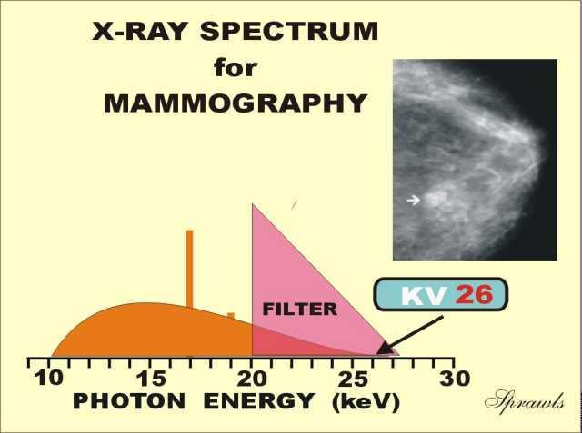
8 Mammomgraphy technical considerations Requires the best image quality Most pathologies either have soft tissue density that is not very different from surrounding tissues or contain microcalcifications Soft x-rays to best differentiate between the soft tissues of the breast
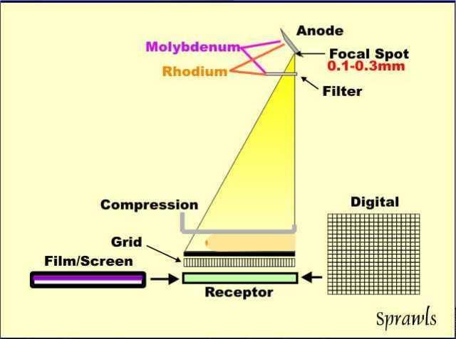
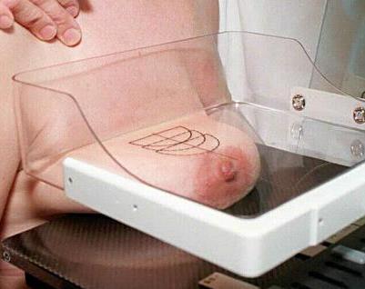
9 Mammomgraphy technical considerations Low energy (25-32 kv) High mas Molybdenum (and rhodium) anode (characteristic x-ray production) Molybdenum filter Two, small focal spots (improved resolution) Compression more uniform breast thickness reduced blurring from patient motion reduced scattered radiation reduced radiation dose better visualization of tissues near the chest wall Grid: filter scattered radiation
10 Mammography
11 Mammography
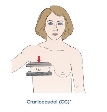
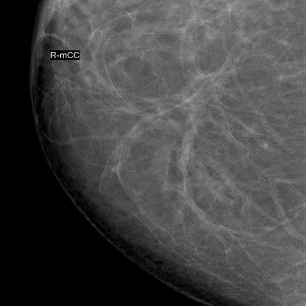
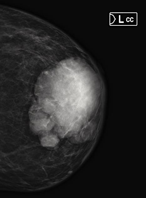
12 Mammography standard views Craniocaudal (CC)


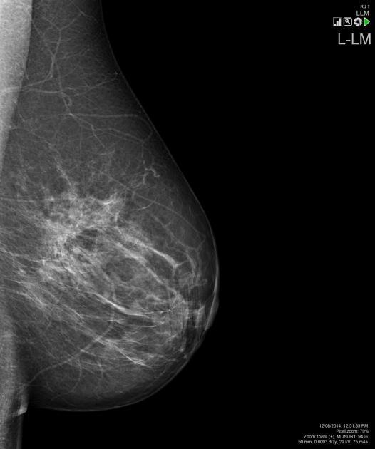
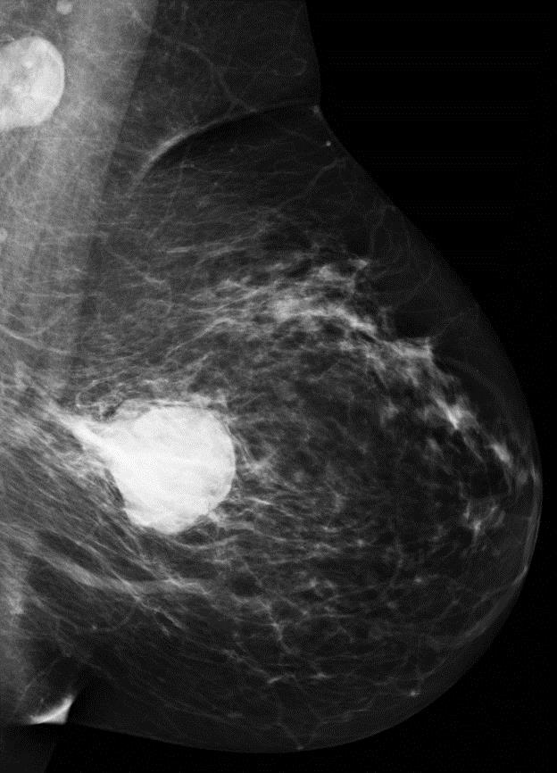
13 Mammography standard views Mediolateral oblique (MLO)
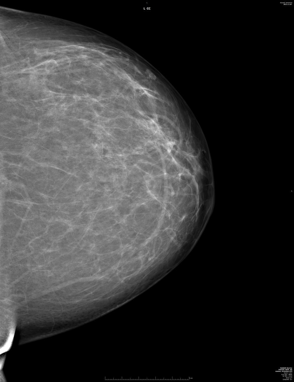
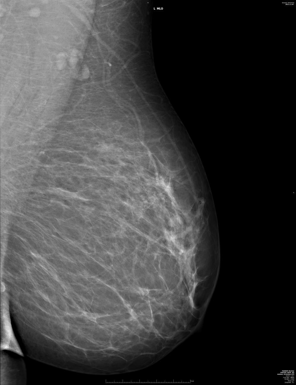
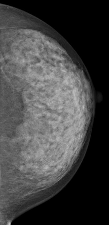
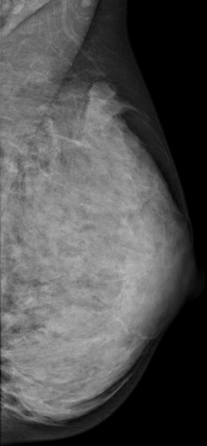
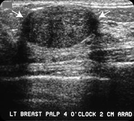
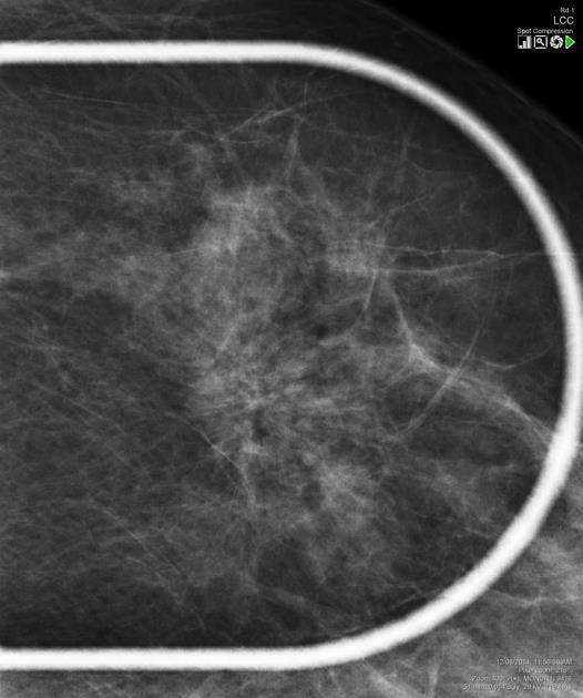
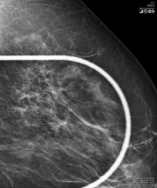
14 Left craniocaudal Left mediolateral oblique
15 Right craniocaudal Right mediolateral oblique

16 Mammography magnification views Mainly to evaluate microcalcifications
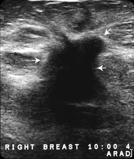
17 Ultrasound First method of choice under 30 year (+ pregnancy and breast feeding, implants) Complementary method for dense breasts (limitation of mammography!) Further characterization of pathologies found on mammograms Axilla!! - Always has to be scanned Differentiate between solid and cystic lesions Post surgical complications (hematoma) Guiding interventions (FNAB or core biopsy)
18 Ultrasound Technical considerations High frequency (10-12 MHz), linear probe Doppler: vascularization of lesions (high vascularization and high flow are suspicious for malignancy) Sonoelastography: compare and quantify tissue elasticity malignant lesions usually show higher stiffness relative to surrounding tissue


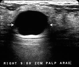
19 Fibroadenoma: well-circumscribed, round to ovoid, or macrolobulated mass with generally uniform hypoechogenicity, pseudocapsule Cyst: anechoic, well circumscribed, acoustic enhancement

20 Breast cancer: echopoor, inhomogeneous, irregular border, poorly circumscribed, acoustic shadow, not compressible
21 Lesion analysis Ultrasound morphology Benign Anechoic or echopoor Round or oval Well circumscribed Homogeneous Acoustic enhancement Lateral shadow sign Compressible Not fixed No hypervascularity Malignant Echopoor Irregular Ill-defined border Inhomogeneous Acoustic shadow Non-compressible Fixed Hypervascularity and flow, irregular vessels

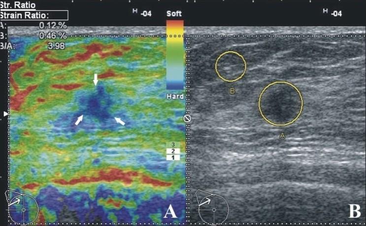
22 Ductal cc: stiff lesion Fibroadenoma: elastic lesion
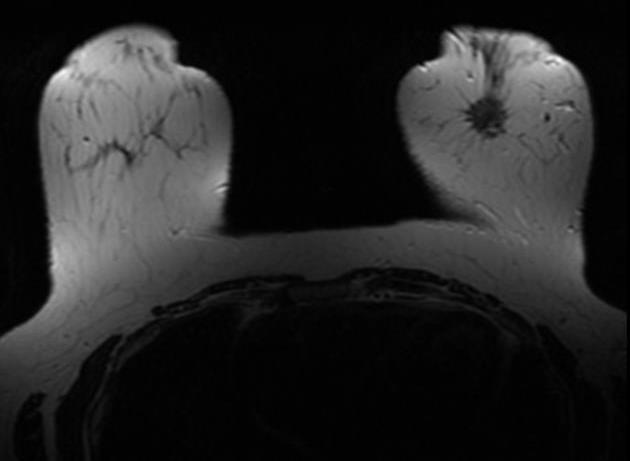
23 MRI Suspected multifocal, bilateral lesions Occult primary tumor Exact extension of infiltrative lesions Suspected lobular cc. Chest wall infiltration To differentiate between recurrence and surgical scar Suspected implant rupture Screening in high risk populations (e.g. Brca mutation) Monitoring the effect of neoadjuvant chemotherapy
24 MRI technical considerations Min. 1.5 T (3T) Breast coils Native + contrast enhanced serials T1, T2, FatSat Dynamic MRI!!!: kinetics of contrast enhancement most important to differentiate between benign and malignant lesion

25 T1 FatSat CE, early phase, subtarction T2

26 Type I curve: Slow rise, continued rise with time. 6 % malignant. Type III curve: Rapid initial rise, followed by washout % malignant.
27 Anatomy Glandular ducts and lobules Connective tissue Fat Basic functional unit: Lobule or terminal ductal lobular unit (TDLU) Most invasive cancers rise from TDLU (+ DCIS, lobular cc in situ or infiltrating lobular cc, fibroadenoma, fibrocystic disease)
28 TDLU
29 How to interpret mammograms? Determine whether film is of diagnostic quality Find the lesion Analyze the lesion Circumscribed Stellate Structural distorsion Calcifications Thickened skin


30 Determine whether film is of diagnostic quality On MLO views, pectoral muscles should be visible at least above the level of the nipple On CC views, the edge of the pectoral muscle should be visible

 trained")
31 Determine whether film is of diagnostic quality The line drawn from the nipple to the pectoral muscle should be the same length on both views (max. difference should be less than 1 cm) trained technitian! 129 mm


32 Determine whether film is of diagnostic quality Nipple should be in profil

33 Determine whether film is of diagnostic quality Image should not be blurred


34 Determine whether film is of diagnostic quality No artefacts! Bra strap Skin folds talcum ointment deodorant patch
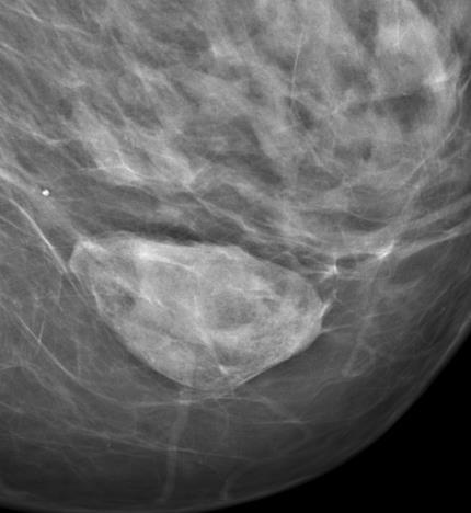
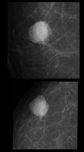
35 Find the lesion Circumscribed easy to recognize; mostly benign Stellate difficult to recognize; most malignant lesions Certain breast types are not easy to evaluate on mammograms
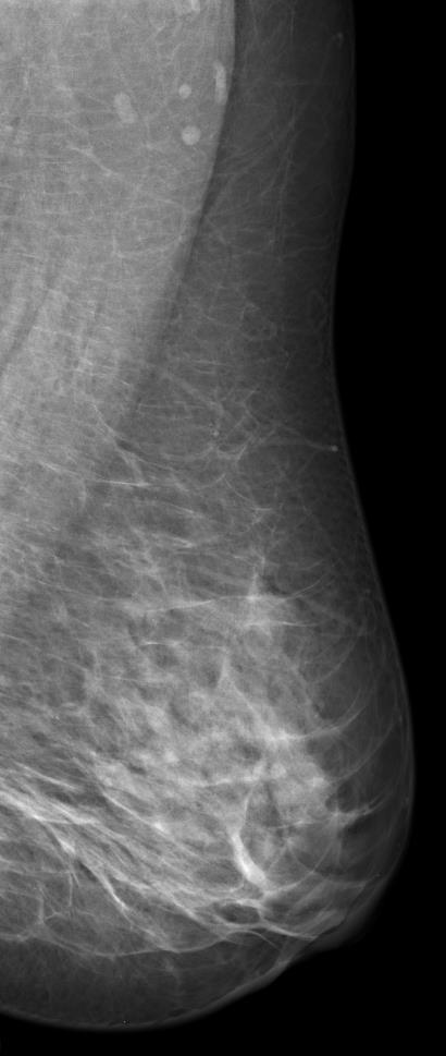
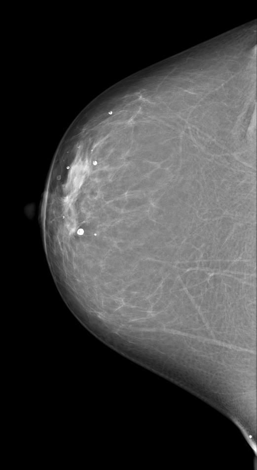
36 Normal breast types (Tabar) Different types of breasts according to mammographic patterns (reflects composition) I: balanced proportion of all components of breast tissue with a slight predominance of fibrous tissue (Fibroglandular; young age) II: predominance of fat tissue (Fatty breast) III: predominance of fat tissue with retroareolar residual fibrous tissue IV: predominantly nodular densities (Adenotic) V: predominantly fibrous tissue (Fibrotic or dense breast) I-III. Can change e.g. with age or hormon therapy IV and V.: genetically coded; difficulte to evaluate on mammograms!!! US!

")
37 Normal breast types (Tabar) Fibroglandular
")
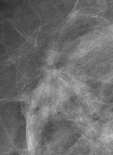
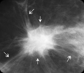
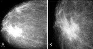
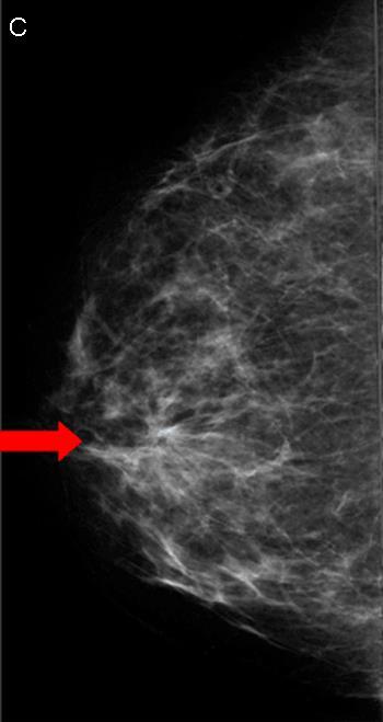
38 Normal breast types (Tabar) Involuted

")
39 Normal breast types (Tabar) Retroaleolar

")
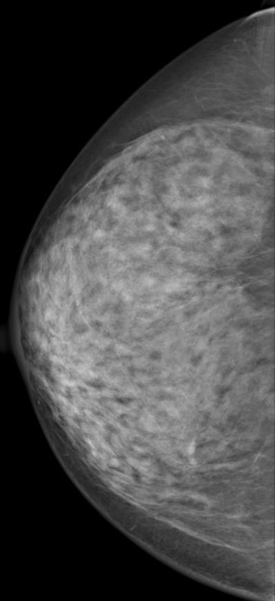

40 Normal breast types (Tabar) Adenotic limitations of mammography!

")

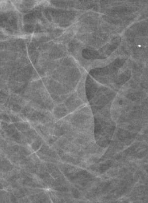
41 Normal breast types (Tabar) Fibrotic - limitations of mammography!
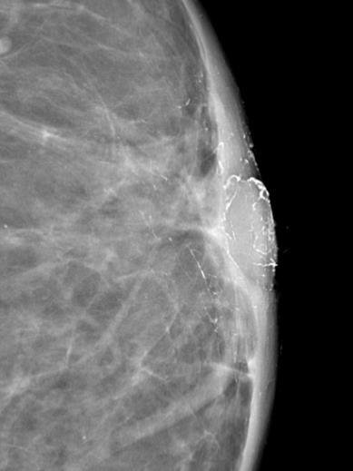
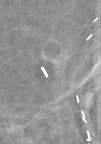
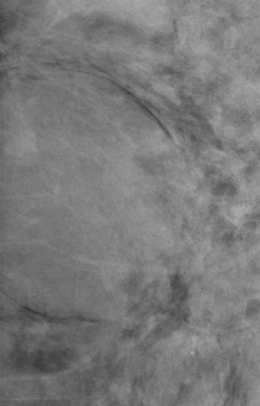
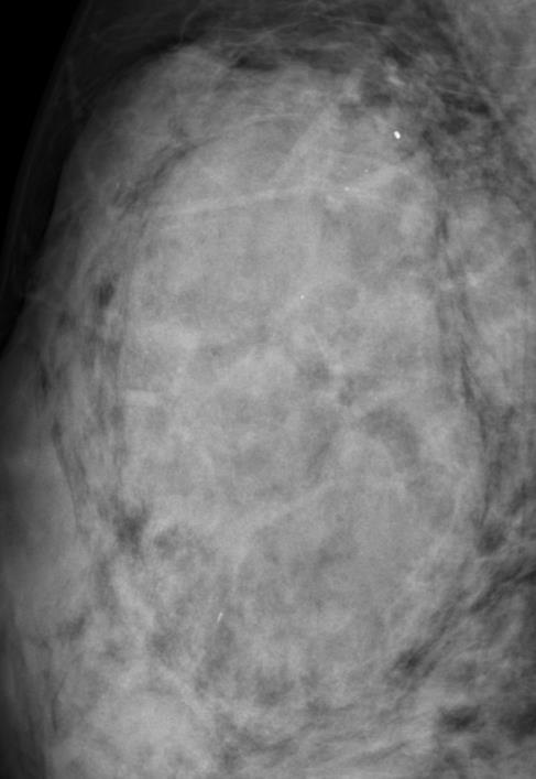
42 Lesion analysis circumscribed lesions cyst, lipoma, fibroadenoma, papillary or mucinous cc, etc Easier to recognize Size Contour (sharp: usually benign feature) Halo, capsule: Benign features Density: radiolucent (lipoma, oil cyst, galactocele); radiolucent and radiopaque combined (l.g., hematoma, fibroadenolipoma); low density radiopaque (fibroadenoma, cyst, but: papillary cc, mucinous cc); high density radiopaque (cc, cyst, abscess, lg...)

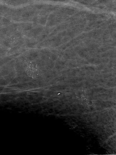
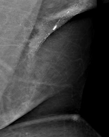
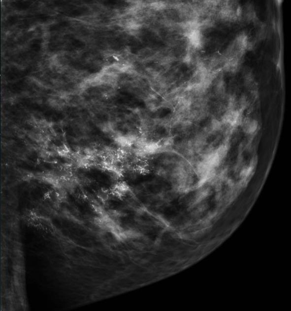
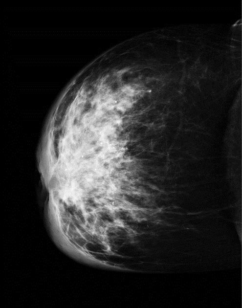
43 Sharp contour - fibroadenoma

44 Sharp contour - Cyst


45 Ill defined contour - cc

46 Capsule Halo


47 High density radiopaque - cyst Lower density radiopaque - fibroadenoma Radiokucent and radiopaque combined - fibroadenolipoma
48 Lesion analysis stellate lesions = radiating structure w ill defined borders More difficult to recognize Center distinct mass - white star : invasive intraductal cc oval or circular radiolucent area - black star : radial scar, fat necrosis, invasive lobular cc Radiating structures sharp dense, fine lines radiating in all directions (invasive ductal cc) Many very fine spicules bunched together (radial scar, fat necrosis) Skin thickening: radial scar never associated w
49 Lesion analysis Radial scar = sclerosing ductal hyperplasia Benign but can be associated with DCIS or tubular cc Mimicker of scirrhous breast cc (invasive ductal cc) y.o. Not palpable Mammo: dark star stellate lesion with translucent, low density center w/o mass No associated skin thickening
50
51
52 Lesion analysis Invasive ductal cc Most frequent type of breast cc Peak: y.o. Can be palpable, immobile Mammo: white star stellate lesion with high density central mass w calcification (granular or casting); the larger the central mass, the longer the spicules; w/o localized skin thickening or retraction
53
54 Lesion analysis Structural distorsion Asymetric densities w architectural distortion Difficult to recognise Lobular carcinoma: often multifoca and bilateral MRI! Mammo: white star Structural distorsion dark star calcification uncommon
55
56 Lesion analysis Calcifications Localization: ductal (malignant) or lobular (benign), extraglandular Terms: cluster, scattered, casting, granular, punctuate Malignant type calcifications: Granular: tiny, dot-like, innumerable, irregularly grouped Casting: casts of ductal lumen, irregular outline, polymorph
57 Lesion analysis Calcifications Benign type calcifications: Egg shell Course or popcorn like Milk of calcium Skin, vascular, etc Large, rod like (plasma cell mastitis)
58 Lesion analysis DCIS Ductal carcinoma in situ % of breast cc on mammomgrams!!! (screening!!!!!) Mammo: 75 % Calcification!!!! Granular or casting
59
60 Lesion analysis Thickened skin sy Diffuse or localized Diffuse: Axillary lymphatic obstruction lg. met Breast cc Lymphoma Advanced gynecologic malignancies (ovarium, uterus) Advanced bronchus or esophagus cc Lymphangitic spread of breast cc Inflammation Right heart failure
61
62 Special type cancers DCIS, invasive ductal cc ~ 70 % Special types ~ 25 % Lobular: diffuse infiltration, structural distortion difficult to recognize Medullary, Mucinous, Papillary cc: usually well circumscribed, round lesions Metastasis: also circumscribed Lymphoma
63
Imaging in breast cancer. Mammography and Ultrasound Donya Farrokh.MD Radiologist Mashhad University of Medical Since
 Imaging in breast cancer Mammography and Ultrasound Donya Farrokh.MD Radiologist Mashhad University of Medical Since A mammogram report is a key component of the breast cancer diagnostic process. A mammogram
Lesion Imaging Characteristics Mass, Favoring Benign Circumscribed Margins Intramammary Lymph Node
 Lesion Imaging Characteristics Mass, Favoring Benign Circumscribed Margins Intramammary Lymph Node Oil Cyst Mass, Intermediate Concern Microlobulated Margins Obscured Margins Mass, Favoring Malignant Indistinct
Leonard M. Glassman MD
 BI-RADS The New BI-RADS Leonard M. Glassman MD FACR Former Chief of Breast Imaging American Institute for Radiologic Pathology Washington Radiology Associates, PC Breast Imaging Reporting and Data System
Amammography report is a key component of the breast
 Review Article Writing a Mammography Report Amammography report is a key component of the breast cancer diagnostic process. Although mammographic findings were not clearly differentiated between benign
Ana Sofia Preto 19/06/2013
 Ana Sofia Preto 19/06/2013 Understanding the underlying pathophysiologic processes leading to the various types of calcifications Description and illustration of the several types of calcifications, according
MEDICAL IMAGING AND BREAST DISEASE HOW CAN WE HELP YOU?
 MEDICAL IMAGING AND BREAST DISEASE HOW CAN WE HELP YOU? Barbara M. Preston, M.D. SCREENING MAMMOGRAPHY AVERAGE RISK PATIENTS KAISER RECOMMENDATION: ALL WOMEN (INCLUDING TRANSGENDER FEMALES) Every 1-21
ACRIN 6666 IM Additional Evaluation: Additional Views/Targeted US
 Additional Evaluation: Additional Views/Targeted US For revised or corrected form check box and fax to 215-717-0936. Instructions: The form is completed based on recommendations (from ID form) for additional
Armed Forces Institute of Pathology.
 Armed Forces Institute of Pathology www.radpath.com Armed Forces Institute of Pathology Breast Disease www.radpath.org Armed Forces Institute of Pathology Interpretation of Breast MRI Leonard M. Glassman
Mammographic imaging of nonpalpable breast lesions. Malai Muttarak, MD Department of Radiology Chiang Mai University Chiang Mai, Thailand
 Mammographic imaging of nonpalpable breast lesions Malai Muttarak, MD Department of Radiology Chiang Mai University Chiang Mai, Thailand Introduction Contents Mammographic signs of nonpalpable breast cancer
ORIGINAL ARTICLE EVALUATION OF BREAST LESIONS USING X-RAY MAMMOGRAM WITH HISTOPATHOLOGICAL CORRELATION
 Available online at www.journalijmrr.com INTERNATIONAL JOURNAL OF MODERN RESEARCH AND REVIEWS IJMRR ISSN: 2347-8314 Int. J. Modn. Res. Revs. Volume 3, Issue 10, pp 807-814, October, 2015 ORIGINAL ARTICLE
Breast Imaging Lexicon
 9//201 200 BI RADS th Edition 201 BI RADS th Edition Breast Imaging Lexicon Mammographic Pathology and Assessment Categories Deborah Thames, R.T.(R)(M)(QM) The Advanced Health Education Center Nonmember:
BI-RADS and Breast MRI. Kathy Borovicka, M.D. Thursday February 15, 2018
 BI-RADS and Breast MRI Kathy Borovicka, M.D. Thursday February 15, 2018 Learning Objectives Be familiar with the Breast Imaging Reporting and Data System (BI-RADS) Understand the components of a breast
Intracystic papillary carcinoma of the breast
 Intracystic papillary carcinoma of the breast Poster No.: C-1932 Congress: ECR 2011 Type: Educational Exhibit Authors: V. Dimarelos, F. TZIKOS, N. Kotziamani, G. Rodokalakis, 1 2 3 1 1 1 2 T. MALKOTSI
Ultrasound of the Breast BASICS FOR THE ORDERING CLINICIAN
 Ultrasound of the Breast BASICS FOR THE ORDERING CLINICIAN Breast Ultrasound Anatomy Skin Breast Parenchyma Pectoralis Fascia Pectoralis Breast Ultrasound Anatomy Indications for Breast Ultrasound Palpable
Diseases of the breast (1 of 2)
") Diseases of the breast (1 of 2) Introduction A histology introduction Normal ducts and lobules of the breast are lined by two layers of cells a layer of luminal cells overlying a second layer of myoepithelial
Diseases of the breast (1 of 2) Introduction A histology introduction Normal ducts and lobules of the breast are lined by two layers of cells a layer of luminal cells overlying a second layer of myoepithelial
Leonard M. Glassman MD Analysis of Breast Calcifications
 Importance of Calcification Leonard M. Glassman MD FACR American Institute for Radiologic Pathology Washington Radiology Associates, PC Washington DC 45% of all breast cancers present as calcification
The radiologic workup of a palpable breast mass
 Imaging in Practice CME CREDIT EDUCTIONL OJECTIVE: The reader will consider which breast masses require further workup and which imaging study is most appropriate Lauren Stein, MD Imaging Institute, Cleveland
Index. C Calcifications fat necrosis 1, 61 fat necrosis 4, 69 nipple/peri-areolar involvement 1, 165
 A ADH. See Atypical ductal hyperplasia (ADH) American College of Radiology (ACR), BI-RADS background parenchymal enhancement, 8, 9, 81, 82 fibroglandular tissue guidelines, 6 American Joint Committee on
Mammographic evaluation of palpable breast masses with pathological correlation: a tertiary care centre study in Nepal
 Original article 21 Mammographic evaluation of palpable breast masses with pathological correlation: a tertiary care centre study in Nepal G. Gurung, R. K. Ghimire, B. Lohani Department of Radiology and
BREAST PATHOLOGY. Fibrocystic Changes
 BREAST PATHOLOGY Lesions of the breast are very common, and they present as palpable, sometimes painful, nodules or masses. Most of these lesions are benign. Breast cancer is the 2 nd most common cause
Imaging the Symptomatic Patient. Avice M.O Connell MD,FACR,FSBI Professor of Imaging Sciences Director, Women s Imaging University of Rochester
 Imaging the Symptomatic Patient Avice M.O Connell MD,FACR,FSBI Professor of Imaging Sciences Director, Women s Imaging University of Rochester The four most common symptoms Mass Pain Discharge Infection
Diagnostic Dilemmas of Breast Imaging
 Diagnostic Dilemmas of Breast Imaging Common Causes of Error in Breast Cancer Detection By: Jason Cord, M.D. Mammography: Initial Imaging The standard for detection of breast cancer Screening mammography
Pitfalls and Limitations of Breast MRI. Susan Orel Roth, MD Professor of Radiology University of Pennsylvania
 Pitfalls and Limitations of Breast MRI Susan Orel Roth, MD Professor of Radiology University of Pennsylvania Objectives Review the etiologies of false negative breast MRI examinations Discuss the limitations
Armed Forces Institute of Pathology.
 Armed Forces Institute of Pathology www.radpath.com Armed Forces Institute of Pathology Breast Disease www.radpath.org Armed Forces Institute of Pathology Evaluation of Breast Calcifications Leonard M.
Breast imaging of benign fat containing lesions
 Breast imaging of benign fat containing lesions Poster No.: C-1870 Congress: ECR 2017 Type: Educational Exhibit Authors: R. Aouini, I. Megdiche, D. Ben Hammadi, N. BEN MAMI, I. Attia, R. Neila, A. Zidi;
S. Murgo, MD. Chr St-Joseph, Mons Erasme Hospital, Brussels
 S. Murgo, MD Chr St-Joseph, Mons Erasme Hospital, Brussels? Introduction Mammography reports are sometimes ambiguous and indecisive. ACR has developped the BIRADS. BIRADS consists of a lexicon in order
COPE Library Sample
 Breast Anatomy LOBULE LOBE ACINI (MILK PRODUCING UNITS) NIPPLE AREOLA COMPLEX ENLARGEMENT OF DUCT AND LOBE LOBULE SUPRACLAVICULAR NODES INFRACLAVICULAR NODES DUCT DUCT ACINI (MILK PRODUCING UNITS) 8420
BI-RADS Update. Martha B. Mainiero, MD, FACR, FSBI Brown University Rhode Island Hospital
 BI-RADS Update Martha B. Mainiero, MD, FACR, FSBI Brown University Rhode Island Hospital No Disclosures BI-RADS History 1980s Quality Issues ACR Accreditation BI-RADS 1994 2003 4 th Edition MRI, US January
BREAST MRI. VASILIKI FILIPPI RADIOLOGIST CT MRI & PET/CT Departments Hygeia Hospital, Athens, Greece
 BREAST MRI VASILIKI FILIPPI RADIOLOGIST CT MRI & PET/CT Departments Hygeia Hospital, Athens, Greece Breast ΜR Imaging (MRM) Breast MR imaging is an extremely powerful diagnostic tool, that when used in
Abid Irshad, MD Director Breast Imaging. Medical University of South Carolina Charleston
 Abid Irshad, MD Director Breast Imaging Medical University of South Carolina Charleston Cases Financial disclosure: I or my family have no financial interest related to the material discussed in this presentation
Here are examples of bilateral analog mammograms from the same patient including CC and MLO projections.
 Good afternoon. It s my pleasure to be discussing Diagnostic Breast Imaging over the next half hour. I m Wei Yang, Professor of Diagnostic Radiology and Chief, the Section of Breast Imaging as well as
Benign, Reactive and Inflammatory Lesions of the Breast
 Benign, Reactive and Inflammatory Lesions of the Breast Marilin Rosa, MD Associate Member Section Head of Breast Pathology Department of Anatomic Pathology Program Director, Breast Pathology Fellowship
Melissa Hartman, DO Women s Health Orlando VA Medical Center
 Melissa Hartman, DO Women s Health Orlando VA Medical Center Most common non-skin cancer and Second deadliest cancer in women Majority are diagnosed by abnormal screening study An approach to breast cancer
University of Washington Radiology Review Course: Strange and Specific Diagnoses. Case #1
 University of Washington Radiology Review Course: Strange and Specific Diagnoses Katherine E. Dee, MD Seattle Breast Center Via Radiology 2014 Case #1 37 year old presents with bilateral palpable lumps.
Non-mass Enhancement on Breast MRI. Aditi A. Desai, MD Margaret Ann Mays, MD
 Non-mass Enhancement on Breast MRI Aditi A. Desai, MD Margaret Ann Mays, MD Breast MRI Important screening and diagnostic tool, given its high sensitivity for breast cancer detection Breast MRI - Indications
Alena Levit MD Avice O Connell MD University of Rochester, Rochester, NY
 Alena Levit MD Avice O Connell MD University of Rochester, Rochester, NY Purpose Review imaging spectrum of both common benign and malignant breast lesions Describe and demonstrate CT features with mammogram,
Breast imaging in general practice
 Breast series CLINICAL PRACTICE Breast imaging in general practice Nehmat Houssami, MBBS, FAFPHM, FASBP, PhD, is Associate Clinical Director, NSW Breast Cancer Institute, Westmead Hospital, Honorary Senior
Breast MRI Update. Jeffrey C. Weinreb, MD, FACR Yale University School of Medicine
 Breast MRI Update Jeffrey C. Weinreb, MD, FACR jeffrey.weinreb@yale.edu Yale University School of Medicine I disclose the following financial relationships with relevant commercial interests: Bracco Bayer
Criteria of Malignancy. Evaluation Score
 30 5 Diagnostic Criteria Criteria of Malignancy Table 5.2 lists criteria in contrast-enhancing MR mammography that strongly indicate the presence of malignancy or are unspecific. Unifactorial evaluation
Breast pathology. 2nd Department of Pathology Semmelweis University
 Breast pathology 2nd Department of Pathology Semmelweis University Breast pathology - Summary - Benign lesions - Acute mastitis - Plasma cell mastitis / duct ectasia - Fat necrosis - Fibrocystic change/
Breast Health. Learning Objectives. Breast Anatomy. Poll Question. Breast Anatomy
 Learning Objectives Describe breast anatomy to a patient Breast Health Answer questions about causes of breast pain and masses Explain breast cancer screening/diagnostic modalities Appropriately triage
Imaging of giant breast masses with pathological correlation
 P i c t o r i a l E s s a y Singapore Med J 2004 Vol 45(3) : 132 Imaging of giant breast masses with pathological correlation M Muttarak, B Chaiwun ABSTRACT Ultrasonography (US) and mammography are the
ROLE OF MRI IN SCREENING, DIAGNOSIS AND MANAGEMENT OF BREAST CANCER. B.Zandi Professor of Radiology
 ROLE OF MRI IN SCREENING, DIAGNOSIS AND MANAGEMENT OF BREAST CANCER B.Zandi Professor of Radiology Introduction In the USA, Breast Cancer is : The Most Common Non-Skin Cancer The Second Leading cause of
Case Scenario 1 History and Physical 3/15/13 Imaging Pathology
 Case Scenario 1 History and Physical 3/15/13 The patient is an 84 year old white female who presented with an abnormal mammogram. The patient has a five year history of refractory anemia with ringed sideroblasts
BREAST IMAGING and NEW IMAGING MODALITIES- A Surgeons view
 BREAST IMAGING and NEW IMAGING MODALITIES- A Surgeons view DR CHANTEL THORNTON SPECIALIST BREAST CANCER SURGEON BMSc (hons) MBBS (hons) FRACS Epworth Hospital, Richmond- Agora Centre for Women s Health
IBCM 2, April 2009, Sarajevo, Bosnia and Herzegovina
 Preoperative diagnosis and treatment planning in breast cancer The pathologist s perspective L. Mazzucchelli Istituto Cantonale di Patologia Locarno, Switzerland IBCM 2, 23-25 April 2009, Sarajevo, Bosnia
Evaluation of Abnormal Screening Mammograms
 342 Evaluation of Abnormal Screening Mammograms Ellen Shaw de Paredes, M.D. The purpose of routine screening mammography is to detect unsuspected cancer that has the potential to be cured. Abnormalities
Excerpts from: The Breast
 Excerpts from: The Breast Jonathan J. James, Robin M. Wilson and Andrew J Evans Grainger & Allison s Diagnostic Radiology Chapter 69 1664-1689.e2. Sheryl Jordan and Sheila Lee June 2018 TABLE OF CONTENTS:
THE MALE BREAST CARCINOMA: EARLY DETECTION HOPE. Author (s) Supreethi Kohli a, Pragya Garg b
 Supreethi Kohli a, Pragya Garg b") Case Report ABSTRACT - Male breast cancer is exceptionally rare and accounts for less than 0.25% of male malignancies and approximately 0.5-1% of all breast cancer (both genders). Mammography of the male
Case Report ABSTRACT - Male breast cancer is exceptionally rare and accounts for less than 0.25% of male malignancies and approximately 0.5-1% of all breast cancer (both genders). Mammography of the male
Advanced Course on Multimodality Detection and Diagnosis of Breast Diseases
 BREAST SEMINAR Advanced Course on Multimodality Detection and 3D image of the breast tissue Invited speaker LÁSZLÓ TABÁR, MD,FACR (Hon) Falun, Sweden Nov 22-23, 2014 ATHENS, Greece Royal Olympic Hotel
CLINICAL SIGNIFICANCE OF BENIGN EPITHELIAL CHANGES
 Papillomas. Papillomas are composed of multiple branching fibrovascular cores, each having a connective tissue axis lined by luminal and myoepithelial cells ( Fig. 23-11 ). Growth occurs within a dilated
Standard Breast Imaging Modalities. Lilian Wang, M.D. Breast Imaging Section Department of Radiology Northwestern Medicine
 Standard Breast Imaging Modalities Lilian Wang, M.D. Breast Imaging Section Department of Radiology Northwestern Medicine Overview Standard breast imaging modalities Mammography Ultrasound MRI Imaging
Imaging Guidelines for Breast Cancer Screening
 Imaging Guidelines for Breast Cancer Screening Sarah Colwick, MD Dr. Sarah Colwick was born and raised in Sikeston, MO. She attended college and medical school at the University of Missouri-Kansas City
Contrast-enhanced Breast MRI RSSA 2013
 Contrast-enhanced Breast MRI RSSA 2013 Prof. dr. Maurice van den Bosch University Medical Center Utrecht, the Netherlands Index 1) Breast cancer 2) Why MRI of the breast 3) Technique 4) Interpretation
Ultrasonography. Methods. Brief Description. Indications. Device-related Prerequisites. Technical Requirements. Evaluation Criteria
 1 Ultrasonography Brief Description Imaging modality using sound waves Tissue-specific wave reflection. Indications Evaluation of palpable breast nodules Evaluation of clinically occult mammographic findings
Breast Imaging Essentials
 Breast Imaging Essentials Module 5 Transcript 2016 ASRT. All rights reserved. Breast Imaging Essentials Module 5 Pathology 1. ASRT Animation 2. Welcome Welcome to Module 5 of Breast Imaging Essentials
Presented by: Lillian Erdahl, MD
 Presented by: Lillian Erdahl, MD Learning Objectives What is Breast Cancer Types of Breast Cancer Risk Factors Warning Signs Diagnosis Treatment Options Prognosis What is Breast Cancer? A disease that
Screening Mammograms: Questions and Answers
 CANCER FACTS N a t i o n a l C a n c e r I n s t i t u t e N a t i o n a l I n s t i t u t e s o f H e a l t h D e p a r t m e n t o f H e a l t h a n d H u m a n S e r v i c e s Screening Mammograms:
of Thyroid Lesions Comet Tail Crystals
 2 Ultrasound Features of Thyroid Lesions There are many different features indicating a certain benign or malignant tumor type, but many of these are overlapping signs. Combining several features is considered
Ductal carcinoma in situ: ultrasound, mammography and MRI features with pathologic correlation
 Ductal carcinoma in situ: ultrasound, mammography and MRI features with pathologic correlation Poster No.: C-2252 Congress: ECR 2013 Type: Educational Exhibit Authors: L. Fernandes, H. A. M. R. Tinto,
Mary Smania, DNP, FNP-BC Clinical Practice Champion Assistant Professor Michigan State University College of Nursing
 Mary Smania, DNP, FNP-BC Clinical Practice Champion Assistant Professor Michigan State University College of Nursing Identify evidence based routine screening guidelines for women of all ages Identify
Fat Necrosis: A Grand Imposter
 Fat Necrosis: A Grand Imposter Poster No.: C-0751 Congress: ECR 2015 Type: Educational Exhibit Authors: L. C. Flores Salinas, Y. A. Ramirez Galvan, A. Garza Báez, C. M. Ferrara Chapa; Monterrey/MX Keywords:
Journal of Medical Imaging and Radiation Oncology
 Journal of Medical Imaging and Radiation Oncology 60 (2016) 506 513 MEDICAL IMAGING PICTORIAL ESSAY Malignant hyperechoic breast lesions at ultrasound: A pictorial essay Stephen Tiang, 1 Cecily Metcalf,
Gynecomastia and Its Mimics: Not All Male Breast Lesions are Benign
 Gynecomastia and Its Mimics: Not All Male Breast Lesions are Benign Poster No.: C-0139 Congress: ECR 2014 Type: Educational Exhibit Authors: S. A. Choudhery, P. Gupta, S. Foshee, F. Garcia-Morales, G.
8/31/2016 HIDING IN PLAIN SITE, ARCHITECTURAL DISTORTIONS AND BREAST ASYMMETRIES ARCHITECTURAL DISTORTIONS ARCHITECTURAL DISTORTIONS
 HIDING IN PLAIN SITE, ARCHITECTURAL DISTORTIONS AND BREAST ASYMMETRIES DEBORAH THAMES R.T. (R)(M)(QM) ARCHITECTURAL DISTORTIONS Definition is disruption of the natural flow of breast pattern towards the
Mammography. What is Mammography? What are some common uses of the procedure?
 Mammography What is Mammography? Mammography is a specific type of imaging that uses a low-dose x-ray system to examine breasts. A mammography exam, called a mammogram, is used to aid in the early detection
Breast Cancer Imaging
 Breast Cancer Imaging I. Policy University Health Alliance (UHA) will cover breast imaging when such services meet the medical criteria guidelines (subject to limitations and exclusions) indicated below.
Contents. Basic Ultrasound Principles and Terminology. Ultrasound Nodule Characteristics
 Contents Basic Ultrasound Principles and Terminology Basic Ultrasound Principles... 1 Ultrasound System... 2 Linear Transducer for Superficial Images and Ultrasound-Guided FNA... 3 Scanning Planes... 4
Case study 1. Rie Horii, M.D., Ph.D. Division of Pathology Cancer Institute Hospital, Japanese Foundation for Cancer Research
 NCCN/JCCNB Seminar in Japan April 15, 2012 Case study 1 Rie Horii, M.D., Ph.D. Division of Pathology Cancer Institute Hospital, Japanese Foundation for Cancer Research Present illness: A 50y.o.premenopausal
BREAST CANCER d an BREAST SELF EXAM
 BREAST CANCER and BREAST SELF EXAM American Cancer Society Statistics: 2009 Invasive breast cancer will be diagnosed in over 192,370 women Carcinoma in situ will be diagnosed in 62,280 women More than
Microcalcifications detected on mammography classified as BIRADS 4 and 5 and their correlations with histopatologic findigns
 Microcalcifications detected on mammography classified as BIRADS 4 and 5 and their correlations with histopatologic findigns Poster No.: C-0401 Congress: ECR 2010 Type: Educational Exhibit Topic: Breast
CPC 4 Breast Cancer. Rochelle Harwood, a 35 year old sales assistant, presents to her GP because she has noticed a painless lump in her left breast.
 CPC 4 Breast Cancer Rochelle Harwood, a 35 year old sales assistant, presents to her GP because she has noticed a painless lump in her left breast. 1. What are the most likely diagnoses of this lump? Fibroadenoma
Breast Health and Imaging Glossary
 Contact: Lorna Vaughan HerSpace Breast Imaging & Biopsy Associates 300 State Route 35 South W. Long Branch, NJ 07764 732-571-9100, ext. 104 lorna@breast-imaging.com Breast Health and Imaging Glossary Women
UW Radiology Review Course Breast Calcifications. BI-RADS 5 th Edition
 UW Radiology Review Course Breast Calcifications Grace Kalish, MD Vantage Radiology BI-RADS 5 th Edition Benign Skin Vascular Large rod like Coarse popcorn Suspicious Amorphous Coarse heterogenous Fine
Breast Evaluation & Management Guidelines
 Breast Evaluation & Management Guidelines Pamela L. Kurtzhals, M.D. F.A.C.S. Head, Dept. of General Surgery Scripps Clinic, La Jolla Objective Review screening & diagnostic guidelines Focused patient complaints
Optimizing Breast Sonography
 Optimizing Breast Sonography Cindy Rapp BS, RDMS, FSDMS, FAIUM Denver, Colorado Breast Sonography general goal to make a more specific diagnosis than can be made with clinical and mammographic findings
Hiding in Plain Sight / Site: Archictectural Distortions and Breast Asymmetries
 Hiding in Plain Sight / Site: Archictectural Distortions and Breast Asymmetries Dianne Georgian-Smith MD Associate Professor Harvard Med School Brigham and Women s Hospital Financial Disclosures Book Publication
Papillary lesions of the breast - Imaging findings and diagnostic challenges
 Papillary lesions of the breast - Imaging findings and diagnostic challenges Poster No.: R-0146 Congress: RANZCR-AOCR 2012 Type: Educational Exhibit Authors: P. Jagmohan, F. J. Pool Keywords: Breast, Mammography,
DISORDERS OF THE BREAST Dated. FIBROADENOSIS Other common names: mastitis, fibrocystic disease, cystic mammary dysplasia.
 DISORDERS OF THE BREAST Dated BENIGN BREAST DISORDERS (Essential Surg 2 nd Ed, pp 540) FIBROADENOSIS Other common names: mastitis, fibrocystic disease, cystic mammary dysplasia. Fibroadenosis is the distortion
MRI BI-RADS: How to make it out?
 MRI BI-RADS: How to make it out? Poster No.: C-1850 Congress: ECR 2016 Type: Educational Exhibit Authors: M. Ben Ammar, A. Ben Miled, O. Ghdes, S. Harguem, A. Gaja, N. Mnif; Tunis/TN Keywords: Breast,
Cytyc Corporation - Case Presentation Archive - March 2002
 FirstCyte Ductal Lavage History: 68 Year Old Female Gail Index: Unknown Clinical History: Negative Mammogram in 1995 6 yrs. later presents with bloody nipple discharge Subsequent suspicious mammogram Suspicious
BREAST PATHOLOGY MCQS
 BREAST PATHOLOGY MCQS 1) :The most important factor in breast enlargement during pregnancy is A. stromal edema B. secretion of chorionic gonadotropin C. glandular hyperplasia D. proliferation of stroma
Pictorial Essay Singapore Med J 2009; 50(9) :
 :") 907 Pictorial Essay CME Article Breast calcifications: which are malignant? Muttarak M, Kongmebhol P, Sukhamwang N ABSTRACT Most calcifications depicted on mammograms are benign. However, calcifications
907 Pictorial Essay CME Article Breast calcifications: which are malignant? Muttarak M, Kongmebhol P, Sukhamwang N ABSTRACT Most calcifications depicted on mammograms are benign. However, calcifications
SIGNIFICANT OTHERS. Miscellaneous Benign Breast Conditions
 SIGNIFICANT OTHERS Miscellaneous Benign Breast Conditions Epworth HealthCare 1 FAT NECROSIS TRAUMATIC Cell rupture Seat-Belt injury Blunt trauma Iatrogenic injury Surgery, Flaps, Radiotherapy Pathology
RSNA, /radiol Appendix E1. Methods
 RSNA, 2016 10.1148/radiol.2016151097 Appendix E1 Methods US and Near-infrared Data Acquisition Four optical wavelengths (740 nm, 780 nm, 808 nm, and 830 nm) were used to sequentially deliver the light
Breast Pathology. Breast Development
 Breast Pathology Lecturer: Hanina Hibshoosh, M.D. Reading: Kumar, Cotran, Robbins, Basic Pathology, 6th Edition, pages 623-635 Breast Development 5th week - thickening of the epidermis - milk line 5th
Breast Cancer Diagnosis, Treatment and Follow-up
 Breast Cancer Diagnosis, Treatment and Follow-up What is breast cancer? Each of the body s organs, including the breast, is made up of many types of cells. Normally, healthy cells grow and divide to produce
EARLY DETECTION: MAMMOGRAPHY AND SONOGRAPHY
 EARLY DETECTION: MAMMOGRAPHY AND SONOGRAPHY Elizabeth A. Rafferty, M.D. Avon Comprehensive Breast Center Massachusetts General Hospital Harvard Medical School Breast Cancer Screening Early detection of
Evolution of diagnostic ultrasound systems Current achievements in breast ultrasound
 Evolution of diagnostic ultrasound systems Current achievements in breast ultrasound Dr. Ayumi Izumori, M. D. Department of Breast Surgery, Takamatsu Heiwa Hospital Tokushima Breast Care Clinic, Japan
Mammography and Other Screening Tests. for Breast Problems
 301.681.3400 OBGYNCWC.COM Mammography and Other Screening Tests What is a screening test? for Breast Problems A screening test is used to find diseases, such as cancer, in people who do not have signs
Invasive lobular carcinoma of the breast; spectrum of imaging findings.
 Invasive lobular carcinoma of the breast; spectrum of imaging findings. Poster No.: C-0847 Congress: ECR 2014 Type: Educational Exhibit Authors: D. Mandich, T. Diaz de Bustamante, L. Koren, M. Arroyo,
Mousa. Israa Ayed. Abdullah AlZibdeh. 0 P a g e
 1 Mousa Israa Ayed Abdullah AlZibdeh 0 P a g e Breast pathology The basic histological units of the breast are called lobules, which are composed of glandular epithelial cells (luminal cells) resting on
Cairo/EG, Khartoum/SD, London/UK Biological effects, Diagnostic procedure, Ultrasound, Mammography, Breast /ecr2015/C-0107
 Role of sono-mammography in the evaluation of clinically palapble breast masses during pregnancy & lactation with differentaition between true patholgical & false physiological lobular hyperlpasia.sudanese
Breast Cancer. Most common cancer among women in the US. 2nd leading cause of death in women. Mortality rates though have declined
 Breast Cancer Most common cancer among women in the US 2nd leading cause of death in women Mortality rates though have declined 1 in 8 women will develop breast cancer Breast Cancer Breast cancer increases
Segmental Breast Calcifications
 Residents Section Pattern of the Month Chen et al. Segmental reast Calcifications Residents Section Pattern of the Month Residents inradiology Po-Hao Chen 1 Erica T. Ghosh 1,2 Priscilla J. Slanetz 1,2
Radiology Review Course Hotel del Coronado Coronado, California
 37 th Annual Radiology Review Course Hotel del Coronado Coronado, California Friday, April 21, 2017 - PM TABLE OF CONTENTS Friday, April 21, 2017 - PM SAM Session - Breast Imaging Update 12:45 PM 1:30
Breast Imaging: Multidisciplinary Approach. Madelene Lewis, MD Assistant Professor Associate Program Director Medical University of South Carolina
 Breast Imaging: Multidisciplinary Approach Madelene Lewis, MD Assistant Professor Associate Program Director Medical University of South Carolina No Disclosures Objectives Discuss a multidisciplinary breast
Breast Cancer. Saima Saeed MD
 Breast Cancer Saima Saeed MD Breast Cancer Most common cancer among women in the US 2nd leading cause of death in women 1 in 8 women will develop breast cancer Incidence/mortality rates have declined Breast
PAAF vs Core Biopsy en Lesiones Mamarias Case #1
 5/19/2014 PAAF vs Core Biopsy en Lesiones Mamarias Case #1 Fine Needle Aspiration Cytology of Breast: Correlation with Needle Core Biopsy 64-year-old woman Mass in breast Syed Hoda, MD CD31 Post-Radiation
Detailed Program of the second BREAST IMAGING AND INTERVENTIONS PROGRAM am am : Clinician s requirements from breast imaging
 Detailed Program of the second BREAST IMAGING AND INTERVENTIONS PROGRAM 2012 Day one, 2 nd November BREAST IMAGING AND INTERVENTIONS PROGRAM 2012 9.00 AM 9.10 am Introduction 9.10 am - 9.30 am : Clinician