PATHOBIOLOGICAL PREDICTORS OF BEHAVIOUR IN INVASIVE LOBULAR CARCINOMA OF THE BREAST. CHEOK POH YIAN (B.Sc., NUS) A THESIS SUBMITTED FOR THE DEGREE OF
|
|
|
- Rudolf Simpson
- 5 years ago
- Views:
Transcription
1 PATHOBIOLOGICAL PREDICTORS OF BEHAVIOUR IN INVASIVE LOBULAR CARCINOMA OF THE BREAST CHEOK POH YIAN (B.Sc., NUS) A THESIS SUBMITTED FOR THE DEGREE OF MASTER OF SCIENCE DEPARTMENT OF ANATOMY NATIONAL UNIVERSITY OF SINGAPORE 2011 i
2 ACKNOWLEDGEMENTS During my graduate studies, several persons and institutions collaborated directly and indirectly with my research. Without their support it would be impossible for me to finish my work and I would like to dedicate this section to recognise their support. I want to start expressing my sincere acknowledgement to my supervisor, Associate Professor Tan Puay Hoon, Head of Pathology Department, Singapore General Hospital (SGH) because she gave me the opportunity to research under her guidance and supervision. I received motivation, encouragement and support from her during my candidature. With her, I have learned how to bring my ideas across effectively. My heart-felt appreciation to Professor Bay Boon Huat, Head of Department of Anatomy, Yong Loo Lin School of Medicine, National University of Singapore (NUS) for his encouragement and valuable suggestions. I also want to thank the motivation and support I received from Dr. Aye Aye Thike. I am completely grateful for her guidance and knowledge in helping me complete my work. I would like to express my sincere thanks to all staff and students of the Department of Anatomy NUS, and Department of Histopathology SGH, for creating such an excellent environment for research and friendship. i
3 The Grant from Singapore Cancer Syndicate MS04 provided the funding and the resources for the progress of this research. Last, but most importantly, I would like to thank my family, for their unconditional support, inspiration and love. ii
4 TABLE OF CONTENTS ACKNOWLEDGEMENTS... I SUMMARY... V LIST OF TABLES... VII LIST OF FIGURES... IX 1 INTRODUCTION BREAST ANATOMY CLASSIFICATION OF BREAST CANCER BREAST CANCER STATISTICS: GLOBAL AND LOCAL Breast cancer statistics (all types breast cancer) Breast cancer statistics on ILC BACKGROUND ON INVASIVE LOBULAR CARCINOMA (ILC) OF THE BREAST HISTOLOGIC VARIANTS OF ILC CLINICAL FEATURES OF ILC RISK FACTORS OF ILC MOLECULAR PATHOLOGY OF ILC E-CADHERIN AND P120 CATENIN SCOPE OF STUDY 15 2 MATERIALS AND METHODS STUDY POPULATION TISSUE MICRO-ARRAY (TMA) CONSTRUCTION PATIENT S CLINICOPATHOLOGICAL CHARACTERISTICS IMMUNOHISTOCHEMISTRY STATISTICAL ANALYSIS 22 3 RESULTS LOBULAR VARIANT Classical Alveolar Solid Tubulo-lobular Pleomorphic PATIENTS AND TUMOUR CHARACTERISTICS IMMUNO-MARKER EXPRESSION AND ITS ASSOCIATION WITH CLINICOPATHOLOGICAL CHARACTERISTICS ASSOCIATION OF HISTOLOGIC TYPE WITH CLINICOPATHOLOGICAL CHARACTERISTICS AND IMMUNO- MARKERS 44 iii
5 3.5 ASSOCIATION OF PLEOMORPHIC VARIANT WITH CLINICOPATHOLOGICAL CHARACTERISTICS AND IMMUNO-MARKERS ASSOCIATION OF TRIPLE NEGATIVITY WITH CLINICOPATHOLOGICAL CHARACTERISTICS AND IMMUNO- MARKERS ASSOCIATION OF BASAL PROTEIN EXPRESSION WITH CLINICOPATHOLOGICAL CHARACTERISTICS AND IMMUNO-MARKERS ASSOCIATION OF MOLECULAR SUBTYPE WITH CLINICOPATHOLOGICAL CHARACTERISTICS AND IMMUNO-MARKERS IMMUNOHISTOCHEMICAL EXPRESSION OF E-CADHERIN AND P120 CATENIN PATIENTS OUTCOME : KAPLAN-MEIER SURVIVAL ANALYSES Disease-Free Survival Overall Survival PATIENTS OUTCOME : UNIVARIATE AND MULTIVARIATE ANALYSES PATTERN OF METASTATIC DISSEMINATION 96 4 DISCUSSION SIGNIFICANCE OF CLINICOPATHOLOGICAL CHARACTERISTICS SIGNIFICANCE OF IMMUNO-MARKER EXPRESSION E-CADHERIN AND P120 CATENIN EXPRESSION INDEPENDENT PROGNOSTIC FACTORS ILC VS MIXED ILC/IDC PLEOMORPHIC VARIANT BASAL PHENOTYPE MODIFIED MOLECULAR CLASSIFICATION LIMITATIONS CONCLUSIONS FUTURE WORK 112 REFERENCES iv
6 SUMMARY Invasive lobular carcinoma (ILC) accounts for approximately 10% of invasive breast carcinoma and its incidence appears to be increasing especially amongst postmenopausal women. Morphologically, ILC is characterised by cells that are bland in appearance, have scant cytoplasm and infiltrate the stroma in single files. Probably due to its diffuse infiltrative growth pattern, ILC tends to be less discrete when presenting as a breast lump. Radiological studies in early diagnosis can be challenging as it tends to permeate imperceptibly through the breast stroma, thus leading to often occult mammographic appearances. ILC is the second most common histologic type of breast cancer and its incidence is reported to be lower in Asian countries compared to the western population. Studies on the clinical outcome and prognostic characteristics of ILC have been few in the Asian population, therefore, warranting a detailed study of their clinical features and outcome in the Singapore population. In this study, the clinicopathological characteristics and immunohistochemical profile of ILC in a large series of Singaporean women were assessed, including its association with triple negativity and basal phenotype. Using immuno-markers Estrogen Receptor (ER), Progesterone Receptor (PR), HER-2, Mammaglobin, Ki-67, Cytokeratin High Molecular Weight (CK HMW), Cytokeratin 14 (CK14) and Epidermal Growth Factor Receptor (EGFR), this study investigated the differences in characteristics and outcome between ILC and mixed ILC/invasive ductal v
7 carcinoma (IDC), the pleomorphic and non-pleomorphic variants of ILC, triple negative ILC and non-triple negative ILC and lastly between basal-like ILC and non basal-like ILC. In these analyses, mixed ILC/IDC was associated with higher histologic grade, tubulolobular variant, absence of associated lobular carcinoma in-situ and HER-2 positivity. Pleomorphic variant was associated with higher histologic grade, increased proliferative activity, positive EGFR status, negative ER and PR status. Triple negativity was associated with older age, higher histologic grade, the pleomorphic variant, negativity for CK HMW and Mammaglobin. Basal phenotype was defined as expression of at least one of the two immuno-markers CK14 or EGFR. This phenotype was associated with older age and presence of accompanying LCIS. Molecular classification using surrogate immunohistochemical markers ER, PR and HER-2 revealed the HER-2 overexpressing molecular subtype to have the worst disease -free outcome. Similar to other Asian countries, incidence of ILC is relatively low in Singapore. The pleomorphic variant, triple negativity and the basal phenotype in ILC were associated with worse characteristics but have no impact with regard to patient survival. Some of the clinicopathological parameters have been re-emphasized to predict patient outcome and in this study, tumour size, histologic grade and lymph node status remained as important independent prognostic indicators. The biology and outcome of ILC between the Asian and Western populations were very similar and this study found no grounds for risk or management stratification based on ethnicity. vi
8 LIST OF TABLES Tables Page TABLE WHO CLASSIFICATION OF BREAST CANCER... 3 TABLE LEADING CANCER SITES OF NEW CASES AND DEATHS WORLDWIDE... 5 TABLE LEADING CANCER SITES OF DEATHS IN SINGAPORE FEMALES FROM TABLE PROPORTION OF ILC DIAGNOSED IN DIFFERENT COUNTRIES... 7 TABLE ANTIBODY DETAILS TABLE CLINICOPATHOLOGICAL CHARACTERISTICS OF THE ENTIRE SERIES (N=345) TABLE IMMUNO-MARKERS STATUS IN ENTIRE SERIES (N = 345) TABLE ASSOCIATION OF HORMONAL MARKER ER STATUS WITH CLINICOPATHOLOGICAL CHARACTERISTICS TABLE ASSOCIATION OF HORMONAL MARKER PR STATUS WITH CLINICOPATHOLOGICAL CHARACTERISTICS TABLE ASSOCIATION OF HER-2 STATUS WITH CLINICOPATHOLOGICAL CHARACTERISTICS TABLE ASSOCIATION OF CK HMW STATUS WITH CLINICOPATHOLOGICAL CHARACTERISTICS TABLE ASSOCIATION OF CK14 STATUS WITH CLINICOPATHOLOGICAL CHARACTERISTICS TABLE ASSOCIATION OF EGFR STATUS WITH CLINICOPATHOLOGICAL CHARACTERISTICS TABLE ASSOCIATION OF KI-67 STATUS WITH CLINICOPATHOLOGICAL CHARACTERISTICS TABLE ASSOCIATION OF MAMMAGLOBIN STATUS WITH CLINICOPATHOLOGICAL CHARACTERISTICS TABLE ASSOCIATION OF HISTOLOGIC TYPE WITH CLINICOPATHOLOGICAL CHARACTERISTICS TABLE ASSOCIATION OF HISTOLOGIC TYPE WITH IMMUNO-MARKERS TABLE DISTRIBUTION OF PLEOMORPHIC VARIANT IN THE ENTIRE SERIES TABLE ASSOCIATION OF PLEOMORPHIC VARIANT WITH CLINICOPATHOLOGICAL CHARACTERISTICS TABLE ASSOCIATION OF THE PLEOMORPHIC VARIANT WITH IMMUNO-MARKERS TABLE DISTRIBUTION OF TRIPLE NEGATIVITY IN THE ENTIRE SERIES TABLE ASSOCIATION OF TRIPLE NEGATIVITY WITH CLINICOPATHOLOGICAL CHARACTERISTICS TABLE ASSOCIATION OF TRIPLE NEGATIVITY WITH IMMUNO-MARKERS TABLE DISTRIBUTION OF BASAL PHENOTYPE IN THE ENTIRE SERIES ACCORDING TO DIFFERENT DEFINITION vii
9 TABLE ASSOCIATION OF BASAL PHENOTYPE WITH CLINICOPATHOLOGICAL CHARACTERISTICS TABLE ASSOCIATION OF BASAL PHENOTYPE WITH IMMUNO-MARKERS TABLE CRITERIA USED FOR MOLECULAR SUBTYPE AND THE DISTRIBUTION OF MOLECULAR SUBTYPE IN THE ENTIRE SERIES TABLE ASSOCIATION OF MOLECULAR SUBTYPE WITH CLINICOPATHOLOGICAL CHARACTERISTICS TABLE ASSOCIATION OF MOLECULAR SUBTYPE WITH IMMUNO-MARKERS TABLE E-CADHERIN AND P120 CATENIN EXPRESSION IN ILC TABLE P120 CATENIN CYTOPLASMIC AND CYTOPLASMIC MEMBRANE LOCALISATION AMONG E-CADHERIN POSITIVE AND NEGATIVE TUMOURS TABLE ASSOCIATION OF E-CADHERIN STATUS WITH CLINICOPATHOLOGICAL CHARACTERISTICS TABLE UNIVARIATE COX REGRESSION MODEL FOR DISEASE-FREE SURVIVAL (DFS) AND OVERALL SURVIVAL (OS) ON CLINICOPATHOLOGICAL CHARACTERISTICS AND IMMUNO-MARKERS TABLE MULTIVARIATE COX REGRESSION MODEL FOR DISEASE-FREE SURVIVAL (DFS) AND OVERALL SURVIVAL (OS) ON CLINICOPATHOLOGICAL CHARACTERISTICS AND IMMUNO-MARKERS TABLE LOCOREGIONAL RECURRENCE AND DISTANT SITES OF FIRST RECURRENCE (N=83) TABLE CLINICOPATHOLOGICAL CHARACTERISTICS OF ILC TUMOURS FROM OTHER STUDIES viii
10 LIST OF FIGURES Figures Page FIGURE TEN MOST FREQUENT CANCER SITES IN SINGAPOREAN WOMEN FIGURE INCIDENCE OF BREAST CANCER IN SINGAPOREAN FEMALE FROM 1968 TO FIGURE (A) BENIGN LOBULES (B) CLASSIC ILC CHARACTERISED BY MONOMORPHIC CELLS THAT HAVE SCANT CYTOPLASM AND INFILTRATE THE STROMA IN SINGLE FILES FIGURE EXPRESSION OF E-CADHERIN IN NORMAL DUCTS AND IN ILC FIGURE SCHEMATIC REPRESENTATION OF TMA CONSTRUCTION AND RESULTING TMA SECTION FIGURE POLYMERIC METHOD FIGURE CLASSICAL VARIANT FIGURE ALVEOLAR VARIANT FIGURE SOLID VARIANT FIGURE TUBULO-LOBULAR VARIANT FIGURE PLEOMORPHIC VARIANT FIGURE IMMUNOHISTOCHEMICAL EXPRESSION OF ER, PR AND HER FIGURE IMMUNOHISTOCHEMICAL EXPRESSION OF BASAL MARKERS CK HMW, CK14 AND EGFR FIGURE IMMUNOHISTOCHEMICAL EXPRESSION OF KI-67 AND MAMMAGLOBIN.. 34 FIGURE VARYING IMMUNOHISTOCHEMICAL EXPRESSION OF E-CADHERIN IN ILC. 62 FIGURE DIFFERENTIAL EXPRESSION OF E-CADHERIN AND P120 CATENIN IN ILC FIGURE RELATIVE CUMULATIVE DFS OF ILC PATIENTS WITH RESPECT TO TUMOUR SIZE FIGURE RELATIVE CUMULATIVE DFS OF ILC PATIENTS WITH RESPECT TO HISTOLOGIC GRADE FIGURE RELATIVE CUMULATIVE DFS OF ILC PATIENTS WITH RESPECT TO LVI FIGURE RELATIVE CUMULATIVE DFS OF ILC PATIENTS WITH RESPECT TO LYMPH NODE STATUS FIGURE RELATIVE CUMULATIVE DFS OF ILC PATIENTS WITH RESPECT TO MOLECULAR SUBTYPE FIGURE RELATIVE CUMULATIVE DFS OF ILC PATIENTS WITH RESPECT TO AGE FIGURE RELATIVE CUMULATIVE DFS OF ILC PATIENTS WITH RESPECT TO LCIS FIGURE RELATIVE CUMULATIVE DFS OF ILC PATIENTS WITH RESPECT TO HISTOLOGIC TYPE FIGURE RELATIVE CUMULATIVE DFS OF ILC PATIENTS WITH RESPECT TO LOBULAR VARIANT FIGURE RELATIVE CUMULATIVE DFS OF ILC PATIENTS WITH RESPECT TO TNBC CATEGORY ix
11 FIGURE RELATIVE CUMULATIVE DFS OF ILC PATIENTS WITH RESPECT TO BASAL PHENOTYPE FIGURE RELATIVE CUMULATIVE DFS OF ILC PATIENTS WITH RESPECT TO PR STATUS FIGURE RELATIVE CUMULATIVE DFS OF ILC PATIENTS WITH RESPECT TO HER-2 STATUS FIGURE RELATIVE CUMULATIVE DFS OF ILC PATIENTS WITH RESPECT TO ER STATUS FIGURE RELATIVE CUMULATIVE DFS OF ILC PATIENTS WITH RESPECT TO KI-67 STATUS FIGURE RELATIVE CUMULATIVE DFS OF ILC PATIENTS WITH RESPECT TO MAMMAGLOBIN STATUS FIGURE RELATIVE CUMULATIVE DFS OF ILC PATIENTS WITH RESPECT TO CK HMW STATUS FIGURE RELATIVE CUMULATIVE DFS OF ILC PATIENTS WITH RESPECT TO CK14 STATUS FIGURE RELATIVE CUMULATIVE DFS OF ILC PATIENTS WITH RESPECT TO EGFR STATUS FIGURE RELATIVE CUMULATIVE DFS OF ILC PATIENTS WITH RESPECT TO E- CADHERIN EXPRESSION FIGURE RELATIVE CUMULATIVE OS OF ILC PATIENTS WITH RESPECT TO AGE FIGURE RELATIVE CUMULATIVE OS OF ILC PATIENTS WITH RESPECT TO TUMOUR SIZE FIGURE RELATIVE CUMULATIVE OS OF ILC PATIENTS WITH RESPECT TO LVI FIGURE RELATIVE CUMULATIVE OS OF ILC PATIENTS WITH RESPECT TO LCIS FIGURE RELATIVE CUMULATIVE OS OF ILC PATIENTS WITH RESPECT TO LN STATUS FIGURE RELATIVE CUMULATIVE OS OF ILC PATIENTS WITH RESPECT TO BASAL PHENOTYPE FIGURE RELATIVE CUMULATIVE OS OF ILC PATIENTS WITH RESPECT TO MOLECULAR SUBTYPE FIGURE RELATIVE CUMULATIVE OS OF ILC PATIENTS WITH RESPECT TO GRADE FIGURE RELATIVE CUMULATIVE OS OF ILC PATIENTS WITH RESPECT TO LOBULAR VARIANT FIGURE RELATIVE CUMULATIVE OS OF ILC PATIENTS WITH RESPECT TO PR STATUS FIGURE RELATIVE CUMULATIVE OS OF ILC PATIENTS WITH RESPECT TO ER STATUS FIGURE RELATIVE CUMULATIVE OS OF ILC PATIENTS WITH RESPECT TO HER-2 STATUS FIGURE RELATIVE CUMULATIVE OS OF ILC PATIENTS WITH RESPECT TO KI-67 STATUS x
12 FIGURE RELATIVE CUMULATIVE OS OF ILC PATIENTS WITH RESPECT TO MAMMAGLOBIN STATUS FIGURE RELATIVE CUMULATIVE OS OF ILC PATIENTS WITH RESPECT TO CK HMW STATUS FIGURE RELATIVE CUMULATIVE OS OF ILC PATIENTS WITH RESPECT TO CK14 STATUS FIGURE RELATIVE CUMULATIVE OS OF ILC PATIENTS WITH RESPECT TO EGFR STATUS FIGURE RELATIVE CUMULATIVE OS OF ILC PATIENTS WITH RESPECT TO E- CADHERIN EXPRESSION xi
13 Introduction 1 INTRODUCTION 1.1 Breast anatomy The female breast rests largely on the pectoralis major muscle and lymph nodes are located around the breast edges or in nearby tissues of the armpits and collarbone. The mature adult breast contains grossly defined lobes. Each lobe, with its corresponding parenchyma, is associated with a major lactiferous duct that terminates in the nipple. At the end of the terminal ducts are lobules which produce milk. The glandular and ductal components of the breast are embedded in tissue which consist of adipose tissue (fats) and collagenous stroma. This fibro-fatty matrix holds and shapes the breast. At puberty, estrogen stimulates the growth of ducts and thickening of epithelium and periductal stroma. Growth hormone and glucocorticoids contribute to ductal growth. Lobuloalveolar differentiation and growth during this period are enhanced primarily by insulin, progesterone, and growth hormone (Topper and Freeman 1980). Lymph nodes play a vital role in the spread of breast cancer. The axillary lymph nodes are particularly important, as they receive more than 75 % of the lymphatic flow (Estourgie et al. 2004). Axillary lymph nodes are likely the first places that metastatic cancer cells are found. Hence, removal of axillary lymph nodes has implications in 1
14 Introduction staging and prognosis of cancer as well as prevention of axillary recurrence (Black et al. 1953). Cancer cells arise from epithelial cells of the terminal duct lobular unit or the ducts. They divide uncontrollably to break through the basement membrane and are no longer confined to the lumens of the ducts or lobules (Barsky et al. 1983). 1.2 Classification of breast cancer The latest World Health Organization (WHO) classification of breast cancer recognises the existence of 18 histologic types of breast cancer and their variants according to Table (Tavassoli and Devilee 2003). This classification includes Invasive Ductal Carcinoma- No Special Type (IDC-NST) and 17 special types. IDC-NST accounts for the majority of all breast carcinomas and it makes up approximately 50-80% of all diagnosed breast cancers. ILC accounts for 5-20% of all invasive breast cancers and it is the most common special type of breast cancer (Weigelt and Reis-Filho 2009). 2
15 Introduction Table WHO classification of breast cancer. WHO classification of breast cancer (2003) Epithelial tumours 1 Invasive ductal carcinomas of no special type Mixed type carcinoma Pleomorphic carcinoma Carcinoma with osteoclastic giant cells Carcinoma with choriocarcinomatous features Carcinoma with melanotic features 2 Invasive lobular carcinomas Classical lobular carcinoma Alveolar lobular carcinoma Solid lobular carcinoma Pleomorphic lobular carcinoma Tubulolobular carcinoma 3 Mucinous carcinomas Mucinous carcinoma Cystadenocarcinoma Signet ring cell carcinoma 4 Medullary carcinoma 5 Invasive papillary carcinoma 6 Invasive cribriform carcinoma 7 Metaplastic carcinomas Pure epithelial metaplastic carcinomas Squamous cell carcinomas Adenocarcinoma with spindle cell metaplasia Adenosquamous carcinoma Mucoepidermoid carcinoma Mixed epithelial/mesenchymal metaplastic carcinomas 8 Tubular carcinoma 9 Adenoid cystic carcinoma 10 Secretory carcinoma 11 Apocrine carcinoma 12 Neuroendocrine tumours Solid neuroendocrine carcinoma Atypical carcinoid tumour Small cell / oat cell carcinoma Large cell neuroendocrine carcinoma 13 Glycogen-rich clear cell carcinoma 14 Lipid-rich clear cell carcinoma 15 Invasive micropapillary carcinoma 16 Acinic cell carcinoma 17 Oncocytic carcinoma 18 Sebaceous carcinoma 3
16 Introduction 1.3 Breast cancer statistics: Global and local Breast cancer statistics (all types breast cancer) Breast cancer is the most frequently diagnosed cancer in women and is the leading cause of cancer death amongst women worldwide (Table 1.3.1)(Garcia et al. 2007). Although breast cancer incidence is on the rise worldwide, mortality rate from this disease has been stable or decreasing in some countries as a result of early detection and improved treatment (Garcia et al. 2007). In Singapore, breast cancer is the most frequently diagnosed cancer in females (Figure 1.3.1). Over the last 4 decades, since the Singapore Cancer Registry started collecting and reporting statistics on cancer, breast cancer incidence has been increasing (Figure 1.3.2). It is also the top cause of cancer mortality in Singaporean females (Table 1.3.2). 4
17 Introduction Table Leading cancer sites of new cases and deaths worldwide. Estimated numbers were taken from Global cancer facts & figures 2007 (Garcia et al. 2007). Worldwide (Female) Estimated new cases Estimated deaths 1 Breast Breast 1,301, ,854 2 Cervix uteri Lung & bronchus 555, ,410 3 Colon & rectum Cervix uteri 536, ,808 4 Lung & bronchus Stomach 440, ,681 Figure Ten most frequent cancer sites in Singaporean women. Taken from Trends in cancer incidence in Singapore with permission from National Registry of Diseases Office. 5
18 Introduction Figure Incidence of breast cancer in Singaporean female from 1968 to Taken from Trends in cancer incidence in Singapore with permission from National Registry of Diseases Office. Table Leading cancer sites of deaths in Singapore females from Taken from Trends in cancer incidence in Singapore with permission from National Registry of Diseases Office. 6
19 Introduction Breast cancer statistics on ILC Incidence of ILC ranges as low as 1-4% to as high as 5-10% in different regions and dependent on how restrictive the diagnostic criteria are. Among the Asian countries, Korea reported its incidence as 2.8% from 2001 to 2008 (Jung et al. 2010) and a Japanese clinical study consisting of 549 cases over 16 years found their incidence to be 1.3% (Tanaka et al. 1987). The breast centre at the Baylor College of Medicine reported 8.2% of breast cancer diagnosed as ILC between 1970 and 1998 (Arpino et al. 2004). A meta-analysis of 15 Internal Breast Cancer Group trials between 1978 and 2002, totalling 1,206 subjects, had 6.2% of breast cancer classified as ILC (Pestalozzi et al. 2008). In Singapore, the incidence of ILC was on the low end of the range. From , ILC comprised 3.9% of all breast cancers (Singapore Cancer Registry 2009). From , the proportion of pure ILC diagnosed is 6.3% and 2.3% for mixed ILC/IDC in the department of Pathology, Singapore General Hospital (SGH). Table Proportion of ILC diagnosed in different countries. Country Proportion of ILC Year Source Korea 2.8% Jung et al., 2010 Japan 1.3% Last 16 years Tanaka et al., 1987 USA (Baylor College of Medicine) 8.2% Arpino et al., 2004 USA 7.6% Christopher I. Li, 2003 Switzerland 6.8% Verkooijen et al., 2003 International (Meta-analysis) 6.2% Pestalozzi et al., 2008 Singapore (National) 3.9% Singapore Cancer Registry SGH 6.3% Department of Pathology 7

.")
20 Introduction 1.4 Background on Invasive Lobular Carcinoma (ILC) of the breast Invasive lobular carcinoma (ILC) of the breast was first fully described and established in 1941 (Foote and Stewart 1941). The in-situ components were described as having uniform cells with non-hyperchromatic nuclei and disorderly arrangement, loosely displaced towards the lumen of the terminal ducts. Their invasive counterparts were described as uniformly sized cells, disorientated arrangement in a circumferential manner around ducts and lobules (targetoid growth) (Figure 1.4.1). Foote and Stewart believed that these cells were indicative of malignancy and had to be radically treated (Foote and Stewart 1941). Their criteria for reporting of ILC have been widely accepted, in particular, the classical variant of ILC. (A) (B) Figure (A) Benign lobules (B) Classic ILC characterised by monomorphic cells that have scant cytoplasm and infiltrate the stroma in single files. 8
21 Introduction ILC has clinicopathological characteristics that are different from invasive ductal carcinoma (IDC). Large population-based studies seen the incidence of IDC kept relatively constant while the incidence of ILC and mixed ILC/IDC have increased over the years (Verkooijen et al. 2003;Li et al. 2003). ILC is often more difficult to detect at an earlier stage due to its reduced tendency to form palpable masses. This may be attributed to its linear pattern of infiltration eliciting little desmoplastic stromal response. There are contradicting findings on prognosis of ILC, with many studies reporting better outcome compared to IDC (Korhonen et al. 2004;Arpino et al. 2004), while a study with long term follow-up reported the contrary (Pestalozzi et al. 2008). 1.5 Histologic variants of ILC The definition of ILC was broadened to include other growth patterns of ILC. In these variants, tumour cells have similar cytologic characteristics as the classical variant but lacks the linear growth pattern (Fechner 1975;Shousha et al. 1986;Fisher et al. 1977;Eusebi et al. 1992;Buchanan et al. 2008). Several variants had been described including alveolar, solid, tubulo-lobular and pleomorphic variants. The pleomorphic variant, in particular, has been shown to be associated with more aggressive clinical behaviour and worse clinical outcome. A study comparing pleomorphic lobular carcinoma with classic ILC and IDC demonstrated that the pleomorphic variant tended to present at a more advanced stage with larger tumour size and more lymph node involvement (Eusebi et al. 1992). Patients with pleomorphic variant of ILC have 9
22 Introduction consistently shown to have less favourable outcome when compared to patients harbouring tumours of the classic ILC variant (Weidner and Semple 1992;Arpino et al. 2004). 1.6 Clinical features of ILC ILC are often diagnosed in older patients compared to IDC (Pestalozzi et al. 2008;Sastre- Garau et al. 1996;Albrektsen et al. 2010). In a Norwegian study of association of histologic types with reproductive trend, proportion of IDC remained constant across age groups while proportion of ILC increased markedly with increasing age (Karl N. Krecke 1983). Radiological studies in early diagnosis of ILC can be challenging as it tends to permeate imperceptibly through the breast stroma. It is less likely to be associated with calcification and its low opacity may contribute to more difficulty in detecting ILC (Yeatman et al. 1995), hence a basis for its underestimation on mammography compared to IDC (Chen et al. 2002). ILC have been reported to be bigger tumours compared to IDC, with increasing number of metastatic lymph nodes associated with tumour size (Yeatman et al. 1995). It has also been shown to be associated with multicentricity and bilaterality (Yeatman et al. 1995). 10
23 Introduction ILC have been reported to have higher incidence of positive margin after breast conservation surgery (Santiago et al. 2005). Breast conserving surgery on ILC can be challenging as it is often associated with more false-negative margins resulting in a higher frequency of conversion to mastectomy subsequently (Moore et al. 2000). Yet, comparing breast conservation surgery for early stage ILC and IDC, there seems to be no significant difference in 10 year overall survival, recurrence and disease-free status. There is also no difference in risk of developing contralateral breast carcinoma (Peiro et al. 2000;Kelsey et al. 1993). 1.7 Risk Factors of ILC Being female without a doubt puts one at risk of developing breast cancer. Wellestablished familial mutations such as BRCA1 and BRCA2 mutations are widely-known inheritable susceptibility genes for breast cancer. High breast tissue density (a mammographic measure of the amount of glandular tissue relative to fatty tissue in the breast) is associated with higher breast cancer risk. Biopsy confirmed hyperplasia of breast tissue especially atypical hyperplasia, and high-dose radiation to the chest as a result of medical procedures are also risk factors. Epidemiologic studies have shown that reproductive and lifestyle exposures are predictive of subsequent breast cancer risk. Non-modifiable long-established reproductive risk factors include a long menstrual history (early age at menarche and late menopause), nulliparity, recent use of oral contraceptives, and having first full term pregnancy after age 30 (Barnes et al. 2010). 11
24 Introduction Other behavioural and lifestyle risk factors, particularly relevant after menopause, include physical inactivity, menopausal hormone therapy use, alcohol consumption, and high body mass index (Biglia et al. 2007). Whereas the incidence of IDC has remained stable, the incidence of ILC appears to be increasing especially amongst post-menopausal women. Recent studies have suggested the association of use of hormone replacement therapy with this trend (Tanaka et al. 1987;Newcomb et al. 2010). Older age at first full term pregnancy and older age at menarche are significantly associated with elevated risk of ILC. This risk is statistically different for ILC and IDC (Albrektsen et al. 2010;Li et al. 2006). Alcohol use has a more pronounced increased risk of developing lobular carcinoma, especially among postmenopausal women. This risk again differs between lobular and ductal tumours (Stange et al. 2006). 1.8 Molecular pathology of ILC Breast cancer is a heterogeneous disease that arises from accumulation of complex array of genomic alterations. Many studies have attempted to characterise these genomic alterations and make sensible relationship to clinical behaviour and morphology. Recent genetic profiling studies have revealed unique changes in ILC at the molecular level. Comparative genomic hybridisation (CGH) is a technique that allows mapping of DNA copy number changes in human tumours. This technique has been 12
25 Introduction frequently employed to characterise breast cancer. Commonly reported DNA copy number changes unique to ILC include gain of 1q and 5p, and loss of 16q, 16p, 17p, 18q12 q21, and 22q. (Loveday et al. 2000;Günther et al. 2001;Zhao et al. 2004) Genome-wide expression profiling techniques have identified abnormal gene regulations in ILC. Downregulation of E-cadherin reflects results of CGH analysis where loss of genetic material in the region of 16q chromosome corresponds to CDH1 gene resulting in its low transcription and expression. Genes related to basal epithelial cell markers (e.g., KRT 5, KRT 17, and EGFR) are also identified to be downregulated in ILC (Weigelt et al. 2010). Other differential expressions include downregulation of genes involved in DNA repair, proliferation/cell cycle activities and up-regulation of genes involved in lipid/prostaglandin biosysnthesis and cell migration (Simpson et al. 2008). Pleomorphic ILC was shown to be more similar to ILC than IDC at the genomic level. Further accumulation of genomic abnormality is associated with the pleomorphic variant and this may explain the aggressive nature of ILC. Loss of BRCA2 is reported at a higher proportion in pleomorphic ILC (Berx et al. 1996). 1.9 E-cadherin and p120 catenin E-cadherins are a class of transmembrane proteins and they are involved in cell to cell adhesion. Loss of function is thought to account for disorientated arrangement of 13
26 Introduction lobular cancer cells and cancer progression by increasing proliferation, invasion and metastasis. Negative E-cadherin expression is one of the major defining features of lobular tumours; rather than a prognostic factor to differentiate between the outcomes of ductal and lobular tumours (Coradini et al. 2002). Lobular carcinomas have diminished, absent or aberrant E-cadherin expression, while presence of complete membrane staining indicates ductal phenotype. p120 catenin belongs to a group of proteins called catenins and it is attached to the juxtamembrane domain of E-cadherin in the intracellular cytoplasm. p120 catenin s expression in the cell membrane is an indication that E-cadherin is intact in the cell membrane, while its localisation to the cytoplasm suggests E-cadherin is non-functional or absent. Cytoplasmic localisation of p120 catenin is a positive marker for lobular neoplasia, from the early stage of atypical lobular hyperplasia to invasive lobular carcinoma (Sarrió et al. 2004). It has been reported to be helpful in diagnosis of metastatic lobular carcinoma (Dabbs et al. 2007). A positive p120 marker may be easier to interpret in some instances compared to negative E-cadherin expression, especially when E-cadherin is also expressed in myoepithelial cell membranes, hence presenting a challenge to interpret precisely which cells are positive with E-cadherin. 14
 Presence of complete E-cadherin membrane staining in normal ductal component (B) E-cadherin negative expression in ILC. 1.")

27 Introduction (A) (B) Figure Expression of E-cadherin in normal ducts and in ILC. (A) Presence of complete E-cadherin membrane staining in normal ductal component (B) E-cadherin negative expression in ILC Scope of study Studies on the clinical outcome and prognostically significant characteristics of ILC have been few in the Asian population. This warrants a detailed study of their clinicopathological features and outcome in the Singapore population including their association with triple negativity and basal phenotype. I propose to document more comprehensively the clinicopathological characteristics and immunohistochemical profile of ILC of in a large series of affected Singaporean women. Hypothesis: The lobular histologic subtypes of breast cancer, although similar in many histological and clinical features, have variable patient outcomes. Triple negative tumours, tumours 15
28 Introduction that exhibit basal-like characteristics and the pleomorphic morphology of tumour cells are predictors that are associated with poor patient outcome. This current work hypothesizes that a subset of lobular cancers which exhibit these characteristics will have worse outcome. The objectives of this study are as follows: 1. To establish prognostic and predictive values of clinicopathological characteristics and immuo-markers in ILC. The immuno-markers to be assessed are ER, PR, HER-2, Mammaglobin, Ki-67, CK HMW, CK14 and EGFR. 2. To investigate the differences in characteristics and outcome of ILC and mixed ILC/IDC. 3. To determine the significance of the pleomorphic variant of ILC. 4. To evaluate the existence and significance of triple negativity in ILC. 5. To ascertain the existence and significance of basal phenotype in ILC. 6. To determine the significance of molecular subtyping using surrogate markers ER, PR and HER-2 7. To survey the usefulness of E-cadherin and p120 expression in interpretation of ILC by documenting the frequency of aberrant E-cadherin staining pattern, p120 catenin cytoplasmic expression and determine the association of E-cadherin expression pattern with outcome. 16
29 Materials and methods 2 Materials and Methods 2.1 Study Population The study population was derived from the database in the archives of the Pathology Department of Singapore General Hospital (SGH). All patients with invasive lobular carcinoma of the breast diagnosed at the Pathology Department (SGH) were identified. A total of 669 cases, including pure ILC and mixed ILC/IDC histologic types were reported. Patients who did not have archival materials available in the department were excluded from the study. Haematoxylin and Eosin (H&E) stain and E-cadherin immunohistochemistry were preformed on whole tumour sections. Tumours that did not express E-cadherin or tumours where E-cadherin expression was aberrant were included in the study and representative tumour areas were selected for Tissue Micro- Array (TMA) construction. A final of 345 cases from 1994 to 2008 were used in this study. Centralised Institutional Review Board approval was obtained. 2.2 Tissue Micro-Array (TMA) Construction In this study, representative areas of tumour were circled on H&E sections. Only tumour areas with a morphologically lobular appearance were chosen. In addition, E-cadherin stained sections of the same areas were screened to verify negativity or aberrant 17
 Beecher Tissue microarrayer. (B) Tissue cores are removed from the donor block and inserted into premade holes of the recipient block. Microtomes are used to cut TMA sections.")
30 Materials and methods expression for E-cadherin. 1mm core and 2 cores per case were used for TMA construction as illustrated in Figure Figure Schematic representation of TMA construction and resulting TMA section. (A) Beecher Tissue microarrayer. (B) Tissue cores are removed from the donor block and inserted into premade holes of the recipient block. Microtomes are used to cut TMA sections. (C) Overview of a H&E stained TMA section. (D) Magnification of H&E spot and immunohistochemistry spot. 18
31 Materials and methods 2.3 Patient s Clinicopathological Characteristics Patients clinicopathological history and tumour characteristics including age, ethnicity, tumour size, histological grade, lobular variant, accompanying lobular carcinoma in-situ (LCIS), lymphovascular invasion (LVI) and lymph node (LN) status were obtained from the database. H&E tumour sections were reviewed and the tumours were classified as classic, alveolar, solid, tubulo-lobular, pleomorphic variant or mixed type. 2.4 Immunohistochemistry Immunohistochemistry was performed by the polymeric-based two-step method. This method consists of a compact polymer to which multiple molecules of enzyme (to catalyse substrate for visualisation) and the secondary antibody (specific for the primary antibody) are attached (Figure 2.4.1). Primary antibody Secondary antibody Antigen Dextran polymer Horseradish peroxidase Figure Polymeric method. 19
32 Materials and methods Immunohistochemical assays were performed on formalin-fixed paraffin embedded sections. Four µm thick sections were cut from the TMA blocks, mounted on silanized glass slides and dried on heating bench for at least 20 minutes. The sections were deparaffinised in 2 changes of xylene for 2 min each. This was followed by rehydration in decreasing concentration of alcohol from 100% followed by 95% to 75% and finally in water. The rehydrated slides were subjected to antigen retrieval according to Table For heat-induced antigen retrieval, slides were heated in 0.01M Tris-0.001M EDTA ph9 antigen retrieval solution for 15 minutes at sub-boiling temperature of 98 C in a microwave (Milestone T/T mega). For enzymatic antigen retrieval, sections were incubated with protease for 10 minutes at 40ºC. Slides were then run on Dako Autostainer Plus according to the following steps: Endogenous peroxidase activity was blocked using hydrogen peroxide (Dako S2022) for 10 minutes. Slides were incubated with the respective optimally diluted primary antibody for 30 minutes at room temperature. Detection was achieved using Dako Envision Detection kit (Dako K5007) which is a dextran backbone conjugated with secondary antibodies to mouse or rabbit immunoglobulin and horseradish peroxidase (Figure 2.4.1). Addition of substrate chromogen, diaminobenzidine (DAB) for 5 minutes will produce an insoluble brown precipitate catalysed by HRP. Slides were removed from the autostainer and countered stained with Mayer s Haematoxylin (Dako S3309) for 1 minute and coverslipped. Appropriate controls were run with each batch of slides. 20
33 Materials and methods Table Antibody Details. Primary Antibody Antigen Clone Host Catalogue no Antibody Dilution Retrieval ER SP1 Rabbit #RM-9101-R7 1:50 TE Monoclonal PR PgR 636 Mouse Dako M3569 1:200 TE Monoclonal HER-2 SP3 Rabbit #RM-9103-R7 1:200 TE Monoclonal Mammaglobin 31A5 Rabbit Monoclonal Cell Marque CMC 903R 1:200 TE Ki-67 SP6 Rabbit Monoclonal CK HMW 34BE12 Mouse Monoclonal CK14 LL002 Mouse Monoclonal EGFR E30 Mouse Monoclonal E-cadherin NCH-38 Mouse Monoclonal p120 catenin 98\pp120 Mouse IgG1 BD transduction TE: Tris-EDTA Thermo Scientific #RM S 1:100 TE Dako M0630 1:200 TE Novocastra NCL- 1:20 TE LL002 Dako M7239 1:50 Protease Dako M3612 1:30 TE 1:100 TE ER, PR, Ki-67 are localised in the nucleus, CK14, CK HMW and Mammaglobin are localised in the cytoplasm. EGFR, HER-2 and E-cadherin have cytoplasmic membrane decoration. p120 catenin stains cytoplasm of lobular tumour cells while ductal cells have cytoplasmic membrane localisation. Scores were semi-quantitated, staining intensity of 0, 1, 2, 3 corresponding to nil, weak, moderate and strong and the proportion of total number of positive tumour cells were recorded. Cut-off values for the different immuno-markers were chosen for statistical analysis. SGH histopathology department 21
34 Materials and methods cut-offs were used for established prognostic immuno-markers. For ER and PR status, at least 1% of tumour cells have to display a minimum of 1+ nuclear staining to be considered positive, according to the American Society of Clinical Oncology/College of American Pathologists (ASCO/CAP) guidelines (Hammond et al. 2010). For HER-2 positive status, more than 30% of tumour cells have to exhibit 3+ uniform intense cytoplasmic membrane staining, according to the ASCO/CAP guidelines ((Wolff et al. 2007). Ki-67 high expression was defined as 10% of positive tumour cells. E-cadherin membranous expression of at least 10% of tumour cells was considered positive for prognostic comparison. For the remaining immuno-markers (CK HMW, CK14, EGFR, mammglobin and p120 catenin), cut-off of at least 1% positive tumour cells stained defined positive status. 2.5 Statistical analysis Associations between categorical variables and status of immuno-markers were assessed using Chi-square and Fisher s exact tests. Survival data including survival time, disease-free interval and development of distant metastasis were retrieved from the database. The primary endpoints of this study were Disease-Free Survival (DFS) and Overall Survival (OS). DFS was defined as the length of time between date of diagnosis and first observation of disease recurrence, either loco-regional recurrence (including ipsilateral and contralateral breast recurrence) or distant metastasis censored at time of last follow-up or death from any cause. OS was defined as the interval between date of 22
35 Materials and methods diagnosis and death from all causes. Survival plots according to selected tumour characteristics and immuno-marker status were drawn using the Kaplan-Meier method. The log rank test was used to assess survival differences between strata. Univariate and multivariate Cox proportional hazards regression analyses were used to assess the independent prognostic significance of clinicopathological parameters and immunomarkers on survival. Initially, a univariate analysis was performed and parameters identified as significant were included in a multivariate analysis. Parameters included in the analyses were age, tumour size, histologic grade, accompanying LCIS, LVI, LN status. Hazard ratio was presented with 95% confidence intervals. Analyses were performed using Statistical Package for the Social Science (SPSS), version 18. All p-values are twosided with p-value significance at <


36 3 RESULTS 3.1 Lobular variant Classical The classical variant as shown in Figure has monomorphic small or medium-sized discohesive cells harbouring centrally positioned nuclei and exhibiting slight hyperchromasia. The tumour cells are arranged as monolayers in a linear fashion and this arrangement may be seen around ducts or lobules in a concentric pattern termed targetoid appearance. Figure Classical variant. 24


37 3.1.2 Alveolar In the alveolar variant (Figure 3.1.2), the tumour cells have the same uniform appearance as the classical type. Architecturally, they are made up of alveolated clusters of about 20 tumour cells separated by a slender bands of stroma (Fechner 1975;Martinez and Azzopardi 1979). Figure Alveolar variant Solid The solid variant (Figure 3.1.3) is made up of irregularly shaped solid sheets of tumour cells with little intervening stroma (Martinez and Azzopardi 1979). 25


38 Figure Solid variant Tubulo-lobular In addition to cords of tumour cells growing in a linear pattern, the tubulo-lobular variant (Figure 3.1.4) is composed of tumour cells organised in a tubular configuration of smaller size, resembling tubular carcinoma (Fisher et al. 1977). Figure Tubulo-lobular variant. 26


39 3.1.5 Pleomorphic The pleomorphic variant (Figure 3.1.5) is distinguished by tumour cells that are more hyperchromatic and have greater degree of nuclear contour irregularities compared to the classic variant. Cytoplasm can be abundant and eosinophilic and nucleoli can be distinct (Middleton et al. 2000;Rosen 2001). Figure Pleomorphic variant. 27
40 3.2 Patients and tumour characteristics A final number of 345 patients with pure ILC and mixed ILC/IDC were included for analysis. Table summarised the clinical and histopathological features for the entire series. Out of these cases, 246 were pure ILCs and 99 were mixed ILC/IDCs. The age ranged from years, mean age was 55 years. Majority of patients were Chinese (80.6%), Malays made up 6.7%, Indians 5.5% and other ethnicities 5.2% of the series. There were 5 (1.4%) cases without ethnicity records. Tumour size ranged from 1mm to 130mm, mean measuring 31 mm. There were more patients with T2 stage tumours (tumour size larger than 20mm) at 55.1%, compared with 38.0% with T1 stage. This series comprised 97 (28.1%) grade 1 tumours, 195 (56.5%) grade 2 tumours and 39 (11.3%) grade 3 tumours. The most frequently detected lobular variant was the classical variant (31.3%), followed by solid (13%), pleomorphic (12.5%), alveolar (9.3%) and tubulo-lobular variant (8.4%). The remaining 25.5% tumours were of mixed subtypes, they mostly comprised classical mixed with other variants. Lymphovascular invasion (LVI) was detected in 68 patients (19.7%) and accompanying LCIS was present in 176 tumours (51.0%). Axillary lymph node metastases were detected in 139 (40.3%) out of 345 patients, 52 (15.1%) of them had 1 to 3 metastatic lymph nodes (pn1), 38 (11.0%) had 4 to 9 metastatic lymph nodes (pn2) and 49 (14.2%) had more than 9 metastatic lymph nodes (pn3). 28
41 Table Clinicopathological characteristics of the entire series (n=345). Characteristics No. of cases (%) Age group - years (mean 55, median 53, range ) (7.5) (30.1) (32.2) (20.3) >70 32 (9.3) Ethnicity Chinese 278 (80.6) Malay 23 (6.7) Indian 19 (5.5) Others 18 (5.2) Unknown 5 (1.4) Tumour size - mm (mean 31, median 25, range 1-130) (38.0) > (55.1) Unknown 24 (7.0) Histologic grade 1 97 (28.1) (56.5) 3 39 (11.3) not available 14 (4.1) Histologic type ILC 246 (71.3) Mixed ILC/IDC 99 (28.7) Lobular variant Classical 108 (31.3) Solid 45 (13.0) Alveolar 32 (9.3) Tubulo-lobular 29 (8.4) Pleomorphic 43 (12.5) Mixed 88 (25.5) Lymphovascular invasion Absent 277 (80.3) Present 68 (19.7) 29
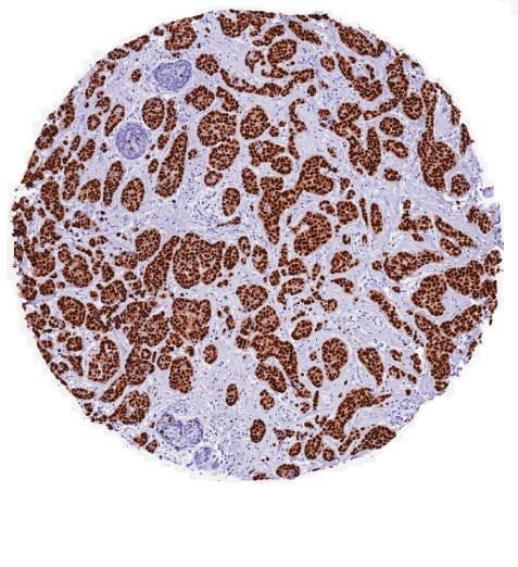
42 Table Clinicopathological characteristics of the entire series (n=345) continued. Characteristics No. of cases (%) Associated LCIS Absent 74 (21.4) Present 176 (51.0) not available 95 (27.5) Lymph node status pn0 163 (47.2) pn1 52 (15.1) pn2 38 (11.0) pn3 49 (14.2) not sampled 43 (12.5) 3.3 Immuno-marker expression and its association with clinicopathological characteristics Localisation of immuno-markers in ILC is shown in Figure to Table shows the distribution of immuno-marker status in the entire series. For hormonal receptors ER and PR, positive tumours comprised 91.6% and 70.1% respectively. A small percentage (5.8%) of tumours overexpressed HER-2. Basal marker CK HMW was positive in 86.4% and CK14 was positive in 15.4% of tumours. EGFR was positive in 3.5% of the tumours. Mammaglobin was positive in 54.8% of tumours. Low proliferative fraction, measured by proportion of Ki-67 positive tumour cells, was detected in 25.5% of the tumours and high proliferative fraction in 11.0% of the tumours. There was absence of Ki-67 staining in 63.5% of the tumours. For prognostic comparison of Ki-67, positive expression in at least 10% of tumour cells was used as cut-off. 30
43 Table Immuno-markers status in entire series (n = 345). Negative status Positive status Immuno-markers No. (%) No. (%) ER 29 (8.4) 316 (91.6) PR 103 (29.9) 242 (70.1) HER (94.2) 20 (5.8) CK HMW 47 (13.6) 298 (86.4) CK (84.6) 53 (15.4) EGFR 333 (96.5) 12 (3.5) Mammaglobin 156 (45.2) 189 (54.8) Ki (63.5) Low fraction (1-9%) 88 (25.5) High fraction ( 10%) 38 (11.0) 31


44 ER PR HER-2 Figure Immunohistochemical expression of ER, PR and HER-2. Localisation of ER and PR in the nucleus and HER-2 in the cytoplasmic membrane. 32


45 CK HMW CK14 EGFR Figure Immunohistochemical expression of basal markers CK HMW, CK14 and EGFR. Cytoplasmic localisation of CK14 and CK HMW and weak cytoplasmic membrane localisation of EGFR. 33


46 Ki-67 Mammaglobin Figure Immunohistochemical expression of Ki-67 and Mammaglobin. Nuclear localisation of Ki-67 and cytoplasmic localisation of Mammaglobin. 34
47 Association of clinicopathological parameters and immuno-markers are illustrated from Table to ER negativity was significantly associated with disease presentation at older age (p=0.031), higher histologic grade (p=0.006), the pleomorphic variant (p<0.001) and the occurrence of lymphovascular invasion (LVI) (p=0.015) as shown in Table PR positivity was associated with younger age (p<0.001), the classical variant (p=0.002), the presence of LVI (p=0.012) and lower LN stage (p=0.020) (Table 3.3.3). Negative HER-2 status was associated with the absence of LVI (p=0.007) (Table 3.3.4). Positive CK HMW status was associated with pn3 (p=0.002) (Table 3.3.5). Presence of accompanying LCIS was associated with positive CK14 status (p=0.002). There was weak association of CK14 negativity with higher LN stage (p=0.044) (Table 3.3.6). Positive EGFR expression was associated with the pleomorphic variant (p=0.038) and negative EGFR expression was associated with absence of LVI (p=0.016) (Table 3.3.7). High Ki-67 positive fraction was associated with the pleomorphic variant (p=0.001) (Table 3.3.8). There was no association of Mammaglobin expression with any better or worse clinicopathological characteristics (Table 3.3.9). 35
48 Table Association of hormonal marker ER status with clinicopathological characteristics. ER status Characteristic - + No. (%) No. (%) p-value Mean Age (55 years) cut off <Mean age 10 (34.5) 178 (56.3) 0.031* Mean age 19 (65.5) 138 (43.7) Tumour size (mm) (34.5) 121 (38.3) >20 16 (55.2) 174 (55.1) Histologic grade 1 2 (7.1) 95 (31.4) 0.006* 2 19 (67.9) 176 (58.1) 3 7 (25.0) 32 (10.6) Lobular variant Classical 5 (17.2) 103 (32.6) <0.001* Solid 2 (6.9) 43 (13.6) Alveolar 2 (6.9) 30 (9.5) Tubulo-lobular 1 (3.4) 28 (8.9) Pleomorphic 14 (48.3) 29 (9.2) Mixed 5 (17.2) 83 (26.3) Lymphovascular invasion Absent 18 (62.1) 259 (82.0) 0.015* Present 11 (37.9) 57 (18.0) Associated LCIS Absent 6 (20.7) 68 (21.5) Present 15 (51.7) 161 (50.9) Lymph node status pn0 14 (48.3) 149 (47.2) pn1 0 (0.0) 52 (16.5) pn2 5 (17.2) 33 (10.4) pn3 6 (20.7) 43 (13.6) Significant associations (p<0.05) are marked with *. 36
49 Table Association of hormonal marker PR status with clinicopathological characteristics. PR status Characteristic - + No. (%) No. (%) p-value Mean Age (55 years) cut off <Mean age 41 (39.8) 147 (60.7) <0.001* Mean age 62 (60.2) 95 (39.3) Tumour size (mm) (34.0) 96 (39.7) >20 60 (58.3) 130 (53.7) Histologic grade 1 22 (22.7) 75 (32.1) (61.9) 135 (57.7) 3 15 (15.5) 24 (10.3) Lobular variant Classical 27 (26.2) 81 (33.5) 0.002* Solid 16 (15.5) 29 (12.0) Alveolar 7 (6.8) 25 (10.3) Tubulo-lobular 9 (8.7) 20 (8.3) Pleomorphic 24 (23.3) 19 (7.9) Mixed 20 (19.4) 68 (28.1) Lymphovascular invasion Absent 74 (71.8) 203 (83.9) 0.012* Present 29 (28.2) 39 (16.1) Associated LCIS Absent 25 (24.3) 49 (20.2) Present 53 (51.5) 123 (50.8) Lymph node status pn0 41 (39.8) 122 (50.4) 0.020* pn1 11 (10.7) 41 (16.9) pn2 14 (13.6) 24 (9.9) pn3 22 (21.4) 27 (11.2) Significant associations (p<0.05) are marked with *. 37
50 Table Association of HER-2 status with clinicopathological characteristics. HER-2 status Characteristic - + No. (%) No. (%) p-value Mean Age (55 years) cut off <Mean age 173 (53.2) 15 (75.0) Mean age 152 (46.8) 5 (25.0) Tumour size (mm) (39.1) 4 (20.0) > (54.8) 12 (60.0) Histologic grade 1 93 (29.8) 4 (21.1) (58.7) 12 (63.2) 3 36 (11.5) 3 (15.8) Lobular variant Classical 103 (31.7) 5 (25.0) Solid 42 (12.9) 3 (15.0) Alveolar 31 (9.5) 1 (5.0) Tubulo-lobular 29 (8.9) 0 (0.0) Pleomorphic 38 (11.7) 5 (25.0) Mixed 82 (25.2) 6 (30.0) Lymphovascular invasion Absent 266 (81.8) 11 (55.0) 0.007* Present 59 (18.2) 9 (45.0) Associated LCIS Absent 69 (21.2) 5 (25.0) Present 166 (51.1) 10 (50.0) Lymph node status pn0 152 (46.8) 11 (55.0) pn1 52 (16.0) 0 (0.0) pn2 36 (11.1) 2 (10.0) pn3 44 (13.5) 5 (25.0) Significant associations (p<0.05) are marked with *. 38
51 Table Association of CK HMW status with clinicopathological characteristics. CK HMW status Characteristic - + No. (%) No. (%) p-value Mean Age (55 years) cut off <Mean age 26 (55.3) 162 (54.4) Mean age 21 (44.7) 136 (45.6) Tumour size (mm) (46.8) 109 (36.6) >20 23 (48.9) 167 (56.0) Histologic grade 1 14 (31.8) 83 (28.9) (54.5) 171 (59.6) 3 6 (13.6) 33 (11.5) Lobular variant Classical 14 (29.8) 94 (31.5) Solid 4 (8.5) 41 (13.8) Alveolar 5 (10.6) 27 (9.1) Tubulo-lobular 3 (6.4) 26 (8.7) Pleomorphic 7 (14.9) 36 (12.1) Mixed 14 (29.8) 74 (24.8) Lymphovascular invasion Absent 40 (85.1) 237 (79.5) Present 7 (14.9) 61 (20.5) Associated LCIS Absent 8 (17.0) 66 (22.1) Present 17 (36.2) 159 (53.4) Lymph node status pn0 20 (42.6) 143 (48.0) 0.002* pn1 8 (17.0) 44 (14.8) pn2 12 (25.5) 26 (8.7) pn3 2 (4.3) 47 (15.8) Significant associations (p<0.05) are marked with *. 39
52 Table Association of CK14 status with clinicopathological characteristics CK14 status Characteristic - + No. (%) No. (%) p-value Mean Age (55 years) cut off <Mean age 153 (52.4) 35 (66.0) Mean age 139 (47.6) 18 (34.0) Tumour size (mm) (37.0) 23 (43.4) > (56.5) 25 (47.2) Histologic grade 1 80 (28.5) 17 (34.0) (60.5) 25 (50.0) 3 31 (11.0) 8 (16.0) Lobular variant Classical 95 (32.5) 13 (24.5) Solid 37 (12.7) 8 (15.1) Alveolar 29 (9.9) 3 (5.7) Tubulo-lobular 25 (8.6) 4 (7.5) Pleomorphic 34 (11.6) 9 (17.0) Mixed 72 (24.7) 16 (30.2) Lymphovascular invasion Absent 235 (80.5) 42 (79.2) Present 57 (19.5) 11 (20.8) Associated LCIS Absent 69 (23.6) 5 (9.4) 0.002* Present 136 (46.6) 40 (75.5) Lymph node status pn0 140 (47.9) 23 (43.4) 0.044* pn1 39 (13.4) 13 (24.5) pn2 36 (12.3) 2 (3.8) pn3 44 (15.1) 5 (9.4) Significant associations (p<0.05) are marked with *. 40
53 Table Association of EGFR status with clinicopathological characteristics. EGFR status Characteristic - + No. (%) No. (%) p-value Mean Age (55 years) cut off <Mean age 178 (53.5) 10 (83.3) Mean age 155 (46.5) 2 (16.7) Tumour size (mm) (37.8) 5 (41.7) > (55.6) 5 (41.7) Histologic grade 1 95 (29.8) 2 (16.7) (58.9) 7 (58.3) 3 36 (11.3) 3 (25.0) Lobular variant Classical 107 (32.1) 1 (8.3) 0.038* Solid 43 (12.9) 2 (16.7) Alveolar 31 (9.3) 1 (8.3) Tubulo-lobular 29 (8.7) 0 (0.0) Pleomorphic 38 (11.4) 5 (41.7) Mixed 85 (25.5) 3 (25.0) Lymphovascular invasion Absent 271 (81.4) 6 (50.0) 0.016* Present 62 (18.6) 6 (50.0) Associated LCIS Absent 72 (21.6) 2 (16.7) Present 170 (51.1) 6 (50.0) Lymph node status pn0 158 (47.4) 5 (41.7) pn1 49 (14.7) 3 (25.0) pn2 37 (11.1) 1 (8.3) pn3 46 (13.8) 3 (25.0) Significant associations (p<0.05) are marked with *. 41
54 Table Association of Ki-67 status with clinicopathological characteristics. Ki-67 status Characteristic Low High No. (%) No. (%) p-value Mean Age (55 years) cut off <Mean age 171 (55.7) 17 (44.7) Mean age 136 (44.3) 21 (55.3) Tumour size (mm) (36.2) 51 (41.1) > (55.7) 67 (54.0) Histologic grade 1 90 (30.7) 7 (18.4) (58.0) 25 (65.8) (11.3) 6 (15.8) Lobular variant Classical 100 (32.6) 8 (21.1) 0.001* Solid 39 (12.7) 6 (15.8) Alveolar 28 (9.1) 4 (10.5) Tubulo-lobular 26 (8.5) 3 (7.9) Pleomorphic 30 (9.8) 13 (34.2) Mixed 84 (27.4) 4 (10.5) Lymphovascular invasion Absent 247 (80.5) 30 (78.9) Present 60 (19.5) 8 (21.1) Associated LCIS Absent 63 (29.3) 11 (31.4) Present 152 (70.7) 24 (68.6) Lymph node status pn0 145 (53.9) 66 (54.5) pn1 46 (17.1) 16 (18.2) pn2 35 (13.0) 10 (9.1) pn3 43 (16.0) 19 (18.2) Significant associations (p<0.05) are marked with *. 42
55 Table Association of Mammaglobin status with clinicopathological characteristics. Mammaglobin status Characteristic - + No. (%) No. (%) p-value Mean Age (55 years) cut off <Mean age 86 (55.1) 102 (54.0) Mean age 70 (44.9) 87 (46.0) Tumour size (mm) (35.9) 75 (39.7) >20 86 (55.1) 104 (55.0) Histologic grade 1 40 (26.8) 57 (31.3) 2 91 (61.1) 104 (57.1) (12.1) 21 (11.5) Lobular variant Classical 42 (26.9) 66 (34.9) Solid 18 (11.5) 27 (14.3) Alveolar 19 (12.2) 13 (6.9) Tubulo-lobular 14 (9.0) 15 (7.9) Pleomorphic 23 (14.7) 20 (10.6) Mixed 40 (25.6) 48 (25.4) Lymphovascular invasion Absent 122 (78.2) 155 (82.0) Present 34 (21.8) 34 (18.0) Associated LCIS Absent 30 (19.2) 44 (23.3) Present 78 (50.0) 98 (51.9) Lymph node status pn0 73 (46.8) 90 (47.6) pn1 22 (14.1) 30 (15.9) pn2 17 (10.9) 21 (11.1) pn3 24 (15.4) 25 (13.2) Significant associations (p<0.05) are marked with *. 43
56 3.4 Association of histologic type with clinicopathological characteristics and immuno-markers Differences between histologic types and clinicopathological characteristics were presented in Table Characteristics that were significantly associated with histology type included grade, accompanying LCIS status and lobular variant. Mixed ILC/IDC was associated with higher grade compared to ILC (p<0.001). Pure ILC was associated with the classical variant (p<0.001), with higher proportion ILC being classical variant compared to mixed ILC/IDC. Accompanying LCIS was presented more frequently in ILC compared to mixed ILC/IDC (p=0.001). 44
57 Table Association of histologic type with clinicopathological characteristics. Histologic Type Characteristic ILC Mixed ILC/IDC No. (%) No. (%) p-value Mean Age (55 years) cut off <Mean age 137 (55.7) 51 (51.5) Mean age 109 (44.3) 48 (48.5) Tumour size (mm) (36.6) 41 (41.4) > (55.7) 53 (53.5) Histologic grade 1 84 (35.6) 13 (13.7) <0.001* (55.9) 63 (66.3) 3 20 (8.5) 19 (20.0) Lobular variant Classical 94 (38.2) 14 (14.1) <0.001* Solid 27 (11.0) 18 (18.2) Alveolar 21 (8.5) 11 (11.1) Tubulo-lobular 15 (6.1) 14 (14.1) Pleomorphic 31 (12.6) 12 (12.1) Mixed 58 (23.6) 30 (30.3) Lymphovascular invasion Absent 204 (82.9) 73 (73.7) Present 42 (17.1) 26 (26.3) Associated LCIS Absent 44 (17.9) 30 (30.3) 0.001* Present 142 (57.7) 34 (34.3) Lymph node status pn0 114 (46.3) 49 (49.5) pn1 38 (15.4) 14 (14.1) pn2 27 (11.0) 11 (11.1) pn3 37 (15.0) 12 (12.1) Significant associations (p<0.05) are marked with *. 45
58 Association of histologic type with immuno-markers was presented in Table There was a weak association of mixed ILC/IDC with HER-2 positivity (p=0.041). Table Association of histologic type with immuno-markers. Histologic Type Immuno-marker Status ILC Mixed ILC/IDC No. (%) No. (%) p-value ER - 18 (7.3) 11 (11.1) (93.1) 88 (88.9) PR - 73 (29.8) 30 (30.3) (70.6) 69 (69.7) HER (96.3) 89 (89.9) 0.041* + 10 (4.1) 10 (10.1) CK HMW - 31 (12.7) 16 (16.2) (87.8) 83 (83.8) CK (83.3) 88 (88.9) (17.1) 11 (11.1) EGFR (97.6) 95 (96.0) (2.9) 4 (4.0) Mammaglobin (44.1) 48 (48.5) (56.3) 51 (51.5) Ki-67 Low 221 (72.0) 25 (65.8) High 86 (28.0) 13 (34.2) Significant associations (p<0.05) are marked with *. 46
59 3.5 Association of pleomorphic variant with clinicopathological characteristics and immuno-markers The pleomorphic variant made up 12.5% (43/345) of all tumours (Table 3.5.1). It was associated with higher tumour grade. From Table 3.5.2, grade 2 tumours made up 73.8% of the pleomorphic variant compared to 56.7% of non-pleomorphic and grade 3 tumours made up 19.0% of the pleomorphic variant versus 10.7% of the nonpleomorphic variant (p=0.002). Table Distribution of pleomorphic variant in the entire series. Pleomorphic category No. (%) Pleomorphic 43 (12.5) Non-pleomorphic 302 (87.5) 47
60 Table Association of pleomorphic variant with clinicopathological characteristics. Pleomorphic Non-pleomorphic Characteristic No. (%) No. (%) p-value Mean Age (55 years) cut off <Mean age 18 (41.9) 170 (56.3) Mean age 25 (58.1) 132 (43.7) Tumour size (mm) (27.9) 119 (39.4) >20 29 (67.4) 161 (53.3) Histologic grade 1 3 (7.1) 94 (32.5) 0.002* 2 31 (73.8) 164 (56.7) 3 8 (19.0) 31 (10.7) Lymphovascular invasion Absent 30 (69.8) 247 (81.8) Present 13 (30.2) 55 (18.2) Associated LCIS Absent 8 (18.6) 66 (21.9) Present 27 (62.8) 149 (49.3) Lymph node status pn0 15 (34.9) 148 (49.0) pn1 4 (9.3) 48 (15.9) pn2 5 (11.6) 33 (10.9) pn3 10 (23.3) 39 (12.9) Significant associations (p<0.05) are marked with *. 48
61 Table shows the association of the pleomorphic variant with immuno-markers. The pleomorphic variant was associated with ER (p<0.001) and PR negative status (p<0.001). Among the pleomorphic tumours, 32.6% were ER negative tumours compared to only 5.0% of non-pleomorphic tumours. PR negativity was present in 55.8% of pleomorphic tumours compared to 26.2% of non-pleomorphic tumours. There were significantly more EGFR positive tumours among the pleomorphic variant (11.6%) than nonpleomprphic variant (2.0%) (p=0.006). There were higher proportions of tumours with high Ki-67 positive fraction among the pleomorphic variant (30.2%) than nonpleomorphic variant (8.3%) (p<0.001). 49
62 Table Association of the pleomorphic variant with immuno-markers. Pleomorphic Non-pleomorphic Immuno-marker Status No. (%) No. (%) p-value ER - 14 (32.6) 15 (5.0) <0.001* + 29 (67.4) 287 (95.0) PR - 24 (55.8) 79 (26.2) <0.001* + 19 (44.2) 223 (73.8) HER-2-38 (88.4) 287 (95.0) (11.6) 15 (5.0) CK HMW - 7 (16.3) 40 (13.2) (83.7) 262 (86.8) CK14-34 (79.1) 258 (85.4) (20.9) 44 (14.6) EGFR - 38 (88.4) 296 (98.0) 0.006* + 5 (11.6) 6 (2.0) Mammaglobin - 23 (53.5) 133 (44.0) (46.5) 169 (56.0) Ki-67 Low 30 (69.8) 277 (91.7) <0.001* High 13 (30.2) 25 (8.3) Significant associations (p<0.05) are marked with *. 50
63 3.6 Association of triple negativity with clinicopathological characteristics and immuno-markers Triple negativity, where ER, PR and HER-2 status is negative, according to defined cutoffs, was seen in 20 (5.8%) tumours. The remaining 325 (94.2%) tumours were grouped as non-triple negative tumours (Table 3.6.1). Table Distribution of triple negativity in the entire series. Triple negative category No. (%) Triple negative ILC 20 (5.8) Non-triple negative ILC 325 (94.2) Triple negativity had less favourable characteristics. As seen in Table 3.6.2, the proportion of older patients was 75.0% for triple negative ILC and 43.7% for non-triple negative ILC (p=0.009). There are more grade 3 tumours among the triple negative ILC (21.1%) compared to non-triple negative ILC (11.2%) (p=0.045). Higher proportion (45.0%) of triple negative ILC were pleomorphic variant compared to 10.5% of non-triple negative ILC (p<0.001). 51
64 Table Association of triple negativity with clinicopathological characteristics. Triple negative Non-triple negative Characteristic No. (%) No. (%) p-value Mean Age (55 years) cut off <Mean age 5 (25.0) 183 (56.3) 0.009* Mean age 15 (75.0) 142 (43.7) Tumour size (mm) 20 8 (40.0) 123 (37.8) >20 10 (50.0) 180 (55.4) Histologic grade 1 1 (5.3) 96 (30.8) 2 14 (73.7) 181 (58.0) 0.045* 3 4 (21.1) 35 (11.2) Lobular variant Pleomorphic 9 (45.0) 34 (10.5) <0.001* Non-pleomorphic 11 (55.0) 291 (89.5) Lymphovascular invasion Absent 13 (65.0) 264 (81.2) Present 7 (35.0) 61 (18.8) Associated LCIS Absent 4 (20.0) 70 (21.5) Present 11 (55.0) 165 (50.8) Lymph node status pn0 9 (45.0) 154 (47.4) pn1 0 (0.0) 52 (16.0) pn2 4 (20.0) 34 (10.5) pn3 4 (20.0) 45 (13.8) Significant associations (p<0.05) are marked with *. 52
65 Table illustrates the association of triple negative ILC with immuno-markers. CK HMW negative status was weakly associated with triple negative ILC (p=0.040), where CK HMW was negative in 30.0% of triple negative ILC and in 12.6% of non-triple negative ILC. Compared with non-triple negative ILC, triple negative ILC were much more likely to be Mammaglobin negative (80.0% of triple negative ILC versus 43.1% of non-triple negative ILC; p=0.002). Table Association of triple negativity with immuno-markers. Triple negative Non-triple negative Immuno-marker Status No. (%) No. (%) p-value CK HMW - 6 (30.0) 41 (12.6) 0.040* + 14 (70.0) 284 (87.4) CK14-16 (80.0) 276 (84.9) (20.0) 49 (15.1) EGFR - 19 (95.0) 315 (96.9) (5.0) 10 (3.1) Mammaglobin - 16 (80.0) 140 (43.1) 0.002* + 4 (20.0) 185 (56.9) Ki-67 Low 19 (95.0) 288 (88.6) High 1 (5.0) 37 (11.4) Significant associations (p<0.05) are marked with *. 53
66 3.7 Association of basal protein expression with clinicopathological characteristics and immuno-markers According to previous criteria established by Thike et al., basal phenotype is defined as tumours expressing positivity for at least one of the following markers: CK HMW, CK14 or EGFR (Thike et al. 2010). Based on this definition, 98.6% of tumours were basal-like. This proportion was too high and might be as a result of the wide spectrum reactivity of CK HMW cocktail (clone: 34βE12). The expression of 34βE12, incorporating CK1, 5, 10 and 14, may be non-specific for basal expression in ILC. Using the criteria of positivity for CK14 and/or EGFR, the proportion of basal phenotype was reduced to a more realistic proportion of 17.4%. This latter definition was adopted to test the association of basal phenotype with clinicopathological characteristics and immuno-markers (Table 3.7.1). Table Distribution of basal phenotype in the entire series according to different definition. Basal-like phenotype No. (%) Based on positivity of CK HMW and/or CK14 and/or EGFR Basal phenotype 340 (98.6) Non-basal phenotype 5 (1.4) Based on positivity of CK14 and/or EGFR Basal phenotype 60 (17.4) Non-basal phenotype 285 (82.6) Comparing tumours with basal and non-basal phenotype, the latter were more frequent in younger patients (66.7% of basal-like versus 51.9% of non basal-like; p=0.046). Basal 54
67 phenotype was significantly associated with presence of LCIS (71.7% of basal-like versus 46.7% of non basal-like; p=0.003). Table Association of basal phenotype with clinicopathological characteristics. Basal phenotype Non-basal phenotype Characteristic No. (%) No. (%) p-value Mean Age (55 years) cut off <Mean age 40 (66.7) 148 (51.9) 0.046* Mean age 20 (33.3) 137 (48.1) Tumour size (mm) (41.7) 106 (37.2) >20 29 (48.3) 161 (56.5) Histologic grade 1 18 (31.6) 79 (28.8) (54.4) 164 (59.9) 3 8 (14.0) 31 (11.3) Lobular variant Pleomorphic 12 (20.0) 31 (10.9) Non-pleomorphic 48 (80.0) 254 (89.1) LVI Absent 47 (78.3) 230 (80.7) Present 13 (21.7) 55 (19.3) Associated LCIS Absent 6 (10.0) 68 (23.9) 0.003* Present 43 (71.7) 133 (46.7) Lymph node status pn0 27 (45.0) 136 (47.7) pn1 14 (23.3) 38 (13.3) pn2 3 (5.0) 35 (12.3) pn3 6 (10.0) 43 (15.1) Significant associations (p<0.05) are marked with *. 55
68 Table shows no association established between basal phenotype and immunomarkers. Table Association of basal phenotype with immuno-markers. Basal-like Non Basal-like Immuno-markers Status No. (%) No. (%) p-value ER - 6 (10.0) 23 (8.1) (90.0) 262 (91.9) PR - 16 (26.7) 87 (30.5) (73.3) 198 (69.5) HER-2-57 (95.0) 268 (94.0) (5.0) 17 (6.0) CK HMW - 4 (6.7) 43 (15.1) (93.3) 242 (84.9) Mammaglobin - 25 (41.7) 131 (46.0) (58.3) 154 (54.0) Ki-67 Low 51 (85.0) 256 (89.8) High 9 (15.0) 29 (10.2) Significant associations (p<0.05) are marked with *. 56
69 3.8 Association of molecular subtype with clinicopathological characteristics and immuno-markers From various gene expression studies, the most powerful discriminator genes for the different molecular subtypes are the expression of luminal epithelial specific gene, ESR1 gene which encodes for estrogen receptor and ERBB2 which encodes for HER-2/neu (van't Veer et al. 2002;West et al. 2001;Sorlie et al. 2003). Stemming from molecular profiling evidence, use of surrogate immunohistochemical markers ER, PR and HER-2 was applied to classify tumours into the modified molecular subtypes. Table shows the criteria used for categorisation of molecular subtypes according to surrogate immunohistochemical markers ER, PR and HER-2 and the distribution of tumours. Majority were luminal A-like (88.4%), 4.1% were luminal B-like and 1.7% were HER-2 overexpressing. Triple negative (ER -, PR - and HER-2 -) breast cancers often overlap significantly with the basal molecular subtype. If we incorporated positivity for basal markers CK14 and/or EGFR in this group, our proportion of basal-like molecular subtype would be 1.2%. This compares against 17.4% of all tumours in the series expressing basal markers CK14 and/or EGFR regardless of triple negativity. Sixteen (4.6%) of the triple negative tumours were also negative for CK14 and EGFR. This group was labeled as unclassified (penta negative). 57
70 Table Criteria used for molecular subtype and the distribution of molecular subtype in the entire series. Molecular subtype Criteria used No. (%) Luminal A Luminal B ER+/PR+/HER-2- or ER+/PR-/HER-2- or ER-/PR+/HER-2- ER+/PR+/HER-2+ or ER+/PR-/HER-2+ or ER-/PR+/HER (88.4) 14 (4.1) HER-2 overexpressing ER-/PR-/HER-2+ 6 (1.7) Basal-like ER-/PR-/HER-2-/CK14+ and/or EGFR+ 4 (1.2) Unclassified (Penta negative) ER-/PR-/HER-2-/CK14-/EGFR- 16 (4.6) Analysis of molecular subtype and clinicopathological characteristics was shown in Table Basal-like and penta negative molecular subtypes were associated with older age (p=0.030). Proportion of patient age older than the mean was 75.0% in the basal-like and penta-negative categories compared to 44.9% in luminal A, 21.4% in luminal B and 33.3% in HER-2 overexpressing subtype. There was a significantly higher proportion of non-pleomorphic variant in luminal A (90.5%) and luminal B (85.7%) subtypes compared to HER-2 overexpressing, basal-like and penta negative molecular subtypes (p<0.001). Lymphovascular invasion was more likely to be absent in the luminal A subtype (p=0.008). 58
71 Table Association of molecular subtype with clinicopathological characteristics. Characteristic Luminal A Luminal B Molecular category HER-2 overexpressing Basal-like Penta negative No. (%) No. (%) No. (%) No. (%) No. (%) p-value Mean Age (55 years) cut off <Mean age 168 (55.1) 11 (78.6) 4 (66.7) 1 (25.0) 4 (25.0) 0.030* Mean age 137 (44.9) 3 (21.4) 2 (33.3) 3 (75.0) 12 (75.0) Tumour size (mm) (39.0) 3 (21.4) 1 (16.7) 2 (50.0) 6 (37.5) > (55.1) 8 (57.1) 4 (66.7) 2 (50.0) 8 (50.0) Histologic grade 1 92 (30.2) 3 (21.4) 1 (16.7) 0 (0.0) 1 (6.3) (55.4) 9 (64.3) 3 (50.0) 3 (75.0) 11 (68.8) 3 32 (10.5) 1 (7.1) 2 (33.3) 1 (25.0) 3 (18.8) Lobular variant Pleomorphic 29 (9.5) 2 (14.3) 3 (50.0) 2 (50.0) 7 (43.8) <0.001* Non-pleomorphic 276 (90.5) 12 (85.7) 3 (50.0) 2 (50.0) 9 (56.3) LVI Absent 253 (83.0) 7 (50.0) 4 (66.7) 2 (50.0) 11 (68.8) 0.008* Present 52 (17.0) 7 (50.0) 2 (33.3) 2 (50.0) 6 (37.5) Associated LCIS Absent 65 (21.3) 3 (21.4) 2 (33.3) 0 (0.0) 4 (25.0) Present 155 (50.8) 8 (57.1) 2 (33.3) 3 (75.0) 8 (50.0) Lymph node status pn0 143 (46.9) 9 (64.3) 2 (33.3) 2 (50.0) 7 (43.8) pn1 52 (17.0) 0 (0.0) 0 (0.0) 0 (0.0) 0 (0.0) pn2 32 (10.5) 1 (7.1) 1 (16.7) 0 (0.0) 4 (25.0) pn3 40 (13.1) 3 (21.4) 2 (33.3) 2 (50.0) 2 (12.5) Significant associations (p<0.05) are marked with *. 59
72 HER-2 overexpressing, basal-like and the penta-negative molecular subtypes were associated with negative Mammoglobin status (p=0.005) according to Table There were significant higher proportions of Mammaglobin negative tumours in the HER-2 overexpressing (83.3%), basal-like (75.0%) and penta negative (81.3%) subtypes compared to luminal A (42.0%) and luminal B (50.0%). Table Association of molecular subtype with immuno-markers. Immunomarker Luminal A Luminal B Molecular category HER-2 overexpressing Basal-like Penta negative No. (%) No. (%) No. (%) No. (%) No. (%) p-value Mammaglobin (42.0) 7 (50.0) 5 (83.3) 3 (75.0) 13 (81.3) 0.005* (58.0) 7 (50.0) 1 (16.7) 1 (25.0) 3 (18.8) Ki-67 low 198 (64.9) 10 (71.4) 3 (50.0) 1 (25.0) 9 (56.3) high 107 (35.1) 4 (28.6) 3 (50.0) 3 (75.0) 7 (43.8) 60
73 3.9 Immunohistochemical expression of E-cadherin and p120 catenin In this series, 111/345 (32.2%) of lobular tumours showed E-cadherin positivity and 67.8% of the tumours were negative for E-cadherin (Table 3.9.1). The percentage of positive cells ranged from 5% to 100%. This positive but aberrant expression of E- cadherin ranged from incomplete membrane to perimembranous cytoplasmic staining (Figure 3.9.1). Majority (80.3%) of the tumours had aberrant cytoplasmic localisation of p120 catenin, 18.8% of the tumours expressed both cytoplasmic and membrane localisation and 3 tumours (0.9%) showed complete loss of p120 catenin cytoplasmic and membrane expression. Table E-cadherin and p120 catenin expression in ILC. Immuno-marker No. (%) E-cadherin Aberrant expression 111 (32.2) Negative expression 234 (67.8) p120 catenin Cytoplasmic only 277 (80.3) Cytoplasmic and membrane 65 (18.8) Complete loss of expression 3 (0.9) 61

 (C) Weak incomplete membrane")

74 (A) (A) Incomplete membranous staining (B) (B) Perimembranous cytoplasmic staining in the absence of complete circumferential membrane staining (C) (C) Weak incomplete membrane staining in a proportion of tumour cells. Figure Varying immunohistochemical expression of E-cadherin in ILC. 62
75 Among the E-cadherin negative tumours, 226/234 (96.6%) had cytoplasmic accumulation of p120 catenin, 5/234 (2.1%) expressed both cytoplasmic and membrane p120 catenin and 3/234 (0.9%) had complete loss of p120 catenin expression. Among the tumours that expressed E-cadherin, 51/111 (45.9%) expressed cytoplasmic p120, 60/111 (54.1%) showed both cytoplasm and membrane expression. The diffuse cytoplasmic expression p120 catenin was strongly associated with negative E-cadherin expression. p120 catenin cytoplasmic localisation ranged from weakly to moderately staining intensity with patchy to homogenous diffuse pattern (Figure 3.9.2). Table p120 catenin cytoplasmic and cytoplasmic membrane localisation among E-cadherin positive and negative tumours. E-cadherin expression p120 catenin expression Negative expression (n=234) Aberrant expression (n=111) No. (%) No. (%) Cytoplasmic only 226 (96.6) 51 (45.9) Cytoplasmic and membrane 5 (2.1) 60 (54.1) Complete loss of expression 3 (1.3) 0 (0.0) 63


 E-cadherin (C)p120 catenin (C)")

76 (A) E-cadherin (A) p120 catenin (A) E-cadherin negative tumour with strong diffuse p120 catenin cytoplasmic localisation. (B) E-cadherin (B) p120 catenin (B) Aberrant E-cadherin patchily positive tumour with weakly positive cytoplasmic membrane and cytoplasmic localisation of p120 catenin. (C) E-cadherin (C)p120 catenin (C) Tumour with negative E-cadherin and p120 catenin expression Figure Differential expression of E-cadherin and p120 catenin in ILC. 64
Carcinoma mammario: le istologie non frequenti. Valentina Guarneri Università di Padova IOV-IRCCS
 Carcinoma mammario: le istologie non frequenti Valentina Guarneri Università di Padova IOV-IRCCS Histological diversity of breast adenocarcinomas Different histological types are defined according to specific
Basement membrane in lobule.
 Bahram Memar, MD Basement membrane in lobule. Normal lobule-luteal phase Normal lobule-follicular phase Lactating breast Greater than 95% are adenocarcinomas in situ carcinomas and invasive carcinomas.
Histological Type. Morphological and Molecular Typing of breast Cancer. Nottingham Tenovus Primary Breast Cancer Study. Survival (%) Ian Ellis
 Ian Ellis") Morphological and Molecular Typing of breast Cancer Ian Ellis Molecular Medical Sciences, University of Nottingham Department of Histopathology, Nottingham University Hospitals NHS Trust Histological Type
Morphological and Molecular Typing of breast Cancer Ian Ellis Molecular Medical Sciences, University of Nottingham Department of Histopathology, Nottingham University Hospitals NHS Trust Histological Type
Maram Abdaljaleel, MD Dermatopathologist and Neuropathologist University of Jordan, School of Medicine
 Maram Abdaljaleel, MD Dermatopathologist and Neuropathologist University of Jordan, School of Medicine The most common non-skin malignancy of women 2 nd most common cause of cancer deaths in women, following
Diseases of the breast (2 of 2) Breast cancer
 Breast cancer") Diseases of the breast (2 of 2) Breast cancer Epidemiology & etiology The most common type of cancer & the 2 nd most common cause of cancer death in women 1 of 8 women in USA Affects 7% of women Peak at
Diseases of the breast (2 of 2) Breast cancer Epidemiology & etiology The most common type of cancer & the 2 nd most common cause of cancer death in women 1 of 8 women in USA Affects 7% of women Peak at
CLINICAL SIGNIFICANCE OF BENIGN EPITHELIAL CHANGES
 Papillomas. Papillomas are composed of multiple branching fibrovascular cores, each having a connective tissue axis lined by luminal and myoepithelial cells ( Fig. 23-11 ). Growth occurs within a dilated
Recent advances in breast cancers
 Recent advances in breast cancers Breast cancer is a hetrogenous disease due to distinct genetic alterations. Similar morphological subtypes show variation in clinical behaviour especially in response
Breast Pathology. Breast Development
 Breast Pathology Lecturer: Hanina Hibshoosh, M.D. Reading: Kumar, Cotran, Robbins, Basic Pathology, 6th Edition, pages 623-635 Breast Development 5th week - thickening of the epidermis - milk line 5th
BREAST PATHOLOGY. Fibrocystic Changes
 BREAST PATHOLOGY Lesions of the breast are very common, and they present as palpable, sometimes painful, nodules or masses. Most of these lesions are benign. Breast cancer is the 2 nd most common cause
CPC 4 Breast Cancer. Rochelle Harwood, a 35 year old sales assistant, presents to her GP because she has noticed a painless lump in her left breast.
 CPC 4 Breast Cancer Rochelle Harwood, a 35 year old sales assistant, presents to her GP because she has noticed a painless lump in her left breast. 1. What are the most likely diagnoses of this lump? Fibroadenoma
Breast pathology. 2nd Department of Pathology Semmelweis University
 Breast pathology 2nd Department of Pathology Semmelweis University Breast pathology - Summary - Benign lesions - Acute mastitis - Plasma cell mastitis / duct ectasia - Fat necrosis - Fibrocystic change/
Surgical Pathology Issues of Practical Importance
 Surgical Pathology Issues of Practical Importance Anne Moore, MD Medical Oncology Syed Hoda, MD Surgical Pathology The pathologist is central to the team approach needed to manage the patient with breast
1 NORMAL HISTOLOGY AND METAPLASIAS
 1 NORMAL HISTOLOGY AND METAPLASIAS, MD Anatomy and Histology 1 Metaplasias 2 ANATOMY AND HISTOLOGY The female breast is composed of a branching duct system, which begins at the nipple with the major lactiferous
Triple Negative Breast Cancer
 Triple Negative Breast Cancer Prof. Dr. Pornchai O-charoenrat Division of Head-Neck & Breast Surgery Department of Surgery Faculty of Medicine Siriraj Hospital Breast Cancer Classification Traditional
Molecular classification of breast cancer implications for pathologists. Sarah E Pinder
 Molecular classification of breast cancer implications for pathologists Sarah E Pinder Courtesy of CW Elston Histological types Breast Cancer Special Types 17 morphological special types 25-30% of all
6/3/2010. Outline of Talk. Lobular Breast Cancer: Definition of lobular differentiation. Common Problems in Diagnosing LCIS in Core Biopsies
 Outline of Talk Lobular Breast Cancer: Common Problems in Diagnosing LCIS in Core Biopsies Definition of lobular differentiation Variants of LCIS that: carry risk for unsampled invasive cancer mimic DCIS
Enterprise Interest None
 Enterprise Interest None What are triple negative breast cancers? A synopsis of their histological patterns Ian Ellis Molecular Medical Sciences, University of Nottingham Department of Histopathology,
Morphological and Molecular Typing of breast Cancer
 Morphological and Molecular Typing of breast Cancer Ian Ellis Molecular Medical Sciences, University of Nottingham Department of Histopathology, Nottingham University Hospitals NHS Trust Histological
LYMPHATIC DRAINAGE AXILLARY (MOSTLY) INTERNAL MAMMARY SUPRACLAVICULAR
 INTERNAL MAMMARY SUPRACLAVICULAR") BREAST LYMPHATIC DRAINAGE AXILLARY (MOSTLY) INTERNAL MAMMARY SUPRACLAVICULAR HISTOLOGY LOBE: (10 in whole breast) LOBULE: (many per lobe) ACINUS/I, aka ALVEOLUS/I: (many per lobule) DUCT(S): INTRA- or
BREAST LYMPHATIC DRAINAGE AXILLARY (MOSTLY) INTERNAL MAMMARY SUPRACLAVICULAR HISTOLOGY LOBE: (10 in whole breast) LOBULE: (many per lobe) ACINUS/I, aka ALVEOLUS/I: (many per lobule) DUCT(S): INTRA- or
Contents 1 The Windows of Susceptibility to Breast Cancer 2 The So Called Pre-Neoplastic Lesions and Carcinoma In Situ
 Contents 1 The Windows of Susceptibility to Breast Cancer... 1 1.1 Introduction... 1 1.2 Risk Factor and Etiological Agents... 2 1.3 The Concept of the Windows of Susceptibility to Carcinogenesis... 5
04/10/2018. Intraductal Papillary Neoplasms Of Breast INTRADUCTAL PAPILLOMA
 Intraductal Papillary Neoplasms Of Breast Savitri Krishnamurthy MD Professor of Pathology Deputy Division Head The University of Texas MD Anderson Cancer Center 25 th Annual Seminar in Pathology Pittsburgh,
University Journal of Pre and Para Clinical Sciences
 ISSN 2455 2879 Volume 2 Issue 1 2016 Metaplastic carcinoma breast a rare case report Abstract : Metaplastic carcinoma of the breast is a rare malignancy with two distinct cell lines described as a breast
Protocol for the Examination of Biopsy Specimens From Patients With Invasive Carcinoma of the Breast
 Protocol for the Examination of Specimens From Patients With Invasive Carcinoma of the Breast Version: BreastInvasive 1.0.0.0 Protocol Posting Date: February 2019 Accreditation Requirements The use of
University of Zurich. Histology and Immunophenotype of Invasive Lobular Breast Cancer. Daily Practice and Pitfalls. Zurich Open Repository and Archive
 University of Zurich Zurich Open Repository and Archive Winterthurerstr. 190 CH-8057 Zurich http://www.zora.uzh.ch Year: 2009 Histology and Immunophenotype of Invasive Lobular Breast Cancer. Daily Practice
Papillary Lesions of the breast
 Papillary Lesions of the breast Emad Rakha Professor of Breast Pathology The University of Nottingham Papillary lesions of the breast are a heterogeneous group of disease, which are characterised by neoplastic
Rare Breast Tumours. 1. Breast Tumours. 1.1 General Results. 1.2 Incidence
 Rare Breast Tumours 1. Breast Tumours 1.1 General Results Table 1. Epithelial Tumours of Breast: Incidence, Trends, Survival Flemish Region 2001-2010 Incidence Trend Survival Females EAPC Relative survival
Proliferative Epithelial lesions of the Breast. Sami Shousha, MD, FRCPath Charing Cross Hospital & Imperial College, London
 Proliferative Epithelial lesions of the Breast Sami Shousha, MD, FRCPath Charing Cross Hospital & Imperial College, London Amman, November2013 Proliferative Epithelial Lesions of the Breast Usual type
XXV Congreso de la Sociedad Española de Anatomía Patológica y División Española de la International Academy of Pathology
 XXV Congreso de la Sociedad Española de Anatomía Patológica y División Española de la International Academy of Pathology NUEVOS FENOTIPOS DEL CÁNCER DE MAMA: NUEVOS PROBLEMAS PARA EL PATÓLOGO? Tienen actualmente
Mousa. Israa Ayed. Abdullah AlZibdeh. 0 P a g e
 1 Mousa Israa Ayed Abdullah AlZibdeh 0 P a g e Breast pathology The basic histological units of the breast are called lobules, which are composed of glandular epithelial cells (luminal cells) resting on
Diseases of the breast (1 of 2)
") Diseases of the breast (1 of 2) Introduction A histology introduction Normal ducts and lobules of the breast are lined by two layers of cells a layer of luminal cells overlying a second layer of myoepithelial
Diseases of the breast (1 of 2) Introduction A histology introduction Normal ducts and lobules of the breast are lined by two layers of cells a layer of luminal cells overlying a second layer of myoepithelial
Implications of Progesterone Receptor Status for the Biology and Prognosis of Breast Cancers
 日大医誌 75 (1): 10 15 (2016) 10 Original Article Implications of Progesterone Receptor Status for the Biology and Prognosis of Breast Cancers Naotaka Uchida 1), Yasuki Matsui 1), Takeshi Notsu 1) and Manabu
LOBULAR CARCINOMA IN SITU: WHAT DOES IT MEAN? THE SURGEON'S PERSPECTIVE
 : WHAT DOES IT MEAN? THE SURGEON'S PERSPECTIVE Benjamin O. Anderson, M.D. Director, Breast Health Clinic Professor of Surgery and Global Health, University of Washington Joint Member, Fred Hutchinson Cancer
Your Guide to the Breast Cancer Pathology. Report. Key Questions. Here are important questions to be sure you understand, with your doctor s help:
 Your Guide to the Breast Cancer Pathology Report Key Questions Here are important questions to be sure you understand, with your doctor s help: Your Guide to the Breast Cancer Pathology Report 1. Is this
Claudin-4 Expression in Triple Negative Breast Cancer: Correlation with Androgen Receptors and Ki-67 Expression
 Claudin-4 Expression in Triple Negative Breast Cancer: Correlation with Androgen Receptors and Ki-67 Expression Mona A. Abd-Elazeem, Marwa A. Abd- Elazeem Pathology department, Faculty of Medicine, Tanta
Clinicopathological Factors Affecting Distant Metastasis Following Loco-Regional Recurrence of breast cancer. Cheol Min Kang 2018/04/05
 Abstract No.: ABS-0075 Clinicopathological Factors Affecting Distant Metastasis Following Loco-Regional Recurrence of breast cancer 2018/04/05 Cheol Min Kang Department of surgery, University of Ulsan
Breast Cancer. Most common cancer among women in the US. 2nd leading cause of death in women. Mortality rates though have declined
 Breast Cancer Most common cancer among women in the US 2nd leading cause of death in women Mortality rates though have declined 1 in 8 women will develop breast cancer Breast Cancer Breast cancer increases
Breast Cancer. Dr Rodney Itaki Anatomical Pathology Discipline Division of Pathology
 Breast Cancer Dr Rodney Itaki Anatomical Pathology Discipline Division of Pathology Muscles Muscles underneath the breasts separating them from the ribs Breast has no muscle tissue 2 Female Breast Anatomy
Ductal Carcinoma in Situ. Laura C. Collins, M.D. Department of Pathology Beth Israel Deaconess Medical Center and Harvard Medical School Boston, MA
 Ductal Carcinoma in Situ Laura C. Collins, M.D. Department of Pathology Beth Israel Deaconess Medical Center and Harvard Medical School Boston, MA Definition of DCIS WHO 2012 A neoplastic proliferation
Lesion Imaging Characteristics Mass, Favoring Benign Circumscribed Margins Intramammary Lymph Node
 Lesion Imaging Characteristics Mass, Favoring Benign Circumscribed Margins Intramammary Lymph Node Oil Cyst Mass, Intermediate Concern Microlobulated Margins Obscured Margins Mass, Favoring Malignant Indistinct
Disclosures 5/27/2012. Outline of Talk. Outline of Talk. When Is LCIS Clinically Significant? Classic LCIS. Classic LCIS
 When Is LCIS Clinically Significant? Disclosures I have nothing to disclose Yunn-Yi Chen, MD, PhD Professor Outline of Talk Outline of Talk Classic LCIS Classic LCIS Definition of lobular differentiation
Breast Cancer. Saima Saeed MD
 Breast Cancer Saima Saeed MD Breast Cancer Most common cancer among women in the US 2nd leading cause of death in women 1 in 8 women will develop breast cancer Incidence/mortality rates have declined Breast
Research Article Stromal Expression of CD10 in Invasive Breast Carcinoma and Its Correlation with ER, PR, HER2-neu, and Ki67
 SAGE-Hindawi Access to Research International Breast Cancer Volume 20, Article ID 47957, 4 pages doi:0.406/20/47957 Research Article Stromal Expression of CD0 in Invasive Breast Carcinoma and Its Correlation
Only Estrogen receptor positive is not enough to predict the prognosis of breast cancer
 Young Investigator Award, Global Breast Cancer Conference 2018 Only Estrogen receptor positive is not enough to predict the prognosis of breast cancer ㅑ Running head: Revisiting estrogen positive tumors
Case study 1. Rie Horii, M.D., Ph.D. Division of Pathology Cancer Institute Hospital, Japanese Foundation for Cancer Research
 NCCN/JCCNB Seminar in Japan April 15, 2012 Case study 1 Rie Horii, M.D., Ph.D. Division of Pathology Cancer Institute Hospital, Japanese Foundation for Cancer Research Present illness: A 50y.o.premenopausal
Note: The cause of testicular neoplasms remains unknown
 - In the 15- to 34-year-old age group, they are the most common tumors of men. - Tumors of the testis are a heterogeneous group of neoplasms that include: I. Germ cell tumors : 95%; all are malignant.
Synchronous squamous cell carcinoma of the breast. and invasive lobular carcinoma
 Sentani K et al. 1 Letter to the editor Synchronous squamous cell carcinoma of the breast and invasive lobular carcinoma Kazuhiro Sentani, 1 Takashi Tashiro, 2 Naohide Oue, 1 Wataru Yasui 1 1 Department
Columnar Cell Lesions
 Columnar Cell Lesions Laura C. Collins, M.D. Department of Pathology Beth Israel Deaconess Medical Center and Harvard Medical School Boston, MA Question? Columnar cell lesions are: a) Annoying lesions
Cytyc Corporation - Case Presentation Archive - March 2002
 FirstCyte Ductal Lavage History: 68 Year Old Female Gail Index: Unknown Clinical History: Negative Mammogram in 1995 6 yrs. later presents with bloody nipple discharge Subsequent suspicious mammogram Suspicious
Papillary Lesions of the Breast A Practical Approach to Diagnosis. (Arch Pathol Lab Med. 2016;140: ; doi: /arpa.
 Papillary Lesions of the Breast A Practical Approach to Diagnosis (Arch Pathol Lab Med. 2016;140:1052 1059; doi: 10.5858/arpa.2016-0219-RA) Papillary lesions of the breast Span the spectrum of benign,
Neoplasia 2018 Lecture 2. Dr Heyam Awad MD, FRCPath
 Neoplasia 2018 Lecture 2 Dr Heyam Awad MD, FRCPath ILOS 1. List the differences between benign and malignant tumors. 2. Recognize the histological features of malignancy. 3. Define dysplasia and understand
Papillary Lesions of the Breast
 Papillary Lesions of the Breast Laura C. Collins, M.D. Associate Professor of Pathology Associate Director, Division of Anatomic Pathology Beth Israel Deaconess Medical Center and Harvard Medical School
Ph.D. THESIS ENDOMETRIAL HYPERPLASIAS IN PERIMENOPAUSE SUMMARY
 UNIVERSITY OF MEDICINE AND PHARMACY OF CRAIOVA FACULTY OF MEDICINE Ph.D. THESIS ENDOMETRIAL HYPERPLASIAS IN PERIMENOPAUSE SUMMARY SCIENTIFIC COORDINATOR: PROF. DR. MIHAI B. BRĂILA, Ph.D. Ph.D. Graduand:
Update on 2015 WHO Classification of Lung Adenocarcinoma 1/3/ Mayo Foundation for Medical Education and Research. All rights reserved.
 1 Our speaker for this program is Dr. Anja Roden, an associate professor of Laboratory Medicine and Pathology at Mayo Clinic as well as consultant in the Anatomic Pathology Laboratory and co-director of
Sentinel nodes. Location: Location: S1.04 Principal clinician. G1.01 Record other relevant information. S2.01 Number of specimens submitted
 Invasive Breast Cancer Histopathology Reporting Proforma Mandatory questions (i.e. protocol standards) are in bold (e.g. S1.01). S1.01 Identification Family name Given name(s) Date of birth DD MM YYYY
Benign Mimics of Malignancy in Breast Pathology
 Arthur Purdy Stout Society of Surgical Pathologists Companion Meeting Benign Mimics of Malignancy in Breast Pathology Stuart J. Schnitt, M.D. Beth Israel Deaconess Medical Center and Harvard Medical School,
Patterns of E.cadherin and Estrogen receptor Expression in Histological Sections of Sudanese Patients with Breast Carcinoma
 Patterns of E.cadherin and Estrogen receptor Expression in Histological Sections of Sudanese Patients with Breast Carcinoma Hadia. Mohammed. Abdalla. Abdalrhman *, Elsadig.A.Adam, Ayda.D.A.Allatif 3,'Namareg.E.Afadul
Urinary Bladder: WHO Classification and AJCC Staging Update 2017
 Urinary Bladder: WHO Classification and AJCC Staging Update 2017 Houston Society of Clinical Pathologists 58 th Annual Spring Symposium Houston, TX April 8, 2017 Jesse K. McKenney, MD Classification
Objectives. Atypical Glandular Cells. Atypical Endocervical Cells. Reactive Endocervical Cells
 2013 California Society of Pathologists 66 th Annual Meeting San Francisco, CA Atypical Glandular Cells to Early Invasive Adenocarcinoma: Cervical Cytology and Histology Christina S. Kong, MD Associate
A712(19)- Test slide, Breast cancer tissues with corresponding normal tissues
- Test slide, Breast cancer tissues with corresponding normal tissues") A712(19)- Test slide, Breast cancer tissues with corresponding normal tissues (formalin fixed) For research use only Specifications: No. of cases: 12 Tissue type: Breast cancer tissues with corresponding
A712(19)- Test slide, Breast cancer tissues with corresponding normal tissues (formalin fixed) For research use only Specifications: No. of cases: 12 Tissue type: Breast cancer tissues with corresponding
BIOLOGICAL AND CLINICAL CHARACTERISTICS OF INVASIVE DUCTAL CARCINOMA WITH LOBULAR PATTERN
 BIOLOGICAL AND CLINICAL CHARACTERISTICS OF INVASIVE DUCTAL CARCINOMA WITH LOBULAR PATTERN Final Degree Project Author: Kyong Bok Rodríguez Rausis Tutor: Cristina Melendez Muñoz FACULTY OF MEDICINE January
Case 1. ACCME/Disclosure. Clinical History. Dr. Mulligan has nothing to disclose
 Breast Evening Specialty Conference USCAP, 2016 Case 1 Anna Marie Mulligan University Health Network, Toronto University of Toronto ACCME/Disclosure Dr. Mulligan has nothing to disclose Clinical History
Salivary Glands 3/7/2017
 Salivary Glands 3/7/2017 Goals and objectives Focus on the entities unique to H&N Common board type facts Information for your future practice Salivary Glands Salivary Glands Major gland. Paratid. Submandibular.
Present Role of Immunohistochemistry in the. Subtypes. Beppe Viale European Institute of Oncology University of Milan Milan-Italy
 Present Role of Immunohistochemistry in the Classification of Molecular Subtypes Beppe Viale European Institute of Oncology University of Milan Milan-Italy We know it is many diseases Breast cancer is
Lesions Mimicking Adenoid Cystic Carcinoma. Diagnostic Problems in Salivary Gland Pathology An Update 5/29/2009
 Diagnostic Problems in Salivary Gland Pathology An Update Lesions Mimicking Adenoid Cystic Carcinoma Stacey E. Mills, M.D. W.S. Royster Professor of Pathology Director of Surgical and Cytopathology University
Presentation material is for education purposes only. All rights reserved URMC Radiology Page 1 of 98
 Presentation material is for education purposes only. All rights reserved. 2011 URMC Radiology Page 1 of 98 Radiology / Pathology Conference February 2011 Brooke Koltz, Cytopathology Resident Presentation
Quality Assurance and Quality Control in the Pathology Dept.
 Quality Assurance and Quality Control in the Pathology Dept. Judith Sandbank M.D. Pathology Assaf-Harofeh Medical Center ISRAEL jsandbank@asaf.health.gov.il 2 nd IBDC, 9 th February, 2012 Pathology as
Imaging in breast cancer. Mammography and Ultrasound Donya Farrokh.MD Radiologist Mashhad University of Medical Since
 Imaging in breast cancer Mammography and Ultrasound Donya Farrokh.MD Radiologist Mashhad University of Medical Since A mammogram report is a key component of the breast cancer diagnostic process. A mammogram
ACRIN 6666 Therapeutic Surgery Form
 S1 ACRIN 6666 Therapeutic Surgery Form 6666 Instructions: Complete a separate S1 form for each separate area of each breast excised with the intent to treat a cancer (e.g. each lumpectomy or mastectomy).
PSA. HMCK, p63, Racemase. HMCK, p63, Racemase
 Case 1 67 year old male presented with gross hematuria H/o acute prostatitis & BPH Urethroscopy: small, polypoid growth with a broad base emanating from the left side of the verumontanum Serum PSA :7 ng/ml
Her-2/neu expression and its correlation with ER status and various clinicopathological parameters
 Original Research Article DOI: 10.5958/2394-6792.2016.00106.X Her-2/neu expression and its correlation with ER status and various clinicopathological parameters Kriti Chauhan 1,*, Monika Garg 2, Abhimanyu
1.Acute and Chronic Cervicitis - At the onset of menarche, the production of estrogens by the ovary stimulates maturation of the cervical and vaginal
 Diseases of cervix I. Inflammations 1.Acute and Chronic Cervicitis - At the onset of menarche, the production of estrogens by the ovary stimulates maturation of the cervical and vaginal squamous mucosa
Diagnosis and Treatment of Patients with Primary and Metastatic Breast Cancer. Pathology. AGO e. V. in der DGGG e.v. sowie in der DKG e.v.
 Diagnosis and Treatment of Patients with Primary and Metastatic Breast Cancer Pathology Pathology Versions 2004 2017: Blohmer / Costa / Fehm / Friedrichs / Huober / Kreipe / Lück / Schneeweis / Sinn /
ARTHUR PURDY STOUT SOCIETY COMPANION MEETING: DIFFICULT NEW DIFFERENTIAL DIAGNOSES IN PROSTATE PATHOLOGY. Jonathan I. Epstein.
 1 ARTHUR PURDY STOUT SOCIETY COMPANION MEETING: DIFFICULT NEW DIFFERENTIAL DIAGNOSES IN PROSTATE PATHOLOGY Jonathan I. Epstein Professor Pathology, Urology, Oncology The Reinhard Professor of Urological
Invasive Papillary Breast Carcinoma
 410 This is an Open Access article licensed under the terms of the Creative Commons Attribution- NonCommercial-NoDerivs 3.0 License (www.karger.com/oa-license), applicable to the online version of the
HISTOMORPHOLOGICAL SPECTRUM OF BREAST LESIONS
 HISTOMORPHOLOGICAL SPECTRUM OF BREAST LESIONS Kiran H. S, Jayaprakash Shetty, Chandrika Rao Assistant Professor, Department of Pathology, Yenepoya Medical College, Mangalore. Professor, Department of Pathology,
Promise of a beautiful day
 Promise of a beautiful day Ductal carcinoma in Situ Lobular Carcinoma in Situ Natural History Manosmed Tartous Oct 2009 Gérard ABADJIAN MD Pathology Department Hôtel-Dieu de France. Associate Professor
The Expression of Basal Cytokeratins in Breast Cancers
 Global Journal of Medical Research: C Microbiology and Pathology Volume 17 Issue 2 Version 1.0 Type: Double Blind Peer Reviewed International Research Journal Publisher: Global Journals Inc. (USA) Online
04/10/2018 HIGH RISK BREAST LESIONS. Pathology Perspectives of High Risk Breast Lesions ELEVATED RISK OF BREAST CANCER HISTORICAL PERSPECTIVES
 Pathology Perspectives of High Risk Breast Lesions Savitri Krishnamurthy MD Professor of Pathology Deputy Division Head Director of Clinical Trials, Research and Development The University of Texas MD
Abid Irshad, MD Director Breast Imaging. Medical University of South Carolina Charleston
 Abid Irshad, MD Director Breast Imaging Medical University of South Carolina Charleston Cases Financial disclosure: I or my family have no financial interest related to the material discussed in this presentation
Immunohistochemical classification of breast tumours
 Immunohistochemical classification of breast tumours Workshop in Diagnostic Immunohistochemistry September 19 th - 21 th 2018 Anne-Vibeke Lænkholm Department of Surgical Pathology, Zealand University Hospital,
Good afternoon everyone. First of all many thanks to Dr. Bonaventura and Dr. Arn for inviting
 PATHOLOGY IN-SITU CARCINOMA, ROHIT BHARGAVA, MD 1 Good afternoon everyone. First of all many thanks to Dr. Bonaventura and Dr. Arn for inviting me here, it s great to be here and I m going to talk about
Mammary analogue secretory carcinoma of salivary gland A case report of new entity
 Case Report Mammary analogue secretory carcinoma of salivary gland A case report of new entity Vaibhav Bhika Bari 1*, Sandhya Unmesh Bholay 2 1 Assistant Professor, 2 Associate Professor Rajiv Gandhi Medical
Post Neoadjuvant therapy: issues in interpretation
 Post Neoadjuvant therapy: issues in interpretation Disclosure: Overview D Prognostic features in assessment of post treatment specimens: Tumor size Cellularity Grade Receptors LN Neoadjuvant chemotherapy:
BREAST CANCER d an BREAST SELF EXAM
 BREAST CANCER and BREAST SELF EXAM American Cancer Society Statistics: 2009 Invasive breast cancer will be diagnosed in over 192,370 women Carcinoma in situ will be diagnosed in 62,280 women More than
Papillary Lesions of the Breast: WHO Update
 Papillary Lesions of the Breast: WHO Update Stuart J. Schnitt, M.D. Department of Pathology Beth Israel Deaconess Medical Center and Harvard Medical School Boston, MA, USA Papillary Lesions of the Breast
Question 1 A. ER-, PR-, HER+ B. ER+, PR+, HER2- C. ER-, PR+, HER2- D. ER-, PR-, HER2- E. ER-, PR+, HER2+
 Triple Negative Breast Cancer Laura C. Collins, M.D. Department of Pathology Beth Israel Deaconess Medical Center and Harvard Medical School, Boston, MA Question 1 The tumor depicted on the next slide
Immunohistochemical Expression of Hormone Receptors and The Histological Characteristics of Biochemically Hormone Receptor Negative Breast Cancers
 Breast Cancer Vol. 14 No. 1 January 2007 Original Article Immunohistochemical Expression of Hormone Receptors and The Histological Characteristics of Biochemically Hormone Receptor Negative Breast Cancers
DISORDERS OF THE BREAST Dated. FIBROADENOSIS Other common names: mastitis, fibrocystic disease, cystic mammary dysplasia.
 DISORDERS OF THE BREAST Dated BENIGN BREAST DISORDERS (Essential Surg 2 nd Ed, pp 540) FIBROADENOSIS Other common names: mastitis, fibrocystic disease, cystic mammary dysplasia. Fibroadenosis is the distortion
FISH mcgh Karyotyping ISH RT-PCR. Expression arrays RNA. Tissue microarrays Protein arrays MS. Protein IHC
 Classification of Breast Cancer in the Molecular Era Susan J. Done University Health Network, Toronto Why classify? Prognosis Prediction of response to therapy Pathogenesis Invasive breast cancer can have
One Breast Cancer Annual Report
 One 2015 Breast Cancer Annual Report One OVERVIEW The Breast Program at Carolinas HealthCare System s Levine Cancer Institute, offers comprehensive care. Patients with benign and malignant disease of the
BREAST PATHOLOGY MCQS
 BREAST PATHOLOGY MCQS 1) :The most important factor in breast enlargement during pregnancy is A. stromal edema B. secretion of chorionic gonadotropin C. glandular hyperplasia D. proliferation of stroma
Breast Imaging: Multidisciplinary Approach. Madelene Lewis, MD Assistant Professor Associate Program Director Medical University of South Carolina
 Breast Imaging: Multidisciplinary Approach Madelene Lewis, MD Assistant Professor Associate Program Director Medical University of South Carolina No Disclosures Objectives Discuss a multidisciplinary breast
Basal phenotype: a powerful prognostic factor in small screen-detected invasive breast cancer with long-term follow-up ...
 21 ORIGINAL ARTICLE Basal phenotype: a powerful prognostic factor in small screen-detected invasive breast cancer with long-term follow-up A J Evans, E A Rakha, S E Pinder, A R Green, C Paish and I O Ellis...
Breast Cancer. Dr. Andres Wiernik 2017
 Breast Cancer Dr. Andres Wiernik 2017 Agenda: The Facts! (Epidemiology/Risk Factors) Biological Classification/Phenotypes of Breast Cancer Treatment approach Local Systemic Agenda: The Facts! (Epidemiology/Risk
04/09/2018. Salivary Gland Pathology in the Molecular Era Old Friends, Old Foes, & New Acquaintances
 Salivary Gland Pathology in the Molecular Era Old Friends, Old Foes, & New Acquaintances Jennifer L. Hunt, MD, MEd Aubrey J. Hough Jr, MD, Endowed Professor of Pathology Chair of Pathology and Laboratory
Rare types of breast carcinoma
 Open Med. 2015; 10: 92-96 Research Article Open Access Daiva Gudaviciene*, Laura Steponaviciene, Raimundas Meskauskas, Giedre Smailyte, Eduardas Aleknavicius Rare types of breast Abstract: Breast cancer
Case Report Basaloid ductal carcinoma in situ arising in salivary gland metaplasia of the breast: a case report
 Int J Clin Exp Pathol 2014;7(9):6370-6374 www.ijcep.com /ISSN:1936-2625/IJCEP0001480 Case Report Basaloid ductal carcinoma in situ arising in salivary gland metaplasia of the breast: a case report Eun
IMMUNOHISTOCHEMICAL EXPRESSION OF TISSUE INHIBITOR OF METALLOPROTEINASE-1 (TIMP-1) IN INVASIVE BREAST CARCINOMA
 IN INVASIVE BREAST CARCINOMA") & IMMUNOHISTOCHEMICAL EXPRESSION OF TISSUE INHIBITOR OF METALLOPROTEINASE-1 (TIMP-1) IN INVASIVE BREAST CARCINOMA Suada Kuskunović*, Svjetlana Radović, Mirsad Dorić, Ajna Hukić, Mirsad Babić, Ivana Tomić,
& IMMUNOHISTOCHEMICAL EXPRESSION OF TISSUE INHIBITOR OF METALLOPROTEINASE-1 (TIMP-1) IN INVASIVE BREAST CARCINOMA Suada Kuskunović*, Svjetlana Radović, Mirsad Dorić, Ajna Hukić, Mirsad Babić, Ivana Tomić,
Resident Short Review. Pleomorphic Lobular Carcinoma of the Breast. A Morphologically and Clinically Distinct Variant of Lobular Carcinoma
 Resident Short Review Pleomorphic Lobular Carcinoma of the Breast A Morphologically and Clinically Distinct Variant of Lobular Carcinoma Pleomorphic lobular carcinoma is an uncommon variant of lobular
Pathology Report Patient Companion Guide
 Pathology Report Patient Companion Guide Breast Cancer - Understanding Your Pathology Report Pathology Reports can be overwhelming. They contain scientific terms that are unfamiliar and might be a bit