Benign Mimics of Malignancy in Breast Pathology
|
|
|
- Stella Foster
- 6 years ago
- Views:
Transcription
1 Arthur Purdy Stout Society of Surgical Pathologists Companion Meeting Benign Mimics of Malignancy in Breast Pathology Stuart J. Schnitt, M.D. Beth Israel Deaconess Medical Center and Harvard Medical School, Boston, MA
2 Many Benign Lesions of the Breast Can Mimic Malignant Lesions
3 BENIGN Sclerosing adenosis Tubular adenosis Microglandular adenosis Radial scar Complex sclerosing lesion Sclerosing papilloma Displaced epithelium s/p core needle biopsy Nipple adenoma Granular cell tumor Myofibroblastoma, epithelioid Mucocele-like lesions Intraductal papilloma MALIGNANT Invasive carcinoma Invasive carcinoma Invasive carcinoma Invasive carcinoma Invasive carcinoma Invasive carcinoma Invasive carcinoma Invasive carcinoma Invasive carcinoma Invasive carcinoma Mucinous carcinoma Papillary carcinoma
4 BENIGN MALIGNANT Collagenous spherulosis Adenoid cystic carcinoma Benign/reactive spindle cell lesions Metaplastic carcinoma, sarcoma Squamous metaplasia Metaplastic carcinoma Chondroid metaplasia Metaplastic carcinoma Toker cells/hyperplasia Paget disease Pseudoangiomatous stromal Angiosarcoma hyperplasia Benign vascular lesions Angiosarcoma Angiolipoma (esp. cellular var.) Angiosarcoma Treatment effects (RT, chemo) DCIS, invasive carcinoma on normal epithelium Benign inclusions in axillary Metastatic carcinoma lymph nodes
5 Factors Contributing to the Difficulty In Distinguishing Benign Mimics from Malignancy in Breast Pathology Lack of awareness of, or familiarity with, the benign entity Failure to recognize key (and sometimes subtle) histologic or cytologic features of value in the differential diagnosis Criteria to distinguish between benign and malignant lesions poorly-defined Failure to use appropriate immunostains Failure to understand the limitations and pitfalls of immunostains that are used Limited sampling (core needle biopsy)
6 Topics to be Discussed (Emphasizing Recent Information) Benign sclerosing lesions vs invasive carcinoma Mucocele-like lesions vs mucinous carcinoma Benign inclusions in lymph nodes vs metastatic carcinoma
7 Topics to be Discussed (Emphasizing Recent Information) Benign sclerosing lesions vs invasive carcinoma Mucocele-like lesions vs mucinous carcinoma Benign inclusions in lymph nodes vs metastatic carcinoma
8 A Variety of Benign Sclerosing Lesions Can Mimic Invasive Breast Cancer

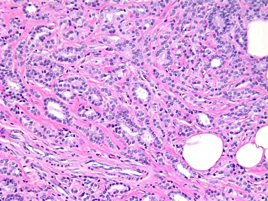
9 A Variety of Benign Sclerosing Lesions Can Mimic Invasive Breast Cancer Sclerosing Adenosis

10 A Variety of Benign Sclerosing Lesions Can Mimic Invasive Breast Cancer Radial Scar

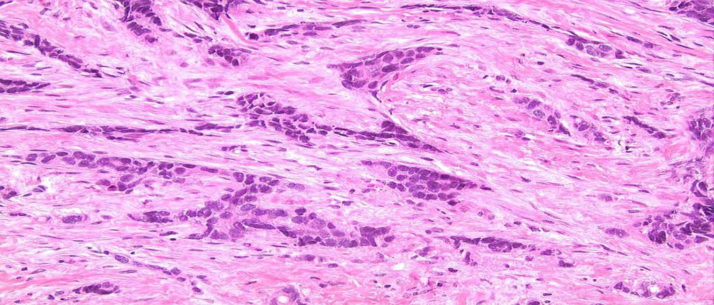
11 A Variety of Benign Sclerosing Lesions Can Mimic Invasive Breast Cancer Complex Sclerosing Lesion

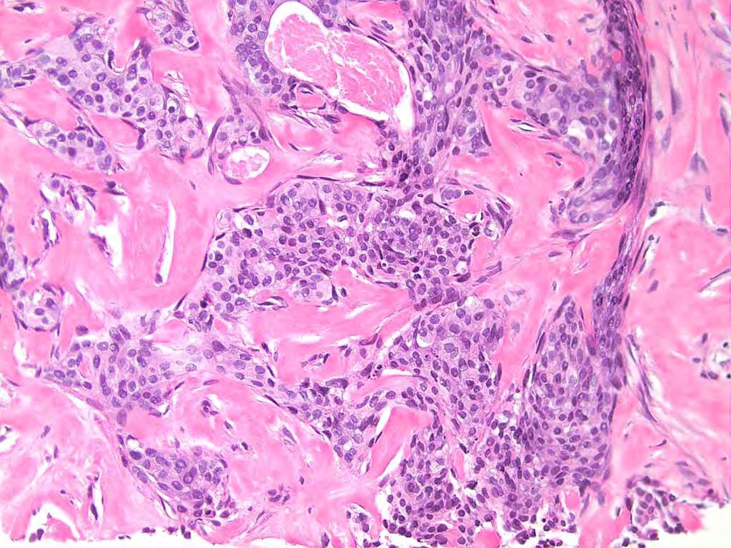
12 A Variety of Benign Sclerosing Lesions Can Mimic Invasive Breast Cancer Sclerosing Papilloma
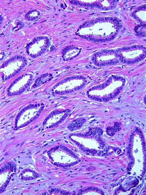
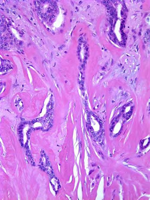
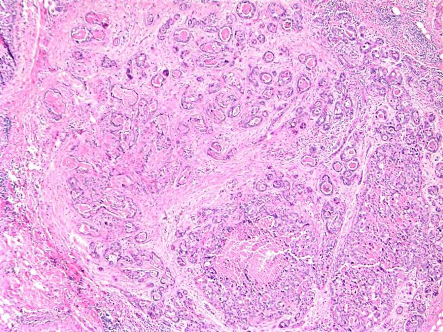
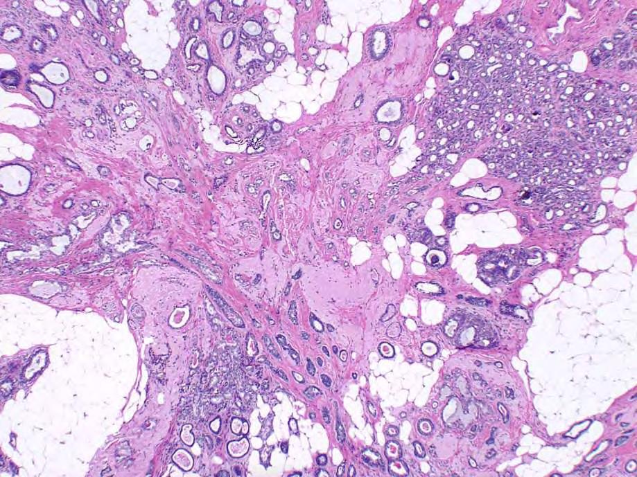
13 Common Theme Presence within fibrous stroma of epithelial/glandular elements; produce patterns mimicking invasive carcinoma (especially low grade carcinomas)

14 Even More Problematic When Sclerosing Lesion Colonized by in situ Carcinoma LCIS involving Sclerosing Adenosis

15 Even More Problematic When Sclerosing Lesion Colonized by in situ Carcinoma DCIS involving Sclerosing Adenosis
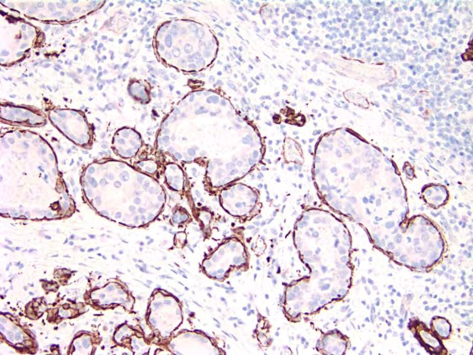
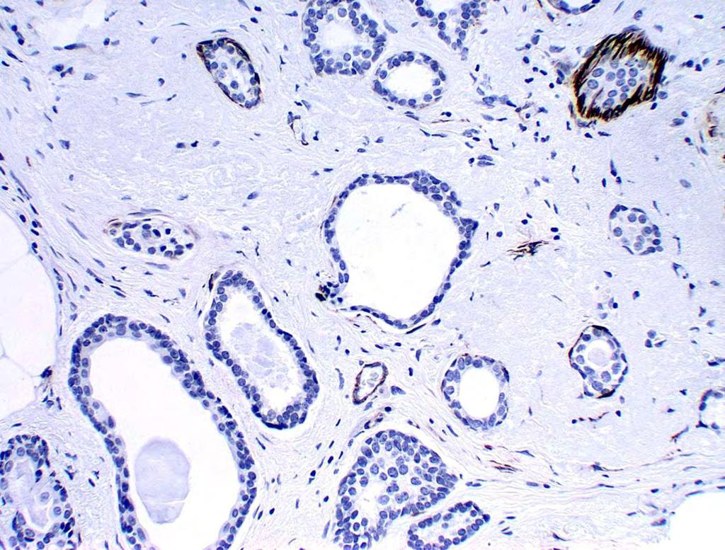
16 Distinguishing Sclerosing Lesions from Invasive Carcinoma Histologic features Immunostains for myoepithelial cells (not basement membrane)
17 Histologic Features Favor sclerosing lesion: Lobulocentricity (sclerosing adenosis) Zonation (radial scar,csl) Dense, hyalinzed, fibroelastotic stroma (radial scar, CSL) Myoepithelial cells around glands/cell nests Associated UDH
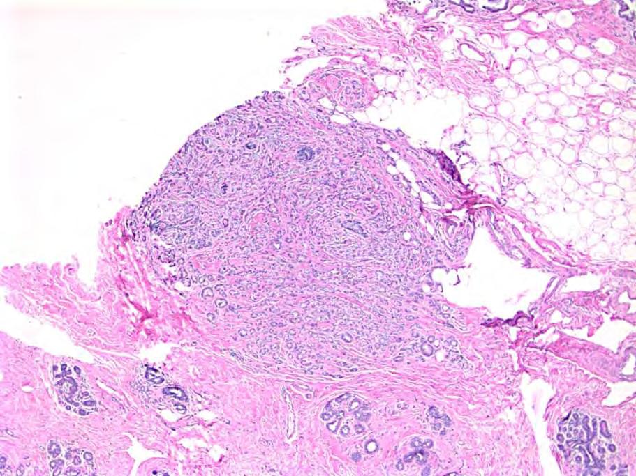
18 Histologic Features Favor sclerosing lesion: Lobulocentricity (sclerosing adenosis) Zonation (radial scar,csl) Dense, hyalinzed, fibroelastotic stroma (radial scar, CSL) Myoepithelial cells around glands/cell nests Associated UDH Favor invasive carcinoma: Haphazard, nonlobulocentric pattern; glands in fat Desmoplastic/celluar stroma No myoepithelial cells around glands/cell nests Associated CIS

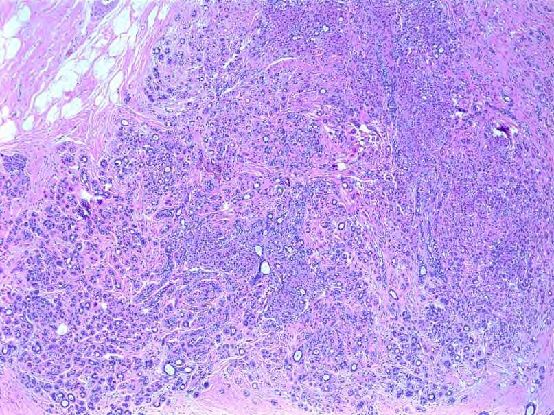
19 Low Power is KEY!! Sclerosing Adenosis


20 Low Power is KEY!! Radial Scar

21 Low Power is KEY!! Tubular Carcinoma

22 Tubular Carcinoma
23 Stroma
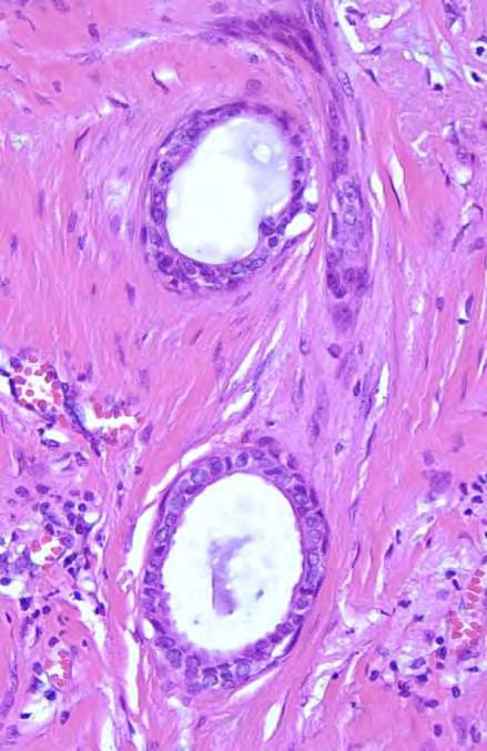
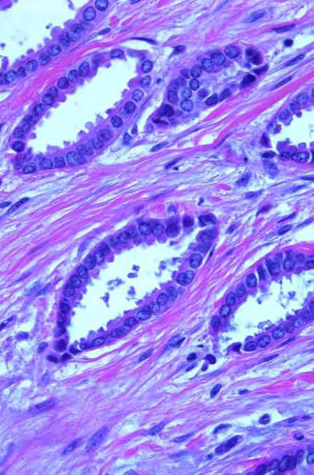
24 Myoepithelial Cells


25 Myoepithelial Cells May Be Difficult to Identify on H&E-Stained Sections No Matter How Good Your Eyes Are Sclerosing Adenosis Complex Sclerosing Lesion
26 Distinguishing Sclerosing Lesions from Invasive Carcinoma Histologic features Immunostains for myoepithelial cells
27 Invasive Cancer No Myoepithelial Cells Notable Exception: Microglandular Adenosis
28 Myoepithelial Cell Markers in the 1980 s Surgical Pathology, 1989
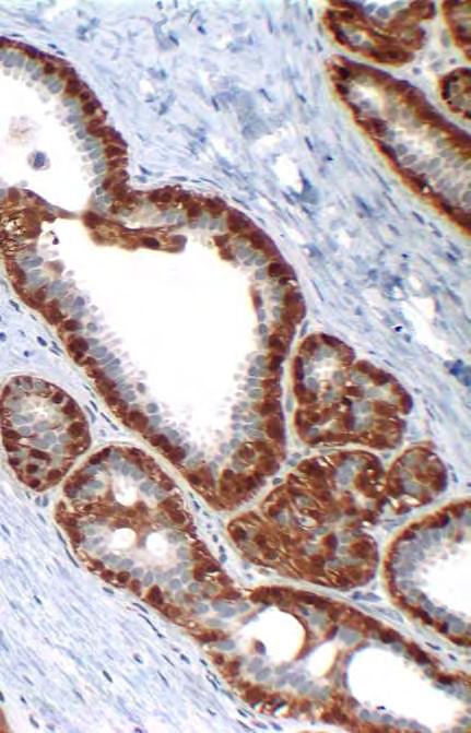
29 Myoepithelial Cell Markers in 2013 Actins S-100 Smooth muscle myosin heavy chain Calponin p63 Maspin CD10 p75 D2-40 WT-1 HMW-CK P-cadherin sigma
30

31 DCIS involving sclerosing adenosis
32

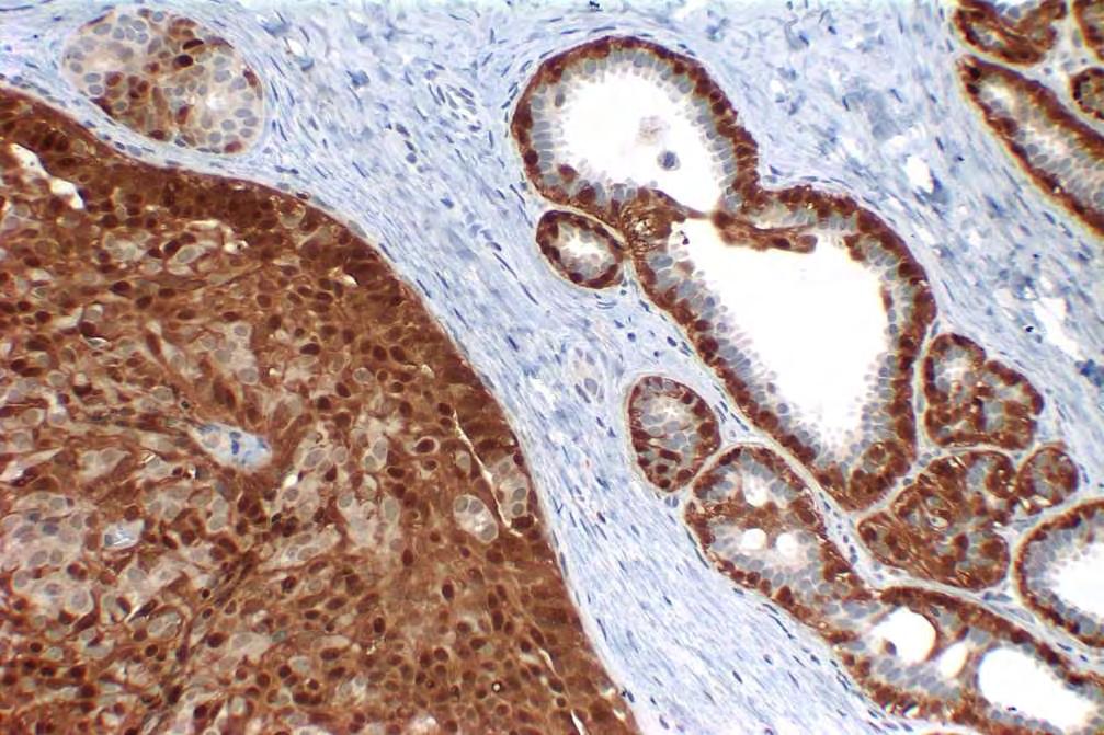
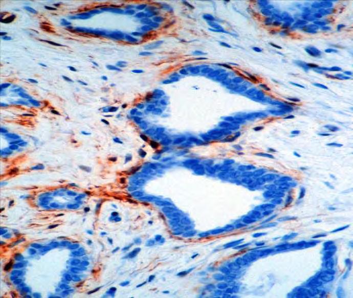
33 SMMHC Invasive ductal carcinoma, grade 1
34
35

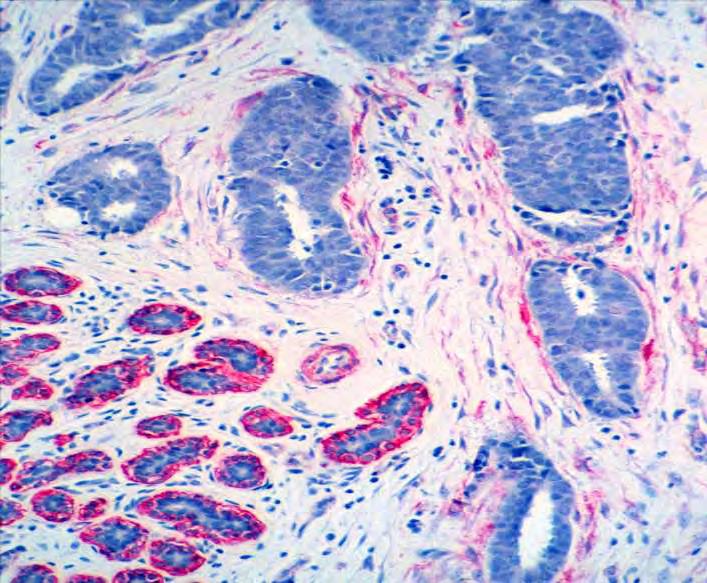
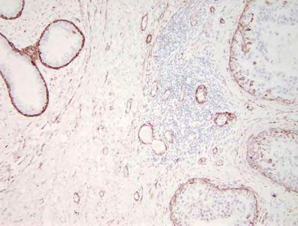
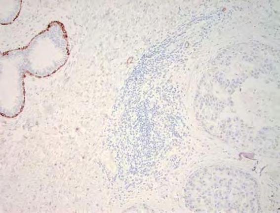
36 p63 Complex sclerosing lesion with invasive ductal carcinoma and DCIS
37 Antibody Cocktails Combinations of MEC markers (e.g., SMMHC+p63) Combination of MEC marker(s) and cytokeratin antibody Most useful for identifying microinvasion Some commercially available
38 Potential Pitfalls in the Use of Myoepithelial Cell Markers Markers vary in specificity and sensitivity Myoepithelial cells may not be uniformly distributed throughout a benign or in situ lesion Myoepithelial cells associated with some lesions may have an immunophenotype that differs from that of myoepithelial cells surrounding normal structures
39 Potential Pitfalls in the Use of Myoepithelial Cell Markers Markers vary in specificity and sensitivity Myoepithelial cells may not be uniformly distributed throughout a benign or in situ lesion Myoepithelial cells associated with some lesions may have an immunophenotype that differs from that of myoepithelial cells surrounding normal structures
40 First and Second Generation Myoepithelial Cell Markers Marker Sensitivity Specificity S-100 Good Unacceptable Actin Good Poor SMMHC Good Excellent Calponin Excellent Very good HMW-CK Very Good Poor adapted from Yaziji, et al, Adv Anat Pathol 2000:7:
41 S-100
42 S-100
43 Myofibroblast Staining
44 Myofibroblast Staining Actin > Calponin > SMMHC

45 Antibody to human podoplanin Used to identify lymphatic spaces Stains MEC in a pattern similar to calponin but less intense Less myofibroblast staining Human Pathology, 2008

46 In Situ Carcinoma Mimicking LVI D2-40 Stain Rabban and Chen, 2008


47 LVI Mimicking DCIS D2-40 Stain Rabban and Chen, 2008
48 Potential Pitfalls in the Use of Myoepithelial Cell Markers Markers vary in specificity and sensitivity Myoepithelial cells may not be uniformly distributed throughout a benign or in situ lesion Myoepithelial cells associated with some lesions may have an immunophenotype that differs from that of myoepithelial cells surrounding normal structures
49 SMMHC
50 Potential Pitfalls in the Use of Myoepithelial Cell Markers Markers vary in specificity and sensitivity Myoepithelial cells may not be uniformly distributed throughout a benign or in situ lesion Myoepithelial cells associated with some lesions may have an immunophenotype that differs from that of myoepithelial cells surrounding normal structures

51 AJSP 2008 DCIS-associated myoepithelial cells show phenotypic differences from normal myoepithelial cells Reduced expression of SMMHC in 3/4 of cases Reduced expression of CD10 and CK 5/6 in 1/3 of cases
52 SMA SMMHC
53 AJSP, 2010 Myoepithelial cells associated with benign sclerosing lesions show phenotypic differences from normal myoepithelial cells Reduced expression of CK5/6 in 1/3 of cases Reduced expression of SMMHC in 16% of cases

54 CD10 CK5/6 SMMHC
55 Summary DCIS-associated MEC and MEC associated with benign sclerosing lesions exhibit immunophenotypic differences from normal MEC?Biological significance Practical implications: sensitivity of MEC markers for DCIS-associated MEC and MEC associated with sclerosing lesions varies and differs from their sensitivity for normal MEC Don t base diagnosis of invasion on absence of MEC using only one marker
56 Topics to be Discussed (Emphasizing Recent Information) Benign sclerosing lesions vs invasive carcinoma Mucocele-like lesions vs mucinous carcinoma Benign inclusions in lymph nodes vs metastatic carcinoma
 Gross:")

57 Mucinous Carcinoma Pure form accounts for ~2% of breast cancers On average, pts older than those with NST carcinomas (but wide age range) Gross: Circumscribed, bosselated; gelatinous cut surface Micro: Neoplastic cell nests/glands in mucin pools Cells usually have low or intermediate grade nuclei; rarely high grade
 Capella C, Eusebi V, Mann B, Azzopardi")

58 Paucicellular (Type A) Hypercellular (Type B) Capella C, Eusebi V, Mann B, Azzopardi JG. Endocrine differentiation in mucoid carcinoma of the breast. Histopathol 1980;4:613

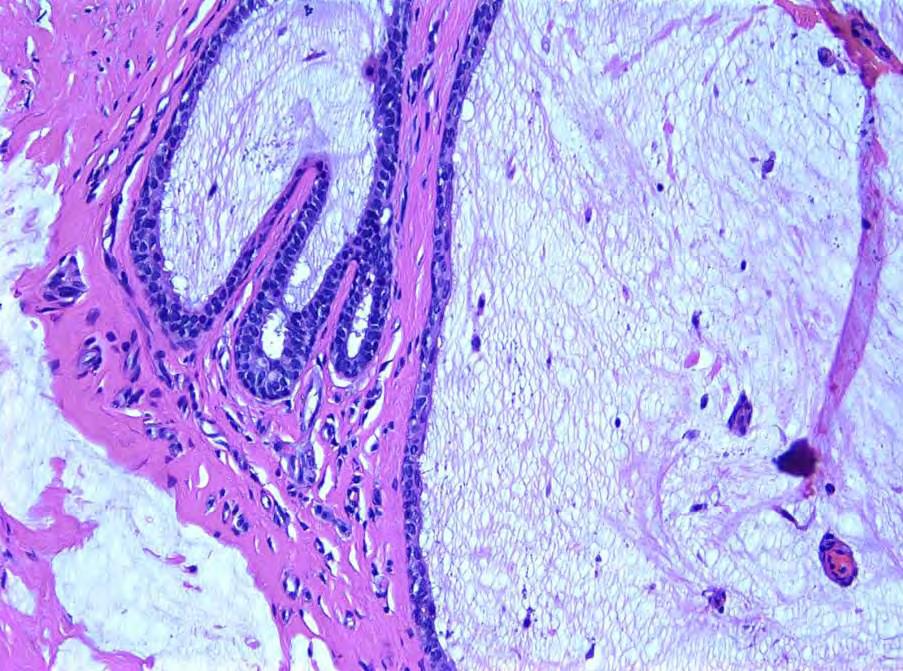
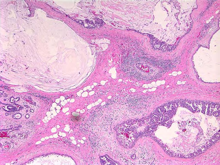
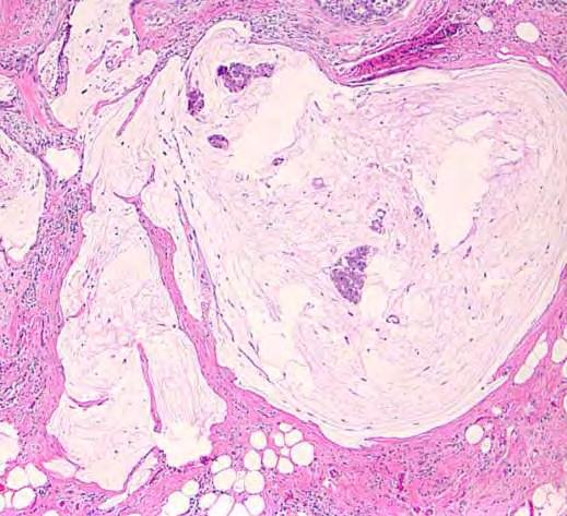
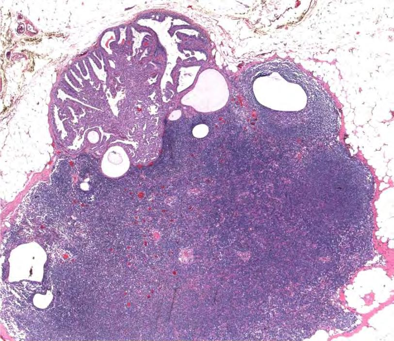
59 Mucocele-Like Lesion Palpable mass, mammographically detected, or incidental Gross: Gelatinous cut surface Micro: Dilated mucin-filled ducts/cysts Stromal mucin extravasation Ductal epithelium can range from attenuated to UDH to ADH to DCIS
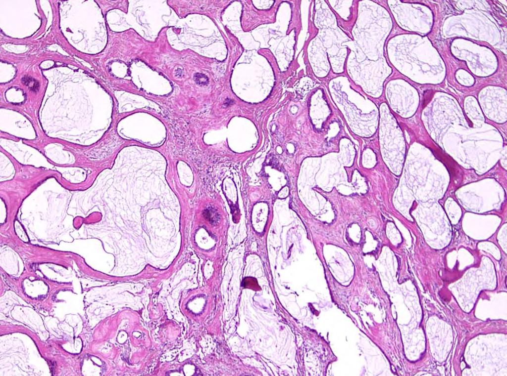
60
61
62
63

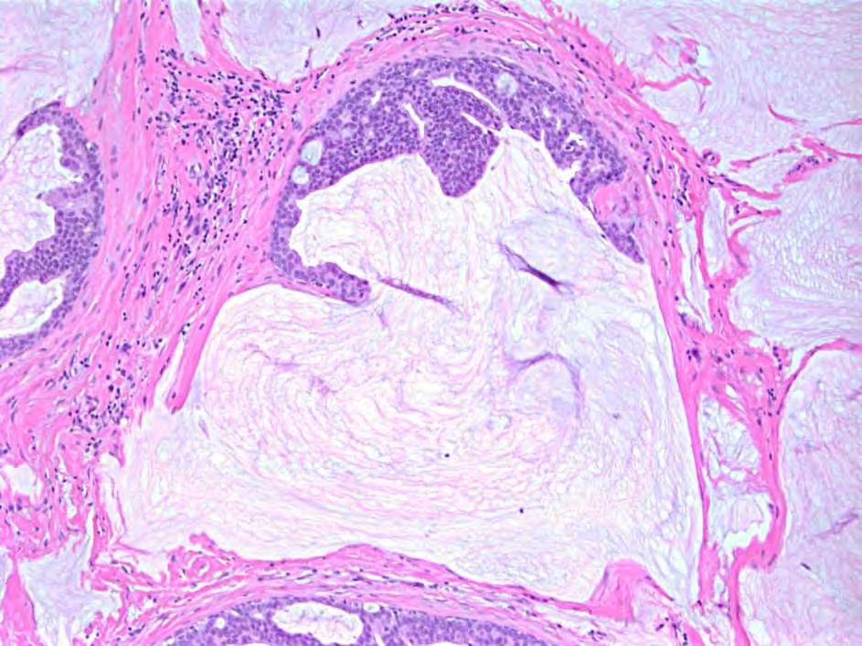
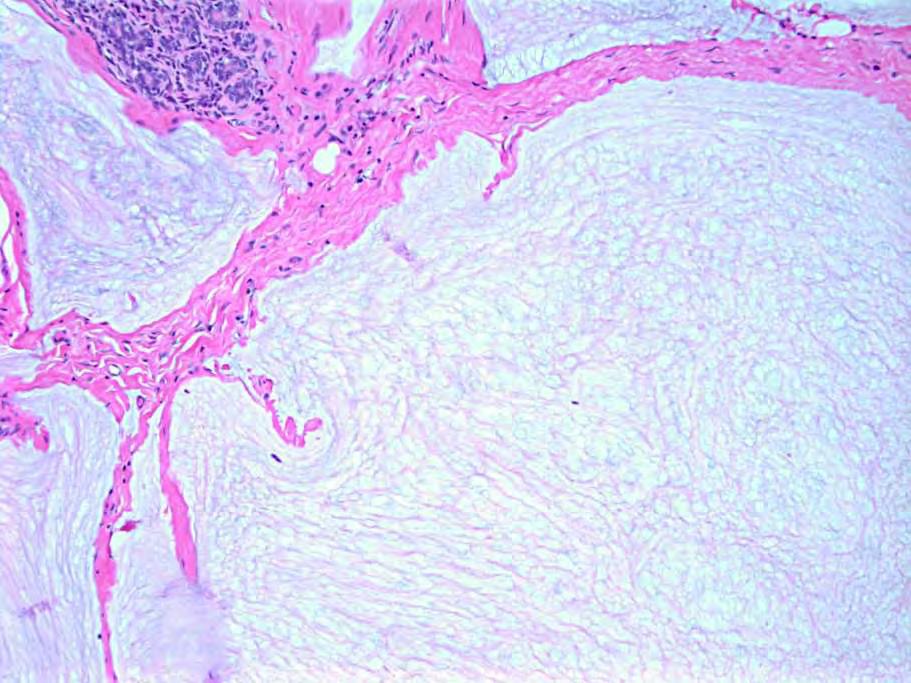
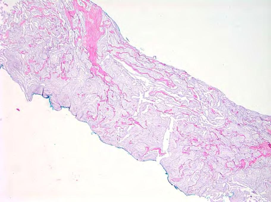
64 CAUTION! Detached strips or fragments of epithelium derived from duct or cyst lining may be present within mucin of mucocele-like lesions Should not be viewed as evidence of mucinous carcinoma
65
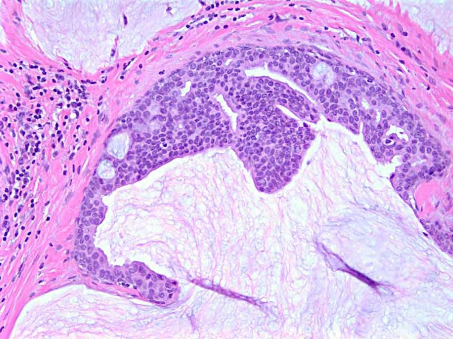
66 But, this may not be so straightforward when the mucocele-like lesion is associated with DCIS
67
68

69 Mucin Pools + Neoplastic Cell Nests = Invasive mucinous carcinoma Or, does it??????
70
71
72 Invasive mucinous ca or Mucocele-like lesion with detached fragments of DCIS?

73 Sometimes you just can t be sure!
74 Potentially Helpful in Distinguishing Mucocele-like Lesion with Displaced Epithelial Cells from Mucinous Carcinoma Myoepithelial cell immunostains Vascularization of mucin

75 Myoepithelial Stains Only Helpful if Positive

76 Myoepithelial Stains Only Helpful if Positive

77 Myoepithelial Stains Only Helpful if Positive p63
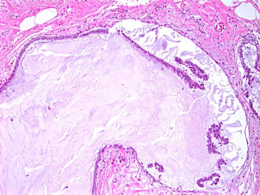
78 Vascularization of Mucin
79 Mucocele-Like Lesion

80 Mucinous Carcinoma

81 Mucinous DCIS

82 Histopathology 2008 Vascularization of mucin seen in invasive mucinous carcinomas and mucinous DCIS but not in extravasated mucin of mucocele-like lesions (but only 4 cases of MLL) Conclusions: Vascularization of mucin cannot be used by itself to distinguish between invasive mucinous carcinoma and mucinous DCIS Vascularization of mucin can be helpful in distinguishing mucocele-like lesion from invasive mucinous carcinoma, especially in small samples
83 Mucocele-like Lesion or Stromal Mucin Pools on Core Needle Biopsy Excision Required in All Cases?
84 Can Mucocele-Like Lesions Be Reliably Diagnosed on CNB? Reported upgrade rates to DCIS or invasive cancer range from 0 to 30% Small numbers Not all patients underwent excision Includes cases of mucocele-like lesions and without atypia/adh
85 What About Mucocele-Like Lesions Without Atypia on CNB? # MLL without atypia # (%) Excised Upgrade to DCIS or Inv CA % Upgrade Renshaw, (60%) 0 0 Carder, (86%) 0 0 Ramsaroop, (100%) 2 22% Wang, (100%) 0 0 Sutton, (100%) 0 0 Carkaci, (32%) 0 0 Begum, (43%) 0 0 Jaffer, (74%) 1 2% Modified from Sutton, et al, 2012
86 Mucinous Lesions on Core Needle Biopsy (that are not obviously mucinous ca or DCIS with mucin production) Excision required if there is pathologic-radiologic discordance epithelial atypia/adh Excision may not be required if the findings are unequivocally those of a mucocele-like lesion without atypia/adh Multiple levels to R/O mucinous carcinoma
87
88
89
90 Topics to be Discussed (Emphasizing Recent Information) Benign sclerosing lesions vs invasive carcinoma Mucocele-like lesions vs mucinous carcinoma Benign inclusions in lymph nodes vs metastatic carcinoma
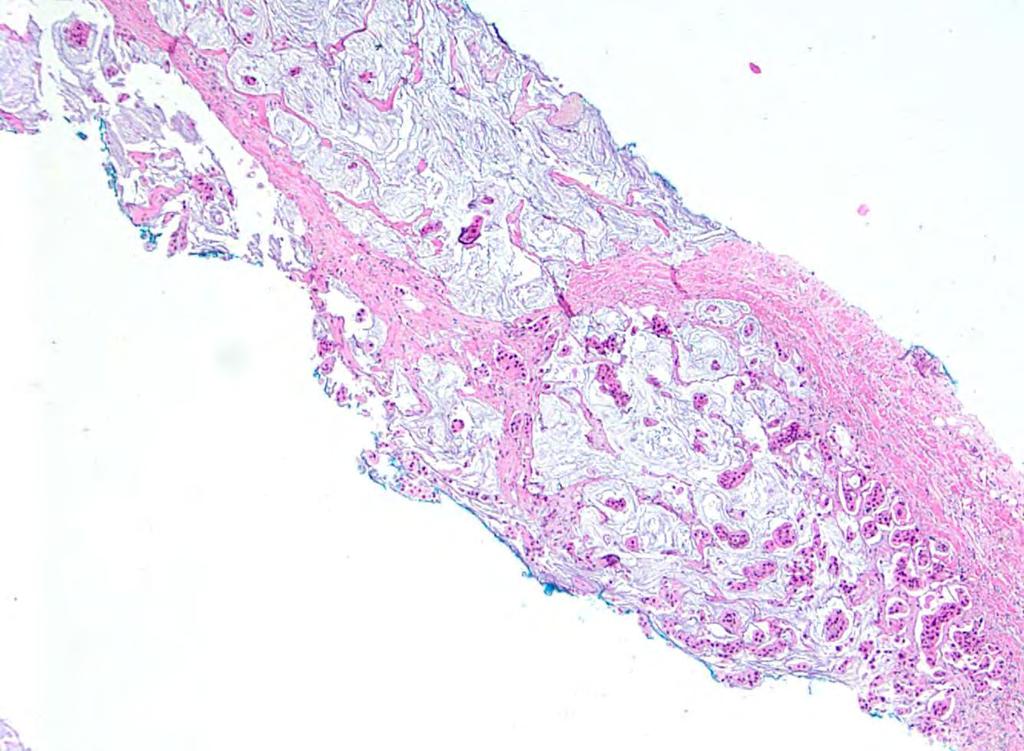
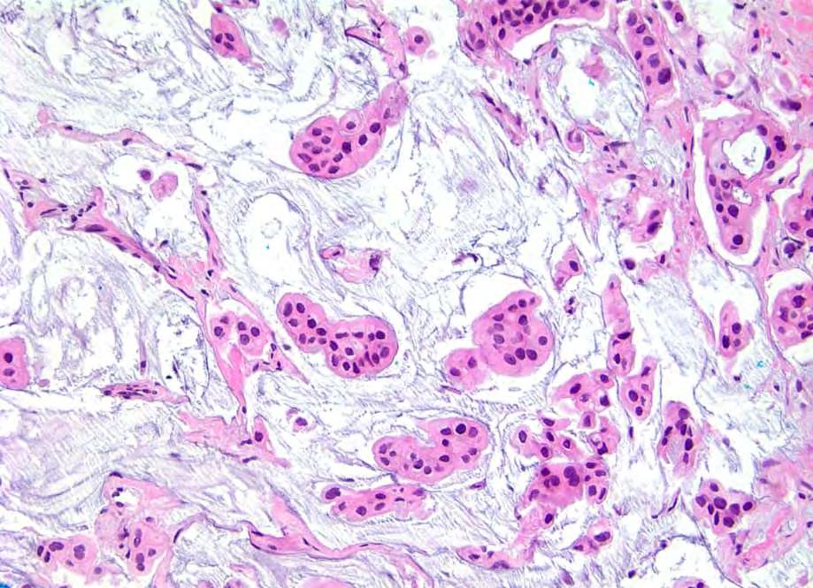
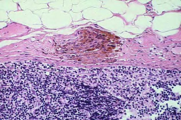
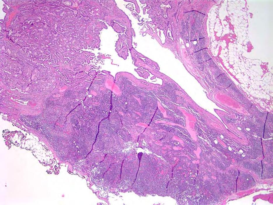
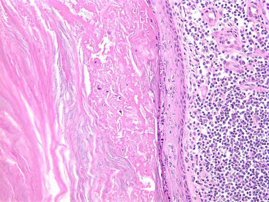
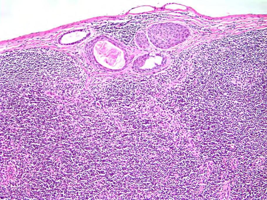
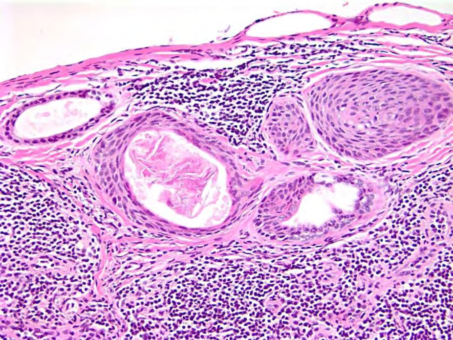
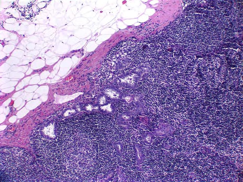
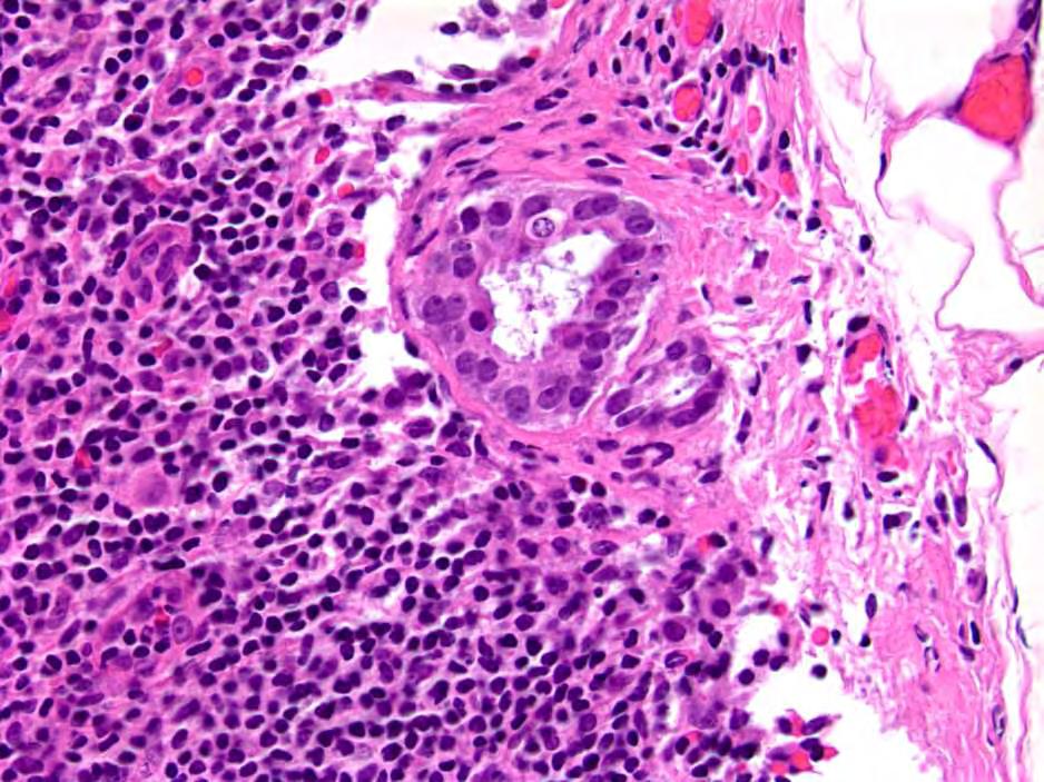
91 Benign Inclusions in Axillary Lymph Nodes Nevus cell aggregates Epithelial inclusions (Displaced epithelium s/p core needle biopsy) Must be distinguished from metastatic carcinoma
92 Nevus Cell Aggregates in Axillary Nodes of Patients with Breast Cancer UNCOMMON! Ridolfi, et al (1977) 17,504 lymph nodes from over 900 mastectomy specimens Nevus cell aggregates in 3 lymph nodes (0.017%) Nodal capsule, fibrous trabeculae But, may be seen in parenchyma Cells most often polygonal/epithelioid, nonpigmented Nodal blue nevi (spindle cells, prominent pigmentation)
93
94

95 S-100 positive (Keratin negative)
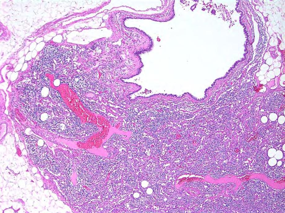
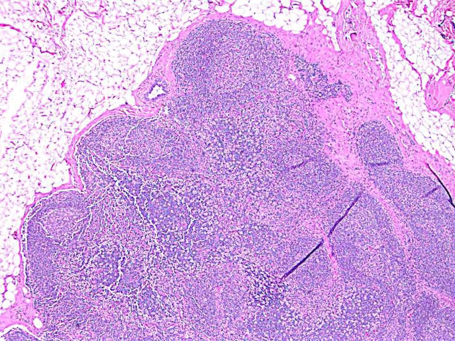
96 Benign Epithelial Inclusions in Axillary Nodes of Patients with Breast Cancer ALSO UNCOMMON! Maiorano, 2003: 7 cases identified among >3500 sentinel node biopsies Nodal capsule, fibrous trabeculae But, may be seen in parenchyma Glandular, squamous, mixed Some glandular inclusions have the appearance of endosalpingiosis


97 AJSP 2003 AJSP 2011 AJSP 2010
98 Benign Glandular Inclusions in Axillary Lymph Nodes May show any changes that occur in mammary epithelium in the breast including: Cysts Apocrine metaplasia Proliferative lesions including UDH, papilloma, sclerosing adenosis, ADH, DCIS
99
100 p63
101
102
103 p63

104
105
106
107
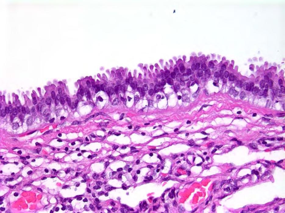
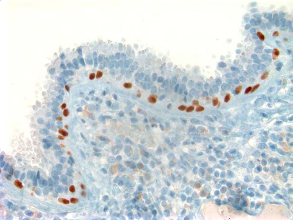
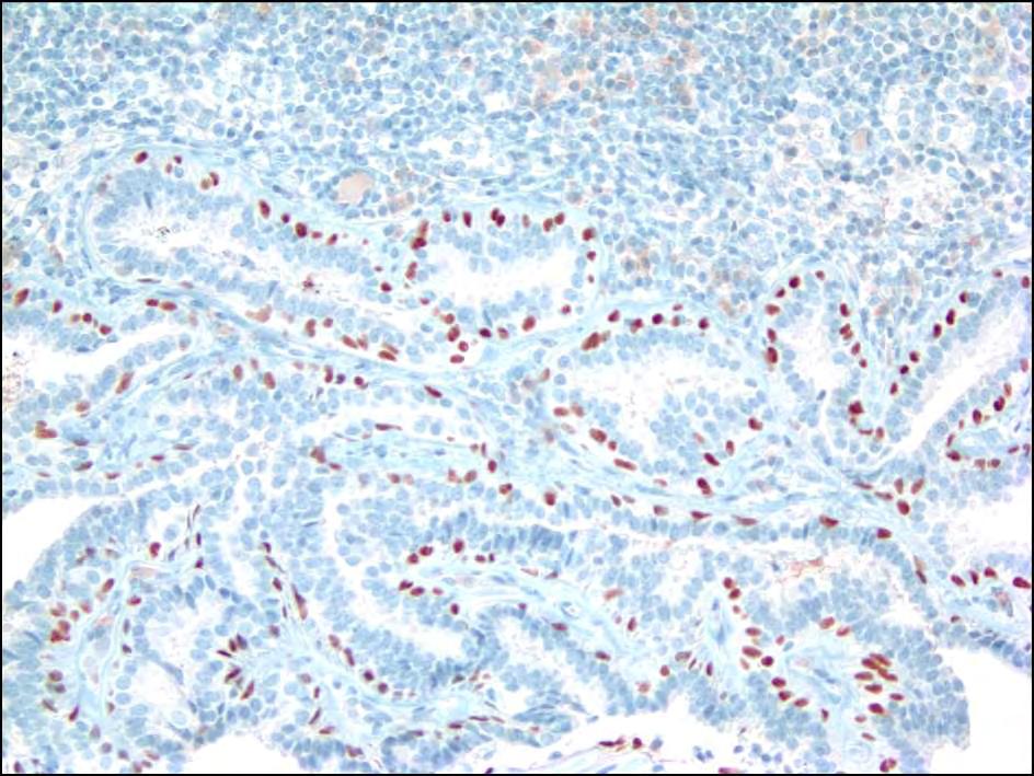
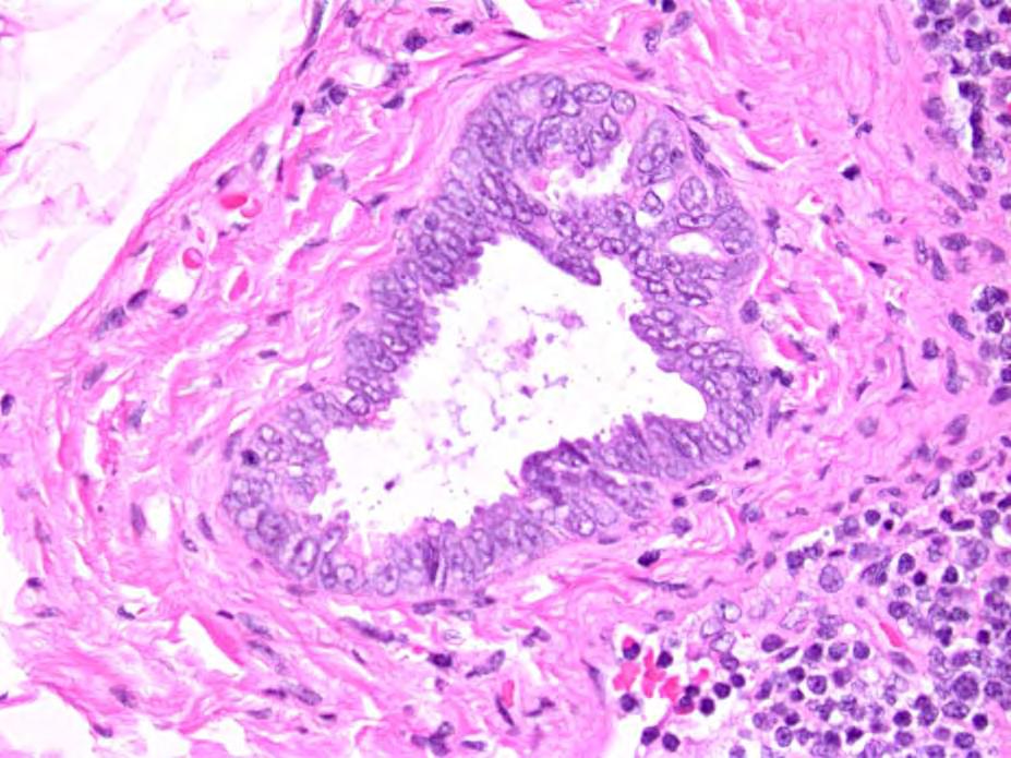
108 Endosalpingiosis in Axillary Lymph Nodes Lymph node capsule or parenchyma Ciliated cells, peg cells No cytologic atypia Immunophenotype No myoepithelial cells Nuclear staining for for WT-1 and PAX8

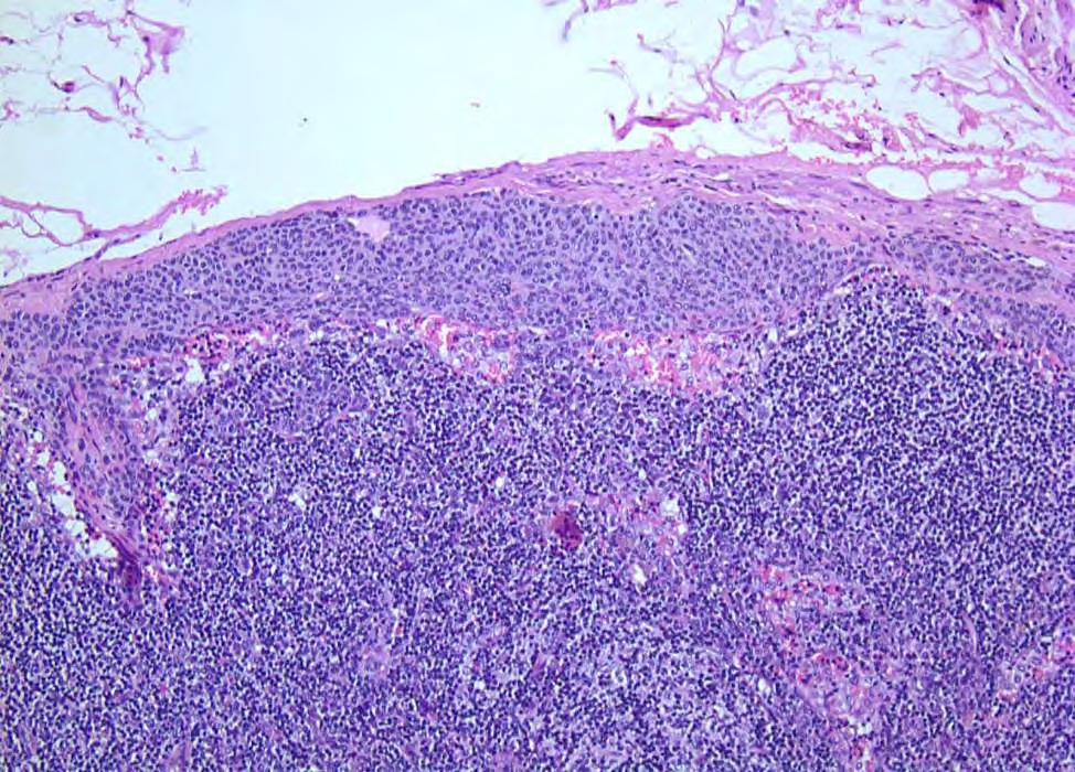
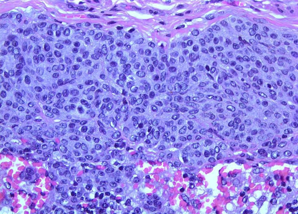
109 Metastatic Carcinoma

110 Benign Glandular Inclusion?

111 Breast Tumor


112 Lymph Node Breast Tumor
113 Benign Glandular Inclusions vs Metastatic Carcinoma Features favoring benign inclusions Location in capsule and fibrous trabeculae Myoepithelial cells Cilia, peg cells Lack of cytologic atypia Compare with breast primary (if available)
114 Many Benign Lesions of the Breast Can Mimic Malignant Lesions

115
Ductal Carcinoma in Situ. Laura C. Collins, M.D. Department of Pathology Beth Israel Deaconess Medical Center and Harvard Medical School Boston, MA
 Ductal Carcinoma in Situ Laura C. Collins, M.D. Department of Pathology Beth Israel Deaconess Medical Center and Harvard Medical School Boston, MA Definition of DCIS WHO 2012 A neoplastic proliferation
Papillary Lesions of the Breast
 Papillary Lesions of the Breast Laura C. Collins, M.D. Associate Professor of Pathology Associate Director, Division of Anatomic Pathology Beth Israel Deaconess Medical Center and Harvard Medical School
Papillary Lesions of the Breast
 Papillary Lesions of the Breast Texas Society of Pathologists 2013 Laura C. Collins, M.D. Associate Professor of Pathology Associate Director, Division of Anatomic Pathology Beth Israel Deaconess Medical
Papillary Lesions of the Breast: WHO Update
 Papillary Lesions of the Breast: WHO Update Stuart J. Schnitt, M.D. Department of Pathology Beth Israel Deaconess Medical Center and Harvard Medical School Boston, MA, USA Papillary Lesions of the Breast
04/10/2018. Intraductal Papillary Neoplasms Of Breast INTRADUCTAL PAPILLOMA
 Intraductal Papillary Neoplasms Of Breast Savitri Krishnamurthy MD Professor of Pathology Deputy Division Head The University of Texas MD Anderson Cancer Center 25 th Annual Seminar in Pathology Pittsburgh,
CURRICULUM FOR THE BREAST PATHOLOGY ROTATION UNIVERSITY OF FLORIDA DEPARTMENT OF PATHOLOGY
 CURRICULUM FOR THE BREAST PATHOLOGY ROTATION UNIVERSITY OF FLORIDA DEPARTMENT OF PATHOLOGY JULY, 2003 The following is a conceptual curriculum and set of guidelines for Pathology Residents on the Breast
1 NORMAL HISTOLOGY AND METAPLASIAS
 1 NORMAL HISTOLOGY AND METAPLASIAS, MD Anatomy and Histology 1 Metaplasias 2 ANATOMY AND HISTOLOGY The female breast is composed of a branching duct system, which begins at the nipple with the major lactiferous
Papillary Lesions of the Breast A Practical Approach to Diagnosis. (Arch Pathol Lab Med. 2016;140: ; doi: /arpa.
 Papillary Lesions of the Breast A Practical Approach to Diagnosis (Arch Pathol Lab Med. 2016;140:1052 1059; doi: 10.5858/arpa.2016-0219-RA) Papillary lesions of the breast Span the spectrum of benign,
Proliferative Breast Disease: implications of core biopsy diagnosis. Proliferative Breast Disease
 Proliferative Breast Disease: implications of core biopsy diagnosis Jean F. Simpson, M.D. Breast Pathology Consultants, Inc. Nashville, TN Proliferative Breast Disease Must be interpreted in clinical and
Columnar Cell Lesions
 Columnar Cell Lesions Laura C. Collins, M.D. Department of Pathology Beth Israel Deaconess Medical Center and Harvard Medical School Boston, MA Question? Columnar cell lesions are: a) Annoying lesions
04/10/2018 HIGH RISK BREAST LESIONS. Pathology Perspectives of High Risk Breast Lesions ELEVATED RISK OF BREAST CANCER HISTORICAL PERSPECTIVES
 Pathology Perspectives of High Risk Breast Lesions Savitri Krishnamurthy MD Professor of Pathology Deputy Division Head Director of Clinical Trials, Research and Development The University of Texas MD
Breast pathology. 2nd Department of Pathology Semmelweis University
 Breast pathology 2nd Department of Pathology Semmelweis University Breast pathology - Summary - Benign lesions - Acute mastitis - Plasma cell mastitis / duct ectasia - Fat necrosis - Fibrocystic change/
Papillary Lesions of the breast
 Papillary Lesions of the breast Emad Rakha Professor of Breast Pathology The University of Nottingham Papillary lesions of the breast are a heterogeneous group of disease, which are characterised by neoplastic
Treatment options for the precancerous Atypical Breast lesions. Prof. YOUNG-JIN SUH The Catholic University of Korea
 Treatment options for the precancerous Atypical Breast lesions Prof. YOUNG-JIN SUH The Catholic University of Korea Not so benign lesions? Imaging abnormalities(10% recall) lead to diagnostic evaluation,
The Hot Topic for today is a biopsy from a 58-year-old woman who had worrisome mammographic calcifications on screening.
 The Hot Topic for today is a biopsy from a 58-year-old woman who had worrisome mammographic calcifications on screening. 1 My name is Dan Visscher; I am a consultant in the Division of Anatomic Pathology
Breast Pathology. Breast Development
 Breast Pathology Lecturer: Hanina Hibshoosh, M.D. Reading: Kumar, Cotran, Robbins, Basic Pathology, 6th Edition, pages 623-635 Breast Development 5th week - thickening of the epidermis - milk line 5th
INDEX. in this web service Cambridge University Press
 abscess. See also subareolar abscess acute mastitis, 44 lactational/puerperal mastitis, 55 mammary tuberculosis, 42 tuberculous, 43 adeno gastric, 198, 200 invasive, 157 lung, 197, 200 prostatic, 199 200
Breast: Difficulties in Core Biopsies
 Breast: Difficulties in Core Biopsies Anna Marie Mulligan, MB, MSc, FRCPath University Health Network and University of Toronto E-mail: annamarie.mulligan@uhn.ca No conflicts of interest Role of Core Needle
Diseases of the breast (1 of 2)
") Diseases of the breast (1 of 2) Introduction A histology introduction Normal ducts and lobules of the breast are lined by two layers of cells a layer of luminal cells overlying a second layer of myoepithelial
Diseases of the breast (1 of 2) Introduction A histology introduction Normal ducts and lobules of the breast are lined by two layers of cells a layer of luminal cells overlying a second layer of myoepithelial
Notice of Faculty Disclosure
 California Society of Pathology Diagnostic Problems in Surgical Pathology December 2015 Case 2 Laura C. Collins, M.D. Associate Professor of Pathology Associate Director of Anatomic Pathology Beth Israel
6/3/2010. Outline of Talk. Lobular Breast Cancer: Definition of lobular differentiation. Common Problems in Diagnosing LCIS in Core Biopsies
 Outline of Talk Lobular Breast Cancer: Common Problems in Diagnosing LCIS in Core Biopsies Definition of lobular differentiation Variants of LCIS that: carry risk for unsampled invasive cancer mimic DCIS
3/27/2017. Disclosure of Relevant Financial Relationships. Papilloma???
 Management of Papillary Lesions Diagnosed at Rad Path Concordant Core Biopsy (CNB) Disclosure of Relevant Financial Relationships USCAP requires that all planners (Education Committee) in a position to
Proliferative Epithelial lesions of the Breast. Sami Shousha, MD, FRCPath Charing Cross Hospital & Imperial College, London
 Proliferative Epithelial lesions of the Breast Sami Shousha, MD, FRCPath Charing Cross Hospital & Imperial College, London Amman, November2013 Proliferative Epithelial Lesions of the Breast Usual type
Enterprise Interest None
 Enterprise Interest None B3 lesions of the breast What are they at surgery? Case 4 Edi Brogi MD PhD Attending Pathologist - Director of Breast Pathology Memorial Sloan Kettering Cancer Center New York
Abid Irshad, MD Director Breast Imaging. Medical University of South Carolina Charleston
 Abid Irshad, MD Director Breast Imaging Medical University of South Carolina Charleston Cases Financial disclosure: I or my family have no financial interest related to the material discussed in this presentation
Lesion Imaging Characteristics Mass, Favoring Benign Circumscribed Margins Intramammary Lymph Node
 Lesion Imaging Characteristics Mass, Favoring Benign Circumscribed Margins Intramammary Lymph Node Oil Cyst Mass, Intermediate Concern Microlobulated Margins Obscured Margins Mass, Favoring Malignant Indistinct
Farid Moinfar Essentials of Diagnostic Breast Pathology
 Farid Moinfar Essentials of Diagnostic Breast Pathology Farid Moinfar Essentials of Diagnostic Breast Pathology A Practical Approach With 116 Figures in 1128 Seperate Illustrations and 6 Tables 123 Farid
LYMPHATIC DRAINAGE AXILLARY (MOSTLY) INTERNAL MAMMARY SUPRACLAVICULAR
 INTERNAL MAMMARY SUPRACLAVICULAR") BREAST LYMPHATIC DRAINAGE AXILLARY (MOSTLY) INTERNAL MAMMARY SUPRACLAVICULAR HISTOLOGY LOBE: (10 in whole breast) LOBULE: (many per lobe) ACINUS/I, aka ALVEOLUS/I: (many per lobule) DUCT(S): INTRA- or
BREAST LYMPHATIC DRAINAGE AXILLARY (MOSTLY) INTERNAL MAMMARY SUPRACLAVICULAR HISTOLOGY LOBE: (10 in whole breast) LOBULE: (many per lobe) ACINUS/I, aka ALVEOLUS/I: (many per lobule) DUCT(S): INTRA- or
Basement membrane in lobule.
 Bahram Memar, MD Basement membrane in lobule. Normal lobule-luteal phase Normal lobule-follicular phase Lactating breast Greater than 95% are adenocarcinomas in situ carcinomas and invasive carcinomas.
Papillary lesions of the breast: selected diagnostic and management issues
 Histopathology 2008, 52, 20 29. DOI: 10.1111/j.1365-2559.2007.02898.x REVIEW Papillary lesions of the breast: selected diagnostic and management issues L C Collins & S J Schnitt Department of Pathology,
Invasive Papillary Breast Carcinoma
 410 This is an Open Access article licensed under the terms of the Creative Commons Attribution- NonCommercial-NoDerivs 3.0 License (www.karger.com/oa-license), applicable to the online version of the
Flat Epithelial Atypia
 Flat Epithelial Atypia Richard Owings, M.D. University of Arkansas for Medical Sciences Department of Pathology Flat epithelial atypia can be a difficult lesion May be a subtle diagnosis Lots of changes
Case Report Basaloid ductal carcinoma in situ arising in salivary gland metaplasia of the breast: a case report
 Int J Clin Exp Pathol 2014;7(9):6370-6374 www.ijcep.com /ISSN:1936-2625/IJCEP0001480 Case Report Basaloid ductal carcinoma in situ arising in salivary gland metaplasia of the breast: a case report Eun
Columnar Cell Lesions. Columnar Cell Lesions and Flat Epithelial Atypia
 Columnar Cell Lesions and Stuart J. Schnitt, M.D. Beth Israel Deaconess Medical Center and Harvard Medical School Boston, MA, USA Columnar Cell Lesions Lesions characterized by columnar epithelial cells
PAAF vs Core Biopsy en Lesiones Mamarias Case #1
 5/19/2014 PAAF vs Core Biopsy en Lesiones Mamarias Case #1 Fine Needle Aspiration Cytology of Breast: Correlation with Needle Core Biopsy 64-year-old woman Mass in breast Syed Hoda, MD CD31 Post-Radiation
Overview of Pathology Evaluation of Breast Lesions and Quality Assurance
 Overview of Pathology Evaluation of Breast Lesions and Quality Assurance 2 Michael O. Idowu, Jaime A. Singh, and Margaret M. Grimes Masses/Densities/Distortions: General Considerations Radiologic evaluation
Controversies and Problematic Issues in Core Needle Biopsies (To excise or not to excise)
") Controversies and Problematic Issues in Core Needle Biopsies (To excise or not to excise) Laura C. Collins, M.D. Beth Israel Deaconess Medical Center and Harvard Medical School Boston, MA Schematic Representation
Controversies and Problematic Issues in Core Needle Biopsies (To excise or not to excise) Laura C. Collins, M.D. Beth Israel Deaconess Medical Center and Harvard Medical School Boston, MA Schematic Representation
Columnar Cell Lesions and Flat Epithelial Atypia
 Columnar Cell Lesions and Flat Epithelial Atypia Laura C. Collins, M.D. Department of Pathology Beth Israel Deaconess Medical Center and Harvard Medical School, Boston, MA Terminology for Columnar Cell
Oncocytic-Appearing Salivary Gland Tumors. Oncocytic, Cystic, Mucinous, and High Grade Salivary Gland Tumors SALIVARY GLAND FNA: PART II
 William C. Faquin, MD, PhD Professor of Pathology Harvard Medical School Director of Head and Neck Pathology Massachusetts Eye and Ear Massachusetts General Hospital SALIVARY GLAND FNA: PART II Oncocytic,
Terminal duct lobular unit (TDLU). A, Diagrammatic representation of this structure. ETD = Extralobular terminal duct; ITD = intralobular terminal
. A, Diagrammatic representation of this structure. ETD = Extralobular terminal duct; ITD = intralobular terminal") Terminal duct lobular unit (TDLU). A, Diagrammatic representation of this structure. ETD = Extralobular terminal duct; ITD = intralobular terminal duct. Mammary gland Mammary gland Terminal duct lobular
Terminal duct lobular unit (TDLU). A, Diagrammatic representation of this structure. ETD = Extralobular terminal duct; ITD = intralobular terminal duct. Mammary gland Mammary gland Terminal duct lobular
Low-grade Adenosquamous Carcinoma Coexisting with Sclerosing Adenosis of the Breast: A Case Report
 31 Case Report J. St. Marianna Univ. Vol. 8, pp. 31 35, 2017 Low-grade Adenosquamous Carcinoma Coexisting with Sclerosing Adenosis of the Breast: A Case Report Ryoko Oi 1, 2, Ichiro Maeda 1, Yoshio Aida
IBCM 2, April 2009, Sarajevo, Bosnia and Herzegovina
 Preoperative diagnosis and treatment planning in breast cancer The pathologist s perspective L. Mazzucchelli Istituto Cantonale di Patologia Locarno, Switzerland IBCM 2, 23-25 April 2009, Sarajevo, Bosnia
COMMON CONSULTATION CONUNDRUMS IN BREAST PATHOLOGY
 COMMON CONSULTATION CONUNDRUMS IN BREAST PATHOLOGY SANDRA J. SHIN CHIEF OF BREAST PATHOLOGY ASOCIATE PROFESSOR OF PATHOLOGY AND LABORATORY MEDICINE NEW YORK PRESBYTERIAN HOSPITAL-WEILL CORNELL MEDICAL
57th Annual HSCP Spring Symposium 4/16/2016
 An Unusual Malignant Spindle Cell Lesion to Involve the Breast Erinn Downs-Kelly, D.O. Associate Professor of Pathology University of Utah & ARUP Laboratories No disclosures Case 39 y/o female with no
BREAST PATHOLOGY. Fibrocystic Changes
 BREAST PATHOLOGY Lesions of the breast are very common, and they present as palpable, sometimes painful, nodules or masses. Most of these lesions are benign. Breast cancer is the 2 nd most common cause
CLINICAL SIGNIFICANCE OF BENIGN EPITHELIAL CHANGES
 Papillomas. Papillomas are composed of multiple branching fibrovascular cores, each having a connective tissue axis lined by luminal and myoepithelial cells ( Fig. 23-11 ). Growth occurs within a dilated
Spindle Cell Lesions Of The Breast. Emad Rakha Professor of Breast Pathology and Consultant Pathologist
 Spindle Cell Lesions Of The Breast Emad Rakha Professor of Breast Pathology and Consultant Pathologist * SCLs comprise a wide spectrum of diseases, ranging from reactive processes to aggressive malignant
Interpretation of Breast Pathology in the Era of Minimally Invasive Procedures
 Shahla Masood, M.D. Professor and Chair Department of Pathology and Laboratory Medicine University of Florida College of Medicine Jacksonville Medical Director, UF Health Breast Center Chief of Pathology
High risk lesions of the breast : Review of the current diagnostic and management strategies
 High risk lesions of the breast : Review of the current diagnostic and management strategies Poster No.: C-1204 Congress: ECR 2016 Type: Educational Exhibit Authors: P. Jagmohan, F. J. Pool, P. G. Pillay,
Surgical Pathology Issues of Practical Importance
 Surgical Pathology Issues of Practical Importance Anne Moore, MD Medical Oncology Syed Hoda, MD Surgical Pathology The pathologist is central to the team approach needed to manage the patient with breast
Disclosures 5/27/2012. Outline of Talk. Outline of Talk. When Is LCIS Clinically Significant? Classic LCIS. Classic LCIS
 When Is LCIS Clinically Significant? Disclosures I have nothing to disclose Yunn-Yi Chen, MD, PhD Professor Outline of Talk Outline of Talk Classic LCIS Classic LCIS Definition of lobular differentiation
OUTLINE FIBROADENOMA FIBROADENOMA. FIBROEPITHELIAL LESIONS OF THE BREAST UCSF Current Issues in Anatomic Pathology 2015 FIBROADENOMA PHYLLODES TUMOR
 OUTLINE FIBROADENOMA FIBROEPITHELIAL LESIONS OF THE BREAST UCSF Current Issues in Anatomic Pathology 2015 Gregor Krings, MD PhD Assistant Professor PHYLLODES TUMOR DIFFERENTIAL DIAGNOSIS CELLULAR FIBROEPITHELIAL
A712(19)- Test slide, Breast cancer tissues with corresponding normal tissues
- Test slide, Breast cancer tissues with corresponding normal tissues") A712(19)- Test slide, Breast cancer tissues with corresponding normal tissues (formalin fixed) For research use only Specifications: No. of cases: 12 Tissue type: Breast cancer tissues with corresponding
A712(19)- Test slide, Breast cancer tissues with corresponding normal tissues (formalin fixed) For research use only Specifications: No. of cases: 12 Tissue type: Breast cancer tissues with corresponding
This case presentation reviews a challenging case of. Metaplastic Carcinomas of the Breast: Diagnostic Challenges and New Translational Insights
 Metaplastic Carcinomas of the Breast: Diagnostic Challenges and New Translational Insights Comprising less than 1% of invasive carcinomas of the breast, metaplastic carcinomas are a heterogeneous group
HISTOMORPHOLOGICAL SPECTRUM OF BREAST LESIONS
 HISTOMORPHOLOGICAL SPECTRUM OF BREAST LESIONS Kiran H. S, Jayaprakash Shetty, Chandrika Rao Assistant Professor, Department of Pathology, Yenepoya Medical College, Mangalore. Professor, Department of Pathology,
Benign Breast Disease and Breast Cancer Risk
 Benign Breast Disease and Breast Cancer Risk Jean F. Simpson, M.D. Vanderbilt University Nashville, Tennessee December 1, 2011 Nashville Nashville Lebanon 1 Cedars of Lebanon State Park The American University
ACRIN 6666 Therapeutic Surgery Form
 S1 ACRIN 6666 Therapeutic Surgery Form 6666 Instructions: Complete a separate S1 form for each separate area of each breast excised with the intent to treat a cancer (e.g. each lumpectomy or mastectomy).
Mody. AIS vs. Invasive Adenocarcinoma of the Cervix
 Common Problems in Gynecologic Pathology Michael T. Deavers, M.D. Houston Methodist Hospital, Houston, Texas Common Problems in Gynecologic Pathology Adenocarcinoma in-situ (AIS) of the Cervix vs. Invasive
Diagnostic Problems in Breast Pathology How to avoid the pitfalls
 FORUM OF PATHOLOGY Diagnostic Problems in Breast Pathology How to avoid the pitfalls Professor C W Elston City Hospital Nottingham, United Kingdom Introduction Almost any breast lesion may produce diagnostic
COMMON BENIGN DISORDERS AND DISEASES OF THE BREAST
 COMMON BENIGN DISORDERS AND DISEASES OF THE BREAST Aberrations of Normal Development and Involution (ANDI). The basic principles underlying the aberrations of normal development and involution (ANDI) classification
Gross appearance of nodular hyperplasia in material obtained from suprapubic prostatectomy. Note the multinodular appearance and the admixture of
 Tiền liệt tuyến Tiền liệt tuyến Gross appearance of nodular hyperplasia in material obtained from suprapubic prostatectomy. Note the multinodular appearance and the admixture of solid and microcystic areas.
Pleomorphic adenoma of breast - a case report and distinction with metaplastic carcinoma D Gupta, S Agrawal, N Trivedi, A Tewari
 of breast - a case report and distinction with metaplastic carcinoma D Gupta, S Agrawal, N Trivedi, A Tewari Introduction, also known as mixed tumour, is a benign tumour which typically presents as a painless,
In situ lobular neoplasia of the breast with marked myoepithelial proliferation
 In situ lobular neoplasia of the breast with marked myoepithelial proliferation Sami Shousha To cite this version: Sami Shousha. In situ lobular neoplasia of the breast with marked myoepithelial proliferation.
Anatomic Pathology / Mucocele-like Lesions on Breast Core Biopsy. Mucocele-like Lesions Diagnosed on Breast Core Biopsy
 Anatomic Pathology / Mucocele-like Lesions on Breast Core Biopsy Mucocele-like Lesions Diagnosed on Breast Core Biopsy Assessment of Upgrade Rate and Need for Surgical Excision Brian Sutton, MD, 1 Simone
Slide seminar. Asist. Prof. Jože Pižem, MD, PhD Institute of Pathology Medical Faculty, University of Ljubljana
 Slide seminar Asist. Prof. Jože Pižem, MD, PhD Institute of Pathology Medical Faculty, University of Ljubljana Case 5 A 57-year-old man with a dermal/subcutaneous lesion on the scalp, which was interpreted
04/09/2018. Salivary Gland Pathology in the Molecular Era Old Friends, Old Foes, & New Acquaintances
 Salivary Gland Pathology in the Molecular Era Old Friends, Old Foes, & New Acquaintances Jennifer L. Hunt, MD, MEd Aubrey J. Hough Jr, MD, Endowed Professor of Pathology Chair of Pathology and Laboratory
Salivary Glands 3/7/2017
 Salivary Glands 3/7/2017 Goals and objectives Focus on the entities unique to H&N Common board type facts Information for your future practice Salivary Glands Salivary Glands Major gland. Paratid. Submandibular.
Image guided core biopsies:
 Recommendations on the Surgical, Radiologic and Pathologic Approaches to Breast Disease: Using best practices based on multidisciplinary methodologies developed through the Allina Breast Committee. Image
Title malignancy. Issue Date Right 209, 12, (2013)
") NAOSITE: Nagasaki University's Ac Title Author(s) A case of intracystic apocrine papi malignancy Hayashi, Hiroko; Ohtani, Hiroshi; Y Citation Pathology - Research and Practice, Issue Date 2013-12 URL Right
NAOSITE: Nagasaki University's Ac Title Author(s) A case of intracystic apocrine papi malignancy Hayashi, Hiroko; Ohtani, Hiroshi; Y Citation Pathology - Research and Practice, Issue Date 2013-12 URL Right
University Journal of Pre and Para Clinical Sciences
 ISSN 2455 2879 Volume 2 Issue 1 2016 Metaplastic carcinoma breast a rare case report Abstract : Metaplastic carcinoma of the breast is a rare malignancy with two distinct cell lines described as a breast
Inflammatory and Reactive Lesions of the Breast
 Inflammatory and Reactive Lesions of the Breast Laura C. Collins, M.D. Vice Chair of Anatomic Pathology Professor of Pathology Beth Israel Deaconess Medical Center and Harvard Medical School Boston, MA
Atypical Ductal Hyperplasia and Papillomas: A Comparison of Ultrasound Guided Breast Biopsy and Stereotactic Guided Breast Biopsy
 Atypical Ductal Hyperplasia and Papillomas: A Comparison of Ultrasound Guided Breast Biopsy and Stereotactic Guided Breast Biopsy Breast Cancer is the most common cancer diagnosed in women in the United
BREAST PATHOLOGY MCQS
 BREAST PATHOLOGY MCQS 1) :The most important factor in breast enlargement during pregnancy is A. stromal edema B. secretion of chorionic gonadotropin C. glandular hyperplasia D. proliferation of stroma
Incidence of ductal lesions
 Ductal Proliferative Lesions of the Breast: From FEA to ADH to DCIS Incidence of ductal lesions Pre-mammography: DCIS < 3% of breast cancers, large palpable masses, with invasion Mammography: DCIS 25%
Enterprise Interest None
 Enterprise Interest None What are triple negative breast cancers? A synopsis of their histological patterns Ian Ellis Molecular Medical Sciences, University of Nottingham Department of Histopathology,
Minimizing Errors in Diagnostic Pathology
 Shahla Masood, M.D. Professor and Chair Department of Pathology and Laboratory Medicine University of Florida College of Medicine-Jacksonville Medical Director, Shands Jacksonville Breast Health Center
RSNA, /radiol Appendix E1. Methods
 RSNA, 2016 10.1148/radiol.2016151097 Appendix E1 Methods US and Near-infrared Data Acquisition Four optical wavelengths (740 nm, 780 nm, 808 nm, and 830 nm) were used to sequentially deliver the light
CASE REPORT Malignant transformation of breast ductal adenoma: a diagnostic pitfall
 Malaysian J Pathol 2015; 37(3) : 281 285 CASE REPORT Malignant transformation of breast ductal adenoma: a diagnostic pitfall Hiroko HAYASHI, Hiroshi OHTANI,* Junzo YAMAGUCHI,** and Isao SHIMOKAWA Department
Benign, Reactive and Inflammatory Lesions of the Breast
 Benign, Reactive and Inflammatory Lesions of the Breast Marilin Rosa, MD Associate Member Section Head of Breast Pathology Department of Anatomic Pathology Program Director, Breast Pathology Fellowship
Diagnosis of Fibroepithelial and Mesenchymal Lesions on Core Needle Biopsy
 Diagnosis of Fibroepithelial and Mesenchymal Lesions on Core Needle Biopsy Emmanuel Agosto-Arroyo, MD Assistant Member Department of Anatomic Pathology 3/3/2018 Disclosure There are no conflicts of interest.
Papillary Lesions in Breast Pathology Practice: Diagnostic Challenges and Practical Approach. A Six- Year Experience from a Tertiary Care Hospital
 Open Access Journal Research Article DOI: 10.23958/ijirms/vol02-i05/12 Papillary Lesions in Breast Pathology Practice: Diagnostic Challenges and Practical Approach. A Six- Year Experience from a Tertiary
Ultrasound of the Breast BASICS FOR THE ORDERING CLINICIAN
 Ultrasound of the Breast BASICS FOR THE ORDERING CLINICIAN Breast Ultrasound Anatomy Skin Breast Parenchyma Pectoralis Fascia Pectoralis Breast Ultrasound Anatomy Indications for Breast Ultrasound Palpable
Update in Salivary Gland Pathology. Benjamin L. Witt University of Utah/ARUP Laboratories February 9, 2016
 Update in Salivary Gland Pathology Benjamin L. Witt University of Utah/ARUP Laboratories February 9, 2016 Objectives Review the different appearances of a selection of salivary gland tumor types Establish
CNB vs Surgical Excision
 Update on Core Needle Biopsy of Non-palpable Breast Lesions Nour Sneige, M.D. UT MD Anderson Cancer Center Houston, Tx Image-Guided CNB of Breast Lesions An alternative to surgical biospy CNB vs Surgical
Original Report. Mucocele-Like Tumors of the Breast: Mammographic and Sonographic Appearances. Katrina Glazebrook 1 Carol Reynolds 2
 Katrina Glazebrook 1 Carol Reynolds 2 Received January 2, 2002; accepted after revision August 28, 2002. 1 Department of Radiology, Mayo Clinic, 200 First St. S.W., Rochester, MN 55905. Address correspondence
University of Washington Radiology Review Course: Strange and Specific Diagnoses. Case #1
 University of Washington Radiology Review Course: Strange and Specific Diagnoses Katherine E. Dee, MD Seattle Breast Center Via Radiology 2014 Case #1 37 year old presents with bilateral palpable lumps.
CPC 4 Breast Cancer. Rochelle Harwood, a 35 year old sales assistant, presents to her GP because she has noticed a painless lump in her left breast.
 CPC 4 Breast Cancer Rochelle Harwood, a 35 year old sales assistant, presents to her GP because she has noticed a painless lump in her left breast. 1. What are the most likely diagnoses of this lump? Fibroadenoma
Disclosure of Relevant Financial Relationships
 Squamous entities of the thyroid: Reactive to Neoplastic Michelle D. Williams Associate Professor Dept of Pathology, Head & Neck Section University of Texas MD Anderson Cancer Center Disclosure of Relevant
Atypical proliferative lesions diagnosed on core biopsy - 6 year review
 Atypical proliferative lesions diagnosed on core biopsy - 6 year review Dr Angela Harris, Dr Julie Weigner & Dr Ricardo Vilain NSW Health Pathology Pathology North, Hunter Anatomical Pathology & Cytology
Salivary Gland Cytology
 Salivary Gland Cytology Diagnostic challenges and potential pitfalls Tarik M. Elsheikh, MD Professor and Medical Director Anatomic Pathology Cleveland Clinic FNA Salivary Gland Lesions Indications Distinguish
DISORDERS OF THE BREAST Dated. FIBROADENOSIS Other common names: mastitis, fibrocystic disease, cystic mammary dysplasia.
 DISORDERS OF THE BREAST Dated BENIGN BREAST DISORDERS (Essential Surg 2 nd Ed, pp 540) FIBROADENOSIS Other common names: mastitis, fibrocystic disease, cystic mammary dysplasia. Fibroadenosis is the distortion
Adenomyoepithelioma A Rare Breast Tumor: Case Studies With Review Of The Literature
 ISPUB.COM The Internet Journal of Pathology Volume 13 Number 2 Adenomyoepithelioma A Rare Breast Tumor: Case Studies With Review Of The Literature V Satyanarayana, S Gole Citation V Satyanarayana, S Gole..
CME. False-Positive Sentinel Lymph Nodes in Breast Cancer Patients Caused by Benign Glandular Inclusions
 Anatomic Pathology / Glandular Inclusions in Sentinel Nodes False-Positive Sentinel Lymph Nodes in Breast Cancer Patients Caused by Benign Glandular Inclusions Report of Three Cases and Review of the Literature
ARIZONA SOCIETY OF PATHOLOGISTS 13 TH APRIL 2013 HEAD AND NECK CYTOPATHOLOGY. F ZAHRA ALY, MD, PhD
 ARIZONA SOCIETY OF PATHOLOGISTS 13 TH APRIL 2013 HEAD AND NECK CYTOPATHOLOGY F ZAHRA ALY, MD, PhD The main areas sites amenable for cytopathology include lymph nodes, thyroid, major salivary glands especially
A712(18)- Test slide, Breast cancer tissues with corresponding normal tissues
- Test slide, Breast cancer tissues with corresponding normal tissues") A712(18)- Test slide, Breast cancer tissues with corresponding normal tissues (formalin fixed) For research use only Specifications: No. of cases: 12 Tissue type: Breast cancer tissues with corresponding
A712(18)- Test slide, Breast cancer tissues with corresponding normal tissues (formalin fixed) For research use only Specifications: No. of cases: 12 Tissue type: Breast cancer tissues with corresponding
Regardless of the type of breast specimen sent for intraoperative
 Intraoperative Assessment of the Breast Guidelines and Potential Pitfalls Rodolfo Laucirica, MD Context. Intraoperative evaluation of breast tissue has changed as newer imaging techniques and surgical
M yoepithelial cells (MEC) are contractile elements
 are contractile elements") 625 ORIGINAL ARTICLE Immunostaining patterns of myoepithelial cells in breast lesions: a comparison of CD10 and smooth muscle myosin heavy chain A N Kalof, D Tam, B Beatty, K Cooper... See end of article
625 ORIGINAL ARTICLE Immunostaining patterns of myoepithelial cells in breast lesions: a comparison of CD10 and smooth muscle myosin heavy chain A N Kalof, D Tam, B Beatty, K Cooper... See end of article
American Journal of Cancer Case Reports. Invasive Papillary Carcinoma of Male Breast: A Rare Case Report
 American Journal of Cancer Case Reports http://ivyunion.org/index.php/ajccr SantraAetal. American Journal of Cancer Case Reports 2014, 3:56-61 Page 1 of 6 Vol 3 Article ID 20140617, 6 pages Case Report
Maram Abdaljaleel, MD Dermatopathologist and Neuropathologist University of Jordan, School of Medicine
 Maram Abdaljaleel, MD Dermatopathologist and Neuropathologist University of Jordan, School of Medicine The most common non-skin malignancy of women 2 nd most common cause of cancer deaths in women, following
Objectives. Atypical Glandular Cells. Atypical Endocervical Cells. Reactive Endocervical Cells
 2013 California Society of Pathologists 66 th Annual Meeting San Francisco, CA Atypical Glandular Cells to Early Invasive Adenocarcinoma: Cervical Cytology and Histology Christina S. Kong, MD Associate