UNC Breast Imaging Division July 2018
|
|
|
- Darlene French
- 5 years ago
- Views:
Transcription

1 UNC Breast Imaging Division July 2018
2 This module is to educate residents on ACR BI-RADS Atlas 2 nd edition Ultrasound Sonomammographic anatomy Sonographic features most associated with malignancy Clinical and sonographic features of common diseases ABR Core Exam Ultrasound blueprint & study guide
3 This module is to educate residents on ACR BI-RADS Atlas 2 nd edition Ultrasound Sonomammographic anatomy Sonographic features most associated with malignancy Clinical and sonographic features of common diseases ABR Core Exam Ultrasound blueprint & study guide
4 ACR Breast Imaging Reporting and Data System Developed in 1993 by ACR to improve the reporting of mammograms BI-RADS - Mammography Fifth Edition 2013 BI-RADS - Ultrasound Second Edition 2013 BI-RADS - MRI Second Edition 2013
5 I. General Considerations II. Breast Imaging Lexicon - Ultrasound III. Reporting System IV. Guidance APPENDIX: ACR BI-RADS - Ultrasound Lexicon Classification Form
6 I. General Considerations - Anatomy II. Breast Imaging Lexicon - Ultrasound III. Reporting System IV. Guidance APPENDIX: ACR BI-RADS - Ultrasound Lexicon Classification Form
7 Fat and fibroglandular tissue of the breast between superficial fascia and pectoral fascia 7-8 to 20 lobes with associated duct segments Duct at nipple then arborizes until most peripheral duct = intralobular terminal duct end in TDLU Vascular lateral thoracic, internal mammary bv Lymphatics 90% to ipsilateral axilla
8 Skin Subdermal fat Subdermal fat Dense parenchyma Pectoral muscle

9 Skin Subdermal fat Skin Subdermal fat Dense parenchyma Dense parenchyma Pectoral muscle Pectoral muscle (Rotate 90 degree for comparable viewing)
10 Skin Subdermal fat Dense parenchyma * Absence of subdermal fat with extremely dense parenchyma Pectoral muscle
11 Enlargement of the male breast due to subareolar duct and stroma proliferation Typically present w subareolar mass tenderness Unilateral or asymmetric in 72% No associated breast cancer risk Subareolar with concentric distribution Nodular, dendritic, diffuse Hormonal effects of medications (anti-hypertensives, antidepressants, H2 blockers, illicit drugs), endocrine-active tumor


 as a CA")
12 * Classic flameshaped retroareolar density. Beware the appearance of gynecomastia on US (irregular and spiculated) as a CA mimicker!
13 I. General Considerations II. Breast Imaging Lexicon - Ultrasound III. Reporting System IV. Guidance APPENDIX: ACR BI-RADS - Ultrasound Lexicon Classification Form
14 A. Tissue composition* screening only B. Masses C. Calcifications D. Associated features E. Special cases
15 Homogeneous background echotexture - Fat Homogeneous background echotexture - Fibroglandular Heterogeneous background echotexture a. can be either focal or diffuse b. multiple areas of incr and decr echogenicity c. shadowing at fat/parenchyma interfaces d. younger breasts and HD parenchyma


16 Fat Composition: Homogeneous on US Dense Composition: Homogeneous on US

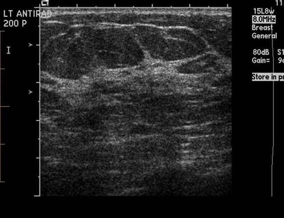
17 Scattered Fibroglandular Densities: Heterogeneous on US
18 A. Tissue composition B. Masses C. Calcifications D. Associated features E. Special cases Mass is space occupying seen in 2 projections and characterized as to: Shape Orientation Margin Echo pattern Posterior acoustic features
19 A. Tissue composition B. Masses C. Calcifications D. Associated Features E. Special cases Lexicon Pictorial Commences... Mass Lexicon Shape: oval, round, irregular Orientation: parallel, not parallel Margin: circumscribed, not circumscribed (indistinct, angular, microlobulated, spiculated) Echo pattern: anechoic, hyperechoic, complex cystic and solid, hypoechoic, isoechoic, heterogeneous Posterior acoustic features: no posterior features, enhancement, shadowing, combined pattern


20 Distinguish a true mass via scanning in > 2 projections and its appearance at real time scan Rib in transverse and longitudinal planes

21 Oval Round Oval Macrolobulated is subtype of oval with 2-3 gentle undulations Irregular Round is sphere Irregular not oval or round

22 Parallel Not Parallel Orientation is a feature unique to US Irregular Parallel vs not parallel to the skin surface Not parallel is taller than wide
23 Circumscribed Not Circumscribed Circumscribed mass margin is welldefined with abrupt transition Irregular Not circumscribed mass margin is defined by its descriptors: indistinct, angular, microlobulated, spiculated


24 Indistinct Angular Microlobulated Spiculated Indistinct has no clear demarcation Angular has sharp corners often acute angles Microlobulated is scalloped Spiculated has projecting sharp lines


25 Anechoic Hyperechoic Complex cystic and solid Isoechoic Anechoic shown is cyst with simple fluid Hyperechoic shown is lipoma Complex cystic and solid contains both anechoic and echogenic tissue (BI-RADS 4 as 25% are malignant as in this case papilloma with DCIS) Isoechoic is relative to fat, acute hematoma in this case

26 Hypoechoic Heterogeneous Hypoechoic masses are both CA Heterogeneous is mixture of echogenic patterns within a solid mass, often due to CA as in this case


27 No Enhancement Shadowing Combined Posterior acoustic feature is the acoustic transmission of a mass ie the attenuation characteristics of the mass Enhancement is echogenic
28 A. Tissue composition B. Masses C. Calcifications D. Associated features E. Special cases Calcifications characterized as: In a mass Outside of a mass Intraductal


29 43yo F with left breast mass 39yo F with left breast mass


30 39yo F with left breast mass fine linear and fine linear branching calcifications right breast mass right breast mass


31 39yo F with left breast mass fine pleomorphic calcifications right breast mass
32 A. Tissue composition B. Masses C. Calcifications D. Associated features E. Special cases Associated features Lexicon Architectural distortion Duct change Skin changes: a. skin thickening b. skin retraction Edema Vascularity: a. absent b. internal vascularity c. vessels in rim Elasticity assessment a. soft b. intermediate c. hard
33 A. Tissue composition B. Masses C. Calcifications D. Associated features E. Special cases NB: Pictorial for Associated features and Special cases slides #43-58 Special cases Lexicon Simple cyst Clustered microcysts Complicated cyst Mass in or on skin Foreign body including implant Lymph node - intramammary Lymph node - axillary Vascular abnormalities - a. AVMs b. Mondor disease Postsurgical fluid collection Fat necrosis
34 I. General Considerations II. Breast Imaging Lexicon - Ultrasound III. Reporting System IV. Guidance APPENDIX: ACR BI-RADS - Ultrasound Lexicon Classification Form
35 ACR Breast Imaging Reporting and Data System Developed in 1993 by ACR to improve the reporting of mammograms BI-RADS - Mammography Fifth Edition 2013 BI-RADS - Ultrasound Second Edition 2013 BI-RADS - MRI Second Edition 2013
36 Quality assurance tool Facilitates outcomes monitoring Standardized lexicon improves clarity of thought Standardized lexicon improves pt care by reducing ambiguity in reports Establishes framework for breast imaging reports BIRADS #0-6 with and 6 as final category designations and 0 as an incomplete category
37 CATEGORY 0: INCOMPLETE - NEED ADDITIONAL IMAGING EVALUATION AND/OR PRIOR MAMMOGRAMS FOR COMPARISON CATEGORY 1: NEGATIVE CATEGORY 2: BENIGN CATEGORY 3: PROBABLY BENIGN CATEGORY 4: SUSPICIOUS CATEGORY 5: HIGHLY SUGGESTIVE OF MALIGNANCY CATEGORY 6: KNOWN BIOPSY-PROVEN MALIGNANCY
38 Category 0: INCOMPLETE - NEED ADDITIONAL IMAGING EVALUATION AND/OR PRIOR MAMMOGRAMS FOR COMPARISON Recall for additional imaging and/or comparison with prior examinations Category 1: NEGATIVE (0% risk) Routine mammography screening Category 2: BENIGN (0% risk) Routine mammography screening Category 3: PROBABLY BENIGN (<2% risk) Short interval 6 month follow-up OR continued surveillance Category 4: SUSPICIOUS (2-95% risk) Biopsy should be performed in the absence of clinical contraindications Category 5: HIGHLY SUGGESTIVE OF MALIGNANCY (>95% risk) Biopsy should be performed in the absence of clinical contraindications Category 6: KNOWN BIOPSY-PROVEN MALIGNANCY (100% risk) Surgical excision when clinically appropriate
39 This module is to educate residents on ACR BI-RADS Atlas 2 nd edition Ultrasound Sonomammographic anatomy Sonographic features most associated with malignancy Clinical and sonographic features of common diseases ABR Core Exam Ultrasound blueprint & study guide
40 Irregular (round) shape Not parallel orientation Not circumscribed (indistinct, angular, microlobulated, spiculated) margins Hypoechoic, isoechoic, complex cystic and solid, and heterogeneous echogenicity No, shadowing, or combined posterior acoustic features Architectural distortion, skin thickening, skin retraction, edema associated features
41 Oval shape Parallel orientation Circumscribed margins Anechoic, Hyperechoic echogenicity Enhanced or no posterior acoustic features
 Biopsy should be performed in the")


42 Category 5: HIGHLY SUGGESTIVE OF MALIGNANCY (>95% risk) Biopsy should be performed in the absence of clinical contraindications 71yo F with screen-detected left breast mass: irregular and spiculated 56yo F with high risk screendetected right breast mass: irregular not parallel and microlobulated 87yo F with screendetected right breast mass: irregular not parallel and spiculated

43 Architectural distortion, skin thickening and skin retraction 81yo F with right breast mass and skin retraction


44 40yo F with left breast mass Skin retraction

45 Edema 49yo F with right breast mass
46 Remember: Special cases Lexicon ie those w pathognomonic appearance Occur Simple in 10% cyst of all women May Clustered be palpable, microcysts painful, grow/regress Complicated quickly cyst Anechoic Mass in mass, or on imperceptible skin wall, Foreign posterior body enhancement including implant Painful Lymph cysts node can -be intramammary aspirated under Lymph ultrasound node --axillary benign type fluid Vascular yellow, green abnormalities - a. AVMs b. Mondor disease Postsurgical fluid collection Fat necrosis

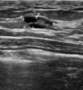
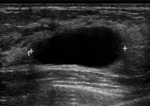
47 Occur in 10% of all women May be palpable, painful, grow/regress quickly Anechoic mass, imperceptible wall, posterior enhancement Painful cysts can be aspirated under ultrasound - benign type fluid yellow, green

48 Etiologies include fibrocystic change and apocrine metaplasia Lesion consists of cluster of tiny anechoic foci, individually smaller than 3 mm Thin intervening septations and no discrete solid component B-R 3 vs B-R 2 lesion

49 Homogeneous low level internal echoes May have a layered appearance Fluid-debris levels may shift with pt position May also contain brightly echogenic foci that scintillate as they shift NOT complex cystic and solid mass


50 Arise in hair follicle Spontaneous or prior trauma Dermal locale, punctum on skin, skin tract on US When inflammed, accompanying edema, hypervascularity, and erythema ensues Visual inspection Utility of US - skin tract and claw sign Skin punctum Skin tract


51 86yo F history treated ovarian CA now presenting w dermal mass and peau d orange


52 43yoF midcycle breast CA neoadjuvant therapy with right mastitis


53 62yoF prior implants, DCIS, skin-sparing mastectomy with breast mass Classic snowstorm of silicone


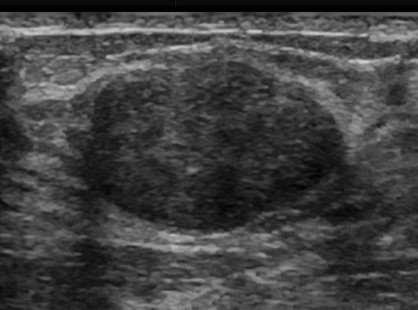
54 Common finding in axilla and breast Distinctive benign appearance: circumscribed oval masses often reniform, contain hilar fat hypoechoic cortex, echogenic hilum < 2-3cm Pathologic appearance: cortex focally or diffusely thickened DDx metastatic disease, lymphoma/leukemia, CTD, granulomatous disease, reactive LN varying appearances, clockwise from top left: Normal intramammary Normal axillary Metastases in eccentric enlarged cortex Diffuse metastatatic involvement LN replaced by tumor
55 BI-RADS 4 Unilateral DDx breast carcinoma, metastatic melanoma, ovarian CA, other CA Careful eval ipsilateral breast Bilateral axillary US to determine if uni/bilateral Clinical eval to exclude mastitis, breast abscess, skin infx, cat scratch fever Proceed to FNA or CNB BI-RADS 2 Bilateral Frequently reactive in inflammatory ds and HIV Sarcoid, SLE, psoriasis, analogous ds Known dx Lymphoma - add wording known lymphoma When bilateral LN new or increasing - rethink BI-RADS 4 and include pass for flow cytometry (saline or RPMI)
 blood")


56 Postoperative seroma May be entirely cystic May contain (mobile) blood products Comparison with pt history and with prior exams crucial BI-RADS Category 2 42yoF with history of treated breast cancer with left breast mass


57 Benign condition Trauma-induced but patient may have no knowledge of precipitating event Typically presents as palpable mass Oil cyst Occasionally - irregular mass that merits CNB


58 Pathology Sclerosing thrombophlebitis of subcutaneous veins of anterior chest wall Fibromuscular hyperplasia of vessel wall Clinical Presentation Typical age 30-60yo Painful breast with cord-like mass Visual Inspection Upper outer quadrant Sudden appearance of red, tender, firm band accompanied by skin retraction CBE findings accentuated by raising arm Imaging Findings Superficial tubular beaded vein on imaging May be seen on mammo and ultrasound US findings characteristic of superficial thrombophlebitis, partially recanalized
59 This module is to educate residents on ACR BI-RADS Atlas 2 nd edition Ultrasound Sonomammographic anatomy Sonographic features most associated with malignancy Clinical and sonographic features of common diseases ABR Core Exam Ultrasound blueprint & study guide

60 Most common breast mass in patients <35 yo Contain stromal tissue and breast ductules Sensitive to hormone changes eg pregnancy Oval parallel circumscribed hypoechoic May contain calcifications Biopsy is recommended for newly palpable or increasing in size or demonstrating suspicious features on exam
 fibroadenoma May be complex")

61 Histologically similar to fibroadenoma Typically present as rapidly enlarging mass Appearance is similar to (large) fibroadenoma May be complex and contain cystic spaces 10% malignant - no distinguishing features Excision of benign and malignant phyllodes Recurrence local. Rare lung, bone, liver mets

62 May be solitary or multiple Solitary - subareolar, multiple - peripheral Classically bloody nipple discharge, also clear Round or oval hypoechoic masses, intraductal, calcifications Atypia on CNB merits excision


63 Common asymptomatic breast masses May present as unilateral palpable soft mass Oval circumscribed homogeneous, hypoechoic, isoechoic, hyperechoic masses Mature lipocytes with at least a thin capsule

64 Antecendent trauma Anticoagulated or bleeding diathesis Mass or bruise Mixed echogenicity mass(es) May have skin involvement Improvement on follow up 87yo anticoagulated M right breast mass and visible discoloration
65 DDx 1. (FA) 2. (Cyst) 3. Lactating adenoma 4. Galactocele 5. Puerperal mastitis / abscess 6. Pregnancy-associated breast cancer (PABC) ULTRASOUND Modality of choice in these women Non ionizing Non invasive Easy to perform Cost effective Majority of lesions are benign and those that aren t typically follow rules ie BI-RADS Ultrasound

66 Common etiologies lactation, post trauma Pain, erythema, edema, mass US study of choice for diagnosis and surveillance Breast parenchyma hyperechoic hypervascular, skin edema, reactive lymphadenopathy Delayed or inadequate antibiotic treatment can progress to abscess


67 Progression of mastitis most common etiology Delayed or inadequate antibiotic treatment Staph aureus in nursing woman Pain, erythema, edema, mass US study of choice for diagnosis and IR guidance Round or irregular complex mass, fluid-debris levels or mobile debris US surveillance


68 Benign mass with milk contents Results from obstructed milk duct During and following cessation of lactation Most regress over time Aspiration can be diagnostic and therapeutic Oval or round, variable internal echogenicity Fat-fluid level

69 Variant of fibroadenoma, tubular adenoma, or lobular hyperplasia as benign stromal tumors Third trimester through lactation Natural course is regression following cessation of breast feeding Oval or lobulated

70 Pregnancy Associated Breast Cancer PABC defined as breast cancer found during pregnancy or in the first year following 1 in 3000 pregnancies complicated by breast CA Increasing incidence 50% are high grade and 80% are lymph node + Poorer prognosis including recurrence < 3yrs 90% present with palpable mass US appearance mass virtually same as nongravid pt
71 Though Cyst and FA are still commonly encountered consider the 4 DDx unique to pregnant and lactating patient Unique clinical presentations with little overlap Puerperal Abscess inflammatory sx early postpartum Lactating Adenoma present like FA as painless, soft, mobile masses. They may also become infarcted and present atypically as a firm tender mass. Unique feature of LA is the tendency to occur earlier then regress after cessation of breast-feeding Galactocele tendency to occur near cessation of breast-feeding PABC defined as breast cancer found during pregnancy or in the first year following. Increasing incidence due to US maternal demographics. 50% are high grade and 80% are lymph node +. Poorer prognosis including recurrence < 3yrs. 90% present with palpable mass
72 This module is to educate residents on ACR BI-RADS Atlas 2 nd edition Ultrasound Sonomammographic anatomy Sonographic features most associated with malignancy Clinical and sonographic features of common diseases ABR Core Exam Ultrasound blueprint & study guide
73 i)sonomammographic anatomy ii)cyst versus solid mass iii)mastitis/abscess iv)characterization of cysts v)lymph node characterization (1) Axillary (2) Supraclavicular (3) Intramammary vi)characterization of solid masses (1) Benign versus malignant (a) Cyst (b) Fibroadenoma (c) Hamartoma (d) Abscess (e) Hematoma (f) Phyllodes tumor (g) Ductal/lobular carcinoma (h) Carcinoma in situ (i) Metastasis (j) Lymphoma (k) Inflammatory carcinoma vii)architectural distortion viii)intraductal masses/abnormalities, galactocele ix)screening x)multifocal/centric malignancy xi)elastography xii)role of IV contrast Breast (10-15%) Relevant and appropriate Diagnostic Ultrasonographic applications and findings in the entities listed in the Breast section of this blueprint a. Normal sonomammographic anatomy b. Cystic versus solid mass c. Mastitis/abscess d. Characterization of cysts e. Lymph node characterization: axillary, supraclavicular, intramammary f. Characterization of solid masses: benign vs. malignant g. Architectural distortion h. Intraductal masses/abnormalities i. Galactocele j. Ultrasound Screening k. Multifocal malignancy
74 de_2018.pdf


75 Think back and test yourself
Leonard M. Glassman MD
 BI-RADS The New BI-RADS Leonard M. Glassman MD FACR Former Chief of Breast Imaging American Institute for Radiologic Pathology Washington Radiology Associates, PC Breast Imaging Reporting and Data System
Lesion Imaging Characteristics Mass, Favoring Benign Circumscribed Margins Intramammary Lymph Node
 Lesion Imaging Characteristics Mass, Favoring Benign Circumscribed Margins Intramammary Lymph Node Oil Cyst Mass, Intermediate Concern Microlobulated Margins Obscured Margins Mass, Favoring Malignant Indistinct
Imaging in breast cancer. Mammography and Ultrasound Donya Farrokh.MD Radiologist Mashhad University of Medical Since
 Imaging in breast cancer Mammography and Ultrasound Donya Farrokh.MD Radiologist Mashhad University of Medical Since A mammogram report is a key component of the breast cancer diagnostic process. A mammogram
Ultrasound of the Breast BASICS FOR THE ORDERING CLINICIAN
 Ultrasound of the Breast BASICS FOR THE ORDERING CLINICIAN Breast Ultrasound Anatomy Skin Breast Parenchyma Pectoralis Fascia Pectoralis Breast Ultrasound Anatomy Indications for Breast Ultrasound Palpable
BI-RADS Update. Martha B. Mainiero, MD, FACR, FSBI Brown University Rhode Island Hospital
 BI-RADS Update Martha B. Mainiero, MD, FACR, FSBI Brown University Rhode Island Hospital No Disclosures BI-RADS History 1980s Quality Issues ACR Accreditation BI-RADS 1994 2003 4 th Edition MRI, US January
ACRIN 6666 IM Additional Evaluation: Additional Views/Targeted US
 Additional Evaluation: Additional Views/Targeted US For revised or corrected form check box and fax to 215-717-0936. Instructions: The form is completed based on recommendations (from ID form) for additional
SIGNIFICANT OTHERS. Miscellaneous Benign Breast Conditions
 SIGNIFICANT OTHERS Miscellaneous Benign Breast Conditions Epworth HealthCare 1 FAT NECROSIS TRAUMATIC Cell rupture Seat-Belt injury Blunt trauma Iatrogenic injury Surgery, Flaps, Radiotherapy Pathology
Imaging the Symptomatic Patient. Avice M.O Connell MD,FACR,FSBI Professor of Imaging Sciences Director, Women s Imaging University of Rochester
 Imaging the Symptomatic Patient Avice M.O Connell MD,FACR,FSBI Professor of Imaging Sciences Director, Women s Imaging University of Rochester The four most common symptoms Mass Pain Discharge Infection
Breast Imaging Lexicon
 9//201 200 BI RADS th Edition 201 BI RADS th Edition Breast Imaging Lexicon Mammographic Pathology and Assessment Categories Deborah Thames, R.T.(R)(M)(QM) The Advanced Health Education Center Nonmember:
Benign, Reactive and Inflammatory Lesions of the Breast
 Benign, Reactive and Inflammatory Lesions of the Breast Marilin Rosa, MD Associate Member Section Head of Breast Pathology Department of Anatomic Pathology Program Director, Breast Pathology Fellowship
Criteria of Malignancy. Evaluation Score
 30 5 Diagnostic Criteria Criteria of Malignancy Table 5.2 lists criteria in contrast-enhancing MR mammography that strongly indicate the presence of malignancy or are unspecific. Unifactorial evaluation
The radiologic workup of a palpable breast mass
 Imaging in Practice CME CREDIT EDUCTIONL OJECTIVE: The reader will consider which breast masses require further workup and which imaging study is most appropriate Lauren Stein, MD Imaging Institute, Cleveland
Amammography report is a key component of the breast
 Review Article Writing a Mammography Report Amammography report is a key component of the breast cancer diagnostic process. Although mammographic findings were not clearly differentiated between benign
Case Scenario 1 History and Physical 3/15/13 Imaging Pathology
 Case Scenario 1 History and Physical 3/15/13 The patient is an 84 year old white female who presented with an abnormal mammogram. The patient has a five year history of refractory anemia with ringed sideroblasts
University of Washington Radiology Review Course: Strange and Specific Diagnoses. Case #1
 University of Washington Radiology Review Course: Strange and Specific Diagnoses Katherine E. Dee, MD Seattle Breast Center Via Radiology 2014 Case #1 37 year old presents with bilateral palpable lumps.
Contents. Basic Ultrasound Principles and Terminology. Ultrasound Nodule Characteristics
 Contents Basic Ultrasound Principles and Terminology Basic Ultrasound Principles... 1 Ultrasound System... 2 Linear Transducer for Superficial Images and Ultrasound-Guided FNA... 3 Scanning Planes... 4
Non-mass Enhancement on Breast MRI. Aditi A. Desai, MD Margaret Ann Mays, MD
 Non-mass Enhancement on Breast MRI Aditi A. Desai, MD Margaret Ann Mays, MD Breast MRI Important screening and diagnostic tool, given its high sensitivity for breast cancer detection Breast MRI - Indications
MEDICAL IMAGING AND BREAST DISEASE HOW CAN WE HELP YOU?
 MEDICAL IMAGING AND BREAST DISEASE HOW CAN WE HELP YOU? Barbara M. Preston, M.D. SCREENING MAMMOGRAPHY AVERAGE RISK PATIENTS KAISER RECOMMENDATION: ALL WOMEN (INCLUDING TRANSGENDER FEMALES) Every 1-21
Diseases of the breast (1 of 2)
") Diseases of the breast (1 of 2) Introduction A histology introduction Normal ducts and lobules of the breast are lined by two layers of cells a layer of luminal cells overlying a second layer of myoepithelial
Diseases of the breast (1 of 2) Introduction A histology introduction Normal ducts and lobules of the breast are lined by two layers of cells a layer of luminal cells overlying a second layer of myoepithelial
BI-RADS and Breast MRI. Kathy Borovicka, M.D. Thursday February 15, 2018
 BI-RADS and Breast MRI Kathy Borovicka, M.D. Thursday February 15, 2018 Learning Objectives Be familiar with the Breast Imaging Reporting and Data System (BI-RADS) Understand the components of a breast
BREAST PATHOLOGY MCQS
 BREAST PATHOLOGY MCQS 1) :The most important factor in breast enlargement during pregnancy is A. stromal edema B. secretion of chorionic gonadotropin C. glandular hyperplasia D. proliferation of stroma
Alena Levit MD Avice O Connell MD University of Rochester, Rochester, NY
 Alena Levit MD Avice O Connell MD University of Rochester, Rochester, NY Purpose Review imaging spectrum of both common benign and malignant breast lesions Describe and demonstrate CT features with mammogram,
Breast Health. Learning Objectives. Breast Anatomy. Poll Question. Breast Anatomy
 Learning Objectives Describe breast anatomy to a patient Breast Health Answer questions about causes of breast pain and masses Explain breast cancer screening/diagnostic modalities Appropriately triage
Mimickers of breast malignancy
 Mimickers of breast malignancy Poster No.: C-0477 Congress: ECR 2010 Type: Educational Exhibit Topic: Breast Authors: S. H. Park, H.-Y. Choi; Incheon/KR Keywords: mimickers, breast malignancy, ultrasound
Index. C Calcifications fat necrosis 1, 61 fat necrosis 4, 69 nipple/peri-areolar involvement 1, 165
 A ADH. See Atypical ductal hyperplasia (ADH) American College of Radiology (ACR), BI-RADS background parenchymal enhancement, 8, 9, 81, 82 fibroglandular tissue guidelines, 6 American Joint Committee on
RADIOLOGIC EVALUATION OF BREAST CANCER
 RADIOLOGIC EVALUATION OF BREAST CANCER Orsolya Farkas, Gabriella Bodrogi and Gábor Szalai Department of Radiology, Pécs University Orsifarkas@yahoo.com Complex evaluation of the breast Patient history
Margaret Ann Mays, M.D. Clinical Instructor in Radiology Vanderbilt University Medical Center Tennessee Radiological Society February 25, 2017
 Breast Imaging in Adolescents and Young Women Margaret Ann Mays, M.D. Clinical Instructor in Radiology Vanderbilt University Medical Center Tennessee Radiological Society February 25, 2017 Disclosures
Abid Irshad, MD Director Breast Imaging. Medical University of South Carolina Charleston
 Abid Irshad, MD Director Breast Imaging Medical University of South Carolina Charleston Cases Financial disclosure: I or my family have no financial interest related to the material discussed in this presentation
BREAST PATHOLOGY. Fibrocystic Changes
 BREAST PATHOLOGY Lesions of the breast are very common, and they present as palpable, sometimes painful, nodules or masses. Most of these lesions are benign. Breast cancer is the 2 nd most common cause
Armed Forces Institute of Pathology.
 Armed Forces Institute of Pathology www.radpath.com Armed Forces Institute of Pathology Breast Disease www.radpath.org Armed Forces Institute of Pathology Interpretation of Breast MRI Leonard M. Glassman
Mammographic imaging of nonpalpable breast lesions. Malai Muttarak, MD Department of Radiology Chiang Mai University Chiang Mai, Thailand
 Mammographic imaging of nonpalpable breast lesions Malai Muttarak, MD Department of Radiology Chiang Mai University Chiang Mai, Thailand Introduction Contents Mammographic signs of nonpalpable breast cancer
ORIGINAL ARTICLE EVALUATION OF BREAST LESIONS USING X-RAY MAMMOGRAM WITH HISTOPATHOLOGICAL CORRELATION
 Available online at www.journalijmrr.com INTERNATIONAL JOURNAL OF MODERN RESEARCH AND REVIEWS IJMRR ISSN: 2347-8314 Int. J. Modn. Res. Revs. Volume 3, Issue 10, pp 807-814, October, 2015 ORIGINAL ARTICLE
S. Murgo, MD. Chr St-Joseph, Mons Erasme Hospital, Brussels
 S. Murgo, MD Chr St-Joseph, Mons Erasme Hospital, Brussels? Introduction Mammography reports are sometimes ambiguous and indecisive. ACR has developped the BIRADS. BIRADS consists of a lexicon in order
Cairo/EG, Khartoum/SD, London/UK Biological effects, Diagnostic procedure, Ultrasound, Mammography, Breast /ecr2015/C-0107
 Role of sono-mammography in the evaluation of clinically palapble breast masses during pregnancy & lactation with differentaition between true patholgical & false physiological lobular hyperlpasia.sudanese
Breast Ultrasound: Improving Your Skills & Patient Care
 Breast Ultrasound: Improving Your Skills & Patient Care Objectives Discuss US techniques available for image optimization. Review & compare the US appearances of benign & malignant masses. Cherie M. Kuzmiak,
Radiology Review Course Hotel del Coronado Coronado, California
 37 th Annual Radiology Review Course Hotel del Coronado Coronado, California Friday, April 21, 2017 - PM TABLE OF CONTENTS Friday, April 21, 2017 - PM SAM Session - Breast Imaging Update 12:45 PM 1:30
Ultrasonography. Methods. Brief Description. Indications. Device-related Prerequisites. Technical Requirements. Evaluation Criteria
 1 Ultrasonography Brief Description Imaging modality using sound waves Tissue-specific wave reflection. Indications Evaluation of palpable breast nodules Evaluation of clinically occult mammographic findings
Ana Sofia Preto 19/06/2013
 Ana Sofia Preto 19/06/2013 Understanding the underlying pathophysiologic processes leading to the various types of calcifications Description and illustration of the several types of calcifications, according
Breast pathology. 2nd Department of Pathology Semmelweis University
 Breast pathology 2nd Department of Pathology Semmelweis University Breast pathology - Summary - Benign lesions - Acute mastitis - Plasma cell mastitis / duct ectasia - Fat necrosis - Fibrocystic change/
THE MALE BREAST CARCINOMA: EARLY DETECTION HOPE. Author (s) Supreethi Kohli a, Pragya Garg b
 Supreethi Kohli a, Pragya Garg b") Case Report ABSTRACT - Male breast cancer is exceptionally rare and accounts for less than 0.25% of male malignancies and approximately 0.5-1% of all breast cancer (both genders). Mammography of the male
Case Report ABSTRACT - Male breast cancer is exceptionally rare and accounts for less than 0.25% of male malignancies and approximately 0.5-1% of all breast cancer (both genders). Mammography of the male
Imaging of giant breast masses with pathological correlation
 P i c t o r i a l E s s a y Singapore Med J 2004 Vol 45(3) : 132 Imaging of giant breast masses with pathological correlation M Muttarak, B Chaiwun ABSTRACT Ultrasonography (US) and mammography are the
Mousa. Israa Ayed. Abdullah AlZibdeh. 0 P a g e
 1 Mousa Israa Ayed Abdullah AlZibdeh 0 P a g e Breast pathology The basic histological units of the breast are called lobules, which are composed of glandular epithelial cells (luminal cells) resting on
AMSER Case of the Month: September 2018
 AMSER Case of the Month: September 2018 60-year-old woman with a left breast mass noted on screening mammography. Catherine McNulty, MS4 Tulane University School of Medicine Dr. Robin Sobolewski Breast
Breast imaging of benign fat containing lesions
 Breast imaging of benign fat containing lesions Poster No.: C-1870 Congress: ECR 2017 Type: Educational Exhibit Authors: R. Aouini, I. Megdiche, D. Ben Hammadi, N. BEN MAMI, I. Attia, R. Neila, A. Zidi;
Benign Breast Disease. David Anderson, MD Assistant Professor of Clinical Surgery
 Benign Breast Disease David Anderson, MD Assistant Professor of Clinical Surgery Overview Nipple Discharge Breast infection Breast Pain Gynecomastia Fibroepithelial lesions High Risk Lesions-Papilloma,
Gynecomastia and Its Mimics: Not All Male Breast Lesions are Benign
 Gynecomastia and Its Mimics: Not All Male Breast Lesions are Benign Poster No.: C-0139 Congress: ECR 2014 Type: Educational Exhibit Authors: S. A. Choudhery, P. Gupta, S. Foshee, F. Garcia-Morales, G.
MRI BI-RADS: How to make it out?
 MRI BI-RADS: How to make it out? Poster No.: C-1850 Congress: ECR 2016 Type: Educational Exhibit Authors: M. Ben Ammar, A. Ben Miled, O. Ghdes, S. Harguem, A. Gaja, N. Mnif; Tunis/TN Keywords: Breast,
OF HEMATOMA INTRACRANIAL HEMORRHAGE
 SCOPE TUTORIAL RADIOLOGY CT BRIAN BREASTS US MRCP Natrada Rawdhetubhai, M.D. Radiology Department, Lerdsin General Hospital CT OF HEMATOMA INTRACRANIAL HEMORRHAGE Intra-axial Extra-axial Intraventricular
04/10/2018. Intraductal Papillary Neoplasms Of Breast INTRADUCTAL PAPILLOMA
 Intraductal Papillary Neoplasms Of Breast Savitri Krishnamurthy MD Professor of Pathology Deputy Division Head The University of Texas MD Anderson Cancer Center 25 th Annual Seminar in Pathology Pittsburgh,
Case Scenario 1: This case has been slightly modified from the case presented during the live session to add clarity.
 Case Scenario 1: This case has been slightly modified from the case presented during the live session to add clarity. Background: 46 year old married premenopausal female with dense breasts has noticed
Validation of the fifth edition BI-RADS ultrasound lexicon with comparison of fourth and fifth edition diagnostic performance using video clips
 Validation of the fifth edition BI-RADS ultrasound lexicon with comparison of fourth and fifth edition diagnostic performance using video clips Jung Hyun Yoon 1, Min Jung Kim 1, Hye Sun Lee 2, Sung Hun
DISORDERS OF THE BREAST Dated. FIBROADENOSIS Other common names: mastitis, fibrocystic disease, cystic mammary dysplasia.
 DISORDERS OF THE BREAST Dated BENIGN BREAST DISORDERS (Essential Surg 2 nd Ed, pp 540) FIBROADENOSIS Other common names: mastitis, fibrocystic disease, cystic mammary dysplasia. Fibroadenosis is the distortion
RSNA, /radiol Appendix E1. Methods
 RSNA, 2016 10.1148/radiol.2016151097 Appendix E1 Methods US and Near-infrared Data Acquisition Four optical wavelengths (740 nm, 780 nm, 808 nm, and 830 nm) were used to sequentially deliver the light
PAAF vs Core Biopsy en Lesiones Mamarias Case #1
 5/19/2014 PAAF vs Core Biopsy en Lesiones Mamarias Case #1 Fine Needle Aspiration Cytology of Breast: Correlation with Needle Core Biopsy 64-year-old woman Mass in breast Syed Hoda, MD CD31 Post-Radiation
Disclaimer no conflict of interest
 Disclaimer no conflict of interest Benign Breast Disease Alison Hayes FRACS Content Clinical assessment of the breast Triple assessment Focal nodularity Breast pain Cysts Infection Nipple discharge Gynaecomastia
Breast Cancer. Most common cancer among women in the US. 2nd leading cause of death in women. Mortality rates though have declined
 Breast Cancer Most common cancer among women in the US 2nd leading cause of death in women Mortality rates though have declined 1 in 8 women will develop breast cancer Breast Cancer Breast cancer increases
Case Scenario 1: This case has been slightly modified from the case presented during the live session to add clarity.
 Case Scenario 1: This case has been slightly modified from the case presented during the live session to add clarity. Background: 46 year old married premenopausal female with dense breasts has noticed
Breast Cancer. Saima Saeed MD
 Breast Cancer Saima Saeed MD Breast Cancer Most common cancer among women in the US 2nd leading cause of death in women 1 in 8 women will develop breast cancer Incidence/mortality rates have declined Breast
Breast Evaluation & Management Guidelines
 Breast Evaluation & Management Guidelines Pamela L. Kurtzhals, M.D. F.A.C.S. Head, Dept. of General Surgery Scripps Clinic, La Jolla Objective Review screening & diagnostic guidelines Focused patient complaints
Breast Pathology. Breast Development
 Breast Pathology Lecturer: Hanina Hibshoosh, M.D. Reading: Kumar, Cotran, Robbins, Basic Pathology, 6th Edition, pages 623-635 Breast Development 5th week - thickening of the epidermis - milk line 5th
Case-based discussion:
 Case-based discussion: Pailin Kongmebhol, M.D. Department of Radiology Faculty of Medicine Chiang Mai University There are many guidelines for managing thyroid nodules Two important guidelines: 2015 American
AB MR Interpretation Overview
 AB MR Interpretation Overview Goal of AB MR interpretation is to maintain high sensitivity and specificity In order to minimize false positives and short term follow ups, it is fundamental to focus only
Mammographically non-calcified ductal carcinoma in situ: sonographic features with pathological correlation in 35 patients
 Clinical Radiology (2009) 64, 628e636 ORIGINAL PAPER Mammographically non-calcified ductal carcinoma in situ: sonographic features with pathological correlation in 35 patients B. Mesurolle a, *, M. El-Khoury
PLACE LABEL HERE. ACRIN 6657 MRI Form: Pre-Treatment (MRI-1)
") M3 ACRIN 6657 MRI Form: Pre-Treatment (MRI-1) If this is a revised or corrected form,indicate by checking box. ACRIN Study 6657 Case # Instructions: In accordance with the protocol, four MRI exams are
M3 ACRIN 6657 MRI Form: Pre-Treatment (MRI-1) If this is a revised or corrected form,indicate by checking box. ACRIN Study 6657 Case # Instructions: In accordance with the protocol, four MRI exams are
PRINCIPLES OF BREAST SURGERY & COMPLICATIONS
 PRINCIPLES OF BREAST SURGERY & COMPLICATIONS Adam Cichowitz The Royal Melbourne Hospital ANATOMY Lies in subcutaneous tissue Base: midline to midaxillary line, 2nd to 6th rib Overlies pec major, serratus
Vesalius SCALpel : Benign breast disease (see also: breast folios)
") Vesalius SCALpel : Benign breast disease (see also: breast folios) Breast cancer risk Imaging Pain non-proliferative: only fibroadenoma may be associated with a slight risk of cancer proliferative: moderate
Vesalius SCALpel : Benign breast disease (see also: breast folios) Breast cancer risk Imaging Pain non-proliferative: only fibroadenoma may be associated with a slight risk of cancer proliferative: moderate
Cystic Masses of the Breast
 Residents Section Pattern of the Month Eisenberg ystic Masses of the reast Residents Section Pattern of the Month Residents inradiology Neely Hines 1 Priscilla J. Slanetz Ronald L. Eisenberg Hines N, Slanetz
Breast Pathology in Men: Radiologic-Pathologic Correlation
 Breast Pathology in Men: Radiologic-Pathologic Correlation Poster No.: C-0243 Congress: ECR 2012 Type: Scientific Exhibit Authors: G. Garrido; Málaga/ES Keywords: Breast, Ultrasound, Mammography, Biopsy,
Practical and illustrated summary of updated BI-RADS for ultrasonography
 Practical and illustrated summary of updated I-RS for ultrasonography Jiyon Lee epartment of Radiology, NYU School of Medicine, NYU ancer Institute, reast Imaging enter, New York, NY, US The merican ollege
Policies, Standards, and Guidelines. Guidelines on Breast Ultrasound Examination and Reporting
 Policies, Standards, and Guidelines Guidelines on Breast Ultrasound Examination and Reporting Approved by Council June 2018 Approved: June 2018 Guidelines on Breast Ultrasound Examination and Reporting
ISSN X (Print) Research Article. *Corresponding author Dr. Amlendu Nagar
 Research Article. *Corresponding author Dr. Amlendu Nagar") Scholars Journal of Applied Medical Sciences (SJAMS) Sch. J. App. Med. Sci., 2015; 3(3A):1069-1073 Scholars Academic and Scientific Publisher (An International Publisher for Academic and Scientific Resources)
Scholars Journal of Applied Medical Sciences (SJAMS) Sch. J. App. Med. Sci., 2015; 3(3A):1069-1073 Scholars Academic and Scientific Publisher (An International Publisher for Academic and Scientific Resources)
Breast Disease: What PCPs Need to Know. Eunice Cho MD FACS
 Breast Disease: What PCPs Need to Know Eunice Cho MD FACS New Breast Cancer Screening Guideline for women with average risk Every other year AGE 40 AGE 45 AGE 55 AGE 55 + Talk with your doctor about when
Optimizing Breast Sonography
 Optimizing Breast Sonography Cindy Rapp BS, RDMS, FSDMS, FAIUM Denver, Colorado Breast Sonography general goal to make a more specific diagnosis than can be made with clinical and mammographic findings
Breast Imaging: Multidisciplinary Approach. Madelene Lewis, MD Assistant Professor Associate Program Director Medical University of South Carolina
 Breast Imaging: Multidisciplinary Approach Madelene Lewis, MD Assistant Professor Associate Program Director Medical University of South Carolina No Disclosures Objectives Discuss a multidisciplinary breast
Treatment options for the precancerous Atypical Breast lesions. Prof. YOUNG-JIN SUH The Catholic University of Korea
 Treatment options for the precancerous Atypical Breast lesions Prof. YOUNG-JIN SUH The Catholic University of Korea Not so benign lesions? Imaging abnormalities(10% recall) lead to diagnostic evaluation,
Case study 1. Rie Horii, M.D., Ph.D. Division of Pathology Cancer Institute Hospital, Japanese Foundation for Cancer Research
 NCCN/JCCNB Seminar in Japan April 15, 2012 Case study 1 Rie Horii, M.D., Ph.D. Division of Pathology Cancer Institute Hospital, Japanese Foundation for Cancer Research Present illness: A 50y.o.premenopausal
ACRIN 6666 Therapeutic Surgery Form
 S1 ACRIN 6666 Therapeutic Surgery Form 6666 Instructions: Complete a separate S1 form for each separate area of each breast excised with the intent to treat a cancer (e.g. each lumpectomy or mastectomy).
Jordan University Faculty Of Medicine. Breast. Dr. Ahmed Salman. Assistant professor of anatomy & embryology
 Jordan University Faculty Of Medicine Breast Dr. Ahmed Salman Assistant professor of anatomy & embryology The breasts are specialized accessory glands of the skin that secretes milk. They are situated
LYMPHATIC DRAINAGE AXILLARY (MOSTLY) INTERNAL MAMMARY SUPRACLAVICULAR
 INTERNAL MAMMARY SUPRACLAVICULAR") BREAST LYMPHATIC DRAINAGE AXILLARY (MOSTLY) INTERNAL MAMMARY SUPRACLAVICULAR HISTOLOGY LOBE: (10 in whole breast) LOBULE: (many per lobe) ACINUS/I, aka ALVEOLUS/I: (many per lobule) DUCT(S): INTRA- or
BREAST LYMPHATIC DRAINAGE AXILLARY (MOSTLY) INTERNAL MAMMARY SUPRACLAVICULAR HISTOLOGY LOBE: (10 in whole breast) LOBULE: (many per lobe) ACINUS/I, aka ALVEOLUS/I: (many per lobule) DUCT(S): INTRA- or
Radiological Appearances of Male Breast Disease
 Radiological Appearances of Male Breast Disease Poster No.: C-1916 Congress: ECR 2017 Type: Educational Exhibit Authors: A. VALDIVIELSO, A. Guma Martinez, R. Ortega Martinez, L. 1 1 1 2 1 1 1 Perez tapia,
Pitfalls and Limitations of Breast MRI. Susan Orel Roth, MD Professor of Radiology University of Pennsylvania
 Pitfalls and Limitations of Breast MRI Susan Orel Roth, MD Professor of Radiology University of Pennsylvania Objectives Review the etiologies of false negative breast MRI examinations Discuss the limitations
BREAST IMAGING and NEW IMAGING MODALITIES- A Surgeons view
 BREAST IMAGING and NEW IMAGING MODALITIES- A Surgeons view DR CHANTEL THORNTON SPECIALIST BREAST CANCER SURGEON BMSc (hons) MBBS (hons) FRACS Epworth Hospital, Richmond- Agora Centre for Women s Health
AMSER Case of the Month: November 2018
 AMSER Case of the Month: November 2018 42 year old with right breast mass Rina Kiyota Petek Lake Erie College of Osteopathic Medicine, OMS-III Kossivi Dantey, MD Bibianna Klepchick, MD Matthew Hartman,
CPC 4 Breast Cancer. Rochelle Harwood, a 35 year old sales assistant, presents to her GP because she has noticed a painless lump in her left breast.
 CPC 4 Breast Cancer Rochelle Harwood, a 35 year old sales assistant, presents to her GP because she has noticed a painless lump in her left breast. 1. What are the most likely diagnoses of this lump? Fibroadenoma
Timby/Smith: Introductory Medical-Surgical Nursing, 9/e
 Timby/Smith: Introductory Medical-Surgical Nursing, 9/e Chapter 60: Caring for Clients With Breast Disorders Slide 1 Infectious and Inflammatory Breast Disorders: Mastitis Pathophysiology and Etiology
Intracystic papillary carcinoma of the breast
 Intracystic papillary carcinoma of the breast Poster No.: C-1932 Congress: ECR 2011 Type: Educational Exhibit Authors: V. Dimarelos, F. TZIKOS, N. Kotziamani, G. Rodokalakis, 1 2 3 1 1 1 2 T. MALKOTSI
Giant Ulcerative Lactating Nodule of Ectopic Breast Mimicking Malignancy.
 57 Giant Ulcerative Lactating Nodule of Ectopic Breast Mimicking Malignancy. I. Roy 1 FRCS(C), E Othieno 2 MMed (Path), R Ssentongo 3 FCS (ECSA) 1 Department of Pathology, St. Raphael of St. Francis Hospital,
Journal of Medical Imaging and Radiation Oncology
 Journal of Medical Imaging and Radiation Oncology 60 (2016) 506 513 MEDICAL IMAGING PICTORIAL ESSAY Malignant hyperechoic breast lesions at ultrasound: A pictorial essay Stephen Tiang, 1 Cecily Metcalf,
Ultrasound 5/1/2017. Ultrasound in the FNA Clinic. Uses of Ultrasound Outside of. Cardiology
 Ultrasound in the FNA Clinic Martha Bishop Pitman, M.D. Director, Cytopathology Massachusetts General Hospital Professor of Pathology Harvard Medical School Boston, MA Uses of Ultrasound Outside of Radiology
Diseases of the breast (2 of 2) Breast cancer
 Breast cancer") Diseases of the breast (2 of 2) Breast cancer Epidemiology & etiology The most common type of cancer & the 2 nd most common cause of cancer death in women 1 of 8 women in USA Affects 7% of women Peak at
Diseases of the breast (2 of 2) Breast cancer Epidemiology & etiology The most common type of cancer & the 2 nd most common cause of cancer death in women 1 of 8 women in USA Affects 7% of women Peak at
Mary Smania, DNP, FNP-BC Clinical Practice Champion Assistant Professor Michigan State University College of Nursing
 Mary Smania, DNP, FNP-BC Clinical Practice Champion Assistant Professor Michigan State University College of Nursing Identify evidence based routine screening guidelines for women of all ages Identify
OPTO-ACOUSTIC BREAST IMAGING
 OPTO-ACOUSTIC BREAST IMAGING A Novel Fusion of Functional and Morphologic Imaging Reni S. Butler, MD A. Thomas Stavros, MD F. Lee Tucker, MD Michael J. Ulissey, MD PURPOSE 1. Explain opto-acoustic (OA)
ROLE OF MRI IN SCREENING, DIAGNOSIS AND MANAGEMENT OF BREAST CANCER. B.Zandi Professor of Radiology
 ROLE OF MRI IN SCREENING, DIAGNOSIS AND MANAGEMENT OF BREAST CANCER B.Zandi Professor of Radiology Introduction In the USA, Breast Cancer is : The Most Common Non-Skin Cancer The Second Leading cause of
Breast Cancer Screening and Surgery. April 26, 2018 Ashley B. Simpson, DO
 Breast Cancer Screening and Surgery April 26, 2018 Ashley B. Simpson, DO Objectives Breast cancer screening Common breast complaints Surgical management of breast cancer Breast Screening Question 1 At
Radiologic and pathologic correlation of non-mass like breast lesions on US and MRI: Benign, high risk, versus malignant
 Radiologic and pathologic correlation of non-mass like breast lesions on US and MRI: Benign, high risk, versus malignant Poster No.: C-1161 Congress: ECR 2013 Type: Educational Exhibit Authors: J. Kwak,
Follow-up of Abnormal Breast Findings. E.J. Siegl RN, OCN, MA, CBCN BCCCP Nurse Consultant January 2012
 Follow-up of Abnormal Breast Findings E.J. Siegl RN, OCN, MA, CBCN BCCCP Nurse Consultant January 2012 Abnormal Breast Findings include the following: CBE results of: Nipple discharge, no palpable mass
Combined mammographic and sonographic evaluation of breast masses
 International Journal of Sciences & Applied Research www.ijsar.in Combined mammographic and sonographic evaluation of breast masses Ramakrishna Narra*, Naganarasimha Raju Jukuri, Bhimeswarao Pasupaleti,
PLACE LABEL HERE BASELINE / PRE-TREATMENT. ACRIN 6657 Extension MRI Form: Baseline / Pre-Treatment MRI 1. o Unknown
 T1 ACRIN 6657 Extension MRI Form: Baseline / Pre-Treatment MRI 1 If this is a revised or corrected form, please box. ACRIN Study 6657 No. Instructions: In accordance with the protocol, four MRI exams are